Cellular reproduction and maintenance
1/122
There's no tags or description
Looks like no tags are added yet.
Name | Mastery | Learn | Test | Matching | Spaced |
|---|
No study sessions yet.
123 Terms
What are the main phases of the cell cycle?
The cell cycle consists of:
G₁ Phase: Cell growth and preparation for DNA replication
S Phase: DNA replication
G₂ Phase: Preparation for mitosis
M Phase: Mitosis (nuclear division) and cytokinesis (cytoplasmic division)
Cells cycle between these phases to ensure proper growth, replication, and division.
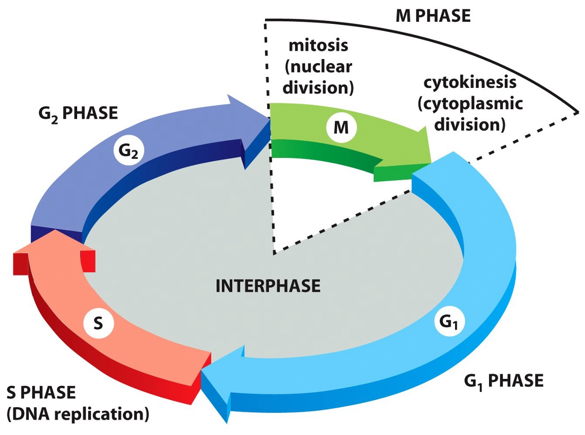
How is the M phase structured?
The M phase is divided into continuous sub-phases.
These sub-phases are artificial divisions for study purposes.
In reality, mitosis is a continuous process without clear-cut transitions.
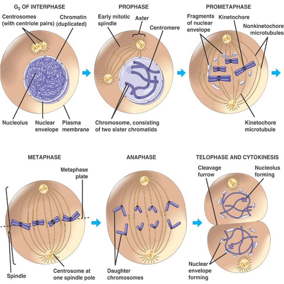
What are the stages of cell division by mitosis?
G₂ of Interphase
Prophase
Prometaphase
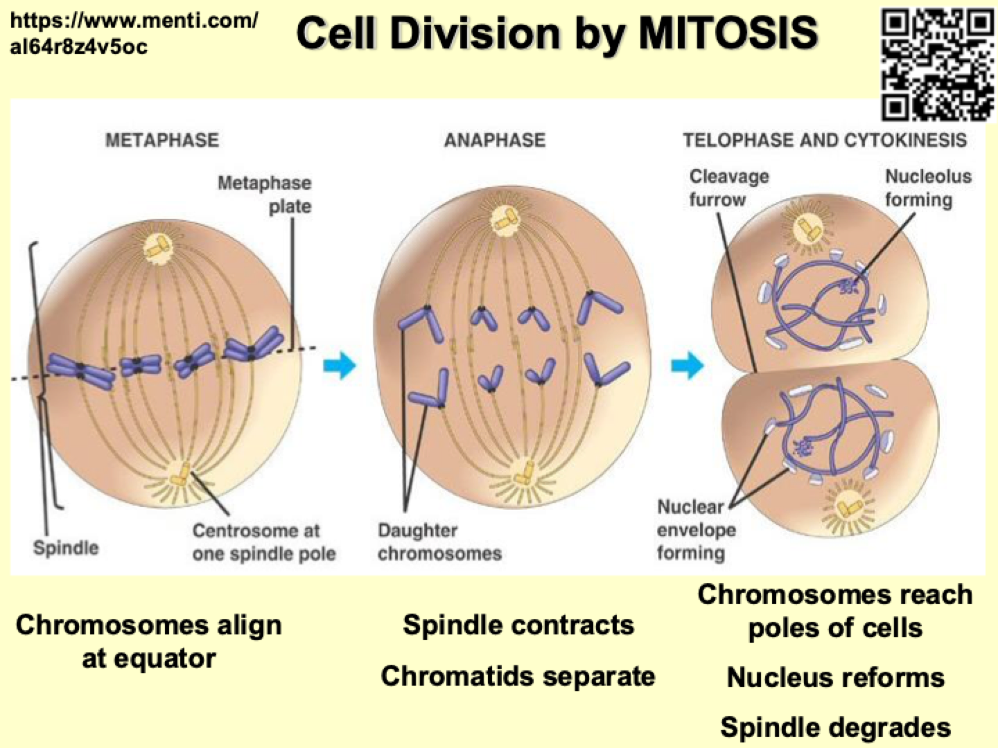
Metaphase
Anaphase
Telophase & Cytokinesis
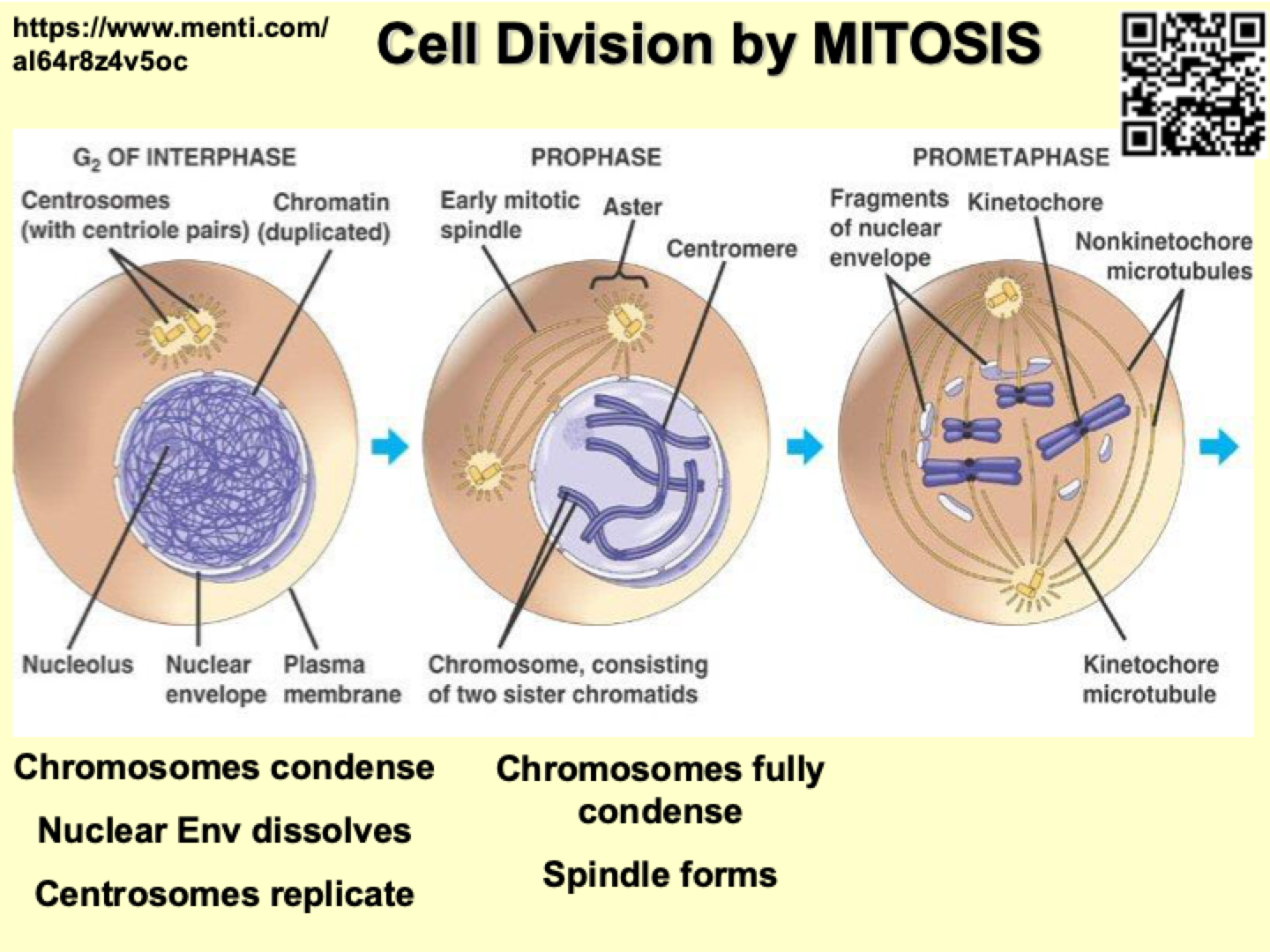
What happens to chromosomes and cellular structures during early mitosis?
Late Interphase: Chromosomes are not condensed yet.
Prophase:
Chromosomes condense and become visible.
Nuclear membrane starts breaking down (due to lamins breaking).
Centrioles replicate.
Centrosomes move to opposite poles, forming the spindle between them.
Prometaphase:
Nuclear membrane is fully broken down.
Chromosomes attach to the spindle.
Each chromosome consists of two sister chromatids (double helices) that have replicated and are aligned.
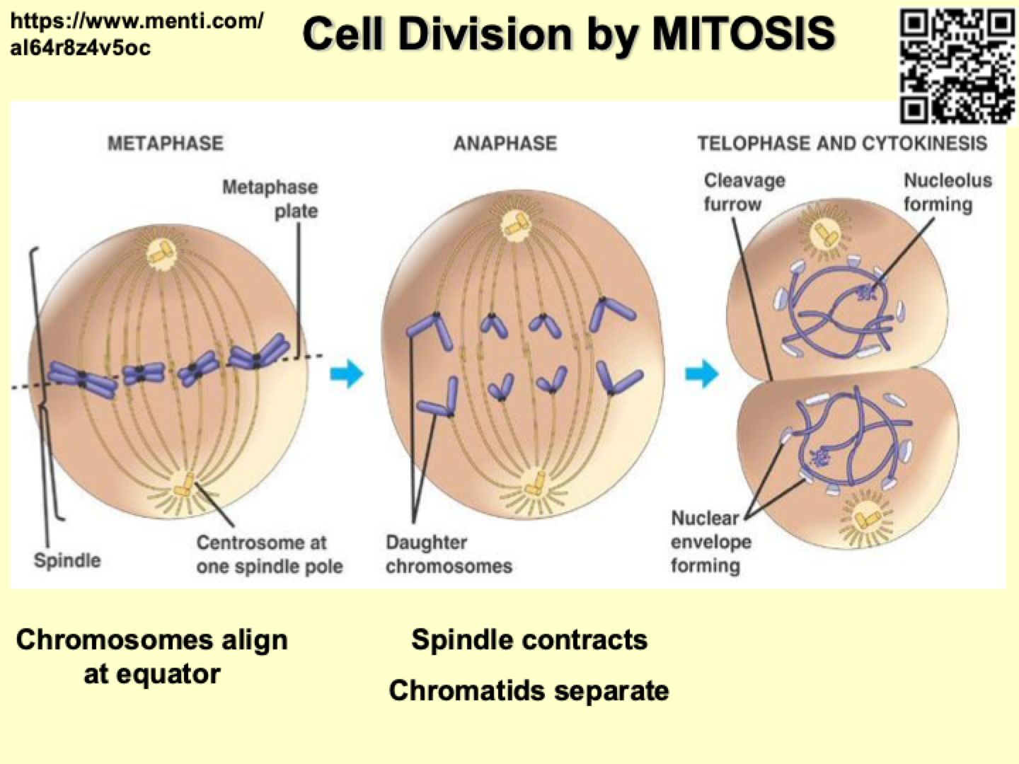
What happens to chromosomes during metaphase and anaphase?
Metaphase:
Chromosomes (each consisting of two sister chromatids) line up individually at the center of the cell (not paired).
Anaphase:
Proteins holding chromatids together break down.
Spindle fibers shorten, pulling sister chromatids apart to opposite poles.
What happens during telophase and cytokinesis?
Telophase:
Chromatids reach opposite poles.
Nuclear membrane reforms around each set of chromosomes.
Chromosomes decondense.
Cytokinesis:
The cytoplasm divides, forming two separate daughter cells.
In animal cells: A contractile ring forms, creating a cleavage furrow.
How does cohesin function in chromosome organization and segregation?
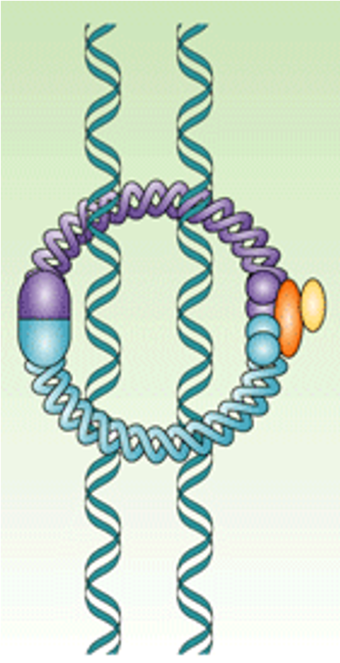
Multi-subunit protein complex.
Forms a loop between subunits, capturing two DNA helices inside.
Acts like a cable tie, holding chromosomes together.
Consists of a head group and a basal group, forming a dimer loop.
Ensures cohesion of chromosomes during cell division.
How does DNA condensation occur during chromosome formation?
DNA undergoes multiple rounds of folding and coiling.
The double helix wraps around histones, forming nucleosomes.
Nucleosomes sometimes coil further to form a solenoid structure.
These structures fold further, leading to condensation during mitosis.
How does condensin contribute to chromosome condensation?
Condensin is a multi-subunit protein that mediates final chromosome condensation.
It dimerizes and works similarly to cohesin.
Functions like a loop fastener, looping DNA together and securing it with a "cable tie" mechanism.
Dimers loop around DNA, securing loops, and DNA passes through the neck of the ring.
How do cohesin and condensin contribute to chromosome condensation?
Cohesin and condensin work together to condense chromatids into 300nm fibers and looped domains.
They are structurally similar, descended from the same bacterial protein.
Cohesin links chromatids together, while condensin forms looped domains and undergoes shape changes to help with condensation.
How does condensin function during chromosome condensation?
Pumping approach: Condensin grabs DNA with its head group, changing conformation to facilitate looping.
Hand-over-head model: Condensin pulls DNA like a rope.
Inchworm model: Condensin moves along DNA sideways, taking one step at a time.
What is the spindle's structure and function in cell division?
The spindle is a network of microtubules made of tubulin, forming the cytoskeleton structure.
It connects the poles of the cell to each other and the cell membrane, facilitating chromosome movement during division.
Centrioles in the centrosome produce microtubules and sit at right angles to each other. They replicate during late G-phase to early S-phase, then separate and move to opposite cell poles.
How does the spindle anchor and organize itself?
The spindle binds to chromosomes and to itself, forming a stable structure.
It also anchors to the plasma membrane at both ends of the cell, securing its position.
There is directionality in the movement of the spindle to help align chromosomes during division.
What are the three different regions of the spindle?
Aster: Made of astral microtubules, with two regions—one anchored in the centrosome and the other bound to the plasma membrane.
Kinetochore: Bound to one chromatid at one end of the cell, with the other chromatid binding to the kinetochore at the opposite end.
Interpolar: Provide structural support for the spindle, bind to each other, and form interlocking poles that extend from one end of the cell to the other.
How are mitotic spindles formed and regulated during the cell cycle?
Microtubules are made from tubulin molecules that polymerize into spirals to form microtubules during cell division.
During interphase, microtubules are depolymerized, preparing for mitosis.
In G2, the microtubules are re-polymerized, forming the mitotic spindle for chromosome alignment and separation during mitosis.
How do motor proteins function in general?
Motor proteins have a head group that binds to a cargo (e.g., chromosomes or other cellular components) and feet that bind to the cytoskeleton (e.g., microtubules).
They "walk" along microtubules, dragging cargo, much like a clown walking along a tightrope.
What is the role of Dynein in mitosis?
Dynein attaches astral microtubules to the plasma membrane.
The head group of dynein has a charge that anchors it to the plasma membrane, fixing the microtubule ends.
This anchoring helps prevent the centrosome from floating off as the spindle contracts during mitosis.
What does Kinesin-14 do in the spindle?
Kinesin-14 is a minus-end-directed motor protein that tightens the spindle.
It pulls microtubules toward each other by crosslinking them and generating inward force, helping bring the spindle poles closer together.
How does Kinesin-5 contribute to spindle formation?
Kinesin-5 is a plus-end-directed motor protein that expands the spindle.
It has motor domains at both ends, moving in opposite directions along antiparallel microtubules.
This action pushes microtubules apart, generating outward force and contributing to spindle elongation.
What is the role of Kinesin-4 and Kinesin-10 during mitosis?
Kinesin-4 and Kinesin-10 are motor proteins that help move chromosomes toward the spindle poles.
They physically walk the chromosome along the microtubules, sometimes flipping them over during the process.
What is the structure and function of the centromere in chromosomes?
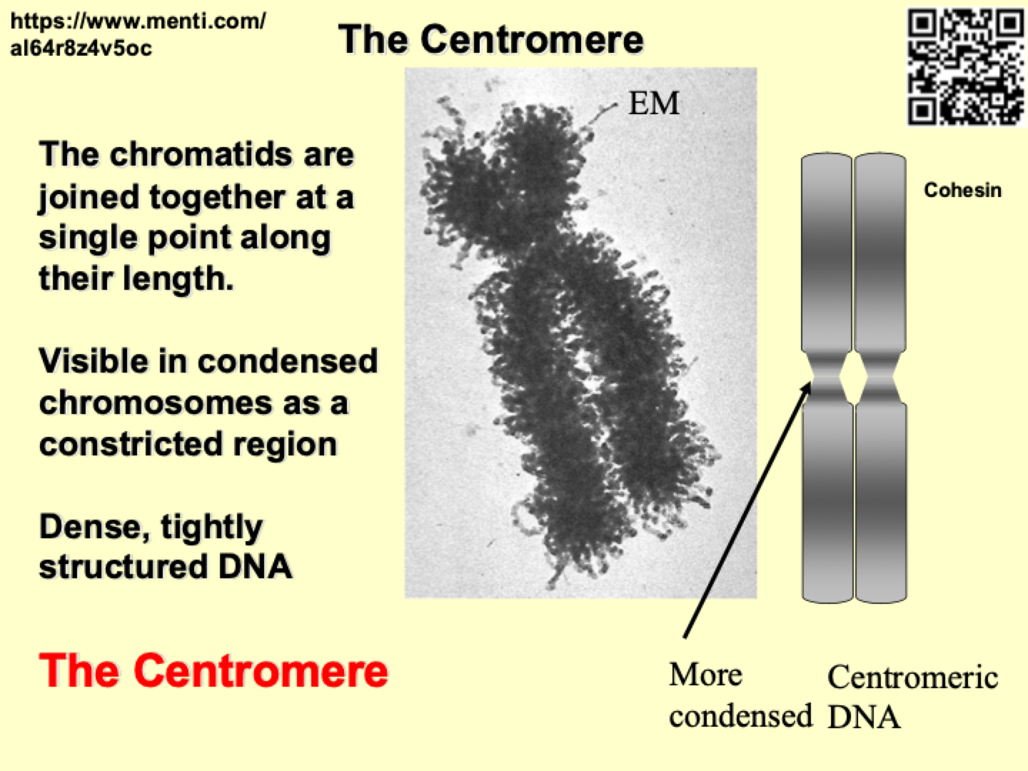
The centromere is the region where chromatids are joined together along their length.
It is visible as a constricted region in condensed chromosomes.
Composed of dense, tightly structured DNA, it is the most condensed part of the chromosome.
Devoid of genes, the centromere is primarily structural and consists of condensed inert DNA.
It has few genes associated with it, making it a key region for chromosome stability and function during cell division.
How does the kinetochore contribute to spindle attachment and chromosome movement?
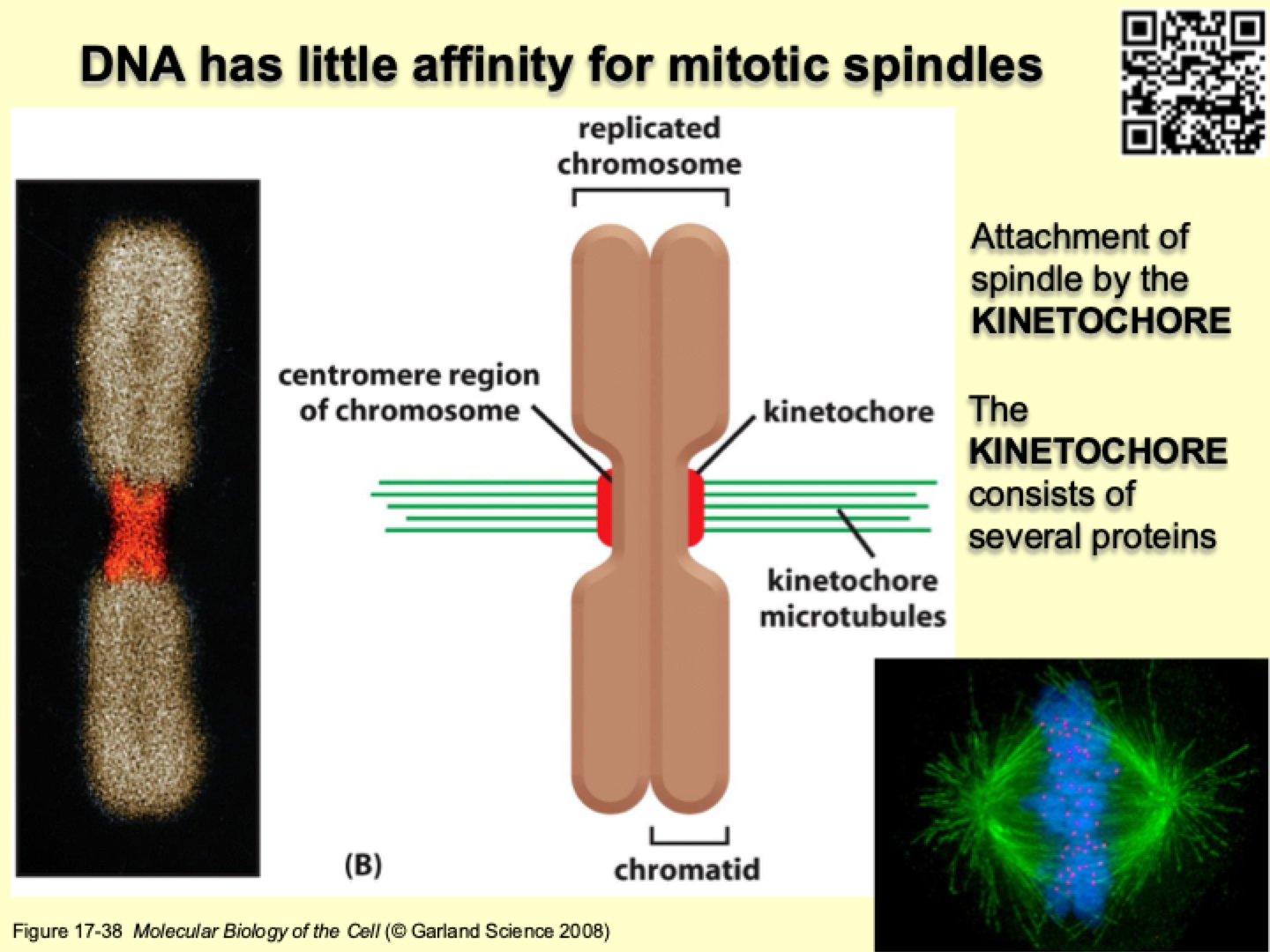
DNA has little affinity for mitotic spindles, so the kinetochore is responsible for attachment.
The kinetochore consists of several proteins organized into three layers:
Inner kinetochore: Binds to DNA and physically attaches to the chromosome.
Outer kinetochore: Binds to the ends of microtubules, anchoring them to the chromosome.
Checkpoint kinetochore: Acts like a ring that grips onto microtubules and holds them in place, playing a role in checkpoint regulation during mitosis.
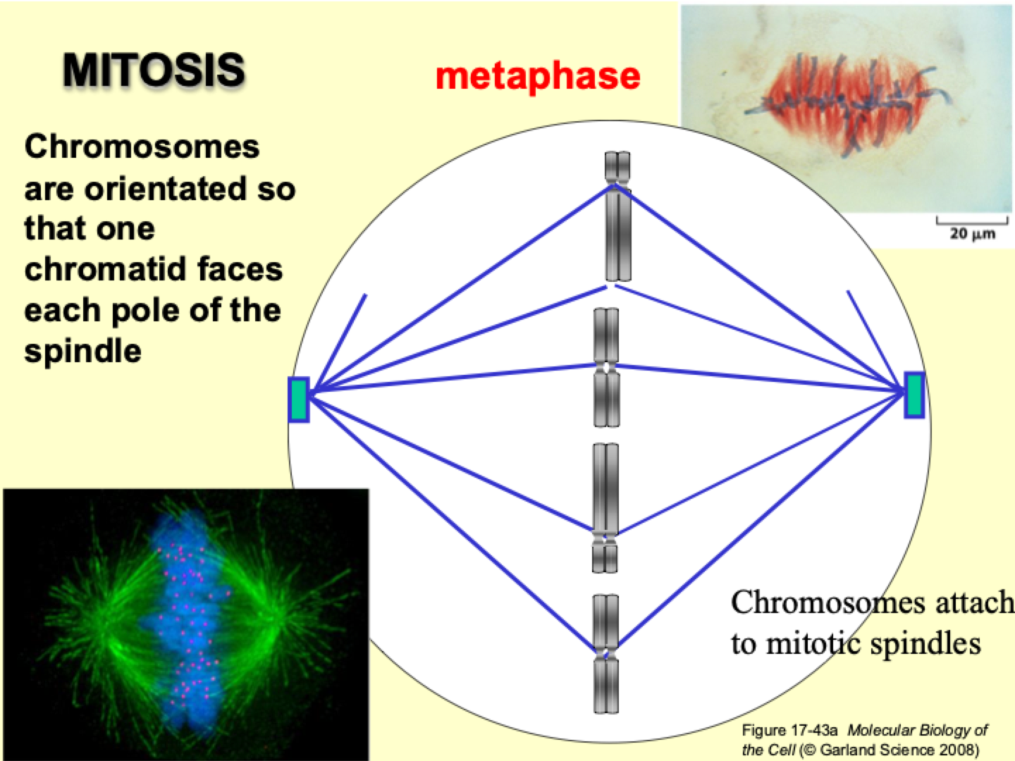
How are chromosomes oriented during mitosis?
Chromosomes are orientated so that one chromatid faces each pole of the spindle.
They attach to the mitotic spindle, and the spindle shortens or lengthens to align the chromosomes at the cell's center.
The kinetochore and kinesins enable the chromosome to “walk” along the spindle fibers, helping position the chromosomes properly for alignment during cell division.
What enzyme allows chromatids to separate?
Separase breaks down cohesin, allowing chromatids to separate between metaphase and anaphase.
What pulls chromatids apart during anaphase?
Mitotic spindle contracts, pulling chromatids to opposite poles.
Microtubules disaggregate by depolymerization at both plus and minus ends.
PacMan-Flux Mechanism: Kinetochore microtubules shorten as tubulin is removed from the + end, pulling chromatids toward the poles.
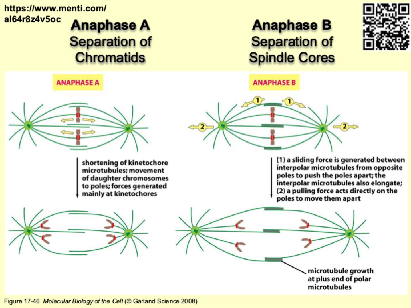
What are the key events in Anaphase A and Anaphase B?
Anaphase A: Kinetochore microtubules shorten, pulling daughter chromosomes to poles.
Anaphase B:
Interpolar microtubules slide apart to push the spindle poles apart.
A pulling force acts on the poles to move them further apart.
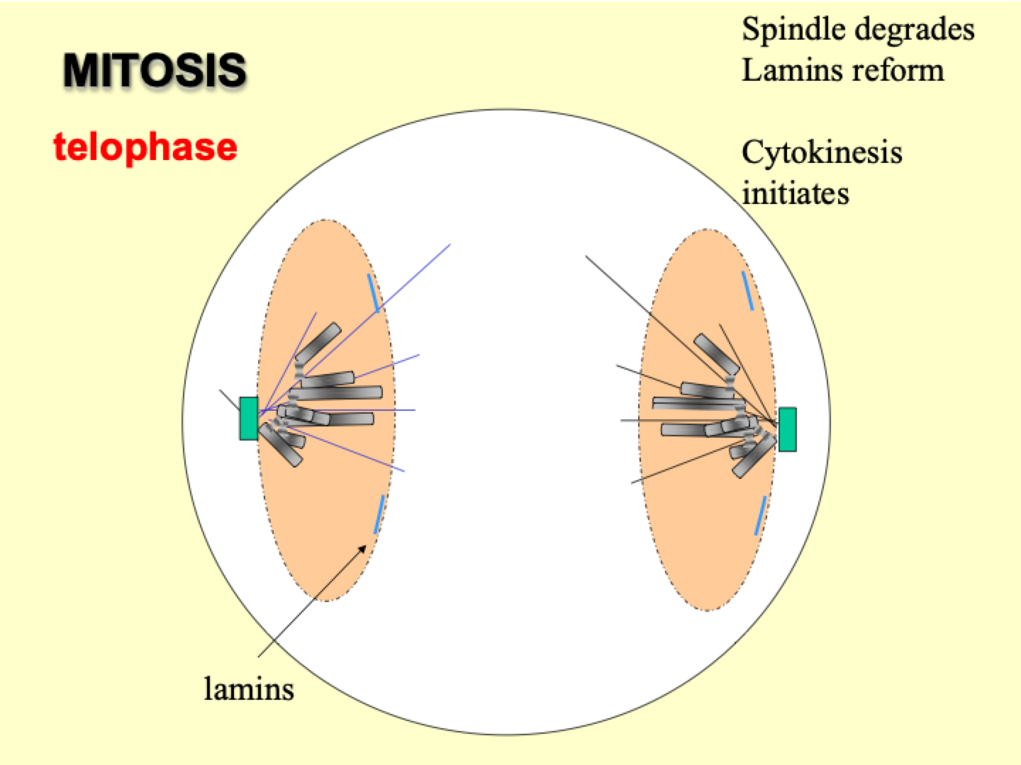
What happens during telophase?
Spindle degrades, nuclear lamina reforms, and cytokinesis begins.
What is the purpose of meiosis, and why is it important?
Meiosis generates gametes (sex cells) that combine to form a new organism.
It occurs mainly in reproductive organs (ovaries and testes in humans).
Rare compared to mitosis, but crucial for genetic variety and biological diversity.
Evolutionary significance:
Asexual reproduction is efficient when resources are abundant.
Sexual reproduction introduces genetic variation, helping species adapt to changing environments.
What are the stages of meiosis?
Prophase I: Leptotene, Zygotene, Pachytene, Diplotene, Diakinesis.
Metaphase I: Eggs are held at this stage.
Anaphase I
Telophase I
Prophase II
Metaphase II
Anaphase II
Telophase II
What is the purpose of meiosis?
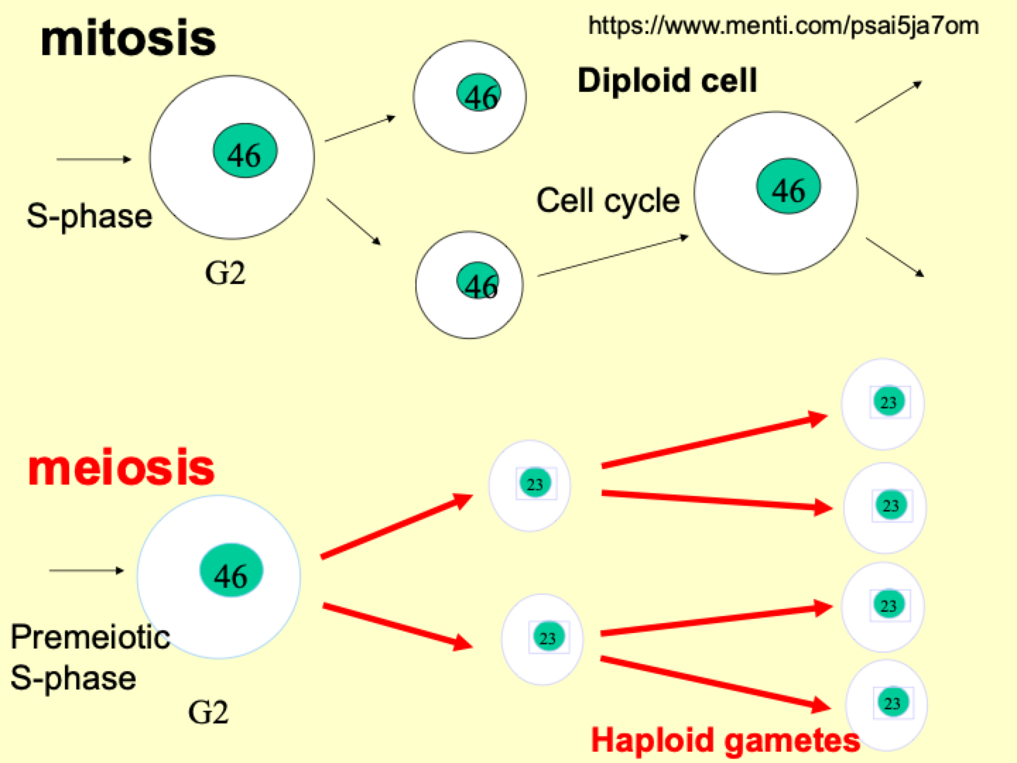
Reduces genetic content to half of a diploid cell.
Produces four haploid gametes.
Ensures each sex cell is unique through genetic recombination.
What happens in Meiosis I?
Homologous chromosomes pair up.
Crossover occurs, swapping DNA between chromosomes.
Creates genetic variety by forming hybrid DNA.
How is Meiosis II different from Meiosis I?
Similar to mitosis, but starts with haploid cells.
Sister chromatids separate, leading to four non-identical haploid cells.
How does meiosis differ from mitosis?
Meiosis: Produces genetically unique haploid cells.
Mitosis: Produces identical diploid cells.
Meiosis reduces chromosome number, mitosis maintains it.
What do diploid, ploidy, and C number mean?
Diploid (2n) = Two sets of chromosomes.
Ploidy = Number of complete chromosome sets.
C number = Amount of DNA in a cell.
Diagram of number of chromosomes in mitosis vs meiosis
What happens during Prophase I of Meiosis I?
Nuclear membrane breaks down.
Chromosomes shorten and become visible.
Centrosomes replicate and move to opposite poles of the cell.
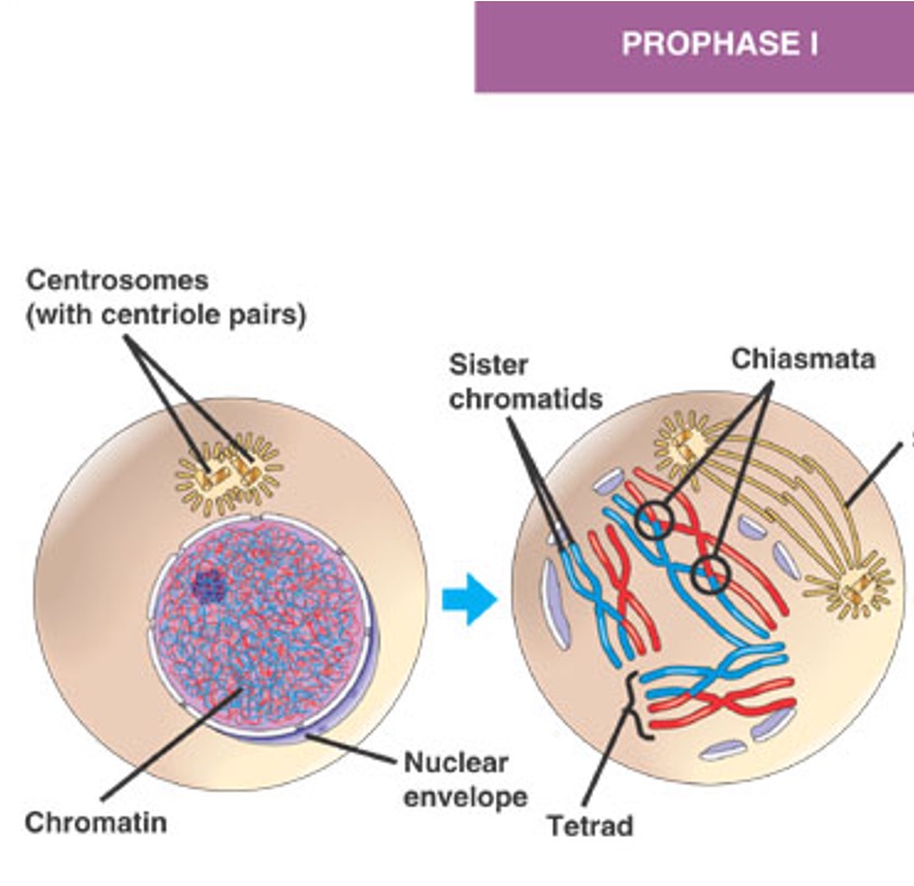
What happens during Bivalent formation in Meiosis I?
Chromosomes pair up to form bivalents, aligning next to each other.
Chiasma (plural: chiasmata) forms where the chromosomes physically exchange DNA.
Crossing over occurs: DNA breaks and fuses between homologous chromosomes.
This leads to genetically different chromatids.

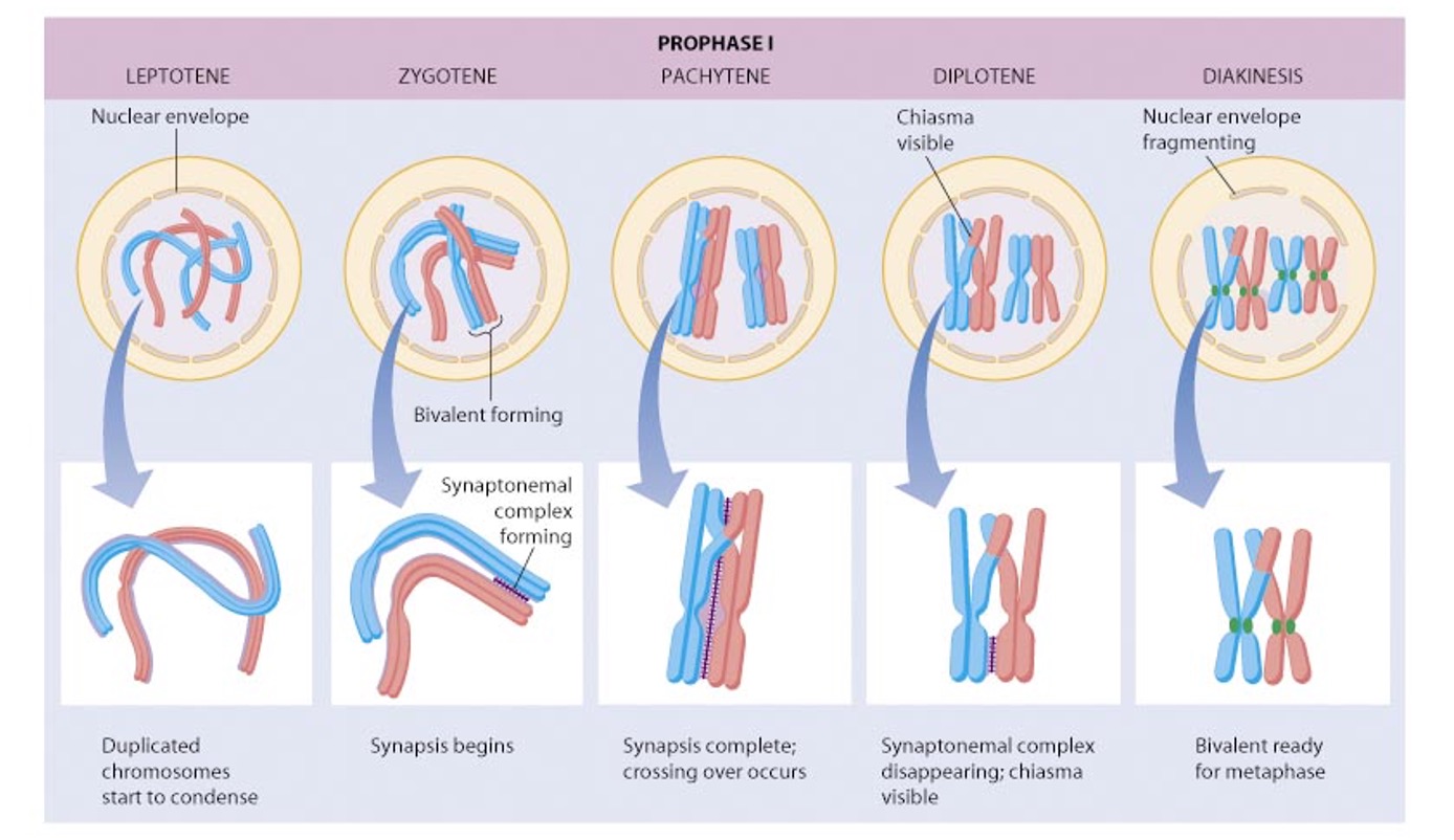
What are the stages of Prophase I in Meiosis I?
Leptotene
Zygotene
Pachytene
Diplotene
Diakinesis
Diagram of meiosis I prophase
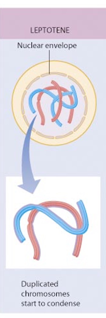
What happens during Leptotene in Prophase I of Meiosis I?
Chromosomes condense.
Nuclear membrane breaks down.
Chromosomes replicate and spindle formation begins.
What happens during Zygotene in Prophase I of Meiosis I?
Chromosomes align with their homologous pair.
The synaptonemal complex begins to form, joining homologous chromosomes together along the length of the chromatids.

What happens during Pachytene in Prophase I of Meiosis I?
Chromosomes align fully in homologous pairs.
Synapsis is complete, with chromatids joined along their lengths.
Crossover events occur, involving the breakage of DNA and re-fusion with the alternate chromatid.

What happens during Diplotene in Prophase I of Meiosis I?
The synaptonemal complex dissolves, leaving chromatids free.
Homologous chromosomes remain joined only at crossover junctions.
Bivalents/Tetrads become visible under the microscope.

What happens during Diakinesis in Prophase I of Meiosis I?
Bivalents associate with the meiotic spindle.
Chromosomes orientate in homologous pairs at the equator of the cell.
Each homologue is attached to one spindle pole.

How does crossover occur in Meiosis I?
The synaptonemal complex acts like a zipper, bringing homologous chromatids together.
Lateral and central elements bind chromatids, aligning DNA loops for recombination.
DNA physically breaks and fuses with DNA from another chromatid, swapping genetic material.
How do crossover events vary between chromosomes?
Bivalents are complex, with larger chromosomes undergoing more crossover events.
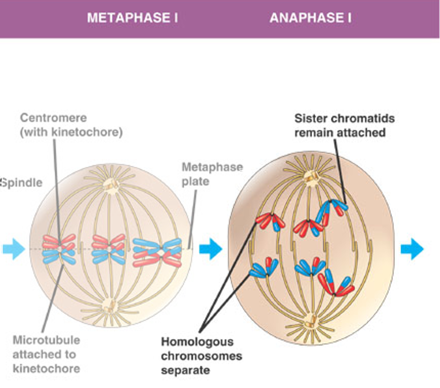
Homologous chromosomes separate during Anaphase I, but chromatids remain attached.
Why is crossover important?
Mixes parental DNA, increasing genetic diversity in offspring.
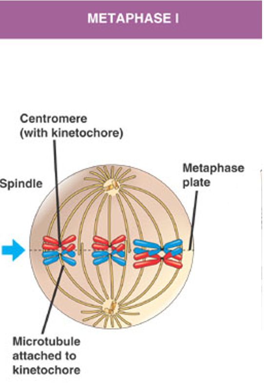
What happens during Metaphase I of Meiosis I?
Homologous pairs (bivalents) line up at the equator.
One kinetochore per chromosome attaches to the spindle.
In humans, most egg cells are suspended at this stage.
What happens during Anaphase I of Meiosis I?
Replicated pairs of chromatids (individual chromosomes) move toward the poles of the cell.
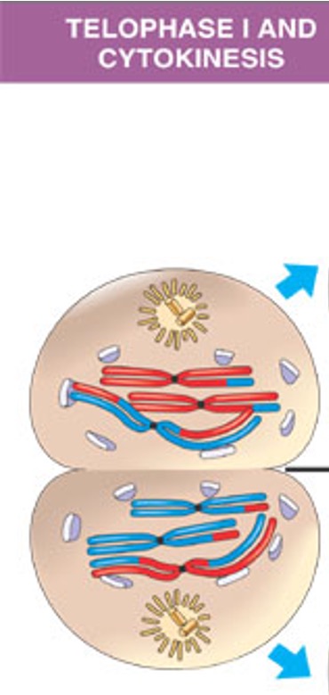
What happens during Telophase I of Meiosis I?
Nuclear membrane begins to reform.
Chromosomes decondense.
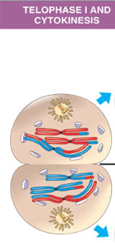
What happens during Cytokinesis after Meiosis I?
Cells divide, and chromosomes enter an interphase of varying length.
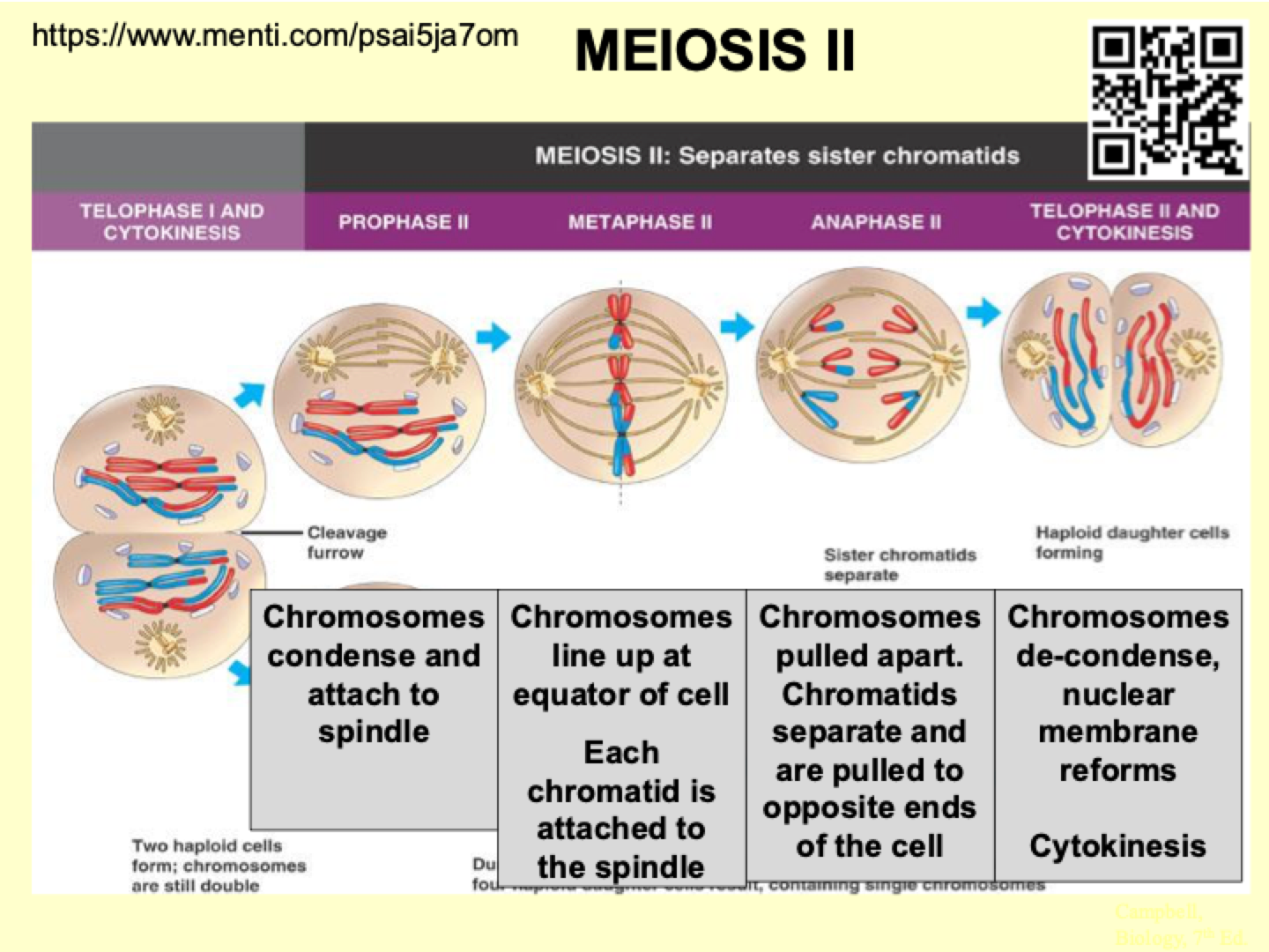
What happens during Meiosis II?
Prophase II:
Chromosomes condense and attach to the spindle.
Metaphase II:
Chromosomes line up at the equator.
Each chromatid is attached to the spindle.
Anaphase II:
Chromatids separate and are pulled to opposite poles.
Telophase II & Cytokinesis:
Chromosomes decondense, and the nuclear membrane reforms.
Cytokinesis divides the cells, resulting in four haploid daughter cells.
Comparison between mitosis and meiosis steps
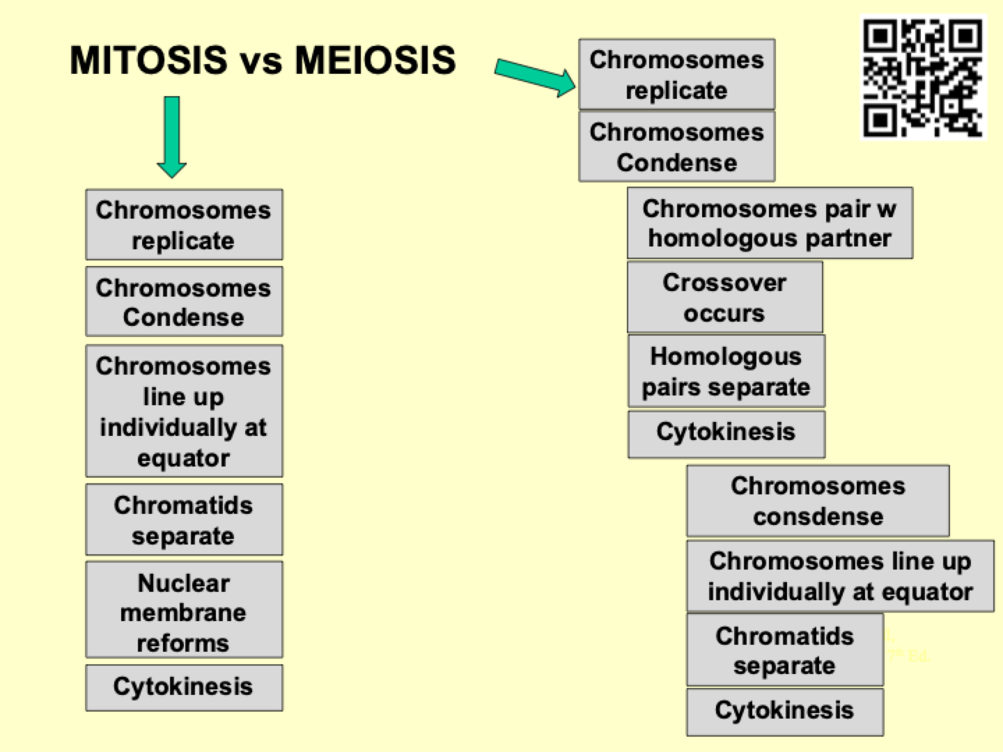
Diagram of chromosomes throughout mitosis vs meiosis
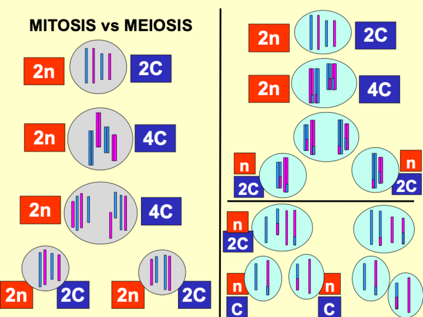
How many cells are produced in mitosis vs meiosis?
mitosis: 2 cells
meiosis: 4 cells
Are cells genetically identical or different from parent in mitosis vs meiosis?
Mitosis: genetically identical to parent
Meiosis: genetically different to parent
Are cells genetically identical or genetically different to each other in mitosis vs meiosis?
Mitosis: Gentically identical to each other
Meiosis: Genetically different to each other
Number of daughter cells in mitosis vs meiosis
mitosis: 2n
meiosis: n
C number in mitosis vs meiosis
Mitosis: 2C
Meiosis: C
What is Cytokinesis?
The final step of cell division.
Happens continuously in replicating cells.
Differs between animals and fungi.
How does the contractile ring function in Cytokinesis?
Forms during Telophase.
Made of cytoskeletal elements and motor proteins.
Wraps around the plasma membrane, tightening to split the cell.
How does membrane fusion occur in Cytokinesis?
The contractile ring tightens until the lipids in the membrane fuse.
Two daughter cells separate.
Some cells divide asymmetrically, especially in epithelial tissues.
How does Cytokinesis differ between animals and plants?
Animals: Microtubules form a contractile ring cleavage furrow in the middle of the cell, dividing it into two.
Plants: A cell wall forms, creating a partition between the two daughter cells.
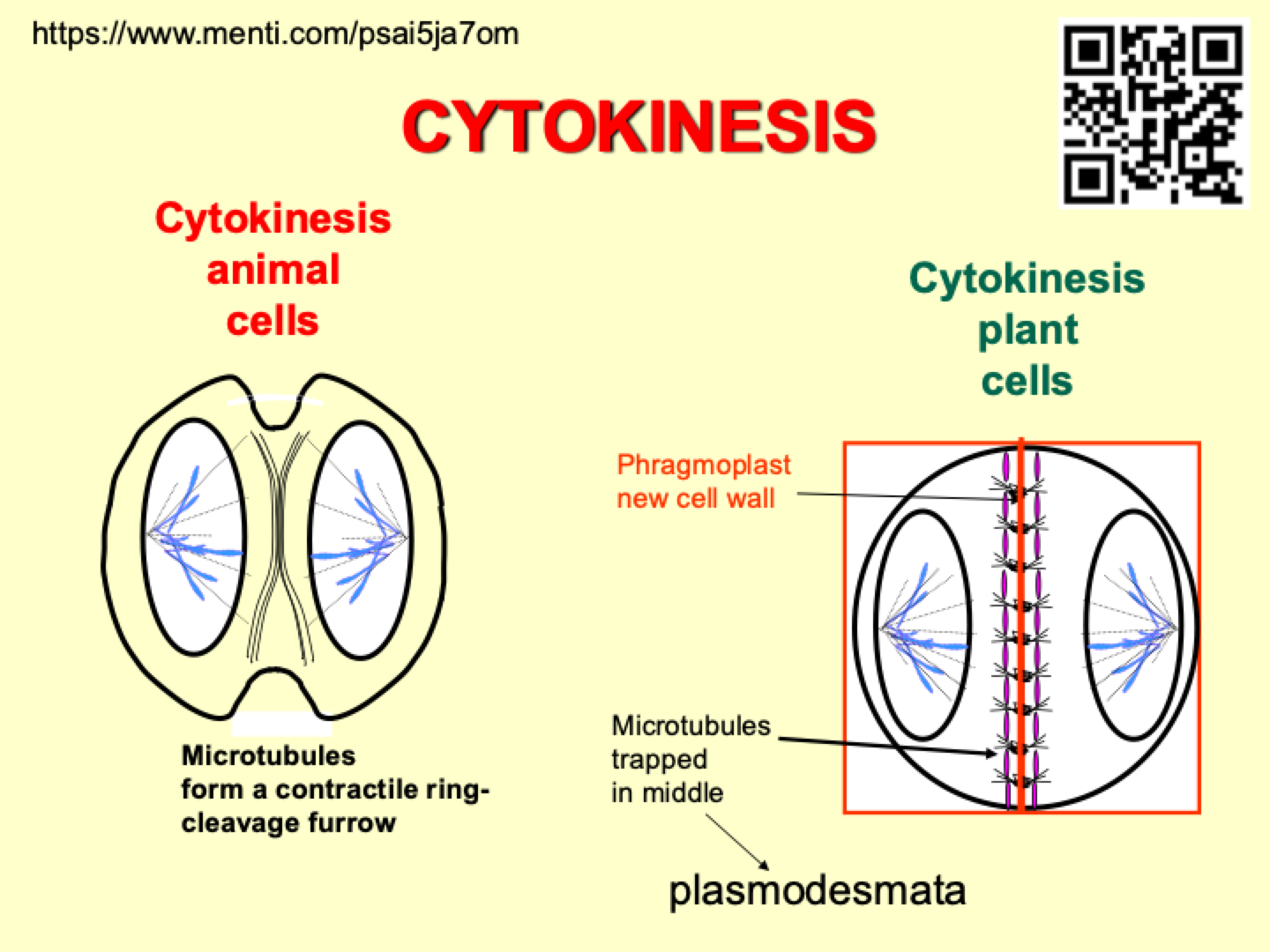
How does Cytokinesis occur in plants?
Cytokinesis in plants occurs by the construction of a dividing wall between cells, called the phragmoplast or cell plate.
What is the role of plasmodesmata in plant cytokinesis?
The wall between two cells has plasmodesmata, which are holes allowing connections and communication between cells.
What happens to the plant cell wall during cytokinesis?
The cell wall loosens, gains volume, and the cell expands.
Is Cytokinesis always equal?
No, Cytokinesis isn’t always equal.
Asymmetric cell divisions occur during development and are common during gamete production.
Does Cytokinesis always follow Mitosis?
No, Cytokinesis doesn’t always follow Mitosis.
In Drosophila fruit fly embryos, mitosis occurs without cell division, replicating several nuclei.
These nuclei form a syncytium, then migrate towards the plasma membrane.
The plasma membrane invaginates to trap the nuclei inside cells.
How is an organism formed?
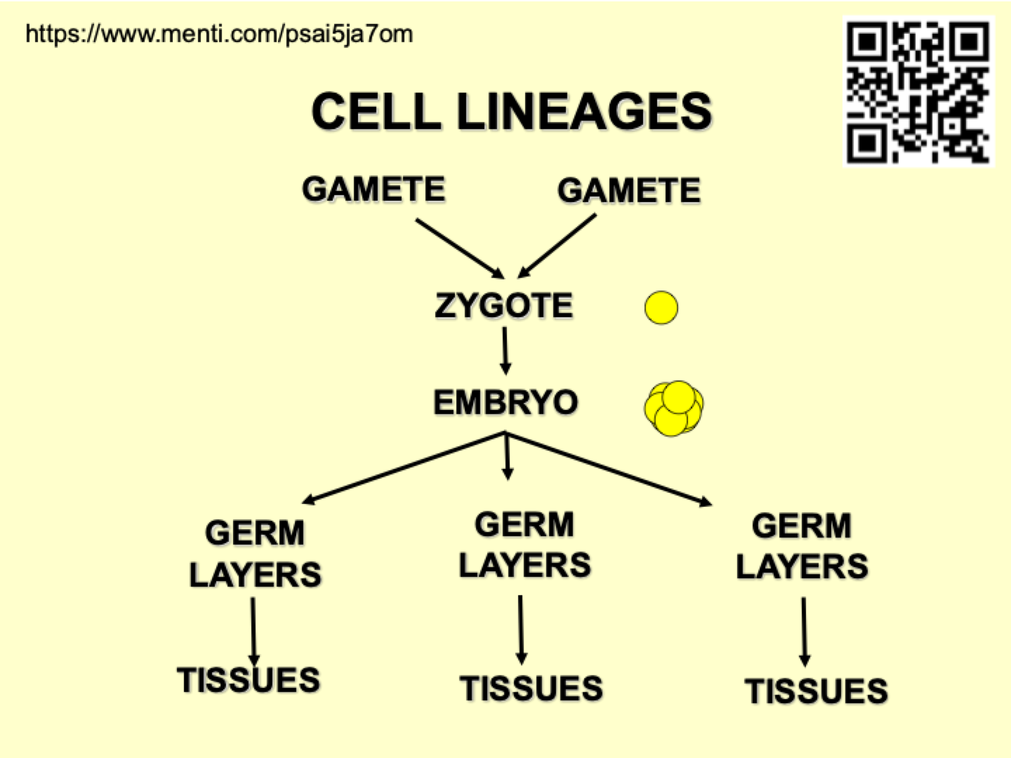
An organism is formed from 2 gametes.
The zygote is formed from the fusion of 2 gametes.
The embryo begins when the zygote starts dividing.
What are germ layers in development?
Germ layers are populations of cells that give rise to different structures in the body.
The three primary germ layers are:
Endoderm (inner layer)
Mesoderm (middle layer)
Ectoderm (outer layer)
What are stem cells?
Stem cells are cells that remain undifferentiated and are partitioned off from the embryo.
They have the potential to develop into various cell types.
Diagram of cell lineages
What are undifferentiated cells?
Undifferentiated cells require genetic determination to develop and differentiate.
They rely on gene expression, which is influenced by pre-existing mRNA and proteins.
How do undifferentiated cells differentiate?
Cells differentiate based on their position in the cell mass, environmental conditions, and pre-existing mRNA laid down during the development of the egg cell.
These factors help produce proteins, which in turn affect gene expression and guide differentiation.
What are the main differentiated cell types found in the epithelial lining of the small intestine?
The four main differentiated cell types in the small intestine epithelial lining are generated from undifferentiated multipotent stem cells near the bottoms of the crypts.
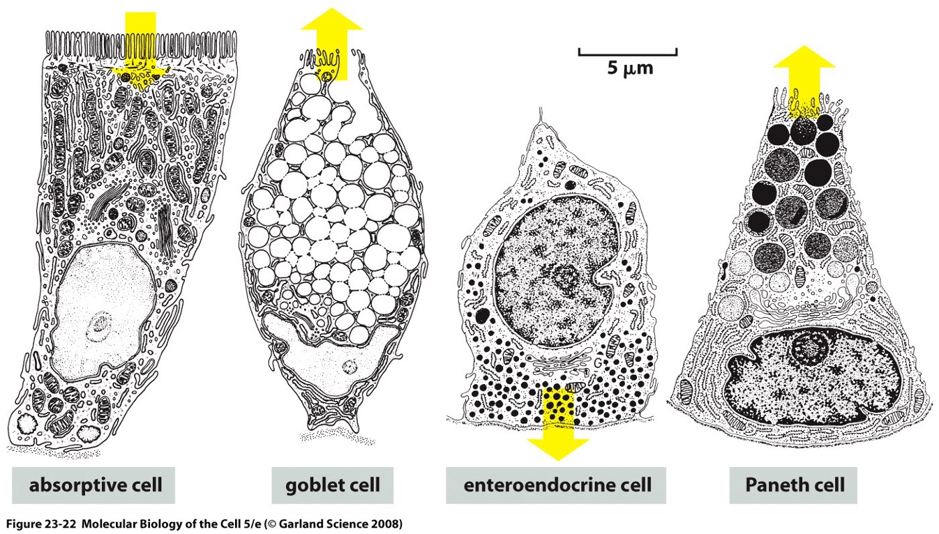
What role do microvilli play in the absorptive cells?
Microvilli on the apical surface of absorptive (brush-border) cells increase the surface area by 30-fold.
This aids in the import of nutrients and provides anchorage for enzymes involved in the final stages of extracellular digestion, breaking down peptides and disaccharides into monomers for transport across the cell membrane.
How do stem cells differentiate?
The zygote is the most undifferentiated cell.
As the zygote divides, different populations of cells start forming.
These cells become committed to differentiating into specific cell types.
This commitment is guided by the expression of different genes in each cell.
How do stem cells differentiate in the skin?
The skin is part of the ectoderm, which primarily forms skin cells and nerve cells.
The outermost layer of the skin consists of dead cells.
Stem cells at the bottom of the epidermis wait for signals to differentiate into skin cells.
When the surface layer of the skin is damaged, these stem cells differentiate to replace lost or damaged skin cells.
What are the different potencies of stem cells?
Totipotent/Omnipotent: Can differentiate into any type of cell, including the whole organism. Found in small number of cells (e.g., embryonic cells, zygote).
Pluripotent: Can differentiate into most cell types in the body, found in many areas of development.
Multipotent: Can differentiate into a wide range of related cell types, such as any type of muscle cell.
Oligopotent: Can differentiate into a few related cell types.
Unipotent: Can only differentiate into one type of cell.
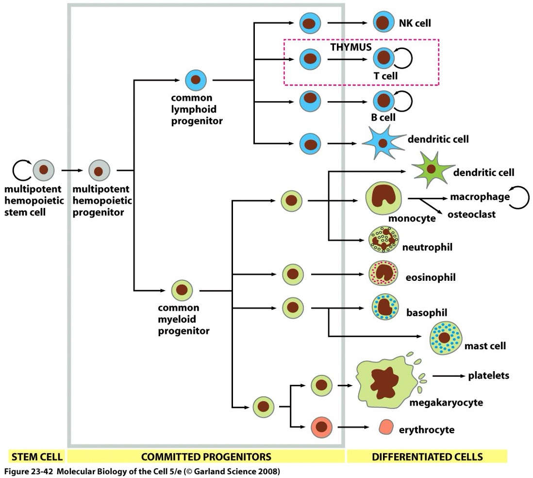
What is the process of hemopoiesis and how do stem cells contribute to it?
Hemopoiesis is the process of blood cell formation. It starts with a multipotent stem cell, which can either self-renew (generate more stem cells) or differentiate into committed progenitor cells.
These progenitor cells can divide a limited number of times before differentiating into mature blood cells.
As progenitor cells divide, they become progressively more specialized, narrowing the range of cell types they can produce (shown as branches in the lineage diagram).
Most blood cells are produced in the bone marrow, except for:
T lymphocytes, which develop in the thymus,
Macrophages and osteoclasts, which arise from blood monocytes,
Some dendritic cells may also derive from monocytes.
What are stem cell niches and what role do they play in stem cell regulation?
Stem cell niches are specific locations in tissues where stem cells are maintained.
These niches regulate the participation of stem cells in tissue generation, maintenance, and repair.
They protect stem cells from environmental damage and restrict over-active stem cells from proliferating uncontrollably throughout the organism.
Genetic and physiological cues within the niche help to maintain stem cell properties and regulate their activity.
What are somatic stem cells?
Somatic stem cells are non-germ cells found in the body that contribute to tissue regeneration and repair.
What are adult stem cells?
can be pluripotent or multipotent, depending on their location e.g. bone marrow
Where do foetal stem cells come from?
Foetal stem cells are derived from the early stages of the foetus or from accessory structures such as the placenta and umbilical cord.
What are amniotic stem cells?
Amniotic stem cells are undifferentiated cells found in amniotic fluid. They originate from the womb, umbilical cord, and embryo and have the potential to differentiate into various cell types. However, obtaining them can be risky for the foetus.
Where are stem cells found in plants?
Stem cells in plants are found in MERISTEMS, which contain undifferentiated, dividing cells.
What is the Shoot Apical Meristem (SAM)?
The Shoot Apical Meristem (SAM) is a dome of cells at the tip of a plant shoot. The inner layers are less differentiated, while cells become more specialized closer to the surface.
Can different parts of a plant give rise to stem cells?
Yes, in plants, stem cells can arise from various parts of the organism, not just from a single specialized location.
How do roots grow using stem cells?
Root growth occurs through two sets of stem cells:
One set near the tip produces the root cap, which is constantly scraped off as the root moves through the soil.
Another set behind it creates new cells, increasing the size of the root.
How do lateral roots form in plants?
Lateral roots arise from stem cell niches similar to apical meristems, allowing branches to develop from the main root.
Are all plant tissues totipotent?
Almost all plant tissues are totipotent, meaning a whole plant can be regenerated from just a few cells.
What hormones are involved in plant cell differentiation?
Auxin and Cytokinin play key roles in regulating plant growth and differentiation.
Where do embryonic stem cells come from?
Embryonic stem cells originate from early embryonic tissues, particularly the inner cell mass of a developing embryo.
Who was the first to culture embryonic stem cells and what was his contribution to genetics?
Professor Sir Martin Evans was the first to culture embryonic stem cells. He won the Nobel Prize for Physiology or Medicine in 2007 (along with Capecchi and Smithies) for his work using homologous recombination in embryonic stem cells to create genetically transformed strains of mice.
Why are stem cells considered more ethical than using animals in research?
Stem cells allow for research with fewer ethical concerns than using animals.
Only a few cells are taken from the early part of the embryo, not the full embryo.
Animal embryos are used to create early cell mass and blastocyst for research.
Stem cells can differentiate into different cell types, reducing the need for entire animals in experiments.
This approach minimizes the use of animals while still enabling vital research.
What is the process of generating iPS cells?
Take a fibroblast cell from a transgenic mouse.
Use a mouse transgenic for an antibiotic resistance gene linked to a pluripotency gene promoter.
Add genes to activate the promoter.
Select for cells with an active promoter.
Inject selected cells into embryos of normal mice.
Cross chimaeric mice with normal mice.
How do iPS cells work?
A differentiated cell (usually multipotent) is taken and dedifferentiated.
Stem cells are cultured and treated with chemicals that activate genes.
Genes help break down differentiation, making the cell pluripotent again.
These new pluripotent stem cells can then be reprogrammed into another cell type.
Why are iPS cells significant in research?
They bypass ethical concerns associated with embryonic stem cells.
They allow researchers to create pluripotent stem cells from adult cells.
However, they are not as effective as embryonic stem cells.
What are the main uses of undifferentiated cells?
Transformation – Genetic modification of cells for research or therapy.
Therapeutics – Potential treatments for diseases like Parkinson’s, diabetes, and spinal injuries.
Research (Cell Culture) – Studying development, drug testing, and disease modeling.
Tissue Engineering – Creating artificial tissues and organs for transplants.
What is CALLUS tissue in plant transformation?
Callus tissue consists of undifferentiated plant cells used for genetic modification.