CHAPTER 3 n ervous sysstem from text
1/63
There's no tags or description
Looks like no tags are added yet.
Name | Mastery | Learn | Test | Matching | Spaced | Call with Kai |
|---|
No analytics yet
Send a link to your students to track their progress
64 Terms
Neurons
structural and ucntional units of the wholenervbous system. they are specc ialsed cells at are desaigneed for rapid communication fo the body
Cell body
is the part of the nucleus that contains the nucleus.responsible for controlling the cucntionning onf the cell.
qaroudn the nuclues is the cytoplasm wiht many of the normal cell organneels
Dendrties
fairly short extensions of the cytoplasm of the cell body.
They are often highly branched and carry messages or nerve impulses intio the vcell body

Axon
a single long extension of the cytoplasm of the cell body of the neruon. and it carries nerve ipmpulses away form the cell body. they are usually longer than dendrites but their size can vary uipon their location lkike in the brain it is samller compared to thespinal cord going towards the legs.
At the end of the axon it diviedes into man yusmall branches each of these branches terminates at the axon terminal

Myelin sheath
most axon are covered inthis fatty t issue called a myelin sheath.

Nerve fibre unmyheline ated and myelinated aswell as others
is used for any long extension of the nerve clel l but usually the axon and dendrites. thsoe
that have a myellin sheat is called myelinated fibres and the ones that do not are said to be unmyelinated
when nerve fibres arre goupred together outside cns they fomra nerve. n
nerve fibres are hled together into bundles by connective tissue with mulitiopoole bundles formign a nerve.d
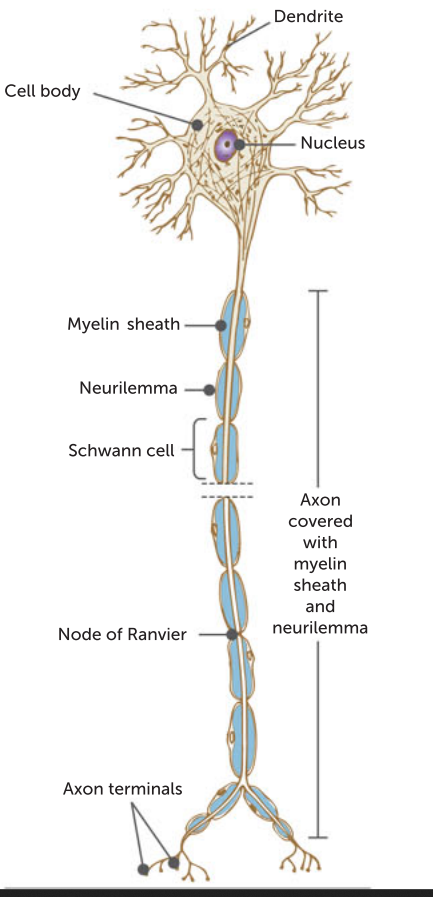
Schwann cell
outside the brain and spinal cord, the myelin sheate is produced by special cells called schwann cells whic wrap aroudn the axon.
At intervals they are grsp in the myelin sheath called the nodes of ranvier

3 functions of myelin sheath
It protects the axon
it insulates the axon
It speeds up the rate of travel of nervce impulses along the axon
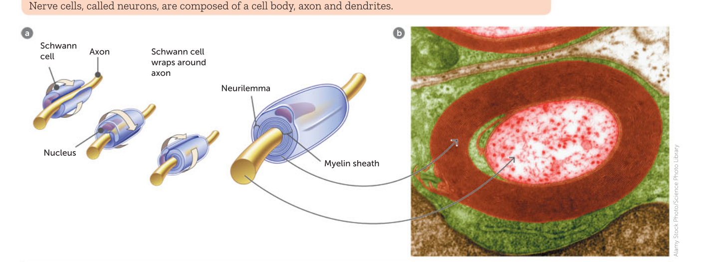
Neurilemma
the outermiost coil of the scwann cell forms a structure called the neurilemma.
it helsp in the repair of injured fibres

oilgodendrocytes
meylin sheath is produced inthe brain and spinal cvord by thius structure
fatty nature of myelin means that the areas containing myelinated fibres appear white and are called white matter.
areas made up of cell bodies and uynmyleinateedd fibreas are called grey amtter due to their g rey colour
Synapses
Nerve impulses must be passed from neuron to neuron.
where one axon terminal joinss witha dendrite or cell body fo antoher.
this juntion is called the snyapse1
neurotransmitters
neyrons do not physciall th=ouc hthere is a gfap in the suynapes.
mewssages have to be carried acros this gap which occurs through the movement of chemical called nuerotransmitters
neuromuscular junction
the simalr synapse whre an axon meets a skeletal muscle cell.
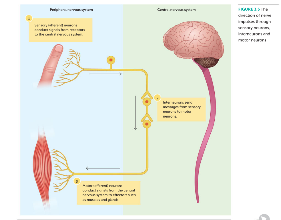
3 fucntional types of neurons list
sensory ( afferent) carry messages from recpetors to cns
motor (efferent) carry messages from cns to effectors liek glands muscles
interneurons - located inside of cns and are the link between sensor yand motor neurons. also called associaten, connector and realay neruosn
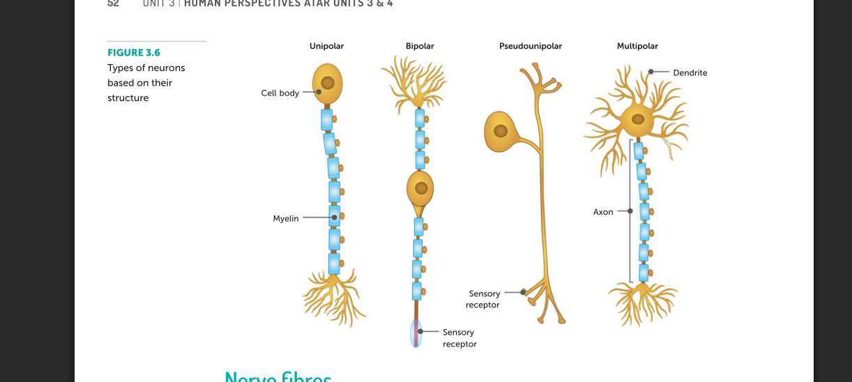
3 structural tyeps of neurons
multipolar neurons - one axon and multiple dendrites form the cell body. these types are most common and include most of the interneurons in the brain as well as motorn neurosn that carry messages tro skeletal msucles
bipolar neurons - have one axon and one dendrite. common in areas like the eyes or nose where they take impulses form ereceptor cells totoehr neurons
unipolare is only a singluar exxtenison which is the axon. its not found in humans or other vertebreates on lyh in insects
pseudounipolar is when there si a single axon which ten separates into two extenstions . one connects to dendrites while the otehr terminates at the axon terminal. most sensory neurons are this type
Potential differenc
the force tghat pulls charges when they are separateed has the potential to release energy if they come together this canvbe measurein voltage
Extracellular vs intracelluls
extracellular has high concentration of siudm chlroide and osmosto fits charge particales are positive sodium ions and negative chloridions
the lfuid inthe intarcellualrfluid has kplus and negatively chatrged subttacnesr releeeased by organnelles
Membrane potential
differnece s in the concenteentrations outwsdie membranes is known as membrane optential,
its inall body cells but mostly in the nerve nad muscle cells
REsting membrane poteinial
of aan unstimujlated nerve cell itis around negative 70mV.
Ions diffusal mehtods
Leakage channesl are open all the times wehereas voltage gate channels are onlyu open when the nerve is stimulated
concentrations of ions in and out
sodium 10 times higher outsidenthan inside. celle mmbembrane on ly slightly permeable ans there is limited aomount of sodium elakeage channes limits facililated diffusio oif sodium ions
potttasium ions 30 timesgreater insdie than outside but lot more pottassium leakgae channels
chloride higer outside thatn inside with cell mbmeraen highly permeable to chlroide ions through prtoein channels
organic ions higher inisde and cell membrteane is impermeable so they stay inside the cell
Sodium pottassium pump
this is a carrier protein that moves 3 sodium ions out of cell and 2 pottasium ions into the cell. therefore there is a net reduction of positive ions inside the cell. this is against the gradient and therefore uses atp
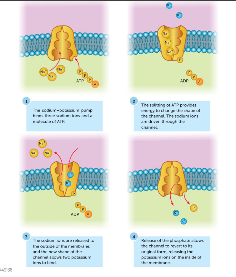
how is resting membrane potential maintained
qdiffernece inthe nubnmer of leakage channels for the sodiu madn pottasium.
\the membrean ebeing impermeable to large orgnaic ions insde the cell
sodium pottassium pump always workign to mainting lower positive ion concentration inside
Action potential
if a stimulis to the neruon is sufficient enought teh voltage gated channels open and close causing the rapid depolaristiaon and repolaristion fothe membrane
Depolarisation
sudden increase inteh membrane potential . this occurs ithe level of stimulatin exceeds 15mV or the threshold.
so when a neuyrotransmitter oi r a sensory recptor stimulates a neuron. some sodium channesl are open these are e claled the LINGAND GATED CHANNELS and they move more doium into the felll makign the potential differene higher
If this levl increassed poitential dfiffernct to -55 then the volatge gate d sodiumchannels open. tihis produces a movemnt of soidum ions into the cell that prodceds independatnley of the stimulus.ALL ORNONE RESONse.
exceeds the otward movment of pottassium inons and mkes hte inside morepositive thatn outsdie and increaserd to 40 mV.
Repolarisation
this is when sodium channels close at the same time pottasium channesl open increasing flow of pottasium ions out of the cell
repolarised but then the pottasisum remains open for lognert thanneeded and this ause thpotential to drop lower thean resting and this iscalled hyper polarisation .
Refractory period
once the sodium channels have opened they quickly becom inactive and wont repsond to stiujkulas.
lasts from -55 mev or thryheshodl till it reaches the resting membrasne potential
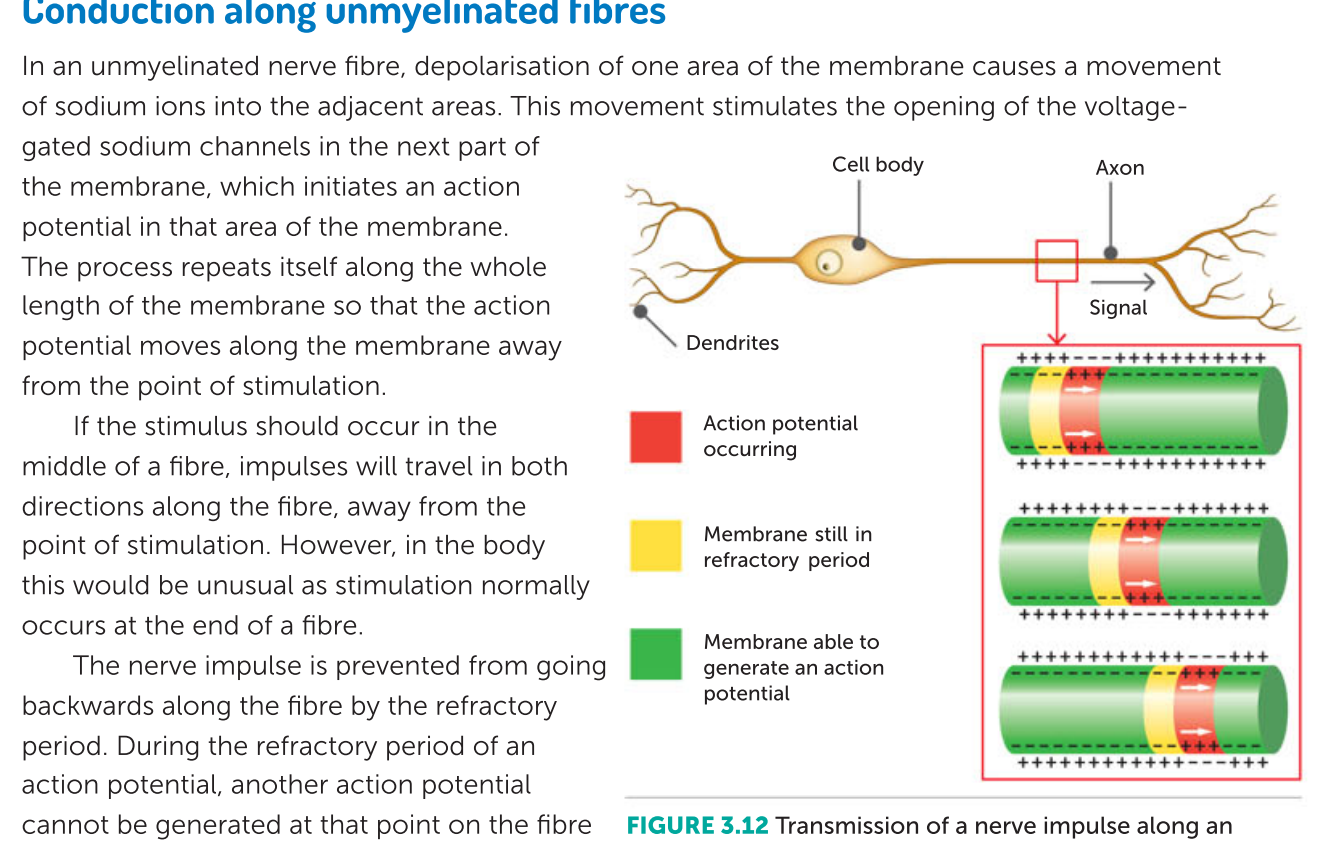
conduction on unmyelinated fibres
cvonduction in myelinated fibrews
the myelin sheat insulates the nerve fibre from extracellular fluid.
hwoever the nodes of ranview are exposed therefore ions cannot flow where theres the myelin sheath.
instead the action potential jumps from node to node this is callleed the jumping conduction known as saltatoryu conduction.
it allows it to travel much faster up to140meters per second whereas unmyelinated fibre max speeed is 2 meters per second
Difference between strong and small stimulaus
a stong stimus causes depolaristion of more nerve fibrese than a weak stimulus.
ansd a strong stimjulus produces more nerve impulses in a give time
Process for transmission acrossa synapsw
1 -when nerve impulse reaches axon terminal it activatersa c oltage gated calcium ion channel
2- as there is a highger concentration of calcium ions in the extracellular fluid they flow into the cell at the pre synaptic axon terminal
3 -this causes synaptic vsicles to fuse with the membrane, releasing special chemicals called neurotransmitters by exocytosis
4- the neurotransmitters diffuse across the gap and attach to receptors on the membrane of the next neuron
5-this stimulates the ligand gated protein channesl to open which allows the influx of sodium ions and inititaes an action potential in the post synaptic membrane
Effect of chemicals on the transmission of nerve impulses
there are many chemicals both natural and synthetic that influence the transmission of nerve impulses mostly at teh synapse or the neuromuscular juction
stimulants like caffeine and benzedrine stimulate transmission at the synapse.
Other drugs like anaesthetics or hypnotics depress the transmission
Venom can also affect the neuromuscular junction
Nerve agents contain organophosphates which cause the build up of acetyl choline at the nueromsuclar juction. adn the overstimulation of contractions leading to repsiratory failure
Receptors
Receptor able to detect a change in the body’s internal or external environment.
Thermo, osmo,chemo, touch and pain receptors
Thermoreceptors
they are able to resond to heat and cold. skin thermorecepotrs inf orrm the brain of changes in the temperature outside the body. In this way we are consciously aware fo the temperature of our surroundings
the temperature inside the body, the core temperature, is monitored by thermoreceptors in hypothalamus , which detect the temperature of the blood that is flowing thrugh the brain
using info from the skin adn its own thermoreceptors the hypothalamus can regulate the body temperature
osmoreceptors
osmotic pressure is determeinged by the concentration of substacnes didssoved in the wter of blood plasma. the hgiher the conecentration, the greater the osmotic pressure.
Osmoreceptors are located in the hypothalamus and are sensitive to even very small changes in osmotic pressure.
they can stimulate the hypothalamus so that the bodys water content is maintained within very narrow limits.
Chemoreceptors
are stimulated by particular chemicals. They are present in the nose and the mouth making us sensitive to odour and tastes.
Internal chemorecepotrs that are senesitive to the composition of body fliuds
chemorecepotrs in blood are important as they are sensitive to the pH of theblood and the concentrations of o2 and co2. these chemoreceptors are involved in h
Touch receptors
are found mainly in the skin
some are close to the surface and sensitive to very light toucho f the skin. Greater conc in places like the lips finger tips and eyelids and external genital organs.
nerves on the ahir adapt rapidlyl like when we wear cloths all day but feeels weird when we first put it on
others are deeper adn are sensitive to eressure adn virbrations
pain recepotrs
are stimulated by damage to the tissues such as from a cut or a heavy bump or poor bloodflow or excessive stimulation such as from heat or chemicals
they occur in most organs but not in the brain. conceentratred though in the skin and mucous membranes
essential forou r welebeing
pain recepotrs adpt little or not at all they keep stimulating pain until tissue damaging situation is stopped
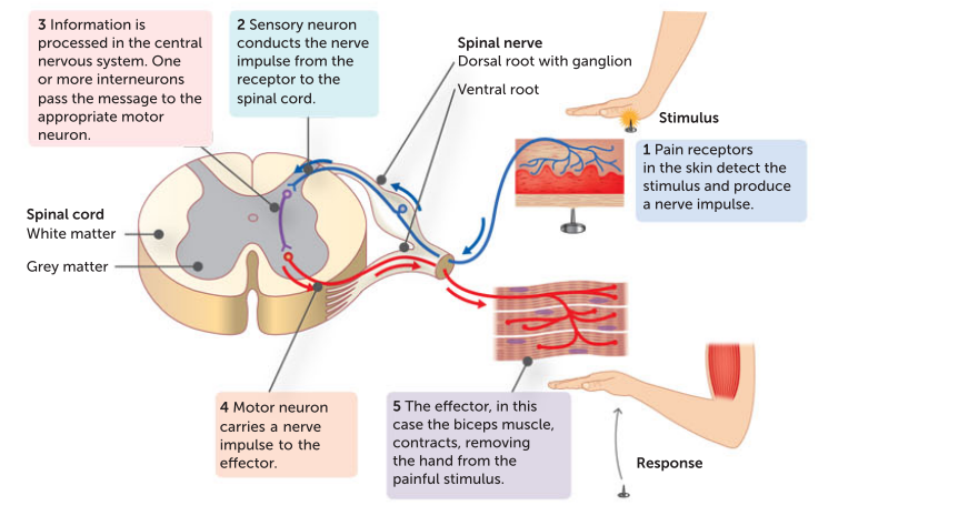
Reflexes - properties
a reflex is a rapid automiatc respons se to a changei nthe externawl or internal environemnt.
a stimulus is required to trigger a reflex
it is involutary and it occurs wihtout an y consiccou thorugh
a reflexx is rapid - on ly a small nubmer orf neuorns involved
a reflex is stereoytyped maeaingin t occurs in the same way each time
reflex arc def along with spinal
reflexses may be conducted by the spinal cord alone is called a soinal reflex.
the pathwya is known as a reflex arc where ta nerve impuls ttravels from a recpetor to an effecotr. or in the spinal refelx its the spinal reflex arc
REFLEX arc basic componnents
a recepotr reacts to the change in internal or external enbironemnt by intitiating a nerve impulse in the sensory neuron
a sensory nueron carries impulses from the receptor to the spinal oord or brian
there is atleast one synapse: the nerve ipulse mayb e passe d directly to a motr neurons, or theyre may be one ore more interneuerons that direte htimpulse to the correct motorr nueruon
a motor neurokn caries the nerve impulse to the effeectors
an effector reveiegs the nerve impulse and carries out the arpportperate repsonse are either muscel cells or secretory cells
Learnt reflexes
protective reflexes are present from birth more compelx like suckling and chewing are during a bbabys developmetn adn they are innate refelexes whwihc are determined genetically
some complex motor patterns are learnt and are called acquired reflexes like balancing bike
Differences between nervous and endocrine system
nervous response reapid and hormonal slower
stimulus ceasese then nnerveimpulses stop almost immediatesly, hormone repsonses can last a long time ven after finisih
nervous messages are electrochemical change t hat travel alogn the membrane of a neuron. endonccrin messages are chemicals that are transbpotrted by the blood
nerveimpulses trave l along a nerve fibre to a sepcicfic part of the body and typically influenfe only one ffector bwhiel homrones travel to all paorts ot f the body and are aaarried by teh blood and they can often ffect a number of diffrent organs
similarities between nervous and endocrine system
some substances fucntion as both hormones and neurotransmitters such as noradrenaline and ADH and dopamine
soe hormones such as oxytocin and adrenaline are secreted by neurons in the exgtracellular fluid
some hormones and neurotransmitters ahve the same effect on target cell
Cranium and vweretebera - form of protection
cranium which is the part of the skull that houses the brain, is the bone that protects the brain. the vertebrae is the bone that protects the spinal cord
Meninges a
are inside the bones and cover the surface of the brain and spinal cord (entire CNS) there are 3 layers of connective tissue forming membranes called the meninges
OUTER Meningeal layer which is the DURA MATER. it is touch and fibrous and therefore provides a layer of protection for the braink. It sticks close to the bones of the skull but on the inside of the vertebral canal it is not so close. SIMILAR TEXTURE TO A HOUSEHOLD RUBBER GLOVE
MIddle layer called the arachnoid mater is a loose mesh of fibres
inner layer the pia mater is far more delicate and contains blood vessels and sticks closely to the surface of the brain and spinal cord.
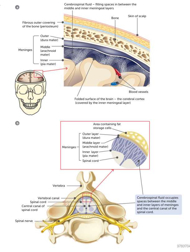
Cerebrospinal fluid
it occupies a space between the middle and inner layers of the menignes.
it also circulates thorugh cavities in the brain and through the central canal in the spinal cord.
CSF is a clear, watery fluid containing a few cells and some glucose, protein, urea and salts
CSF 3 functions
protection as it acts as a shock absorber cushioning any blows or shocks the CNS may sustain
support : the brain is suspended inside the cranium and floats in the fluid that surrouds it
transprot : the csf is formed from the blood and circulates around and through teh CNS before evenetuall y reneetnerign the blood capillaries./ DUring its ciruculation it takes nutreints to the cell s of the brain and spinal cord and carries away wastes
Cerebrum
the biggest part of brain..
contains outer sruface of 2 to 4 mm thick of grey matter known as the cerebral cerebral cortex. they grey matter consists of neuron cell bodies, dendrites and unmylelinated axons.
below the cortex is white matter which is madee up of myelinated axons, the fatty nature of myelin gives its colour and textture. deep inside the cerebrum is additional grey matter called the basal ganglia
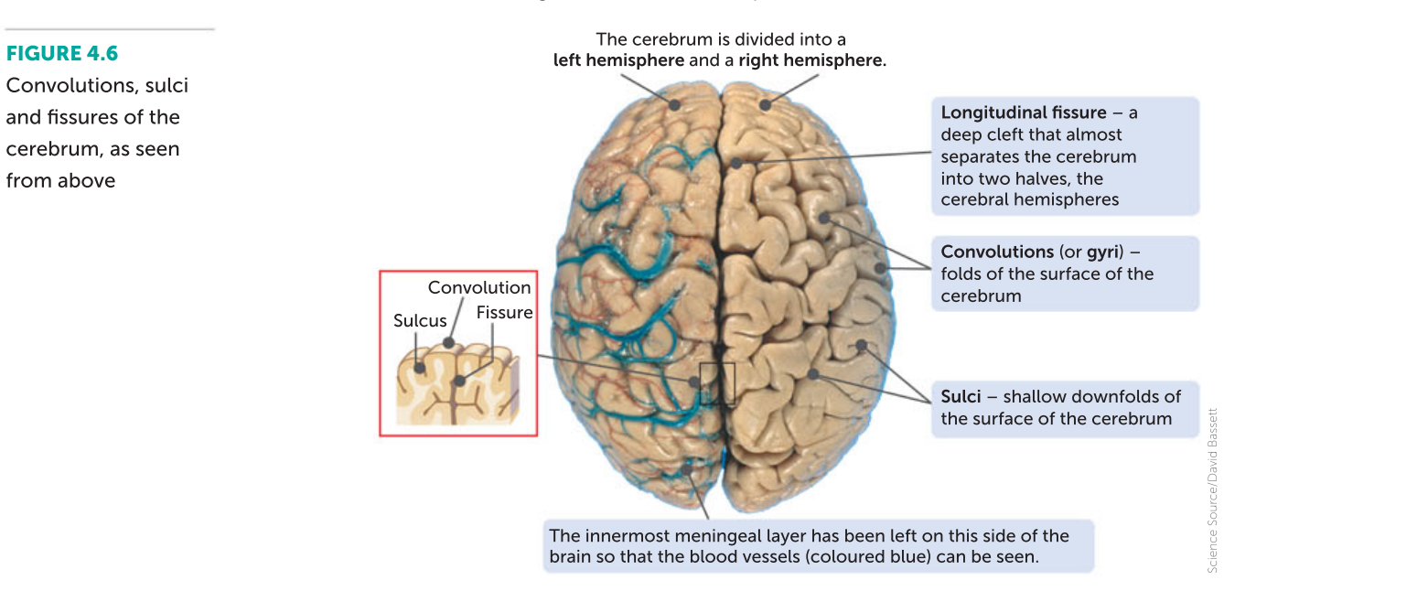
cerebru mcortex fold
increases its surface area and there fore the cereubrm corttex ontains 70 ercent of all the neurons in the CNS. the convolutions are separated by either shallow downfolds called sulci or deep downfolds called fissures
longitudinal fissure serpates teh cerebrum into 2 halves teh left and right cerebral hemispheres. Joining the two hemispheres at the base of the longitudinal fissure is a large bundle of transverse fibres known as the corpus callosum
Frontal lobe
forward thinking, problem solving, and control of movement and emotions

parietal lobe
processing temperature touch taste pain and movement

temporal lobe
processing memories and linking them with senses, receives auditory information
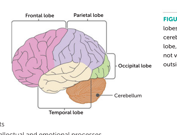
occipital lobe
vision

insula
re cognition of different senses and nemotion and addiction and psychiatric disorders

corpus callossum
is a wide band of nerve fibers that lie underneath the verebrum at the base of the longitudinal fissure. nerve fibers cross form one hemispehre to the other allowing the two sides to communicate
cerebellum
lies under the rear part of the cerebrum. It is the second largest part of the brain
outer folds are gre y matters and the inside is wehite matter that branches to all lparts of the cerebellum rather like brancehs of a tree
Controlv oveer posture, balance and fine coordination of voluntary muscles. to carry out these funmcthion the verebllulm reviecves sensor yinfomration from the
inner ear for info on posture and abalance and stretch receptors in the skeletal muscles for information about the length of muscles
hypothalamus
lies in the middle of the brain and cannot be seen form the outside .
maintains homeostasis regulation fo the autonomic nervous system like heart rate blood pressure and the secretion of digestive juices
bodyt temperature
food and water intake
contraction of unirary bladder
secretion of hormones and coordination of parts of parts of the endocrine system; acting through the pituitary gland, the hypothalamus regulates metabolism, growth, reproudction and repsonses to stress
medulla oblongata
is a continuation oc the spinal cord
contains the cardiac center which regulates the heart rate and force of heart besat
respiratiory center whcih controls the rate and depth of breathing
vasomotor ceneter whic hregulates the diameterf of blood vessels
Under the influence of mostly the hypothalamus
spinal cord
contains gray matter in the middles in the shape of the letter H. there is a small space called the central canal which runs the length of the spinal cord and contains cerebrospinal fluid
the myelinated fibres of the white matter are arranged in bundles known as ascending and descending tracts
ascending are sensory axons that carry impulses upwards towards the brain
descending contain motor axons that conduct impulses downwards away from the brain