3.2.1.3 - methods of studying cells
1/54
There's no tags or description
Looks like no tags are added yet.
Name | Mastery | Learn | Test | Matching | Spaced |
|---|
No study sessions yet.
55 Terms
what is the magnification formula
image size / actual size (remember I AM triangle)
order of units
m, cm, mm, µm, nm
how to calculate just the magnification of a diagram from a scale bar
image size = found by measuring the ACTUAL length of the scale bar on the sheet, using a ruler. convert to mm to make calculations easier
actual size = is the length given by the scale bar
as magnification = image / actual, do image / actual with these values. make sure they are in the SAME UNITS!
after calculating magnification of a diagram from a scale bar, how do you find the actual size of the diagram?
actual size = image size / magnification
so measure image size of DIAGRAM (not the scale bar, like before) using a ruler
then do image size / magnification (that you found before!)
what is the resolution of microscope
the minimum distance apart that two objects can be in order for them to appear as separate items
does increasing magnification always increase resolution
no
bc every microscope = has a limit of resolution
up to this point, increasing mag. = will reveal more detail
but beyond this point, increasing mag. = will not do this. object may appear larger, but may just be more blurred
what is an eyepiece graticule
an engraved ruler visible when looking through the eyepiece of a microscope
does the eyepiece graticule stay in the eyepiece
yes
does the eyepiece graticule change depending on the magnification?
no - it remains constant
but it does need to be calibrated every time the magnification is changed, i.e. every time the objective lens is changed (because the size of the specimen has changed. we need to work out what the distance between each division represents at THIS magnification)
does the eyepiece graticule have a known scale / known values?
no
the scale can be found by c___________ the graticule
what can this be done with
calibrating
a stage micrometre
what is a stage micrometre
a piece of graph paper with KNOWN values
it is required to calculate the eyepiece graticule
placed on the stage of the microscope
how would you use the stage micrometre to calibrate the eyepiece graticule
place SM on stage. looking through the eyepiece, line up the SM with EG (so the divisions are lined up)
count how many divisions on the EG scale fit into 1 division on the SM scale.
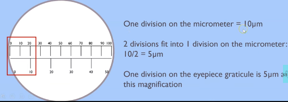
if each division on the SM is a certain length (e.g. 10 micrometers), use this to calculate what one division on the EG is. (e.g. with this example: if 2 EG divisions fit into 1 division on the SM, we do 10 / 2 = 5 micrometers. so 1 division on the eyepiece graticule is 5 micrometers)
then you can measure the specimen as normal using the EG. (i.e. how many divisions long is the specimen? for this example, imagine a nucleus is 13 divisions long). then work out the actual length by multiplying (or maybe dividing) by how much one division is (so 13 × 5 = 65 micrometres)
diagram that shows step 2 from last flashcard
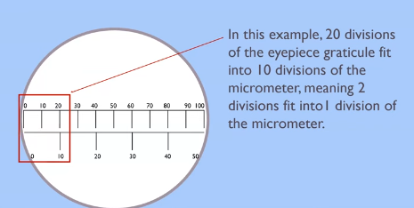
diagram that shows step 3 from last flashcard
name the 2 types of microscope
optical (light) microscope
electron microscope
two types of electron microscope
transmission electron microscope (TEM)
scanning electron microscope (SEM)
know how to label a diagram of a light microscope (find in folder)
!!!
how does an optical microscope work
uses visible light + a series of lenses = to produce a magnified image of specimen
pros of optical microscope
easy to use —→ preparation of specimens is fairly simple
produces coloured images
cheap + affordable
portable —→ usually small + lightweight
can view living specimens
cons of optical microscope
poor resolution —→ uses a relatively long wavelength of light
potential distortion —→ some details may be unclear (due to light scattering)
limited magnification —→ typically up to about 1000x - 2000x. this limits viewing very small structures
thin samples required —→ so light = can pass through
why do electron microscopes have a high resolving power
electron beam = has a v. short wavelength
so microscope can resolve objects well
can resolve objects that are just 0.1nm apart —→ 2000x better than an optical microscope
HOW A TEM WORKS:
the TEM consists of an electron gun. what does this produce?
what is this focused onto the specimen by
what does the beam pass through
why do denser parts of the specimen appear dark?
why do other parts of the specimen appear bright?
an image of the specimen is produced on the screen. this can be photographed to give a….
a beam of e-
a condenser electromagnet
a thin section of the specimen
they absorb more e-
they allow e- to pass through
photomicrograph
pros of TEM
extremely high resolution (0.1nm) because electrons have a short wavelength
high magnification (1,000,000×, greater than optical microscopes)
cons of TEM (5)
complex sample prep —→ must be ultra thin (to allow e- to penetrate) and able to withstand a vacuum
image = not in colour
image = 2D
operates in a high vacuum —→ living organisms cannot be observed
high-energy electron beam can damage delicate samples
expensive
training required to operate
artefacts present (explained in next card)
what may appear on the photomicrograph from a TEM
what are these
what does this mean (a con)
artefacts
things that result from the way the specimen was prepared, but are not part of the natural specimen
can’t always be sure that what we see on photomicrograph = really exists in that form
how does an SEM work
scans a focused beam of e- (from an e- gun) back and forth across the surface of a specimen.
the e- are scattered by the specimen. this interaction emits signals
the SEM detects these, and a computer converts them into a detailed image of the specimen’s surface structure
pros of an SEM
high resolution
high magnification
image = 3D
resolution = 10x better than that of a light microscope
cons of an SEM
lower resolving power than a TEM (around 20nm)
image = not in colour
image = does not show internal structures
rest of the cons = same as a TEM! (except the fact that specimens need to be extremely thin, since electrons don’t penetrate)
remember: in exam Qs, you can refer to the microscopes by their abbrievations!
EXAM Q: why is resolution of an image obtained using an e- microscope = higher than the resolution of an image obtained using an optical microscope (1 mark)
M1: shorter wavelength between e-
OR longer wavelength in light rays (so they can’t zoom into smaller structures)
what does cell fractionation do
separates individual organelles from the rest of the cell
3 stages of cell fractionation
homogenisation
filtration
ultracentrifugation
what is homogenisation
cell membrane is broken to release the organelles
how to prepare the tissue for homogenisation
the tissue is cut up
and placed in a cold, isotonic, buffered solution
why is the solution the tissue placed in cold, isotonic and buffered? (give a reason for each property)
cold = prevent (slows) enzyme activity which may break down organelles
isotonic = stops organelles bursting / shrinking due to osmotic gains / losses
buffered = maintains pH to stop protein / enzyme denaturation
what is used to break down the cells further
what exactly does this do
a blender
disrupts cell membrane —→ breaking open cells and releasing organelles/cytoplasm/cytosol into a solution
what is the solution the organelles are released into called
homogenate (this is IN the cold, isotonic, buffered solution)
during filtration, the h_____________ c____ s____________ is filtered through a g_______. this separates large, unwanted c____ d______ and c____________ t______.
during filtration, the homogenised cell suspension is filtered through a gauze. this separates large, unwanted cell debris and connective tissue.
where do the organelles remain after filtration
in the filtered homogenate (filtrate)
what happens during ultracentrifugation
homogenate is placed into a tube
tube is placed into a centrifuge
what is a centrifuge
a machine that separates materials by spinning
during ultracentrifugation, the homogenate is spun at i______________ s_______ to separate organelles by d_______.
during ultracentrifugation, the homogenate is spun at increasing speeds to separate organelles by density. (heaviest organelles separated first, lightest last)
(just for your understanding) therefore, as density of organelle decreases, speed of centrifuge…
increases
what do the heaviest organelles form at the bottom of the tube
where do the rest of the organelles go?
a pellet
they stay suspended in the solution above the pellet
what is the solution above the pellet now known as
the supernatant
what is then done with the supernatant
it is removed from the pellet, put into a new centrifuge tube, and respun at higher speeds
this causes the next-heaviest organelles to form a new pellet
this process is repeated several times, each time removing the supernatant from the previous tube and spinning it faster to separate smaller and lighter organelles.
give the order of separation of organelles from most dense (1st spin) to least dense (5th spin)
nucleus (most dense)
chloroplasts + mitochondria
lysosomes
endoplasmic reticulum
ribosomes (least dense)
most important ones to know are the most and least dense (in bold)
MS answer for how to obtain undamaged chloroplasts from a sample of leaves (not completely accurate but you can get a general gist. update when PPQs are completed)
chop up sample
place in a cold, isotonic, buffered solution
homogenise and filter
centrifuge filtrate
remove supernatant and centrifuge
at a higher speed
chloroplasts should be in the second pellet
EXAM Q: A student used an optical microscope to observe a piece of tissue from the lower surface of an orchid leaf.
The piece of leaf tissue observed was very thin. Explain why this was important. (2)
M1: Single/few layer(s) of (cells/tissue)
M2: So light can pass through
EXAM Q: The student produced a biological drawing of the leaf tissue they viewed through an optical microscope.
Give three ways the student could ensure they produce a correct biological drawing of the leaf tissue.
Assume the student uses a sharp pencil. (3)
Use single/continuous/joined lines
OR Do not use sketching
OR No crossing or hanging lines;
2. Do not use shading/hatching;
3. Draw (only) the outline of tissues
OR Do not draw cells (detail);
4. Draw parts to the same scale/relative size;
5. Show labels/annotations/title;
6. Show magnification/scale (bar)
Give the uncertainty associated with taking a measurement using a ruler with 1 mm graduations.
1mm
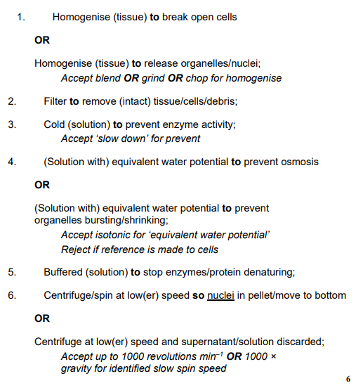
Describe and explain how you would use cell fractionation and ultracentrifugation to obtain a sample of nuclei from muscle tissue (6)
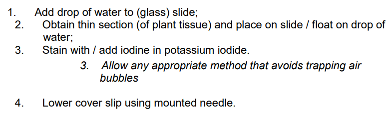
Describe how you could make a temporary mount of a piece of plant tissue to observe the position of starch grains in the cells when using an optical (light) microscope. (4)
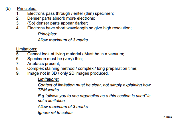
Describe the principles and the limitations of using a transmission electron microscope to investigate cell structure. (5)