Diagram of Week 1: Overview of Anatomy of Thorax | Quizlet
1/51
There's no tags or description
Looks like no tags are added yet.
Name | Mastery | Learn | Test | Matching | Spaced |
|---|
No study sessions yet.
52 Terms
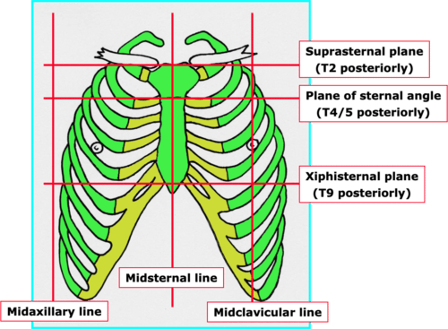
Describe the different lines and planes of the thorax. (6)
1. Midaxillary line.
2. Midsternal line.
3. Midclavicular line.
4. Suprasternal plane (at T2 posteriorly).
5. Plane of sternal angle (at T4/5 posteriorly).
6. Xiphisternal plane (T9 posteriorly).
What are the intercostal muscles? (4)
1. In intercostal spaces.
2. Have 3 layers: external, internal and innermost.
3. Neurovascular bundles are located between the internal and innermost layers.
4. Important for respiration and keeping the intercostal space rigid.

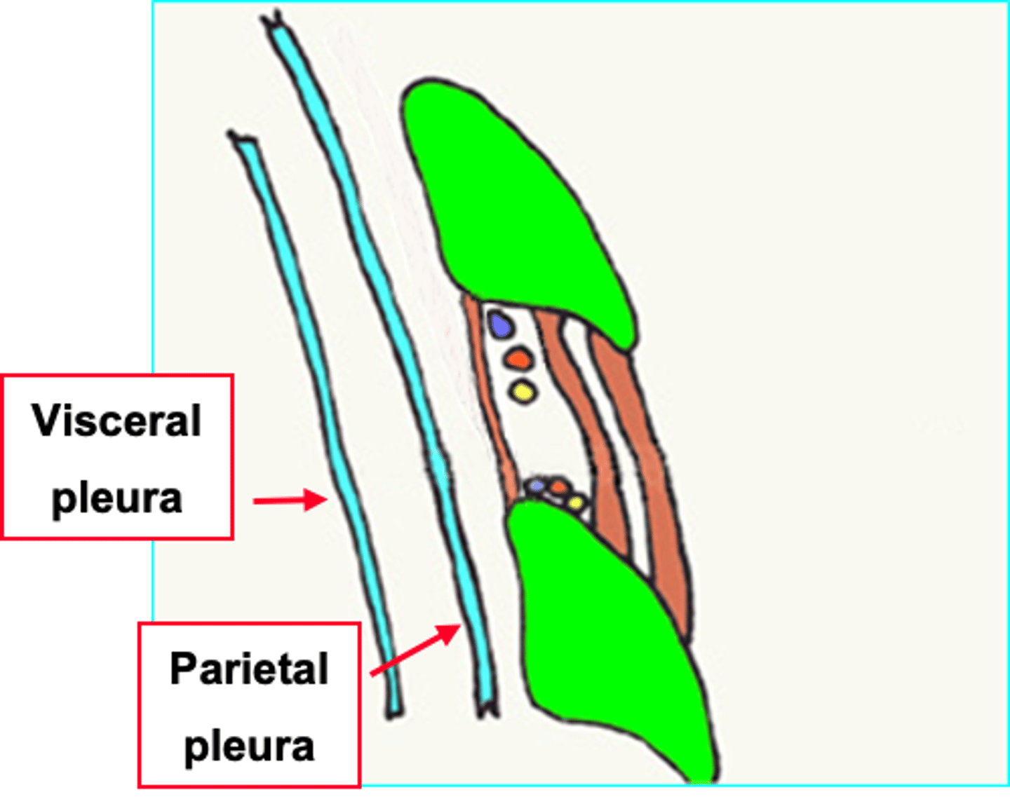
What are the pleura of the lungs?
Each lung is enclosed in a serous pleural sac, which has 2 continuous membranes: visceral and parietal.
What are the 2 types of pleura?
Visceral: covers surface of lungs.
Parietal: lines pulmonary cavities (inside of ribcage).
They are continuous with each other.
What is the purpose of the fluid in the pleural cavity?
Fluid is a potential space, used for friction.
Why may you need to insert a needle into an intercostal space? (2)
1. To drain or sample fluid (pleural fluid, blood, pus) from pleural cavity.
2. Anaesthetise ('block') an intercostal nerve.
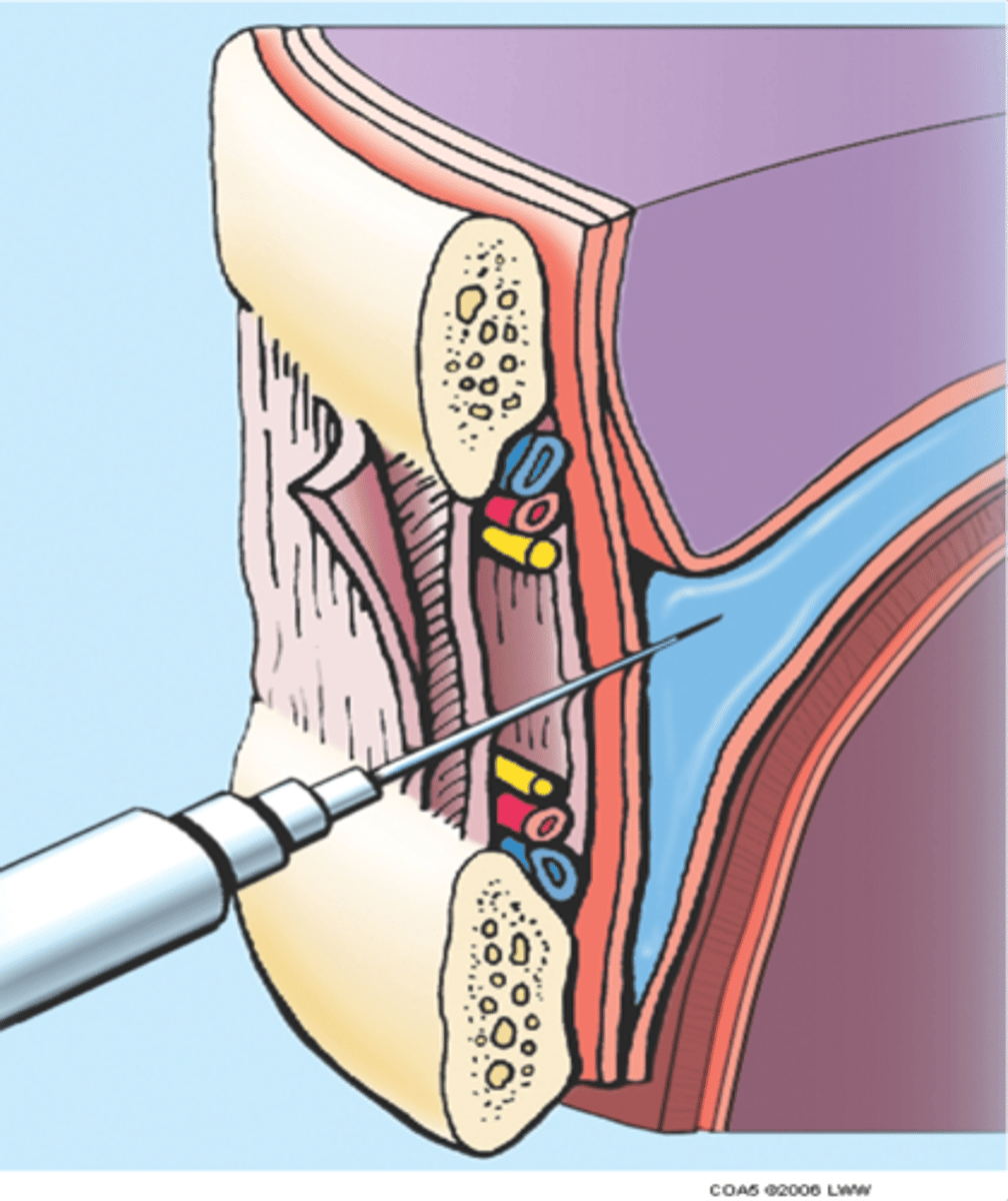
How can you avoid the main neurovascular bundle when inserting a needle?
Insert needle close to upper border of lower rib.
What is the superior thoracic aperture?
Opening for structures to enter/leave the neck/thorax. It is quite a narrow opening - which can lead to problems.

What is the inferior thoracic aperture?
Opening at lower part of thoracic cavity (closed by diaphragm).
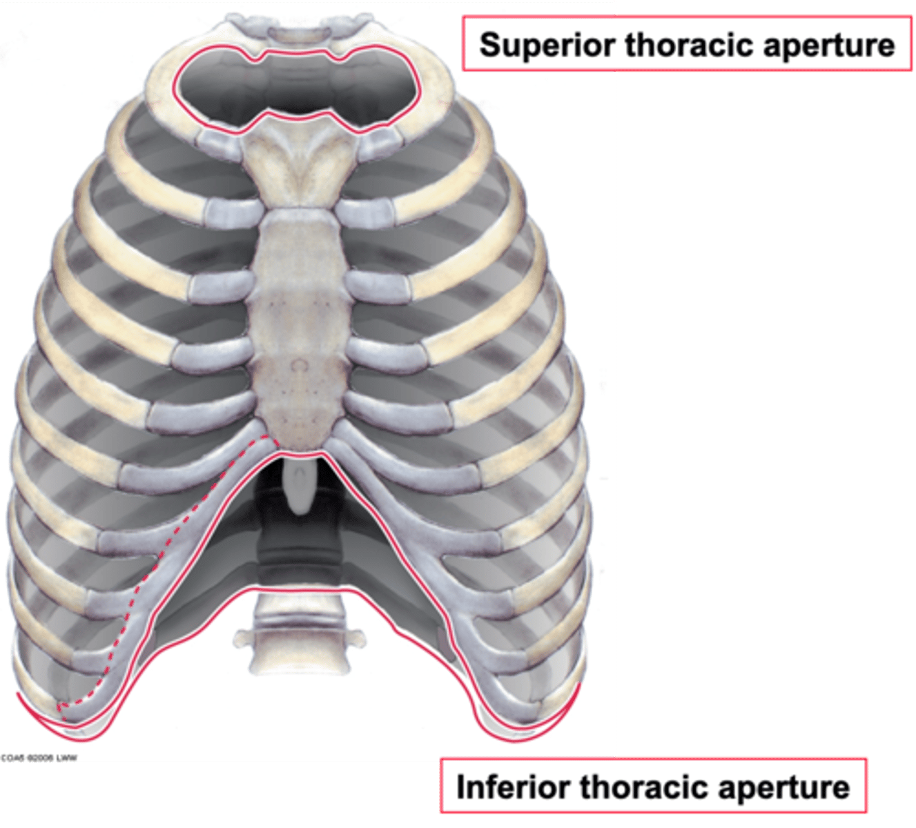
What are the boundaries of the superior thoracic aperture?
Anterior: superior aspect of manubrium.
Posterior: T1 of thoracic vertebrae.
Lateral: costal cartilage.
What are the boundaries of the inferior thoracic aperture?
Anterior: xiphisternum.
Posterior: T12 of thoracic vertebrae.
Lateral: floating ribs (11, 12) and rib 10, costal margin.
Why are the boundaries of the superior and inferior thoracic apertures clinically relevant?
x
What is Thoracic outlet syndrome and what are the different types? (2)
1. The compression of important arteries and nerves which pass through into neck and upper limb. Can lead to shoulder and neck pain and numbness in fingers.
2. 3 different types: neurogenic (neck nerves compressed), venous (compressed vein > upper body thrombosis) and arterial (compressed artery).
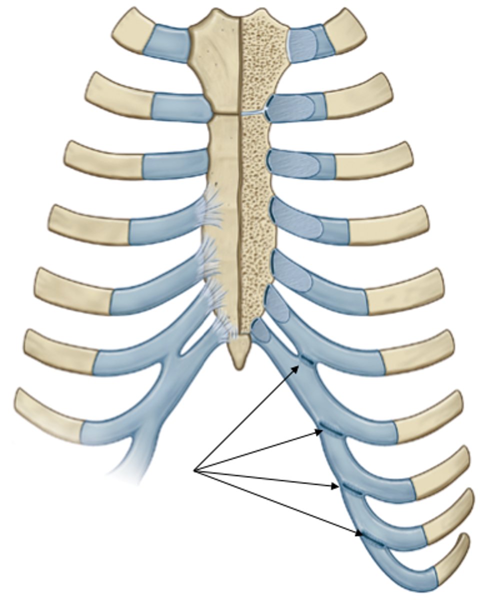
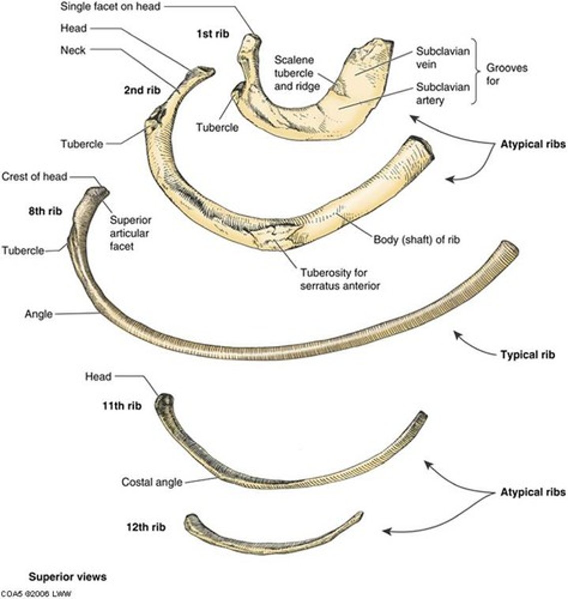
Distinguish the different types of ribs.
Ribs 1-7: true ribs - costal cartilages articulate directly with sternum.
Ribs 8-10: 'false' ribs as costal cartilage articulates with one above.
Ribs 11-12: 'floating' ribs - don't articulate at all.
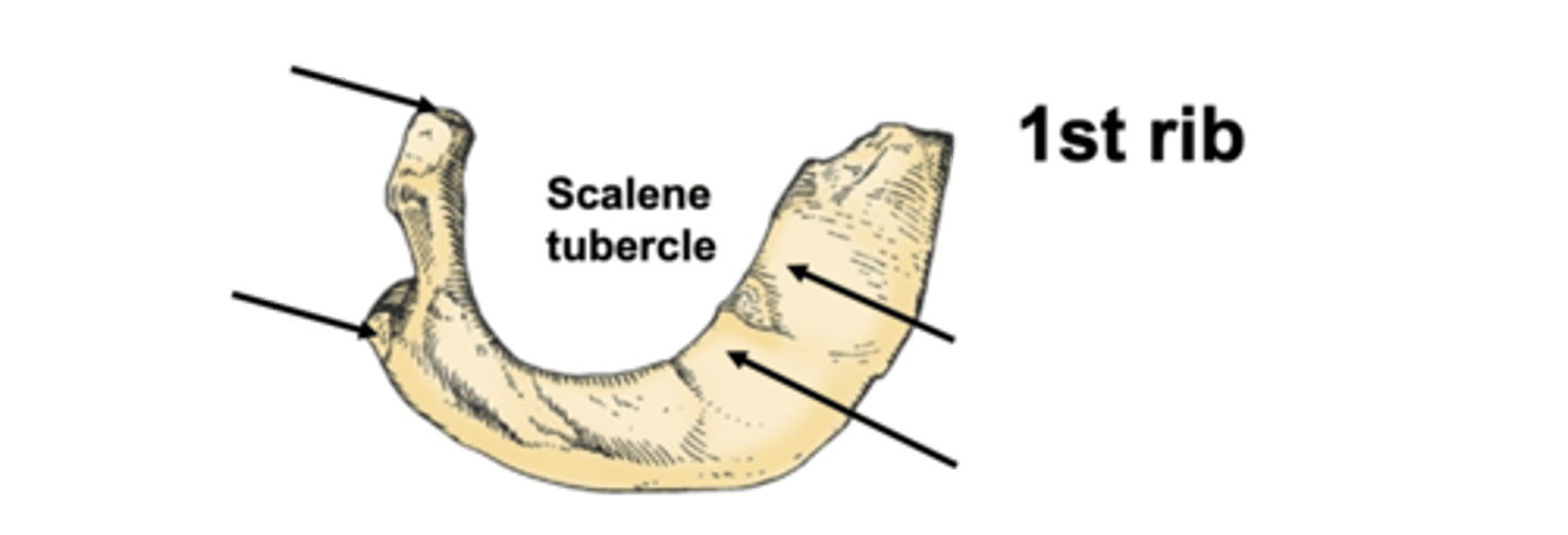
What are the features of the 1st rib? (5)
Rib 1 is shorter and wider than other ribs. Has only one facet on head for articulation with corresponding vertebra and 2 grooves for subclavian vessels.
1. Facet on the head (articulate with vertebral body of corresponding vertebra).
2. Tubercle.
3. Below costal end, there are 2 grooves: for subclavian vein and for subclavian artery (these 2 structures pass through thoracic aperture). A tumour in this region can lead to compression of these vessels.
4. Scalene tubercle: in between the 2 grooves
5. The end of the rib articulates with its costal cartilage.
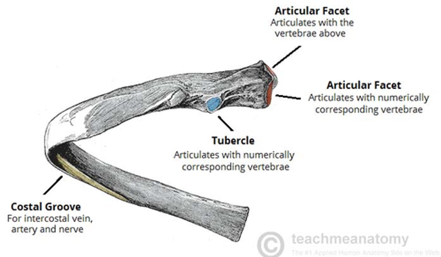
What are the features of the 8th rib (typical rib)? (4)
1. Head - wedge-shaped, 2 articular facets separated by bone wedge. One articulates w/ corresponding vertebra, other with the vertebra above.
2. Neck - no bony prominences (tuberosities) but simply connects head w/ body. There is a roughed tubercle where neck meets the body, and contains facet which articulates with the transverse process of corresponding vertebra.
3. Angle
4. Body/shaft - flat and curved. Internal surface has a costal groove for neurovascular supply of thorax, protecting vessels and nerves from damage.
What are the features of the 12th rib (floating rib)?
An atypical rib, it has no tubercle, angle or costal groove. Does not articulate with the sternum at all.
Outline 4 parts of the vertebrae.
1. 7 Cervical
2. Thoracic
3. 5 Lumbar
4. 5 Sacral
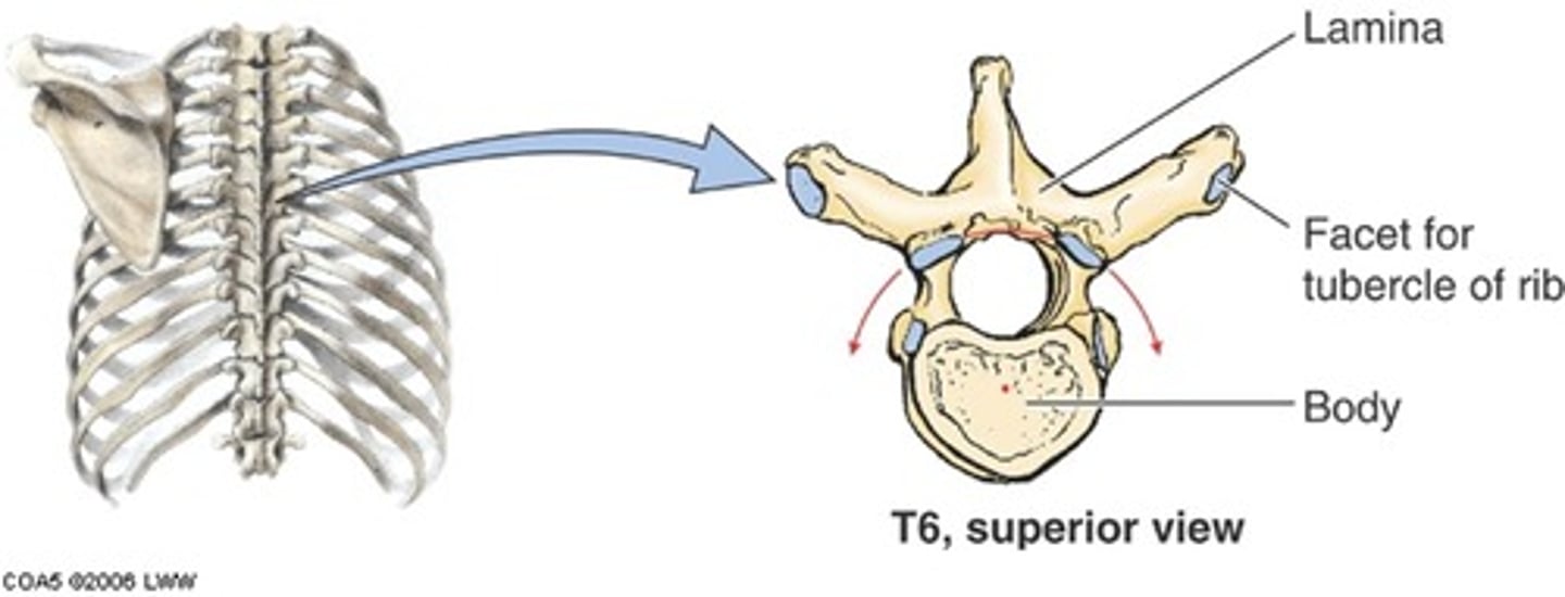
Outline features of T6 - typical vertebrae. (9)
1. Has a cylindral mass of bone in front > vertebral body which supports weight.
2. Set of bony plates/projections with 3 functions: protect spinal cord, give muscle attachment/ligaments + articulate with joining vertebrae.
3. Contain a neural arch which encloses spinal cord.
4. Vertebral foramina: tubular space containing spinal cord - vertebral canal.
5. Lamina and pedicle.
6. Small notch in upper edge of pedicle and larger notch in lower edge form the invertebral foramen.
7. Spinous process in midline and transverse process on each side.
8. 4 articular processes - 2 above and 2 below (lower ones face forward, upper ones face backwards).
9. Articular processes of adjoining vertebrae interlock and form pair of synovial joints > movement between adjoining vertebrae.
How are the vertebrae different?
T1 - T12 changes - they change shape based on the load - they increase in size.
What are the attachments of the pectoralis major? (3)
1. Clavicular head from medial half of clavicle.
2. Sternocostal head (lateral part) from sternum and upper 6 costal cartilages.
3. All fibres converge on the intertubercular (or bicipital) groove of humerus.
What are the actions of pectoralis major? (4)
1. Adductor.
2. Medial rotator of arm at shoulder joint.
3. Can also be a flexor (when extended) and extensor.
4. Can also be accessory muscle of respiration when fixing pectoral girdle.
What is the nerve supply for pectoralis major?
Medial and lateral pectoral nerves (C5-8, T1).
What are the attachments of pectoralis minor?
1. Coracoid process of scapula.
2. Ribs 3-5 near their cartilages.
How is pec major arranged to pec minor?
Superficial to pec minor. Pec minor is deep to pec major.
What are the functions of pectoralis minor? (3)
1. Depressor of scapula.
2. Protractor of scapula.
3. When fixing girdle, it can be an accessory muscle for respiration.
What is the nerve supply of pectoralis minor?
Medial pectoral nerve (mostly C8, T1).
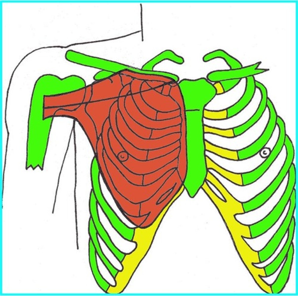
Where is the breast located? (3)
Base of breast is from ribs 2-6 and from lateral margin of sternum to midaxillary line.
2. Breast sits on pec major and pec minor deep to that.
3. An axillary tail runs superiorly and laterally towards axilla.

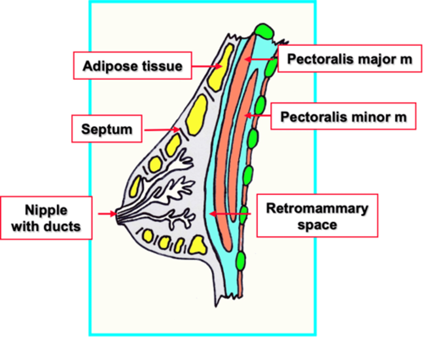
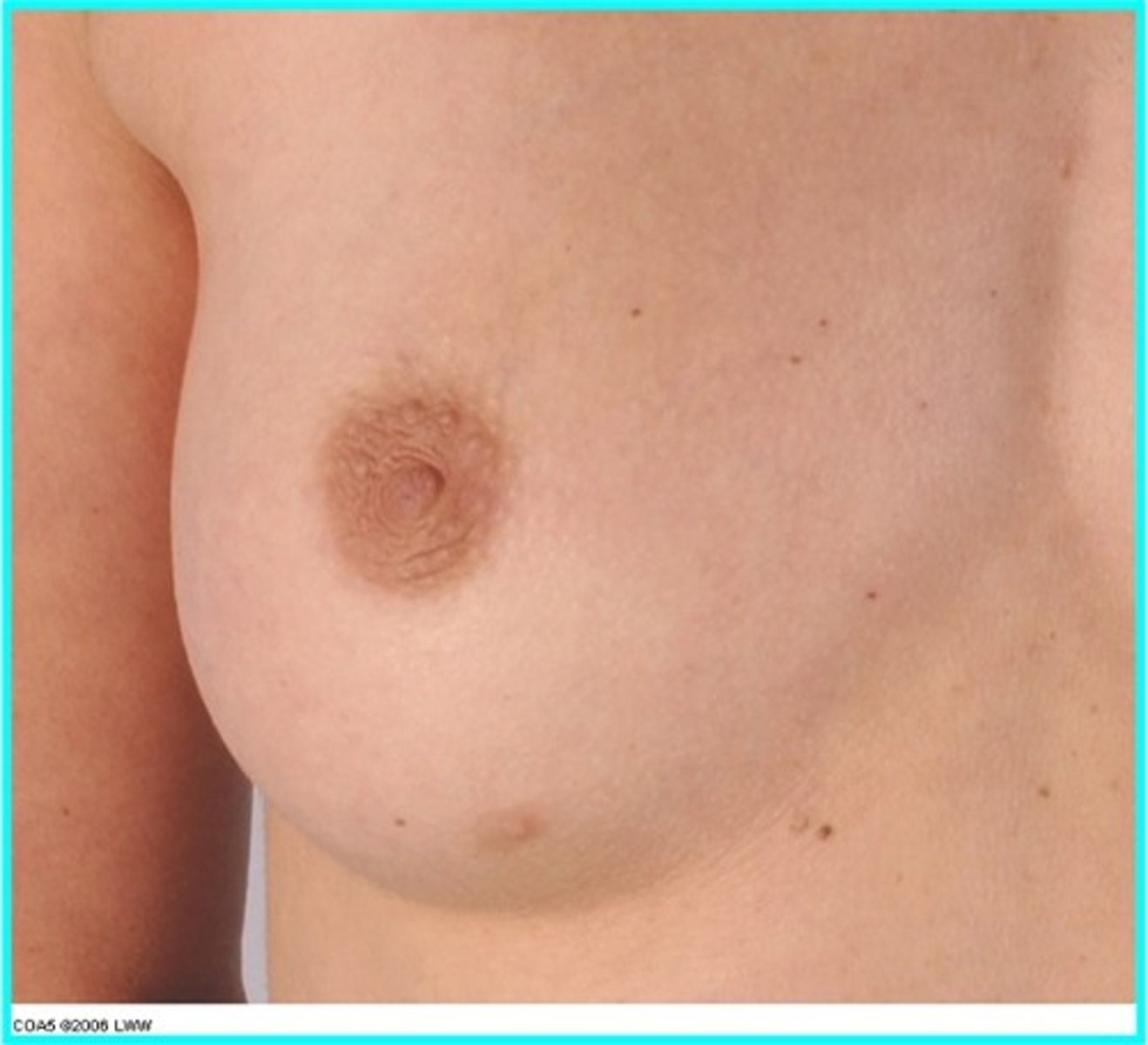
What are the features of the breast? (3)
1. Modified sebaceous gland with 15-20 lobes sending lactiferous ducts to nipple.
2. Lobes have glands and adipose tissue separated by fibrous septa (suspensory ligaments).
3. Breast is separated from deeper pec muscle by a retromammary space.
Why is lymphatic drainage clinically important to the breast?
The frequency of breast cancer and its spread to other parts of body by lymph and blood vessels. You would palpate areas around breast and to umbilicus.
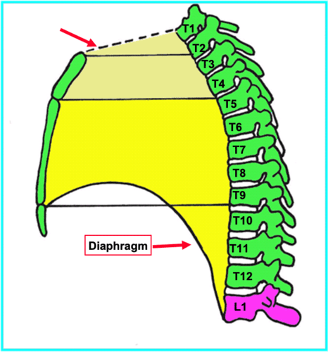
What is the mediastinum?
1. Central part of thoracic cavity between right and left pleural cavities. Contains heart and other structures.
2. Superior boundary is superior thoracic aperture and inferior boundary is diaphragm.
What are the 4 different boundaries of mediastinum?
Anterior: sternum
Posterior: thoracic vertebral column
Superior: thoracic inlet/root of neck.
Inferior: diaphragm.
What are the different contents of the mediastinum? (7)
1. heart and pericardium
2. great vessels
3. oesophagus
4. trachea
5. thymus
6. lymph nodes
7. various nerves and other blood vessels.
What is the mediastinum divided into and where do these areas lie? (3)
1. Divided into superior and inferior parts by sternal angle plane (manubriosternal joint to T4/5 disc).
2. Superior mediastinum lies behind manubrium sterni.
3. Inferior mediastinum lies behind body and xiphoid process of sternum (between sternal angle plane and diaphragm).
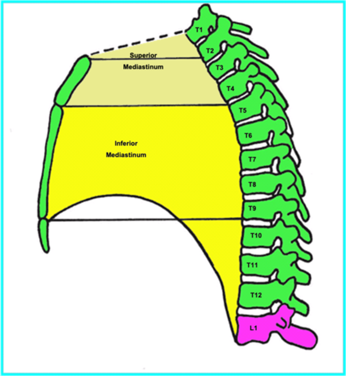
Outline the contents of the superior mediastinum. (8)
1. Lymphoid organ (involutes in adult).
2. Great veins - brachiocephalic and subclavian.
3. Phrenic nerves and vagus nerve.
4. Arch of aorta and branches.
5. Origins of internal thoracic arteries.
6. Pulmonary arteries and veins.
7. Recurrent laryngeal branches (branch of vagus controls the larynx), trachea.
8. Oesophagus and thoracic duct.
What are the 3 parts of the inferior mediastinum?
1. Anterior
2. Middle
3. Posterior.
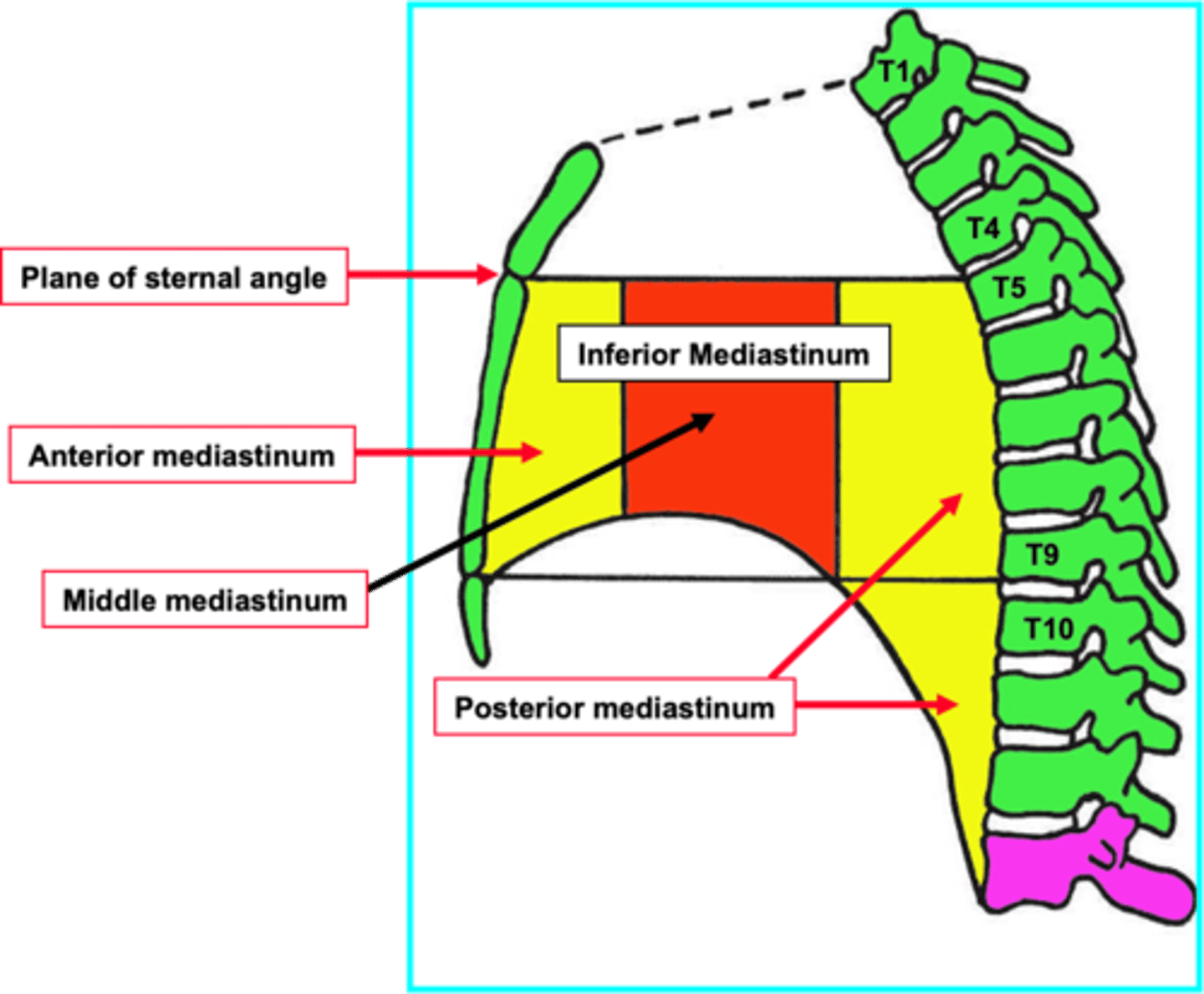
Outline the contents of the 3 parts of the inferior mediastinum.
Anterior:
1. Internal thoracic arteries and veins and thymus and sternopericardial ligaments.
Middle:
1. Heart and pericardium
2. Phrenic nerves and pericardioprenic arteries and nerves.
3. Inferior vena cava.
Posterior:
1. Descending aorta and branches.
2. Azygos nerve,
3. Oesophagus, thoracic duct, sympathetic trunks (and branches).
TERM
What is the manubrium?
DEFINITION
The manubrium is the most superior region of the sternum and articulates with the clavicles or collarbones and the first pair of ribs. The manubrium is the thickest portion of the sternum as it carries the greatest physical load.
TERM
What is the body of the sternum?
DEFINITION
The sternum is a partially T-shaped vertical bone that forms the anterior portion of the chest wall centrally. The sternum is divided anatomically into three segments: manubrium, body, and xiphoid process.
TERM
What is the clavicle?
DEFINITION
The clavicle is located between the ribcage (sternum) and the shoulder blade (scapula). It is the bone that connects the arm to the body.
TERM
What is the sternal angle?
DEFINITION
It marks the point at which the costal cartilages of the second rib articulate with the sternum. This is particularly useful when counting ribs to identify landmarks as rib one is often impalpable i.e. top part of sternum.
TERM
What is the xiphoid process (xiphisternum)?
DEFINITION
The xiphoid process articulates with the superiorly located sternum corpus at the xiphisternal joint i.e. the bottom part of the sternum.
TERM
What is the intercostal space?
DEFINITION
The intercostal spaces (spaces between the ribs) are largely occupied by muscles (intercostal muscles), which, in association with other muscles of the thorax, move the ribs during breathing.
TERM
What is the costal margin?
DEFINITION
The lower edge of the chest (thorax), formed by the bottom edge of the rib cage.
TERM
What is the suprasternal/jugular notch?
DEFINITION
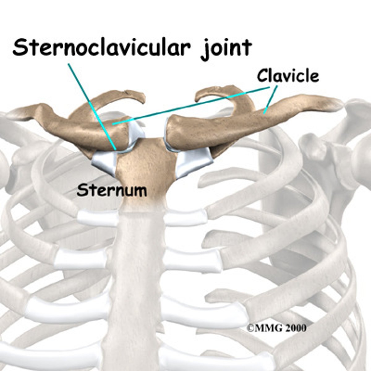
1. A depression in the superior aspect of manubrium, which has a large fossa either side lined with cartilage.
2. Fossa articulate w/ medial ends of clavicles, forming sternoclavicular joints.
What is the intervertebral joint?
- Articulation between adjacent vertebrae.
- With a secondary cartilaginous joint.
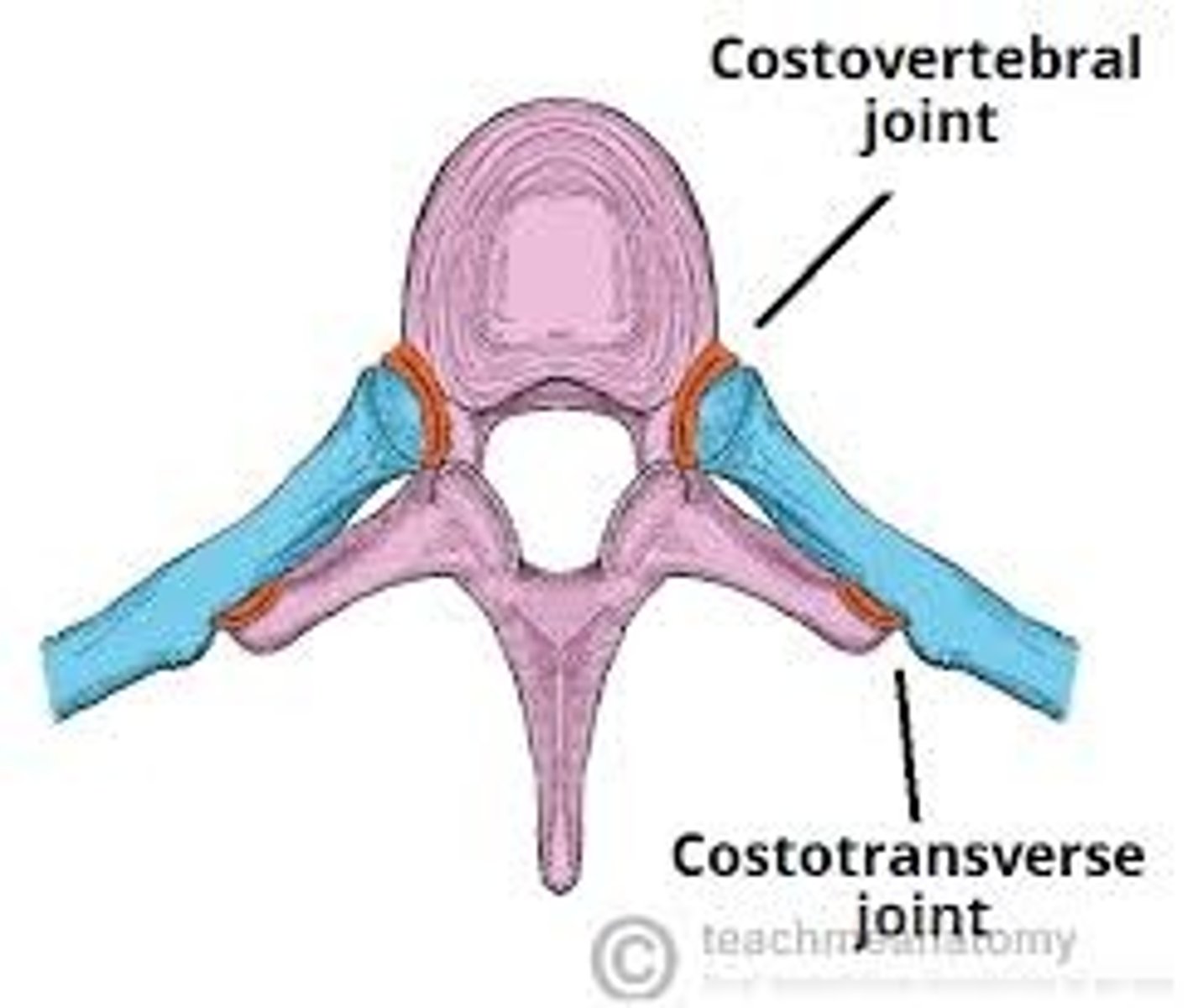
What is the costotransverse joint?
- Between ribs and vertebrae - the transverse process of the vertebrae and the head facet of the corresponding rib.
- It is a synovial plane joint.
- Allows for movement of rib during respiration.

What is the costovertebral joint?
- Between head of rib and superior costal demifacet of corresponding vertebra.
- Synovial plane joint.
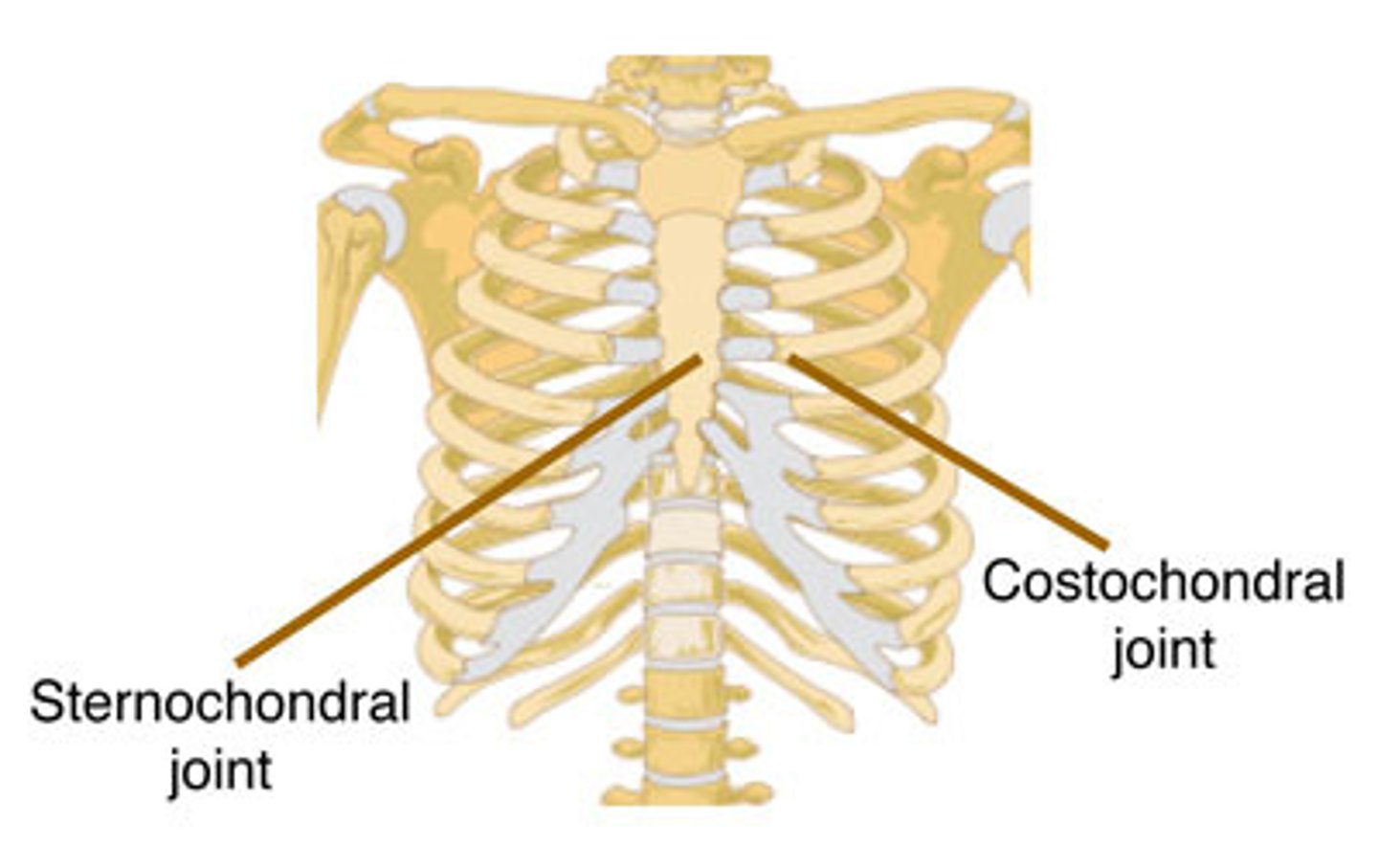
What is the sternochondral joint?
- Between sternum and costal cartilages.
- Primary cartilaginous (1st) and synovial plane joints for 2nd-7th cartilages.
What is the sternoclavicular joint?
- Articulation between the sternum and the clavicle.
- A saddle-shaped synovial joint.
What is the costochondral joint?
- Articulation between rib and costal cartilage.
- A primary cartilaginous joint.
What is the interchondral joint?
- Joint between adjacent costal cartilages of ribs 6-10.
- Synovial plane joint.