CHAPTER 7: ACCESS CAVITY PREPARATION
1/50
There's no tags or description
Looks like no tags are added yet.
Name | Mastery | Learn | Test | Matching | Spaced | Call with Kai |
|---|
No analytics yet
Send a link to your students to track their progress
51 Terms
access cavity preparation
create an opening to the tooth
essential to achieve proper biomechanical preparation (BMP) cleaning and shaping of the root canal system, and obturation sealing and closure after BMP
used by:
high/low speed handpiece
endodontic access burs (long shank)
characteristics of ideal access
straight entry into the canal orifice
line angles form a funnel that drops smoothly into the canal(s)
projection of the canal center line to the occlusal surface:
indicates location of line angles
connection of line angles:
creates the outline form
objectives of access cavity preparation
to remove all caries
to locate all root canal orifices
to remove all coronal pulp tissue
to conserve sound tooth structure
to completely unroof the pulp chamber
to establish restorative margins to minimize marginal leakage of the restored tooth
to achieve straight- or direct line access to the apical foramen or to the initial curve of the canal
[ SLA is the main objective – the opening that you create would help you enter the RCS w/out hindrances ]
fissure carbide & diamond burs (with safety tip)
safer for axial wall extensions
can extend to pulp floor safely
produce axial walls free of gouges
used to:
extend axial walls
favorably orient axial walls
level cusp tips
level incisal edges (reference points for working length)
round carbide burs
create initial external outline
penetrate pulp chamber roof
remove chamber roof & caries
risk (in inexperienced hands):
gouging pulp floor and axial walls
round bur #2 & #4
used to access through porcelain or ceramometal restorations
advantages:
less traumatic to porcelain than carbide burs
less likely to crack or fracture porcelain
reminders:
must be used with water spray
after penetrating porcelain:
switch to carbide bur for metal or dentin (greater cutting efficiency)
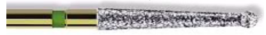
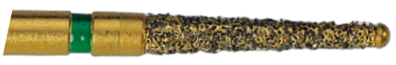
medium- or fine-grit diamond burs
bur used for zirconia restorations as carbide burs do not cut zirconia efficiently or safely with the use of copious water spray
zirconia is brittle:
cutting may create cracks
cracks may propagate and cause restoration failure
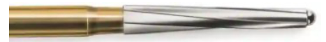
transmetal bur
bur used for metal restorations
benefits:
uses new bur
excellent cutting efficiency
use with water spray for maximal cutting effect
extended-shank round burs
improves visibility
moves handpiece head away from tooth
alternative:
ultrasonic units
examples:
LN bur
mueller bur
munce discovery bur
bur used for intial RCT
round bur & fissure bur to enlarge and create a proper form
—regular bur will block visualization
special endodontic burs
diamendo
endo Z bur
howard martin bur
howard martin bur
round bur and a taper-fissured body
diamendo
diamond bur for refining the walls of the canals
endo Z bur
cutting the walls of the access prep
used for canal exploration
K-files (patency files)
canal probe: #12 orange
endodontic explorer (DG-16)
endodontic explorer (DG-16)
identify canal orifices
determine canal angulation
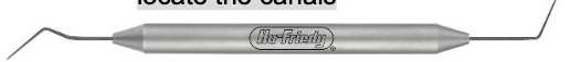
endodontic explorer (JW-17)
thinner, stiffer tip than DG-16
useful for:
identifying calcified canals
patency files of K-files
initial files that would enter the canal
#6 → pink, #8 → gray, #10 → violet
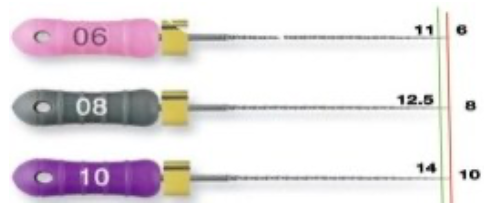
canal probe: #12
created to bridge the gap between #10 & #15 files for smoother canal enlargement
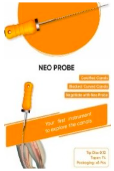
NeoProbe → tip diameter of 0.12 and a taper of 1% (varies on the brand)
pulp extirpation
the removal of vital pulp using nerve broach
when inserting nerve broach, avoid touching dentin
canal preparation
used of irrigating syringe and irrigating needles
size of irrigating syringe used
10–20mL
size of Irrigating needles used for anterior
25-gauge
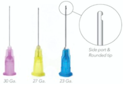
size of irrigating needles used for posterior
27 or 30 gauge

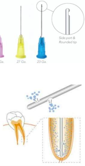
side-venting needles
23 gauge
to flush out debris or root canals in a whirlpool effect w/out hitting the apical foramen = efficient cleaning
reminders for canal preparation
isolation – to prevent contamination
preoperative x-ray – to assess canal anatomy
canal patency – use small files (#6, #8, #10) to locate and confirm canals
instrumentation/shaping – use hand or rotary files, noting canal bifurcations and curvatures
obturation – final step, filling the canal (the “end game” of RCT)
laws of access cavity prep
law of centrality
law of concentricity
law of the CEJ
law of symmetry
law of color change
law of orifice location

law of centrality
floor of the pulp chamber is always located in the center of the tooth at the level of the CEJ → most stable landmark as reference during access prep
file – prepare ; bur – access

law of concentricity
walls of the pulp chamber are always concentric to the external surface at the CEJ
—> that is, the external root surface anatomy reflects the internal pulp chamber anatomy

law of the CEJ
distance from the external surface of the clinical crown to the wall of the pulp chamber is the same throughout the circumference at the CEJ
making the CEJ the most consistent repeatable landmark for locating the position of the pulp chamber

first law of symmetry
except for the mx molars
canal orifices are equidistant from a line drawn in a mesiodistal direction through the center of the pulp chamber floor

second law of symmetry
except for the mx molars
canal orifices lie on a line perpendicular to a line drawn in a mesiodistal direction across the center of the pulp chamber floor

law of color change
the pulp chamber floor is always darker in color than the walls.
from enamel: white – yellow → dentin: brownish – yellow → pulpal floor: grayish color

first law of orifice location
the orifices of the root canals are always located at the junction of the walls and the floor
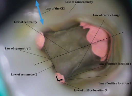
second law of orifice location
the orifices of the root canals are always located at the angles in the floor-wall junction

third law of orifice location
the orifices of the root canals are always located at the terminus of the developmental fusion lines of roots

distal
rubber clamp bow is always located at the __
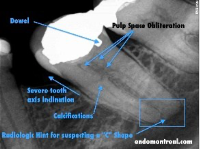
fast break
sudden disappearance of the canal along its length
signifies:
splitting of the canals
presence of a bifurcation / trifurcation
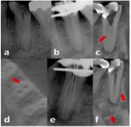
pulp canal obliteration (PCO)
calcify itself as a defense-mechanism
pulp canal has been calcified in response to injury
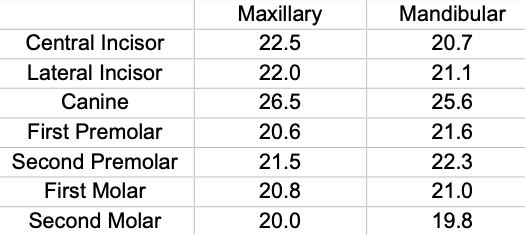
average length of teeth (mm)
oval access opening
mx canine
mx 1st premolar
mx 2nd premolar
mn canine
mn 1st premolar
mn 2nd premolar
mn incisors
oval-triangular access opening
mx incisors (central/lateral)
triangular access opening
mx 1st molar
mx 2nd molar
trapezoidal access opening
mn 1st molar
mn 2nd molar
from the lingual surface to the incisal surface
entry of access for mn incisors
benefits:
better access to the lingual canal
improved straight-line access
improved canal debridement
reminders for cavity access preparation
recommended average length is the actual canal length
rubber stopper placed at the highest reference point of the clinical crown, not on the orifice
slight extrusion of root canal sealer through the apical foramen is generally not a major concern, as macrophages and the body’s immune system can gradually dissolve it
apical constriction: natural stop for RCT
0.5–1.5 mm from the apical foramen (AF)
0.5–1 mm short of the radiographic apex
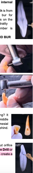
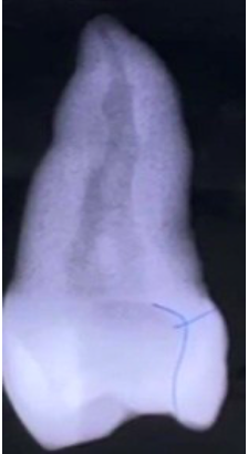
access cavity prep by doc g
area of internal resorption due to trauma (that burot gi mark ni doc g)
anterior tooth:
initial entry: lingual surface
use: long shank round bur
penetration technique:
advance slowly
you will feel a sudden drop when entering the pulp chamber
then reorient the bur perpendicular to the long axis
posterior tooth:
initial entry: occlusal surface
based on the law of centrality
after entering the chamber:
extend the preparation to form a triangular outline form (especially in anterior teeth)
use an explorer to:
remove debris
locate canal orifices
how do you know you’ve found the canal?
use patency files (small K-files)
if the file advances smoothly down the canal → you are in the canal
why not an oval access opening?
an oval prep:
removes only the middle pulp chamber
may leave mesial and distal pulp horns behind
has big opening access but orifice is small!
use gates glidden drill or SX (canal orifice opener) → to create a smooth transition
gouging
iatrogenic mistake during access cavity preparation on the lateral walls or pulp chamber floor, it should be straight
treatment: fill it
root canal walls
must guide instruments, not access cavity walls
failure to follow root canal walls may cause:
root perforation
ledge formation
apical transportation
incorrect canal shape
instrument separation
micro-openers
offset handles enhance visualization
flexible stainless steel hand instruments
used for locating canal orifices before dam placement