Looks like no one added any tags here yet for you.
What is a Zygote?
a diploid cell resulting from the fusion of two haploid gametes; a fertilized ovum.
Organisms start as sets of _____ and ______
tubes, sheets
How many different types of cells do humans have?
~350
What are the basic cellular components?
membrane, cytoplasm, and nucleus
What are the 4 basic cellular functions?
Multiplication, Movement, Differentiation, Signal
What are polar bodies and their significance?
One of the oocytes created via oogenesis that has no cytoplasm. This happens to maximize the cytoplasm in the future zygote for increased energy reserves in a cleaving cell as the cell doesn't grow any bigger.
What is cleavage? When does it first happen?
Where the zygote goes through repeated cell divisions where it splits but does not grow, it becomes a blastocyst. First cleavage is 30 hrs post ovulation.
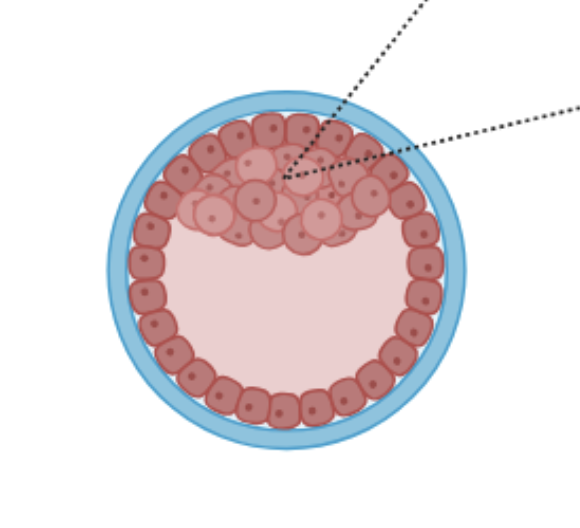
What is a blastocyst? (Label diagram)
A structure the same size as a zygote but with around 70-100 cells and composed of two distinct regions: trophoblast and embryoblast.
What is a trophoblast?
The outer layer of blastocyst, which later develops into the placenta and other support structures
What is the embryoblast?
The inner cell mass in the blastocyst that gives rise to the embryo. First, during implantation, it flattens into a bilaminar disc with 2 layers: epiblast and hypoblast.
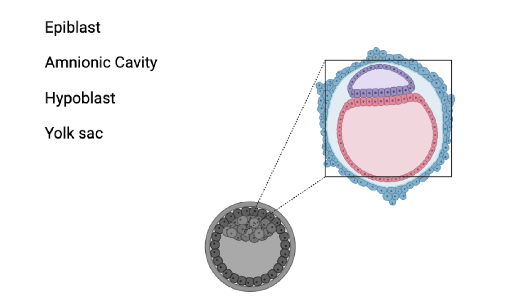
Label the components of this diagram
Grey - blastocyst, trophoblast, embryoblast
Blue - amniotic cavity(purple), epiblast(bottom layer of purple), hypoblast(pink)
What is the hypoblast?
The lower layer of the bilaminar disc of the embryoblast. Becomes the yolk sac and early nutrients before formation of placenta.
What is the epiblast?
The top layer of the bilaminar disc of the embryoblast; the future embryo. This layer forms the 3 primary germ layers during gastrulation. Contains all stem cells necessary.
What is Gastrulation?
The formation of 3 germ layers (ectoderm, mesoderm, and endoderm) common to all animals. A process of migration and differentiation. This process kicks off with the primitive streak.
How does epiblast create 3 germ layers?
Cells in the midline of epiblast dive down & spread out (primitive streak - caudal to cranial). As they migrate, they form two new layers: mesoderm and endoderm. The epiblast layer becomes the ectoderm.
What is the notochord?
In all chordates, it is the stiffened rod of mid-layer mesoderm that becomes the future "core" of axial skeleton. Patterns the development of central tissue. Still present in small amounts in adult human within intervertebral discs.
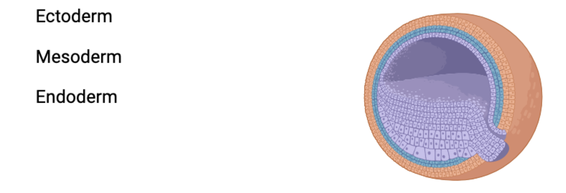
Label this diagram
Ectoderm (orange), mesoderm (blue), purple (endoderm)
What is the ectoderm?
The outer germ cell layer. During neuralation, folds into the neural tube that later becomes the nervous system. Also forms the epidermis, and most of the head (eyes, ears, nose)
What is the mesoderm?
The middle germ cell layer. In early development, the middle layer of the mesoderm stiffens into the notochord. It also creates somites. Also forms the circulatory, muscle, skeletal, lymph, and reproductive systems.
What is the endoderm?
The innermost germ cell layer. In ealry development, becomes the coelom and the gut tube. Also forms the digestive, respiratory, and endocrine system.
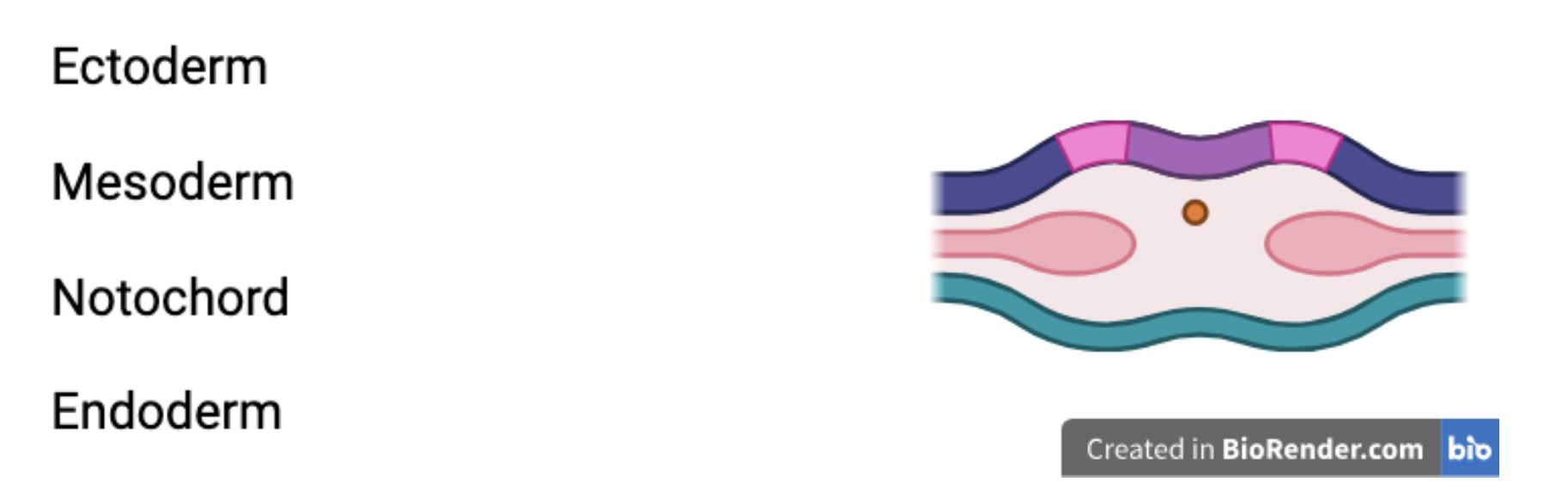
Label this diagram
ectoderm (dark blue), mesoderm (light pink + orange), ectoderm (cyan)
What is neuralation?
When the central plate of ectoderm folds and zips itself into the neural tube, releasing neural crest cells in the process. Yolk sac gets used up in this process.
What is Spina Bifida?
Incomplete fusion of neural tube
Anterior/Ventral
Front
Posterior/Dorsal
Back
What is the most similar developmental stage across species?
Phylotypic Stage
What process is happening here?
Neuralation
What are somites?
Paired segments of mesoderm on the left and right sides of the neural tube and notochord (initially, just swellings)
What do somites give rise to?
Vertebrae and associated muscle and tendons, skeletal muscles of the body wall and limbs, Dermis, some of the PNS
Why are somites crucial for the body plan?
SEGMENTATION: HOX Genes and transcription factors
Set up the central axis of the body. Patterning the body plan anterior - posterior axis
Collinear expression
formation and pattern of body plan
Permutation (order matters)
Location of Homeobox genes on chromosomes determine somite position and identity
What are homeobox (HOX) genes? Why are they significant?
An array of 39 genes on 4 chromosomes. Their order & pattern of expression determine position and identity of somites
How many somites do humans have?
42 (4 occipital, 8 cervical, 12 toracic, 5 lumbar, 5 sacral, 8-10 coccygeal)
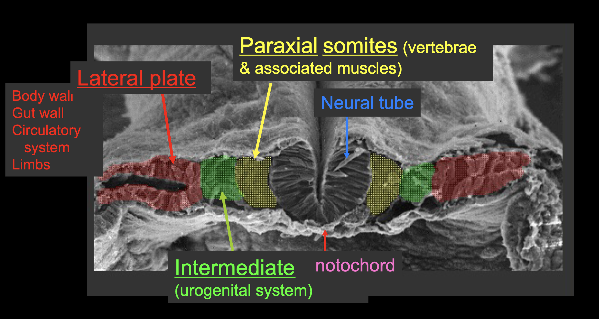
What are the medial to lateral mesoderm differentiations (on either side of somites)?
Paraxial - somites - vertebrae & muscles
Intermediate - urogenital, kidneys, gonads
Lateral plate - body walls, gut wall, circulatory, limbs
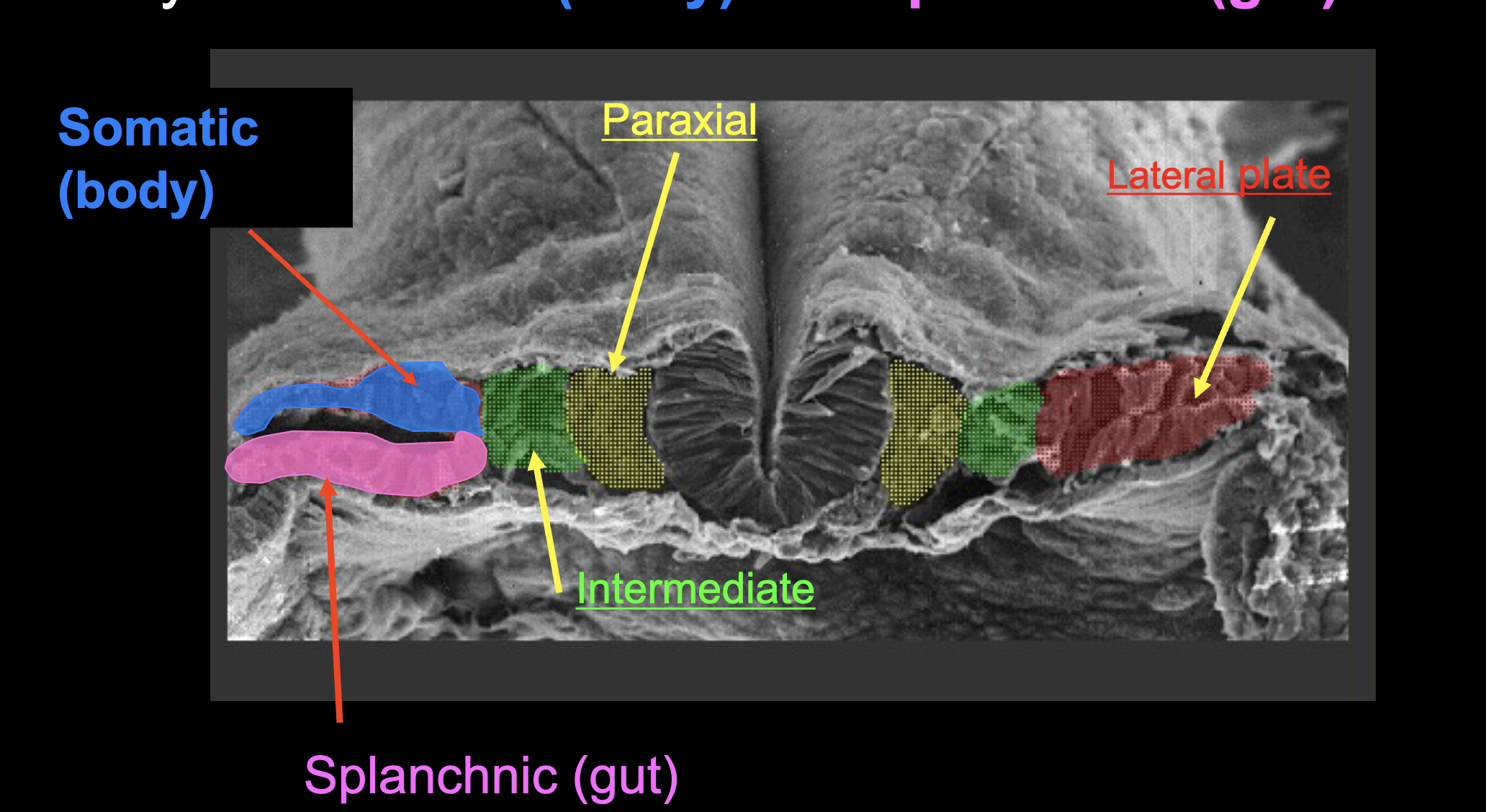
What are the components of the lateral plate mesoderm?
Somatic (top) & Splanchnic (bottom)
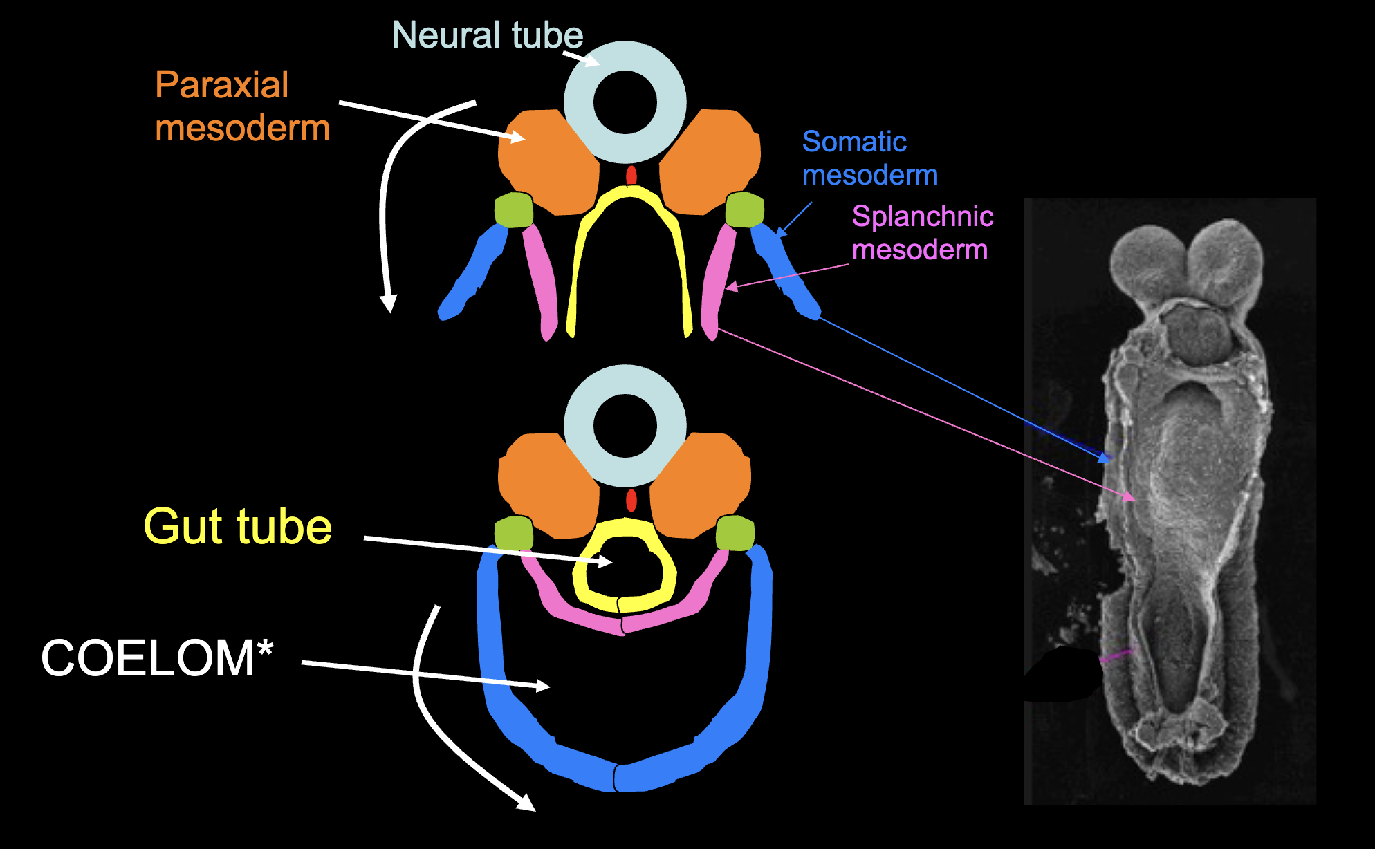
How does the gut tube and coelom form?
When the splanchnic and the somatic mesoderm on either side fold over and meet at the midline. This causes the endoderm layer to fold into the gut tube, and the remaining space to become the coelom (body cavity).
Explain the “Blind Watchmaker” Analogy
-Evolution by Natural Selection is agentless (blind)
- Organisms are complex (like a watch)
- Having nothing “guide” evolutionary changes to
development is like assembling a watch “blind”
What are the two solutions to the “blind watchmaker” problem?
1. Evolutionary changes that generate
complexity occurred from many, many
intermediate steps.
2. The processes by which development
occur permit & promote evolutionary change
and complexity
Body has many parts (modules) that develop via hierarchical
(branching) process with many interactions between modules:
creates integration of many parts into a single, funtional whole
How do we have variation?
Genetic mutations = module variation = novelties!
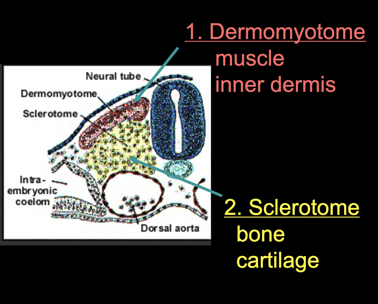
What do the somites (paraxial mesoderm) differentiate into?
Dermomyotome (inner dermis, muscle) and Sclerotome (bone, cartilage)
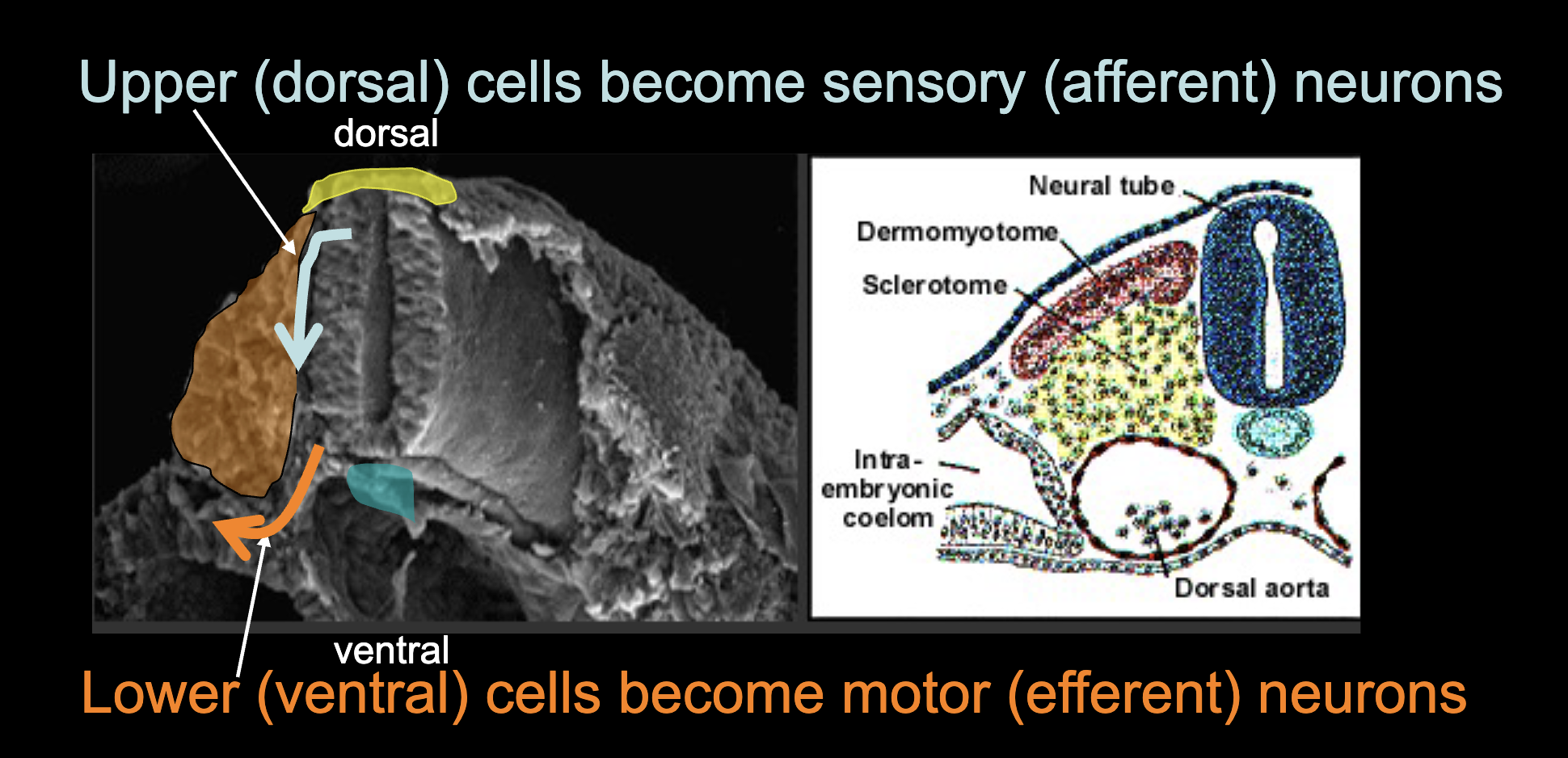
What types of neurons do upper(dorsal)/ lower(ventral) cells of the neural tube and notochord become?
upper(dorsal) - sensory (afferent) neurons
lower(ventral) - motor (efferent) neurons
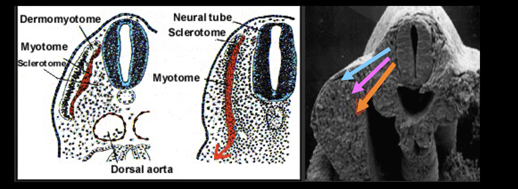
What is happening here?
the myotome cells migrate laterally, bringing with them their bundle of sensory and motor nerves. In this way, muscles and
nerves are connected to each other and to the neural tube as they migrate!
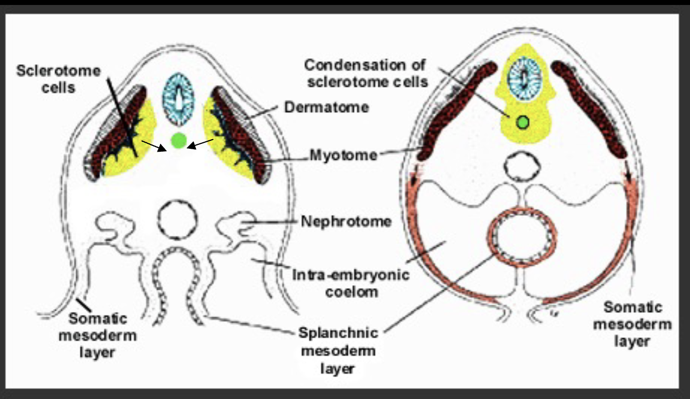
What does the sclerotome become?
The sclerotome condenses around the notochord and neural tube and becomes vertebral cells.
Muscles and nerves subdivide to what sides?
epaxial (dorsal side) - pink, hypaxial (ventral side) - green
Explain ‘resegmentation’ in vertebral cells?
Each vertebra is half of one segment plus half of another. In this way spinal nerves emerge between vertebrae and muscles cross vertebrae.
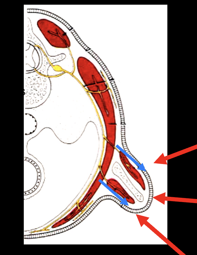
What are primitive fins?
specialized flaps of the body wall (lateral plate mesoderm)
What is happening here?
Limb formation: As the fin grows, the muscles on the ventral side of the animal (hypaxial muscles) split around the fin (each carrying its own nerve supply). Elevators (top arrow), Fin (middle arrow), Depressors (bottom arrow)
What do the bony fish serve to prove in embryology?
Bony fish (Osteichthyes) evolved ≈400 million years ago including one group of lobe-finned fish (Sarcopterygii) with bones in their fins
-LIMBS, THEREFORE, ARE MODIFIED FINS
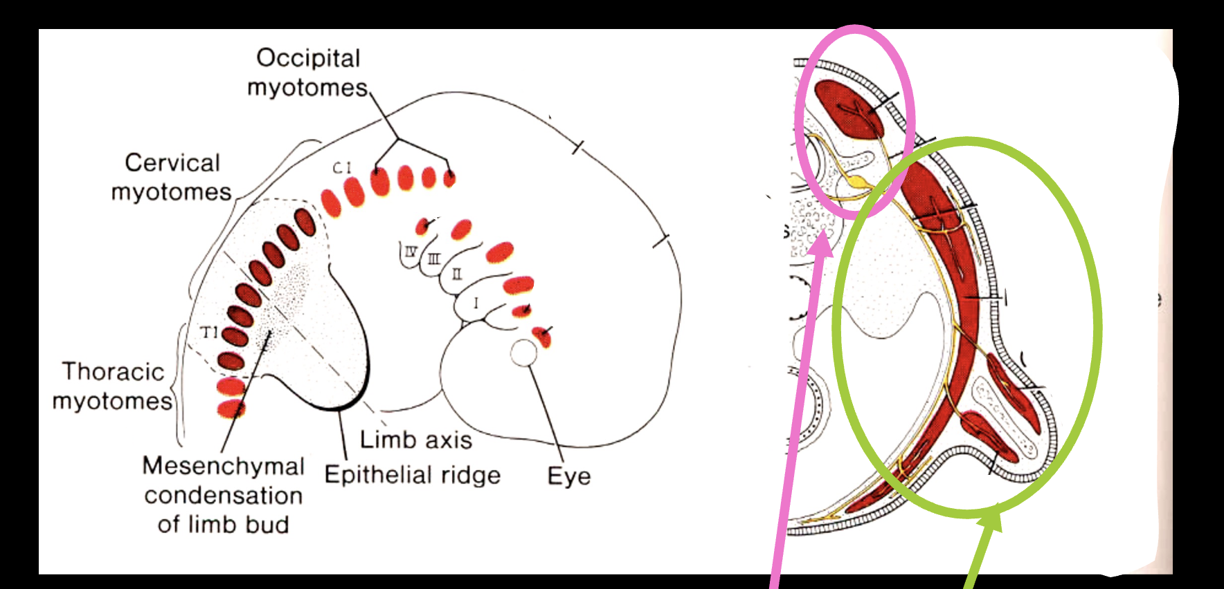
Explain the historical accounts for how limbs develop?
1.BEGIN AS A BUD IN LATERAL PLATE MESODERM
(also muscle cells from myotome)
2.BUD POSITION DETERMINED BY SIGNALING FROM SOMITES (Forelimb vs. Hindlimb)
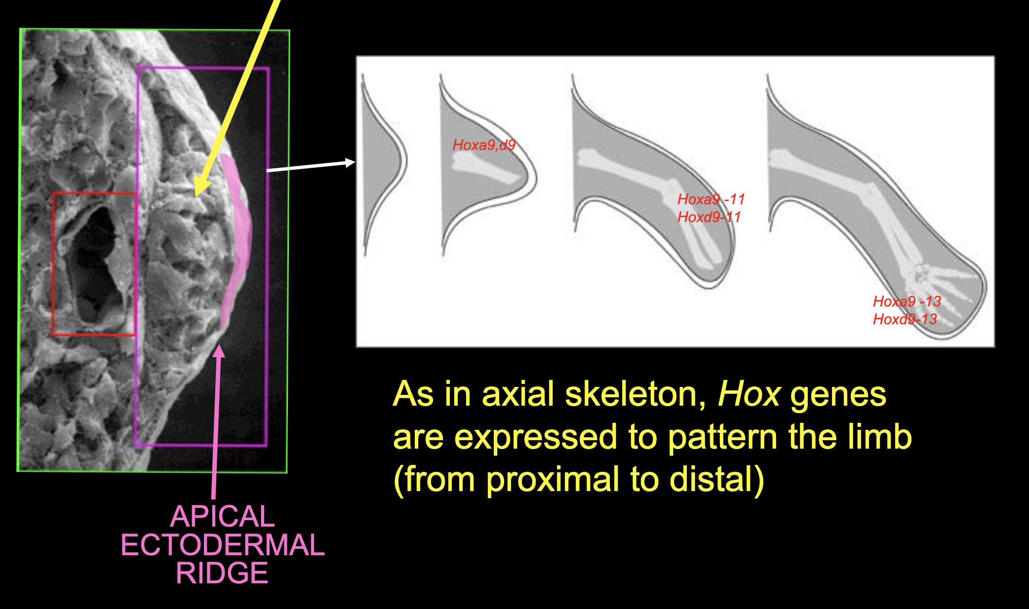
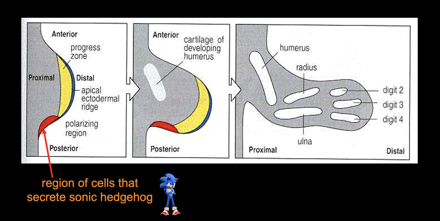
What is the apical ectodermal ridge (AER)?
the “organizing center” - sends out signals that cause pattern of cell differentiation of limbs from proximal to distal
Other than the apical ectodermal ridge, can other genes pattern other axes?
Yes, posterior vs anterior (shh)
dorsal vs ventral (wnt7a)
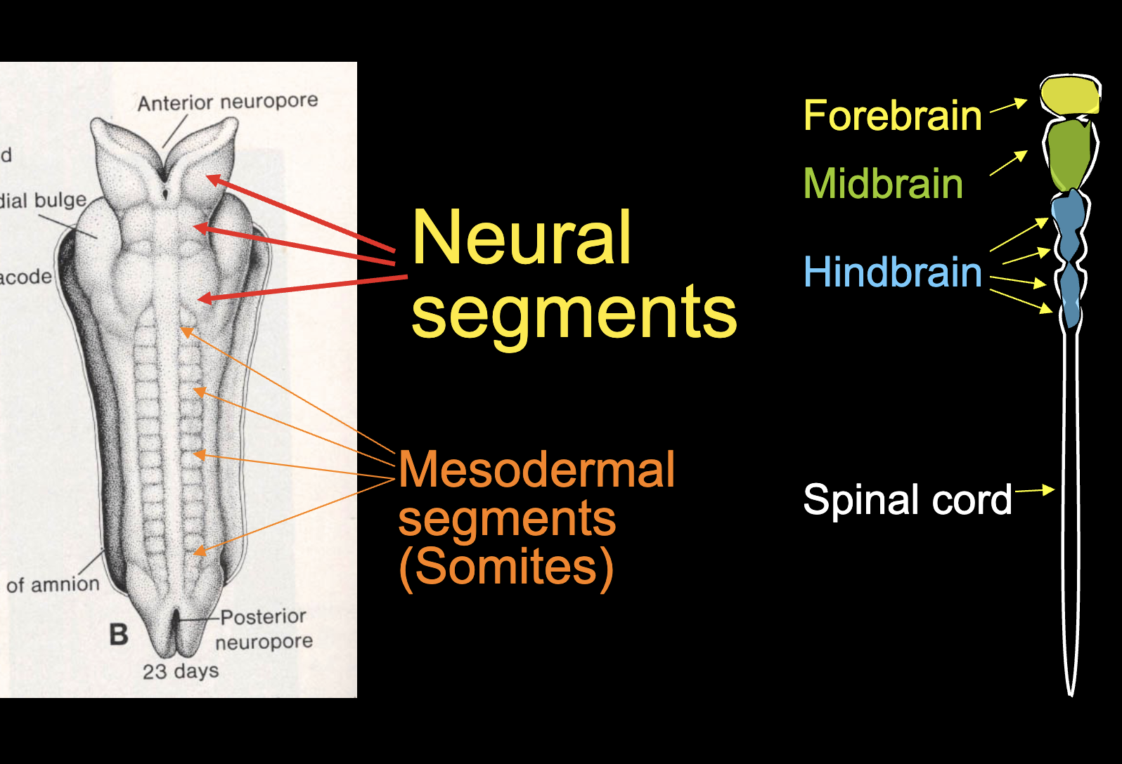
Does the neural tube form segments?
Yes, 3. Forebrain (prosencephalon), Midbrain(mesencephalon), Hindbrain (rhombencephalon). These parts also differentiate into primordial sensory organs (eyes, olfactory cells, etc.)
What are neural crest cells?
Amazing stem cells that are migratory, can differentiate into
many types (pluripotent).
Form much of the head (especially the face and neck)
Also, throughout body: Pigment cells, Adrenal glands, Autonomic (involuntary) nervous system, etc
Describe the movement of neural crest cells?
They stream from back of head towards the front:
-over the top (like a hood) —> cranial vault & upper face (eyes, nose)
- around the sides (like collars) BRANCHIAL ARCHES —> rest of face and neck
What are branchial arches?
The streams of cells that go round the sides and become the rest of the face and neck (around the mouth and gut tube)
- Originally gills, first arch got co-opted as lower jaw
-Each arch includes a nerve, blood vessel, ectoderm, mesoderm and endoderm
Where are branchial arches I & II?
Branchial arch I goes from inner ear to lower jaw
Branchial arch II goes from inner ear to hyoid (above “voicebox”)
The pouch between arches I & II remains a tube (Eustachian
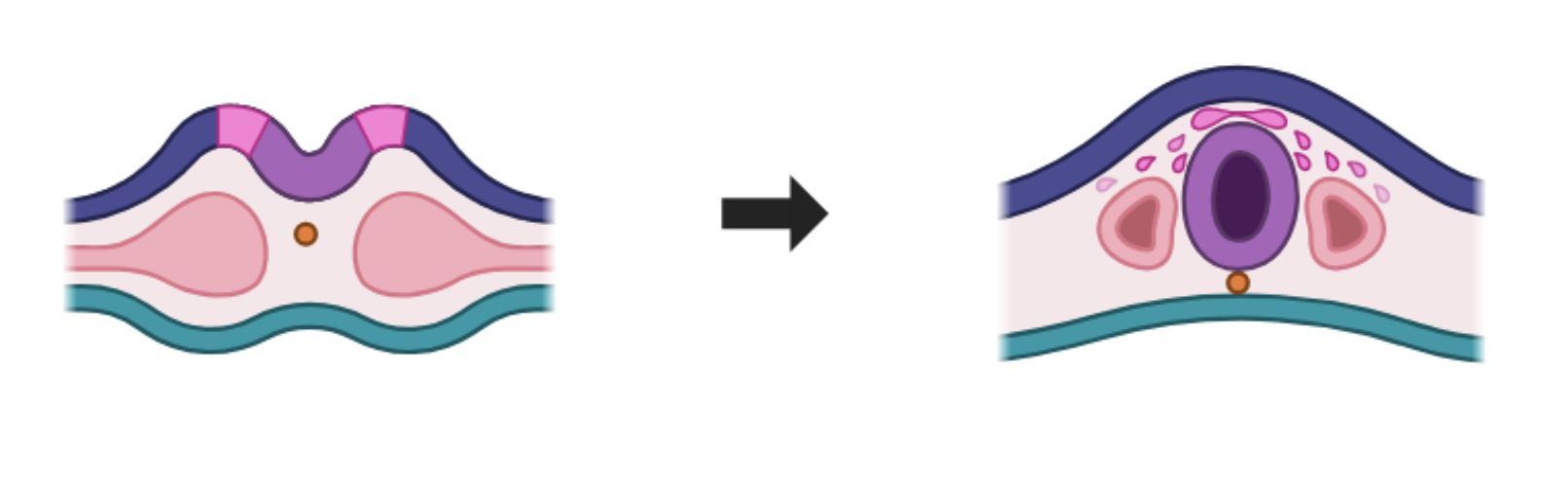
What is a cleft palate an example of?
Incorrect fusion of facial structures
What is the big picture of embryology?
1.How all vertebrates share
basic body plan, 2. Hierarchical pattern of
differentiation and interaction
among derivatives of 3 germ
layers permits integration &
evolvability, 3. We really are modified fish!