Organs of Special Sense Pathology Image-Based Learning
1/176
There's no tags or description
Looks like no tags are added yet.
Name | Mastery | Learn | Test | Matching | Spaced |
|---|
No study sessions yet.
177 Terms
Investigating deathness - BAER test
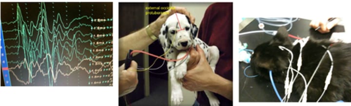
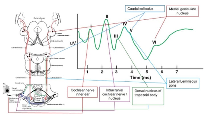
Brainstem Auditory Evoked Response Results
I – cochlear nerve of the inner ear
II – intracranial cochlear nerve/ nucleus
III – dorsal nucleus of trapezoid body
IV – lateral lemniscus pons
V – caudal colliculus
VI – medial geniculate nucleus
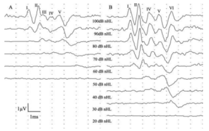
BAER slope shifted to the right with conductive hearing loss
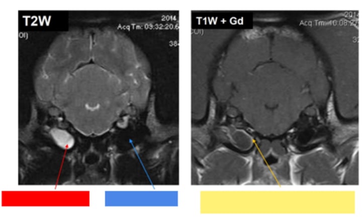
Otitis media/interna
Red - fluid filled bullae
Blue - air filled bullae
Yellow - gadolinium enhancement (increased blood supply with inflammation)
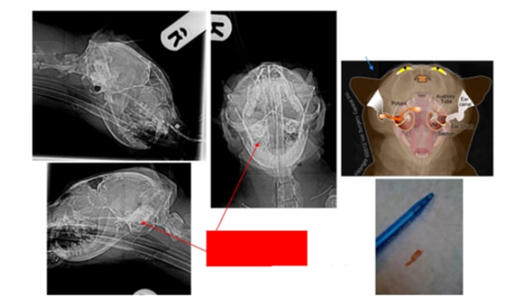
Bulla radiographs - Loss of air filled bullae
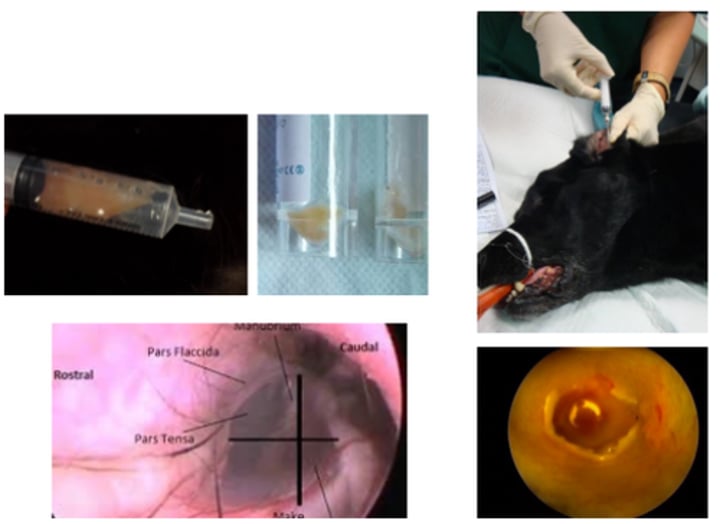
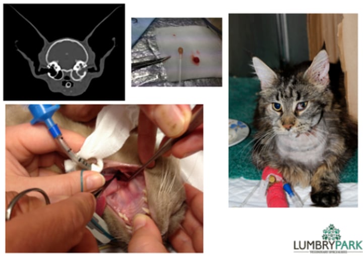
Myringotomy
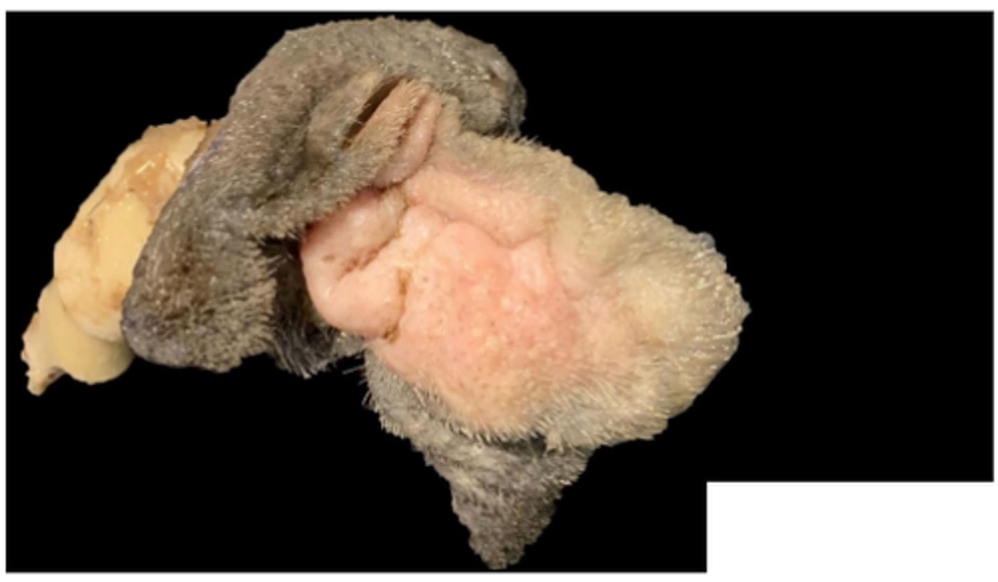
Feline nasopharyngeal polyp
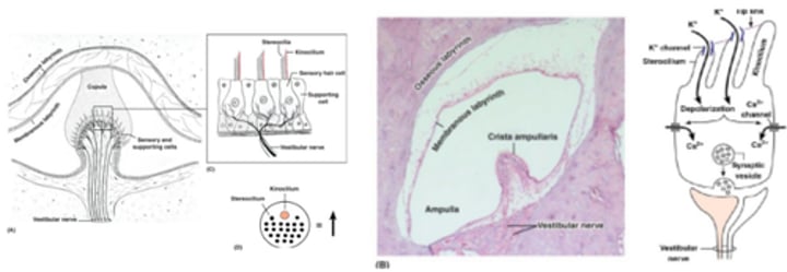
Sensory hair cells of crista ampulla (semicircular canal)
Movement: endolymph bends hair cells
Different orientations mean directionally sensitive
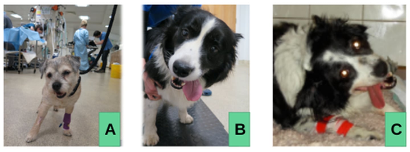
Peripheral vestibular disease
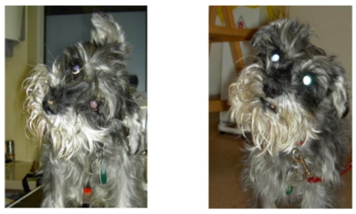
Peripheral vestibular disease - hypothyroidism
Vestibular disease and bilateral facial palsy
Second image: after 2 weeks of Levothyroxine therapy
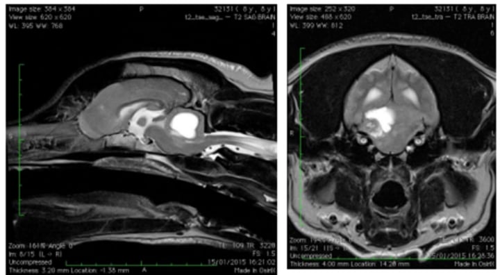
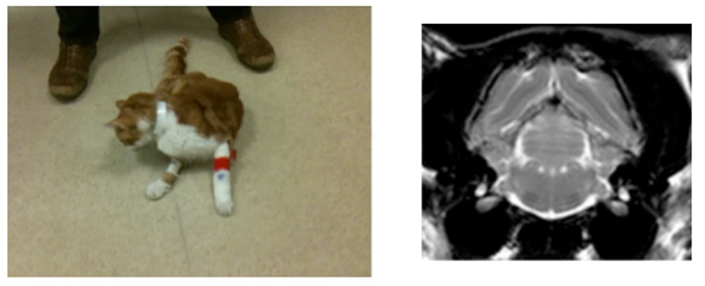
Central vestibular disease
Paradoxic vestibular syndrome
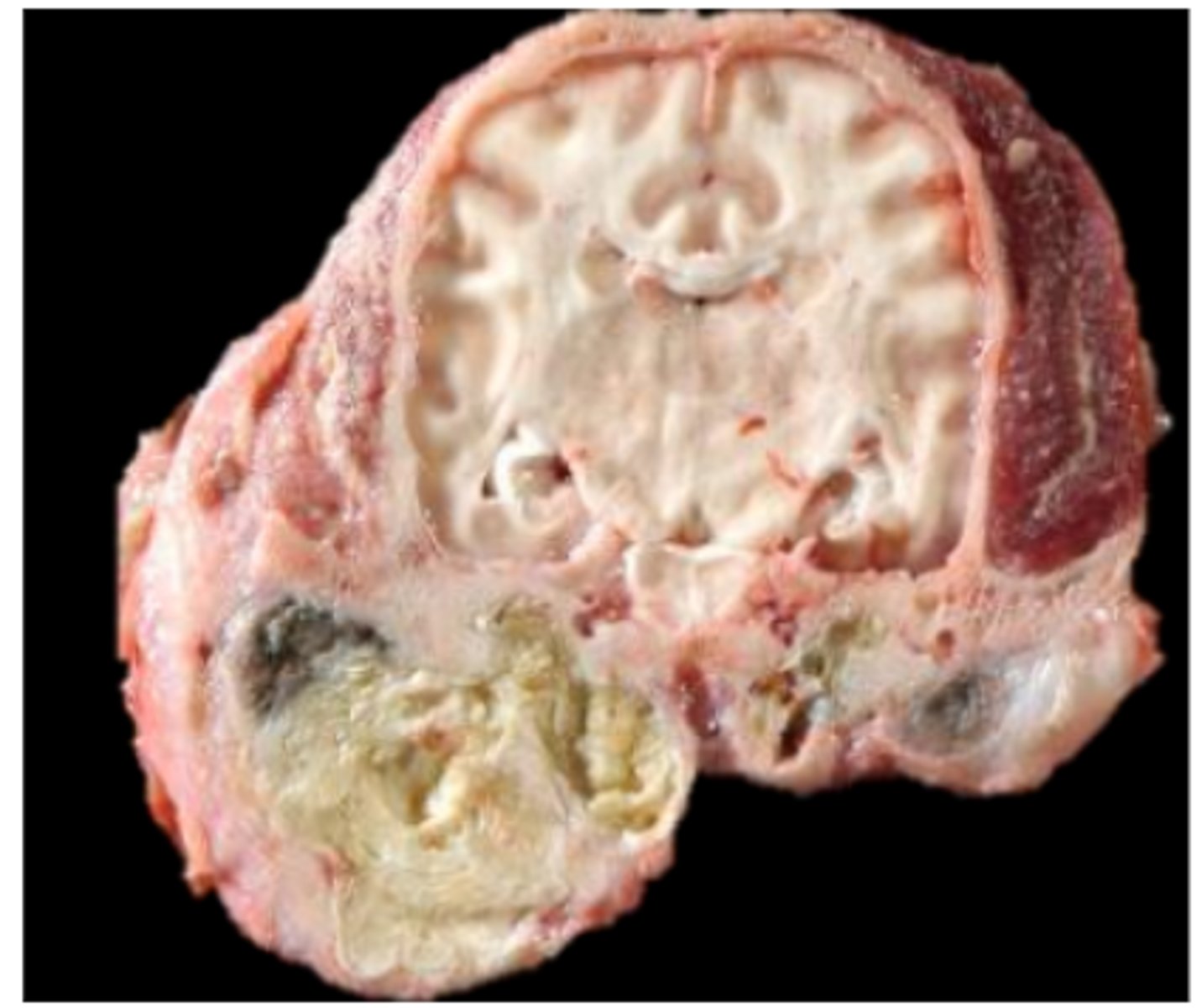
Carcinoma in 4th ventricle
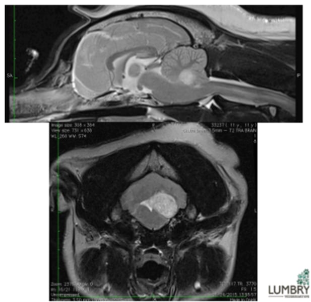
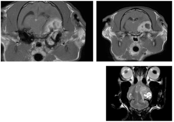
Otitis media-interna – secondary meningoencephalitis
Infection of middle ear cavity where peripheral disease has progressed to central disease
Paradoxical vestibular disease
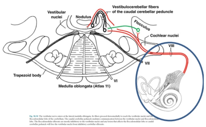
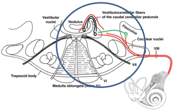
Lesions resulting in loss of inhibitory influence of cerebellum
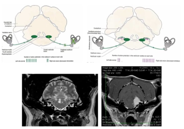
Paradoxical vestibular disease
Cystic tumour between the feedback from the cerebellum and brainstem
Tilting of the head due to nerve impulses firing only on one side resulting in contraction of extensor muscles on the other side
Thiamine deficiency - Bilateral vestibular disease
Malassezia infection
Sebaceous gland hyperplasia, hyperkeratosis, ceruminous debris
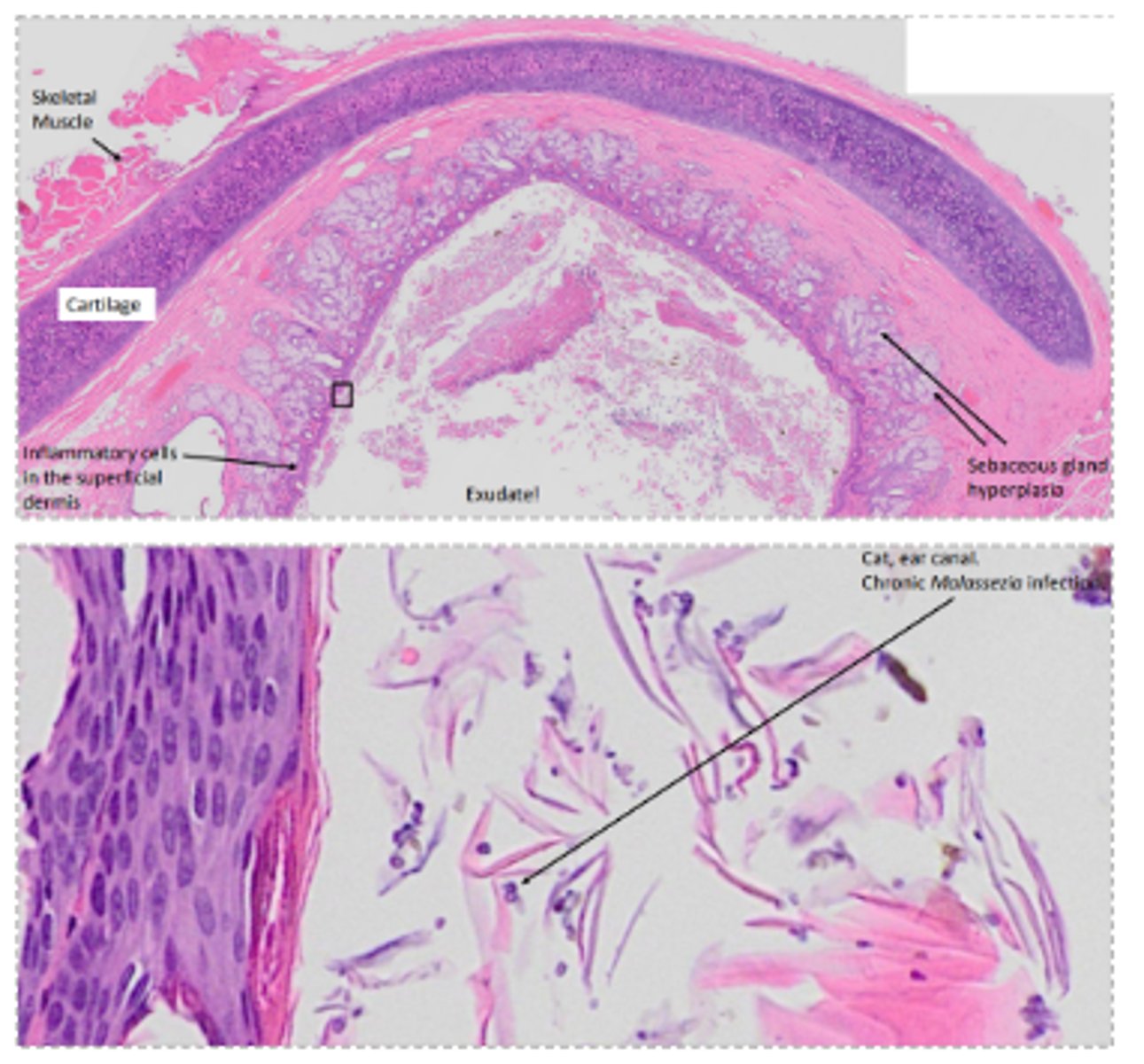
Otitis externa - chronic
Thickened, leathery, narrowing of the external acoustic meatus (stenosis), hyperplasia, dermal changes, osseous metaplasia of soft tissues
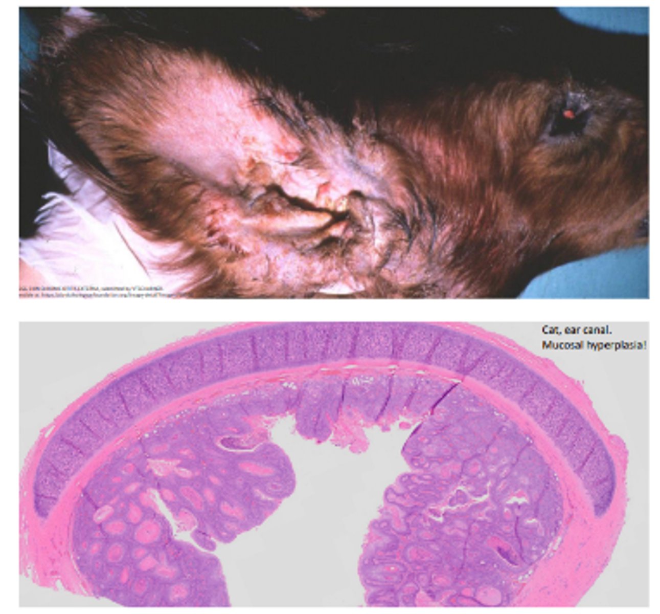
Otitis externa
Narrowing of the external acoustic meatus (stenosis), hyperplasia, dermal changes, oedema (acute), fibrosis (chronic), mixed inflammation (plasma cells, lymphocytes, mast cells, neutrophils), osseous metaplasia of soft tissues
Otitis externa
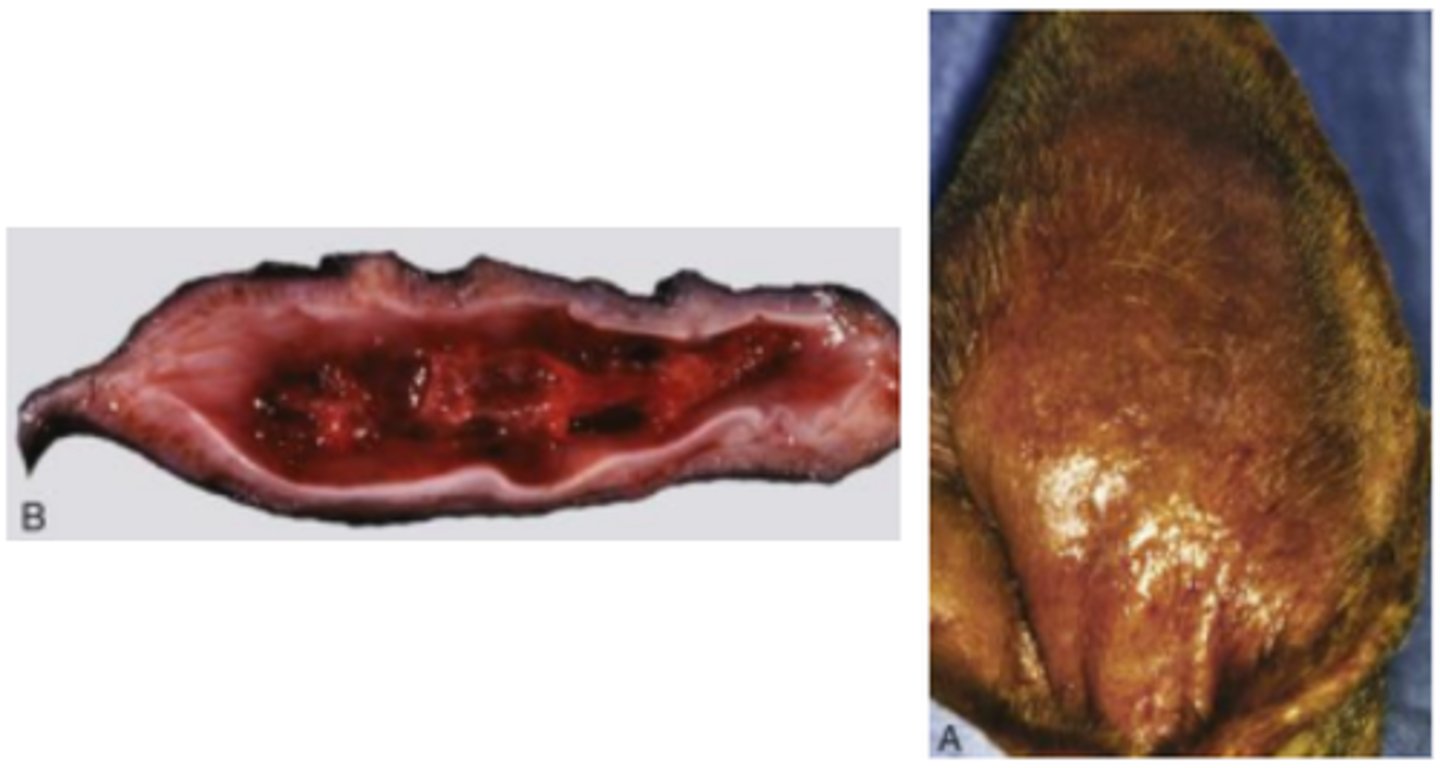
Cartilage splitting (fracture), fibrosis, healed haematoma
Aural haematoma
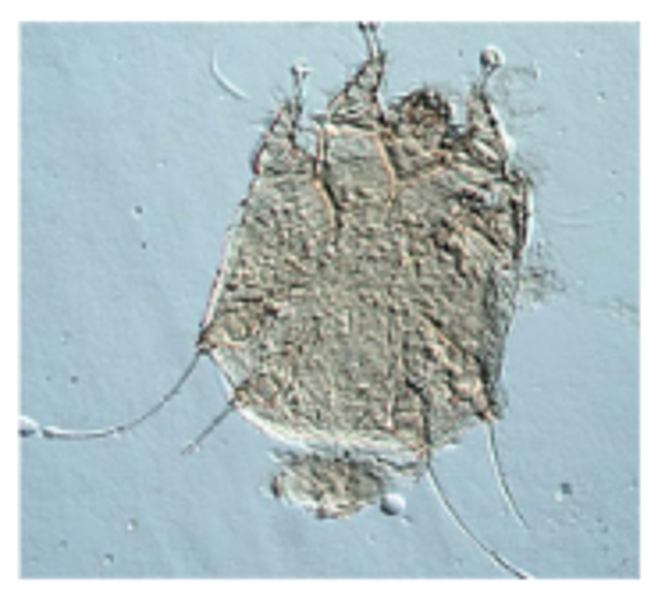
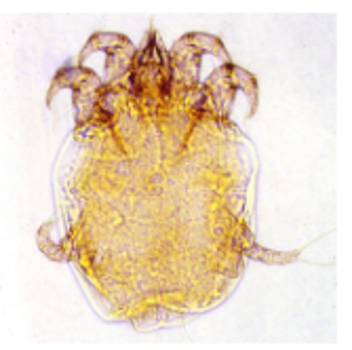
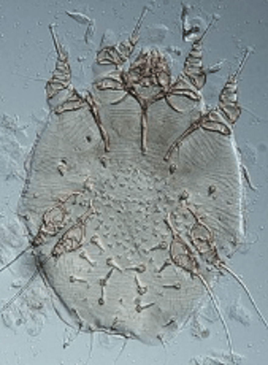
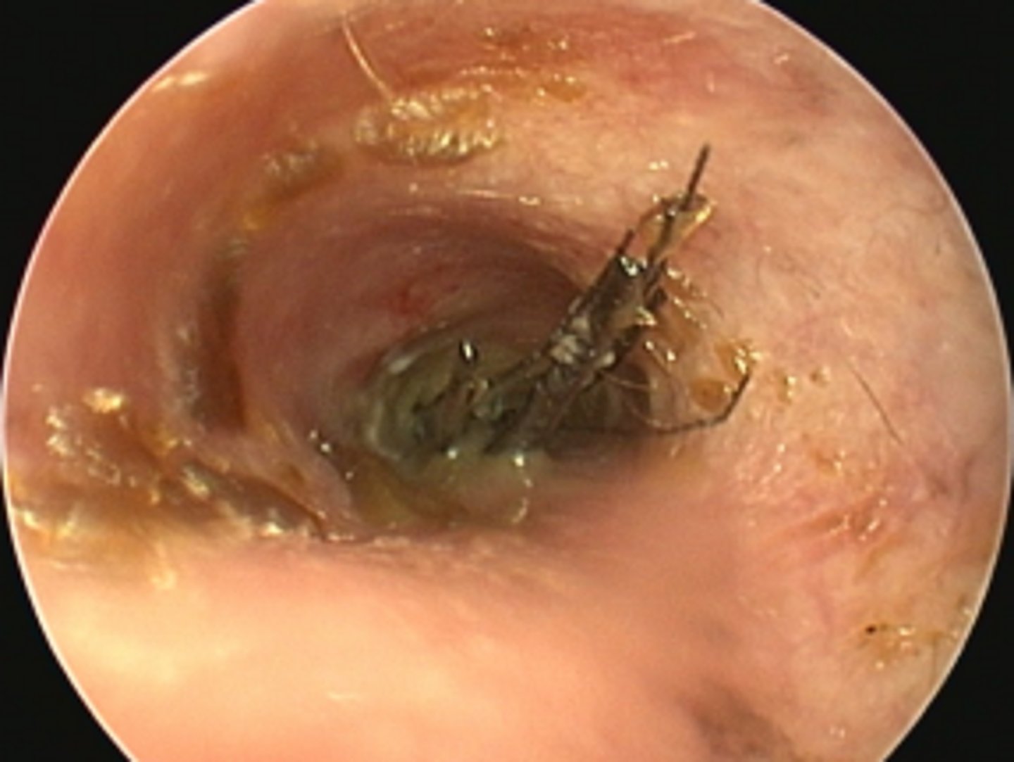
Notoedres cati
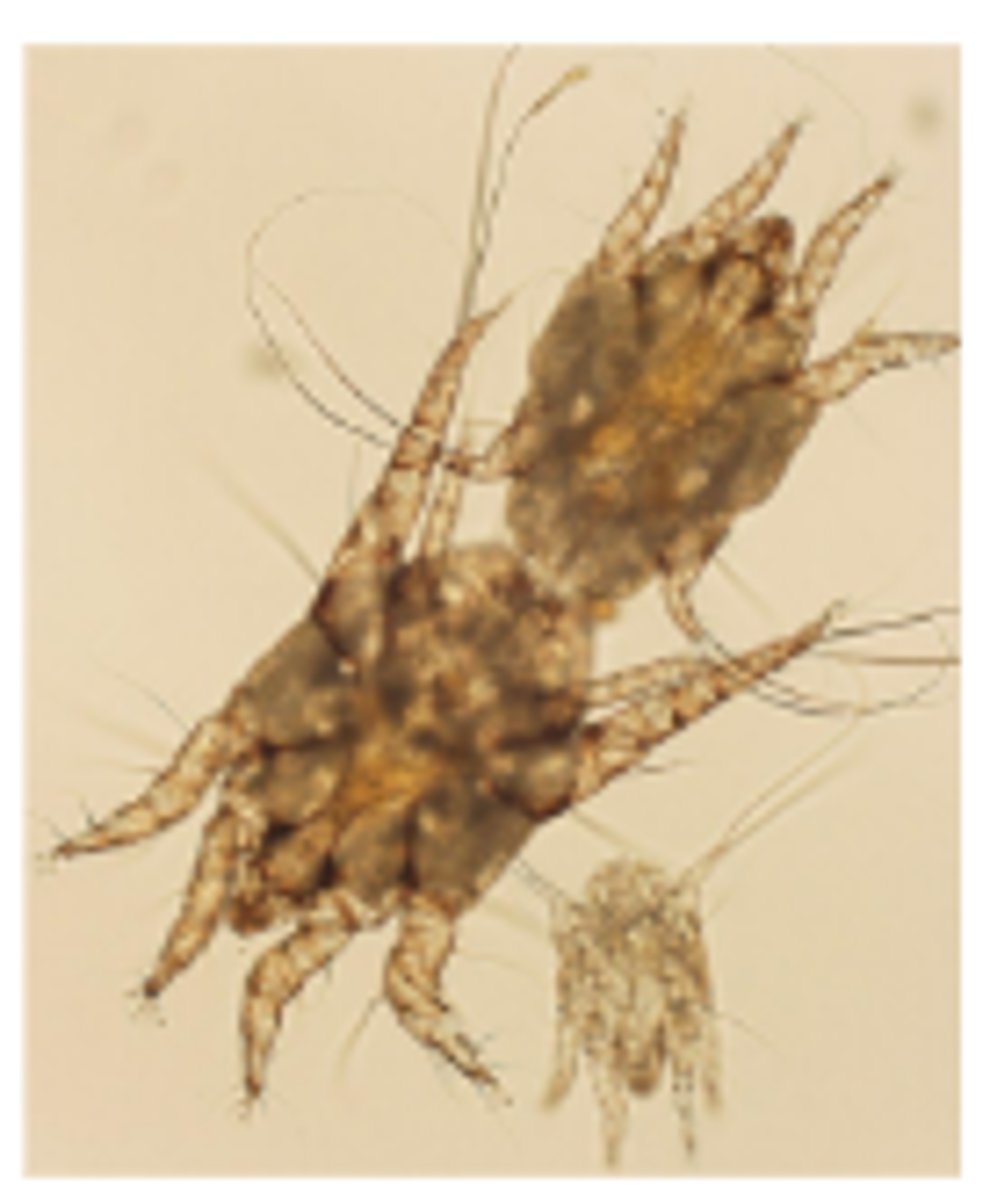
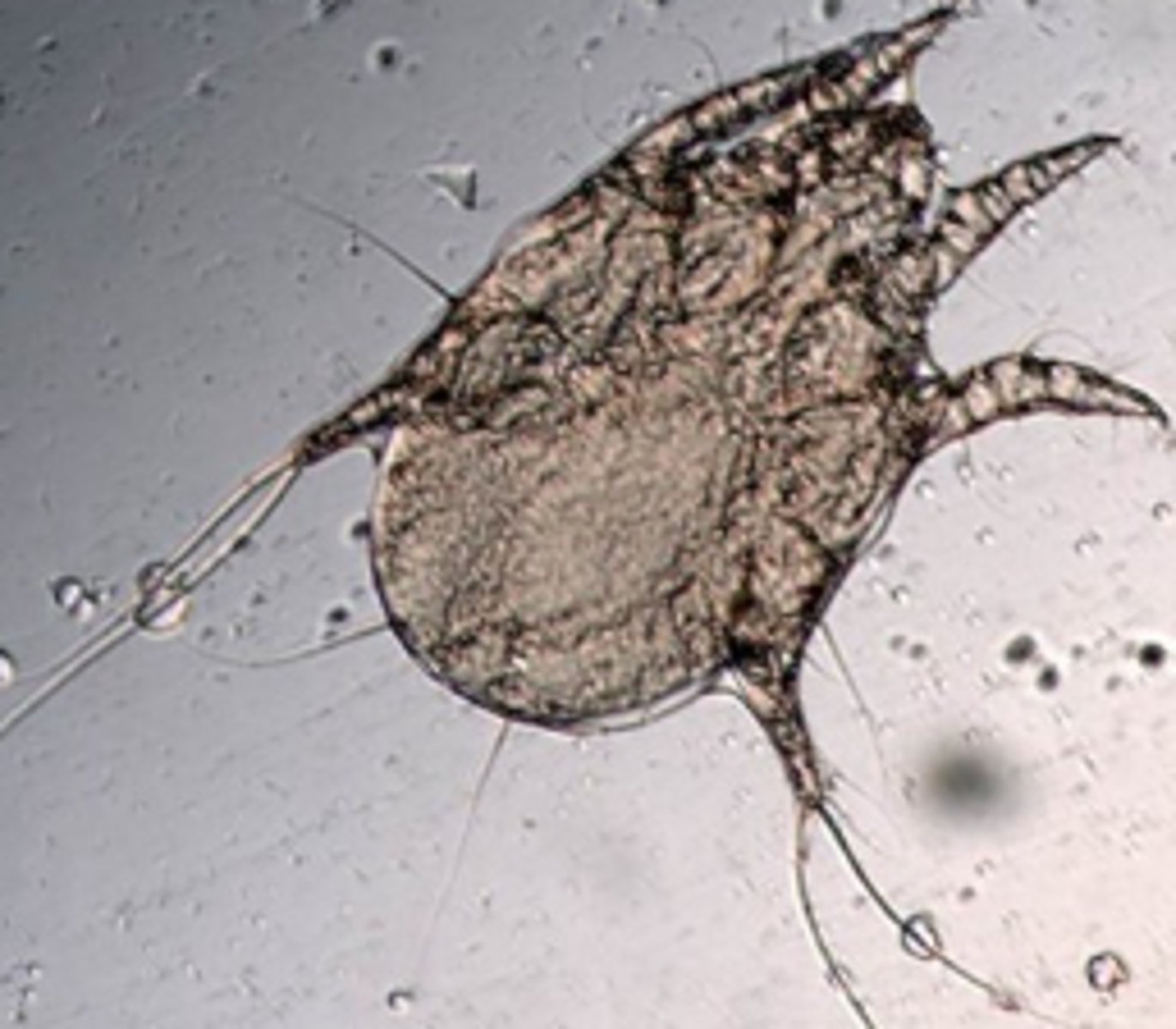
Otodectes cyanotis
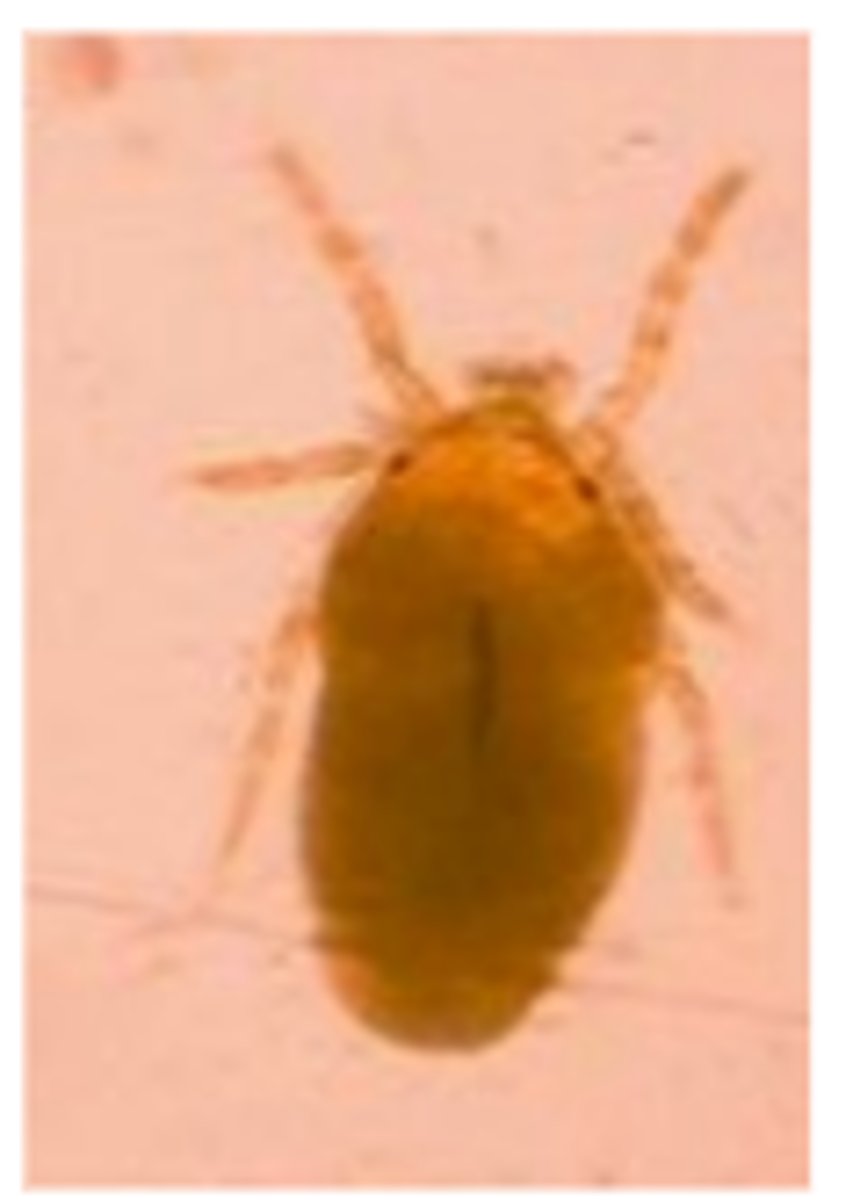
Psoroptes cuniculi
Neotrombicula autumnalis (Harvest mite)
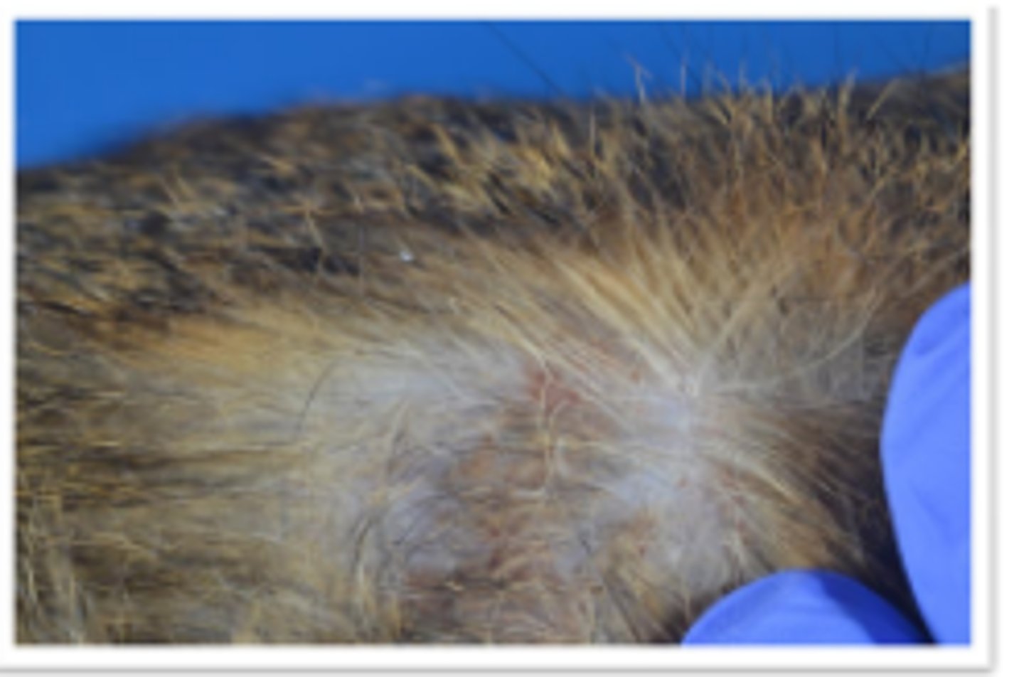
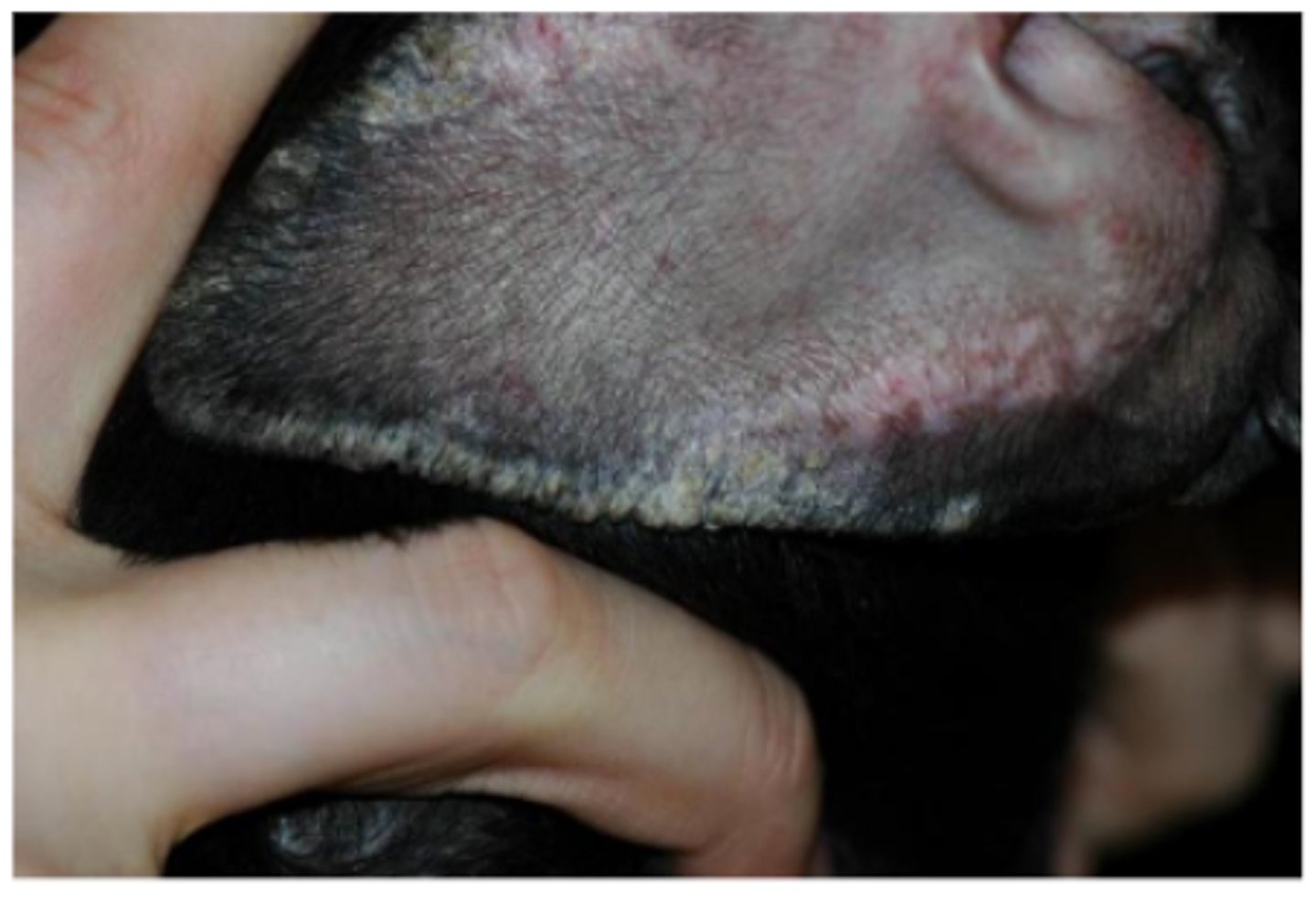
Atopic dermatitis - Primary cause of otitis externa (OE)
Hyperpigmentation, lichenification, pyoderma secondary to pruritus
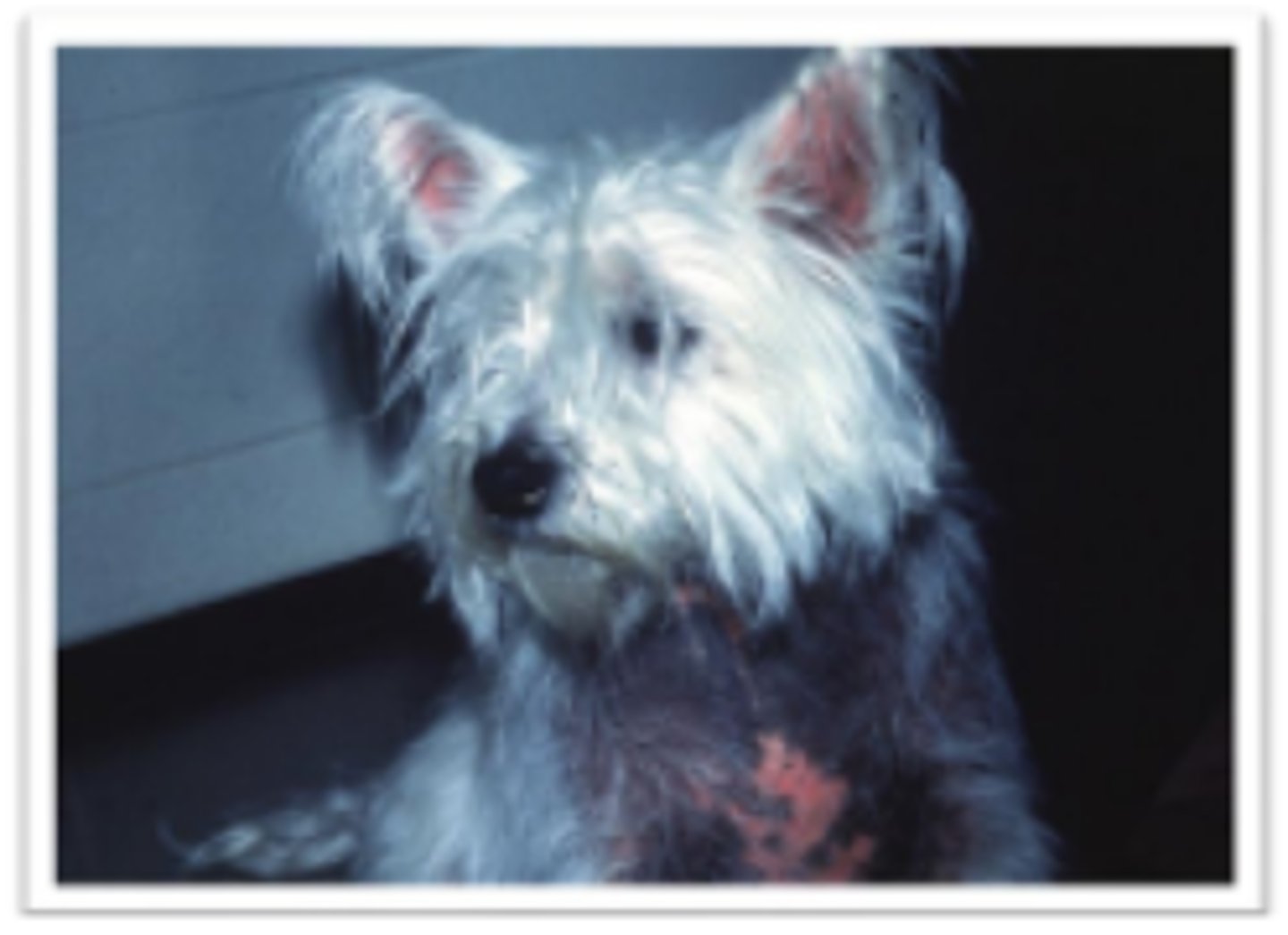
Allergic skin disease - Primary cause of otitis externa (OE)
Hyperpigmentation, lichenification, pyoderma secondary to pruritus
Ear margin seborrhea
Waxy crusts, small fissures
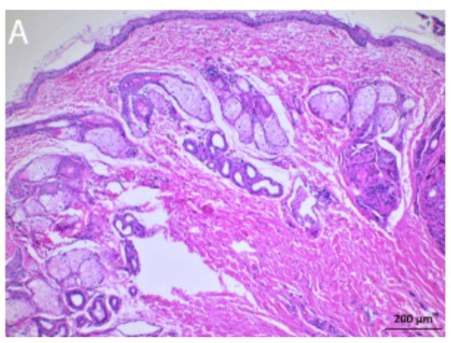
Dog, ears. Normal ceruminous glands in clinically healthy dog
Increased keratin thickness layers, increased size and numbers of glands
Mild epidermal hyperplasia and compact orthokeratotic hyperkeratosis, mild infundibular hyperkeratosis, numerous sebaceous glands and multifocal ceruminous gland ectasia (L)
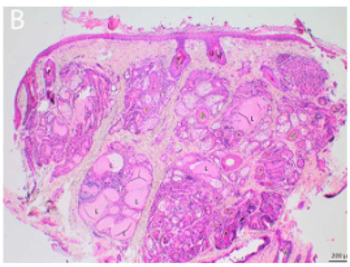
Otitis externa, dog pinna
Marked multifocal ceruminous gland hyperplasia and ectasia, and periadnexal pigment-laden macrophages
Lupus erythematosus
Scaling, crusting (multifocal to coalescing)
Pemphigus foliaceus
Scaling, crusting (multifocal to coalescing)
Vascular disease leading to primary otitis externa
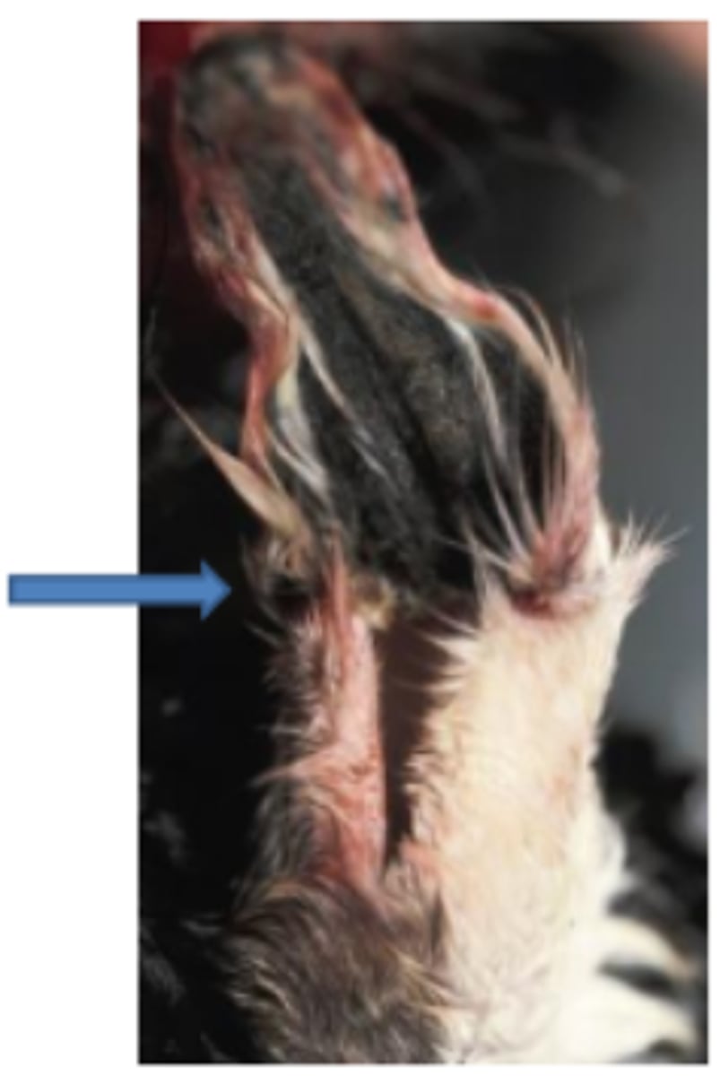
Auricular infarction, frost bite, Goat. Dry gangrene. Note the clear line of demarcation between dead and living tissue
Cat ear canal, Malassezia chronic infection
Otitis media
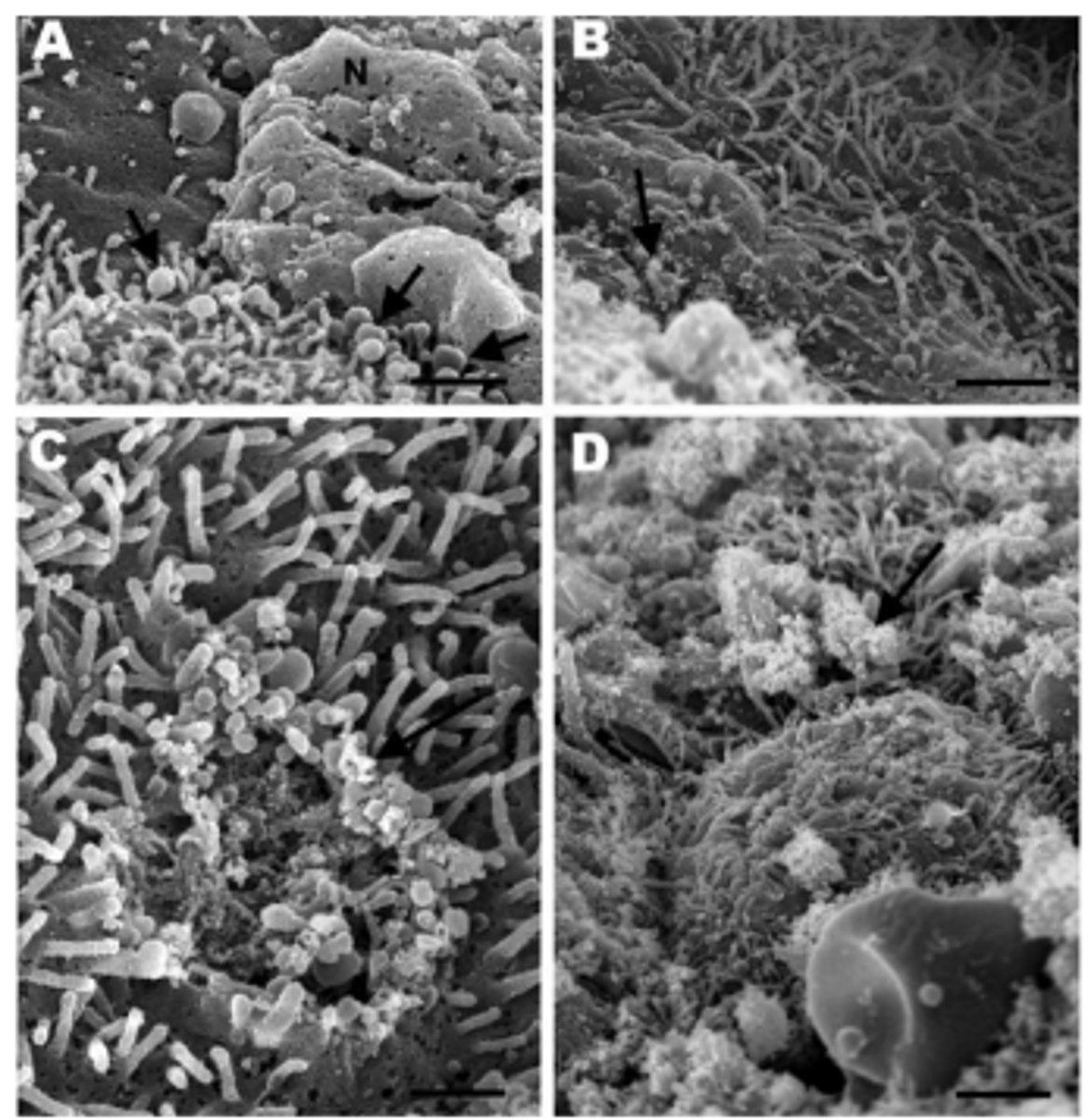
Ferret, ciliated epithelium (lung) with mycoplasmosis
Multifocal loss of cilia with mycoplasma-like organisms (arrows)
Mycoplasm trying to blend in with the cilia, but ultimately causing cilia death (c)
Otitis media gross pathology (Mycoplasma bovis)
Caseous, necrotic exudate, suppurative inflammation
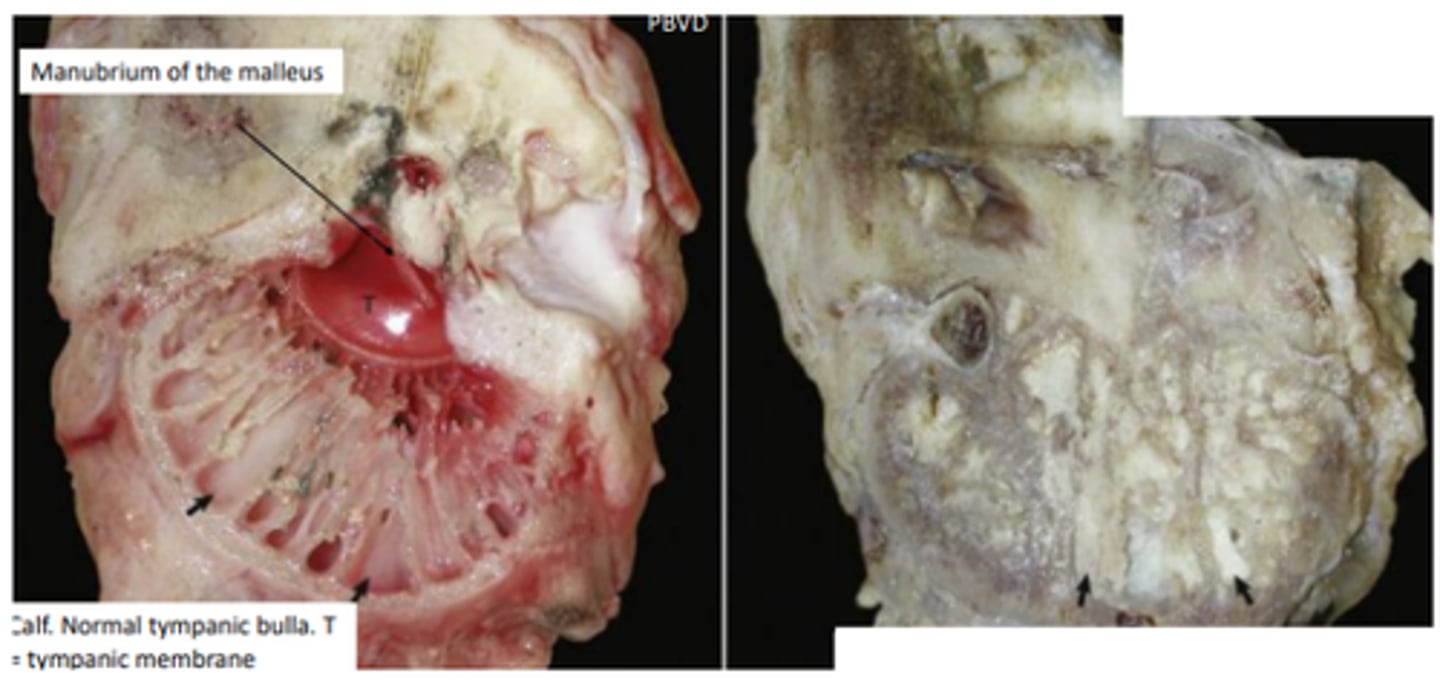
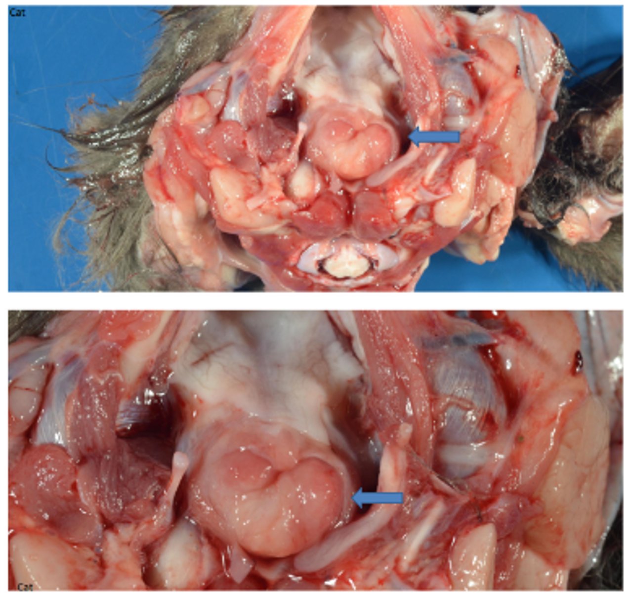
Bilateral suppurative otitis media
Tympanic cavities bilaterally filled with soft, yellow-tan exudate (pus; otitis media).
Arrow; eustachitis
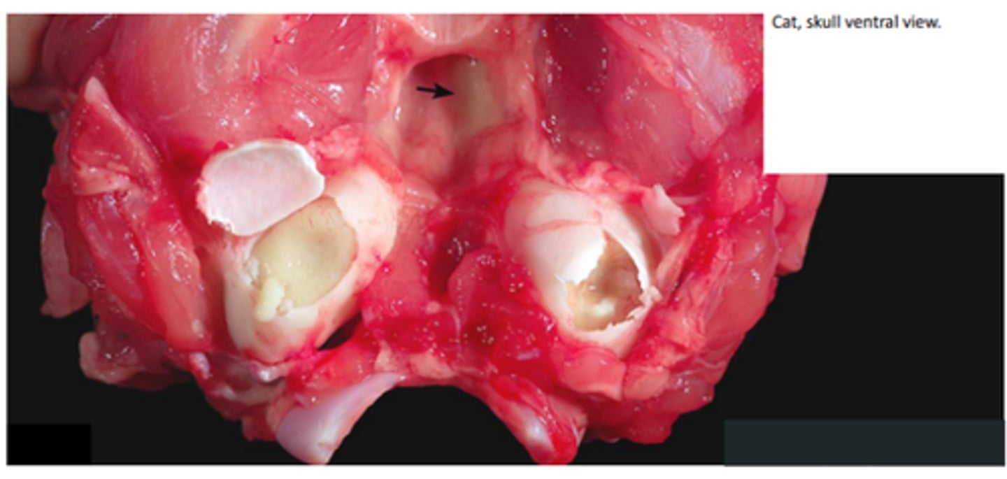
Otitis media
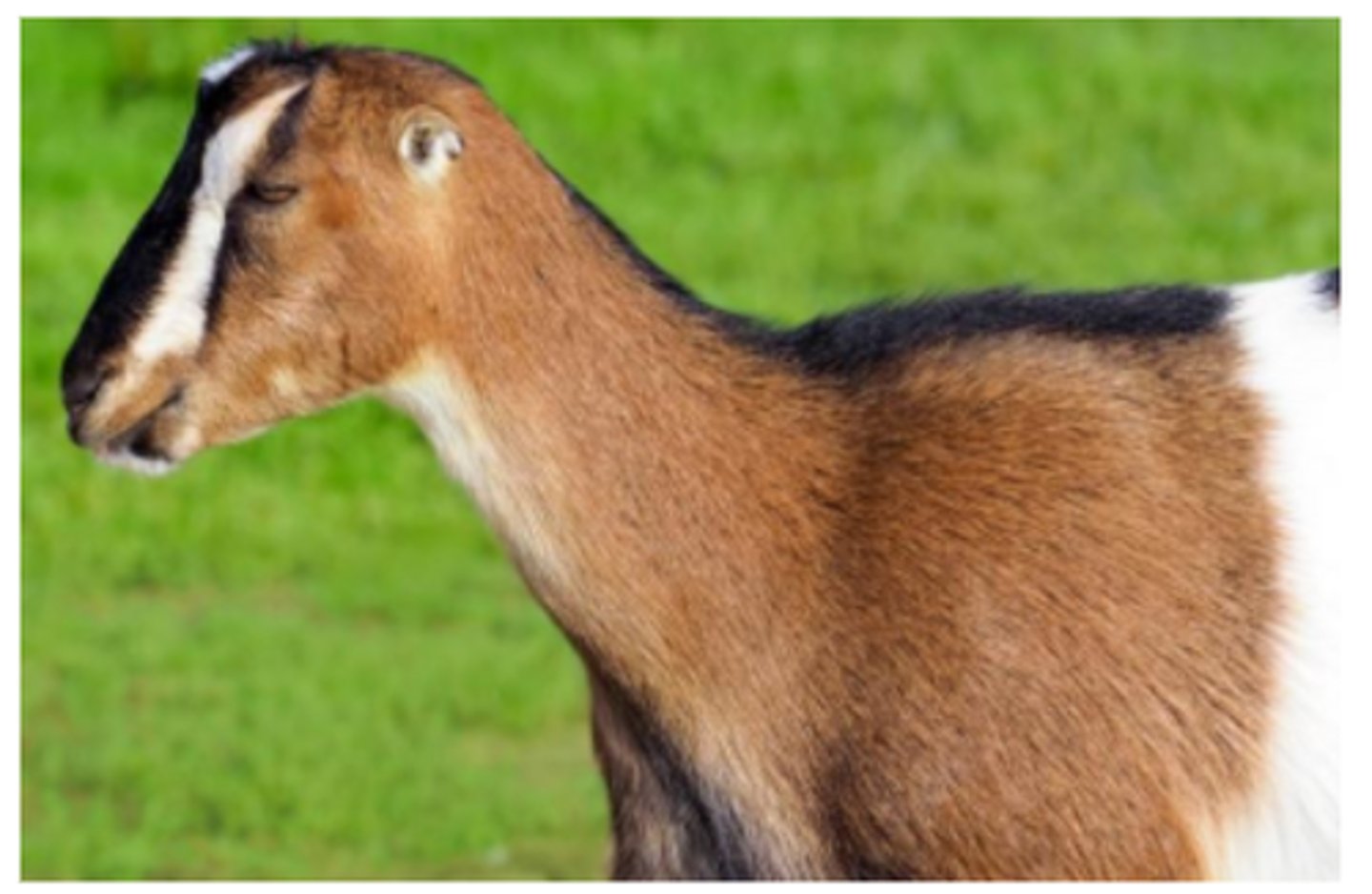
Microtia
Small pinna - La Mancha goat
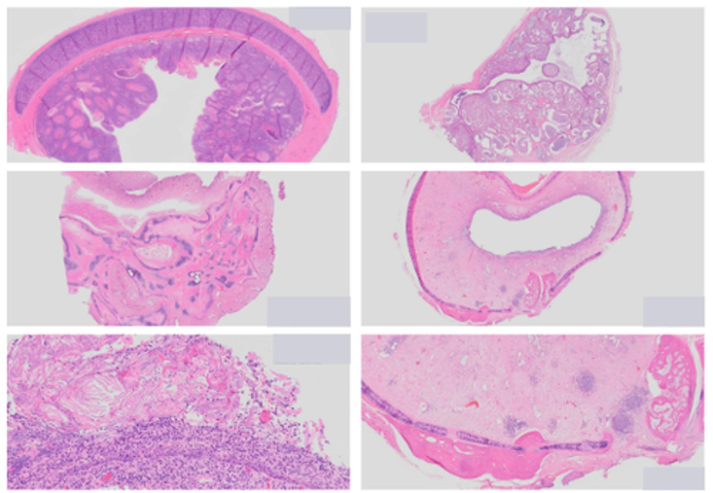
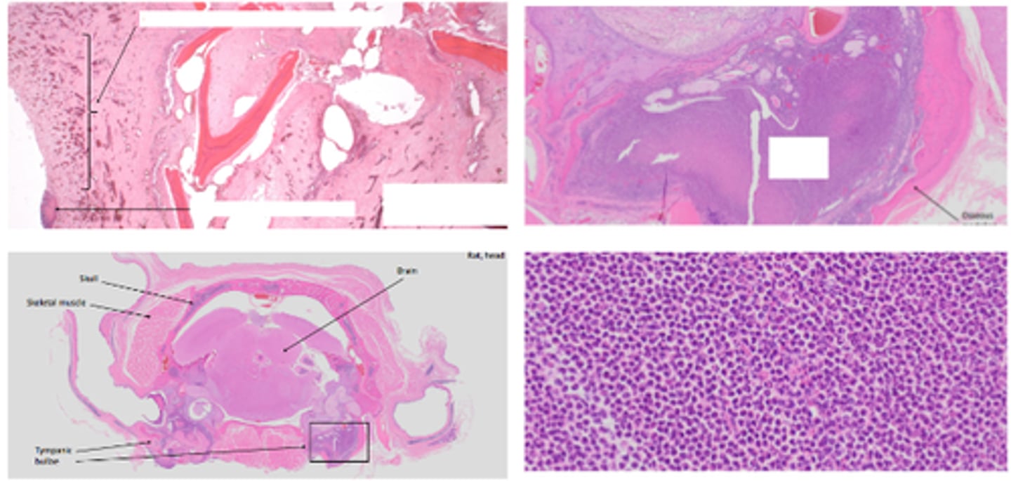
Nasopharyngeal polyps
Gross pathology: pedunculated to polypoid mass with smooth surface, variably ulcerated mucosal surface
Histopathology: fibrovascular core, lymphocytes, plasma cells, histiocytes
Nasopharyngeal polyps
Hyperplasia
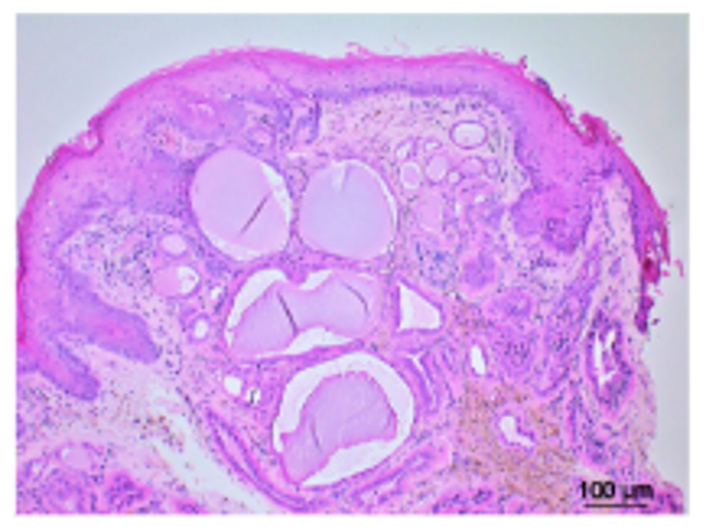
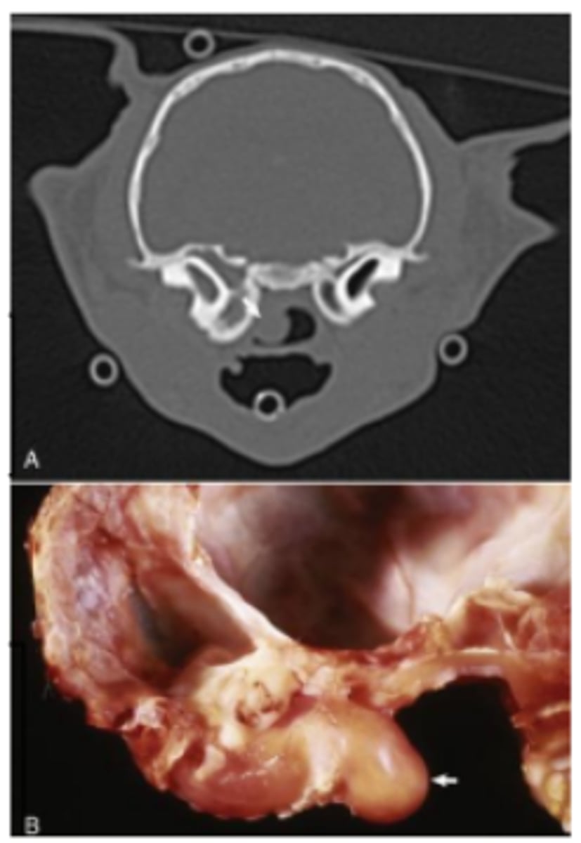
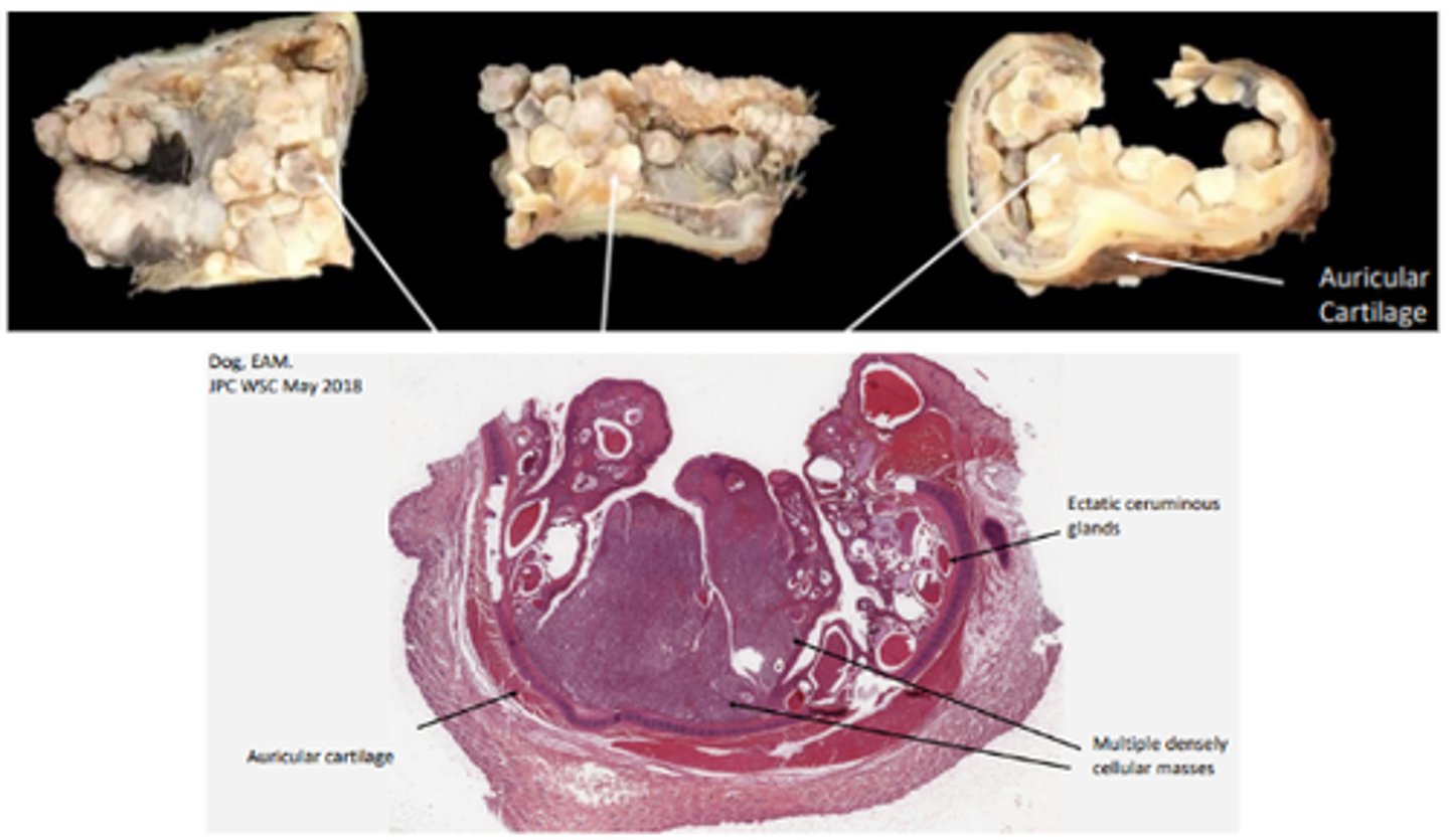
Tympanokeratoma
Gross pathology: tympanic cavity filled and expanded with pasty, yellow to pale-tan material, loss of normal structures (ossicles)
Histopathology: keratin surrounded by squamous epithelium, granulation tissue +/- otitis media (+/-) cholesterol clefts and granulomatous inflammation (cholesterol granulomas)
Ceruminous gland adenocarcinoma
Multifocal to coalescing nodules; firm, pale tan; EAM obstruction (perpetuates otitis externa!)
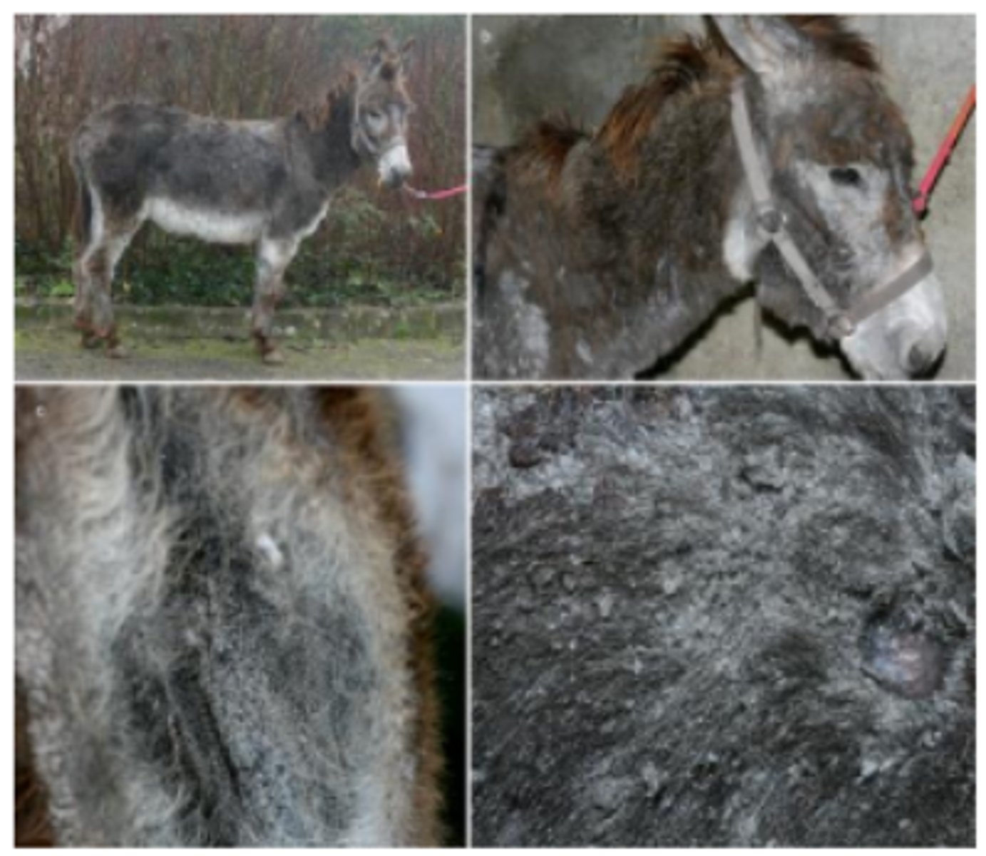
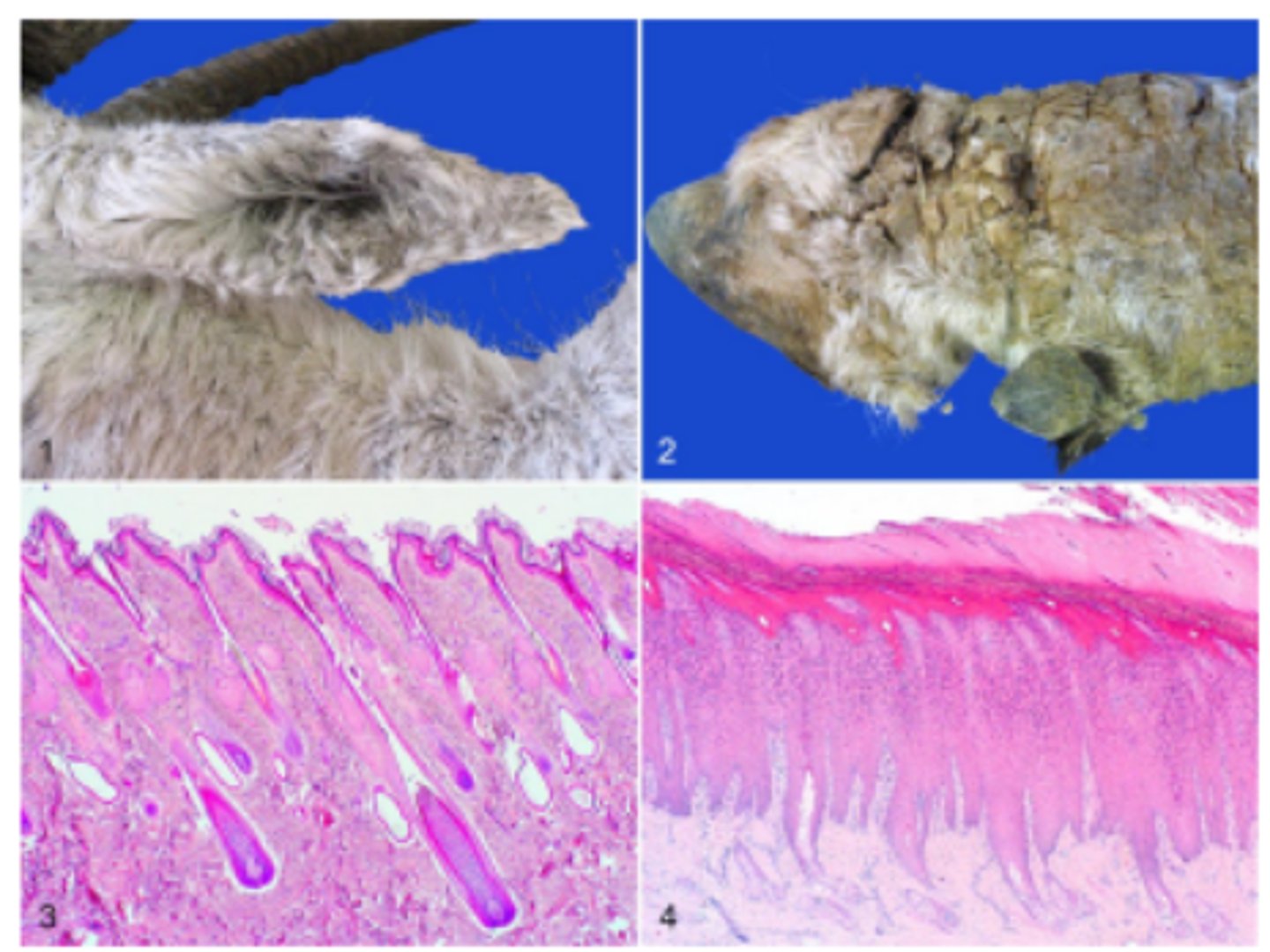
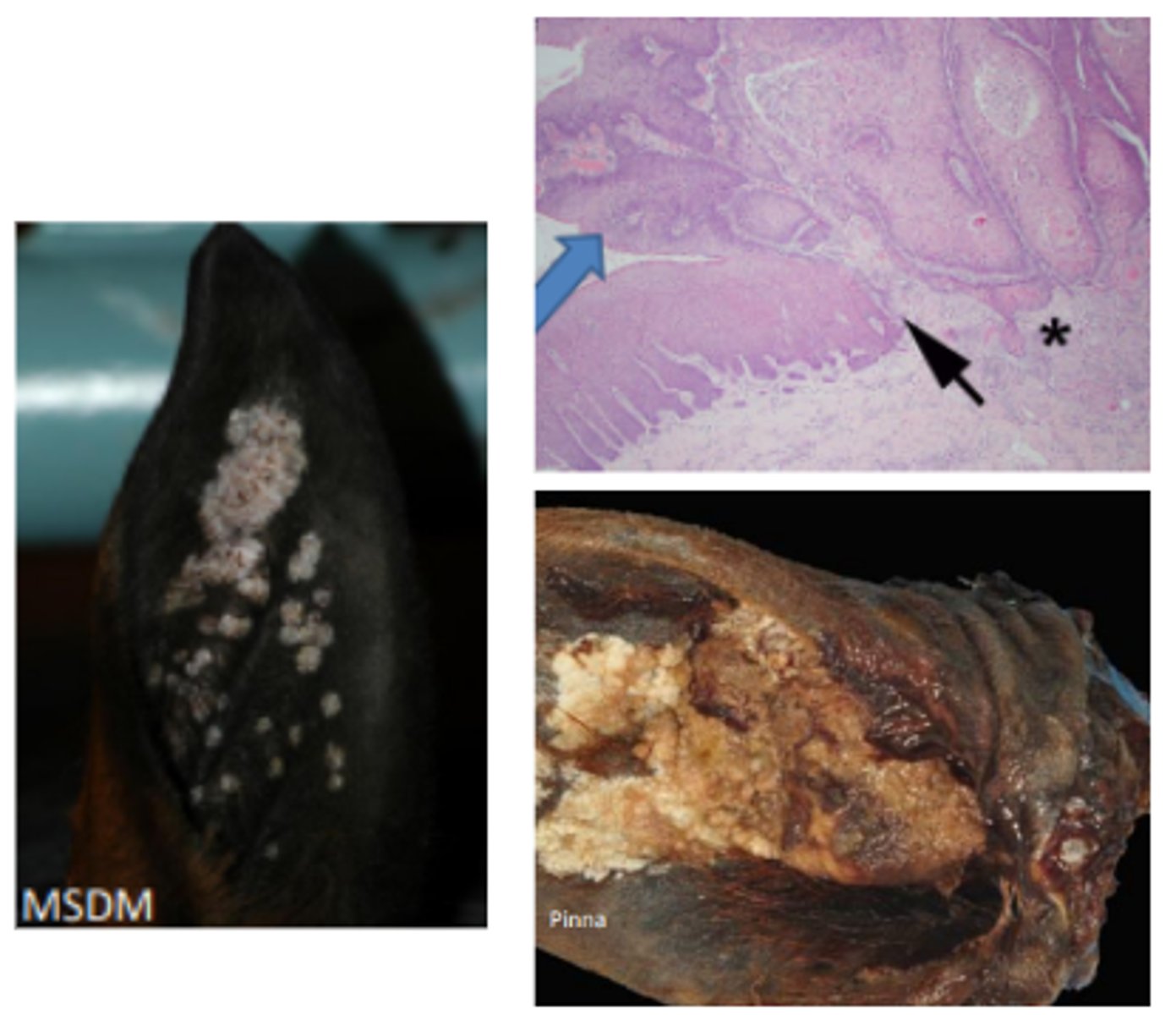
Equine aural plaques (papillomatosis)
Transition of viral plaque (black arrow) into SCC. Note the proliferative mass with disorganization of the keratinocyte layers and the invasive islands of neoplastic epithelium (*)
Demodex
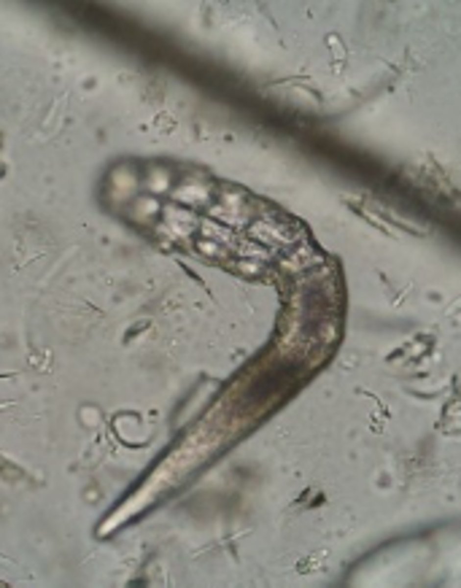
Sarcoptes scabiei
Otodectes
Dermatophytosis
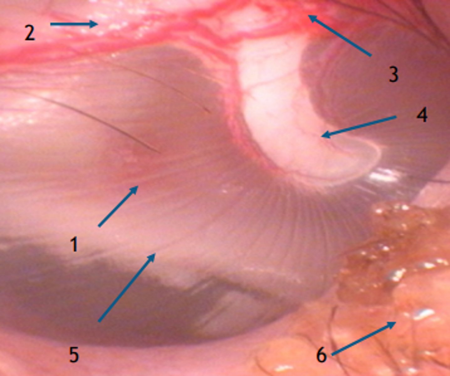
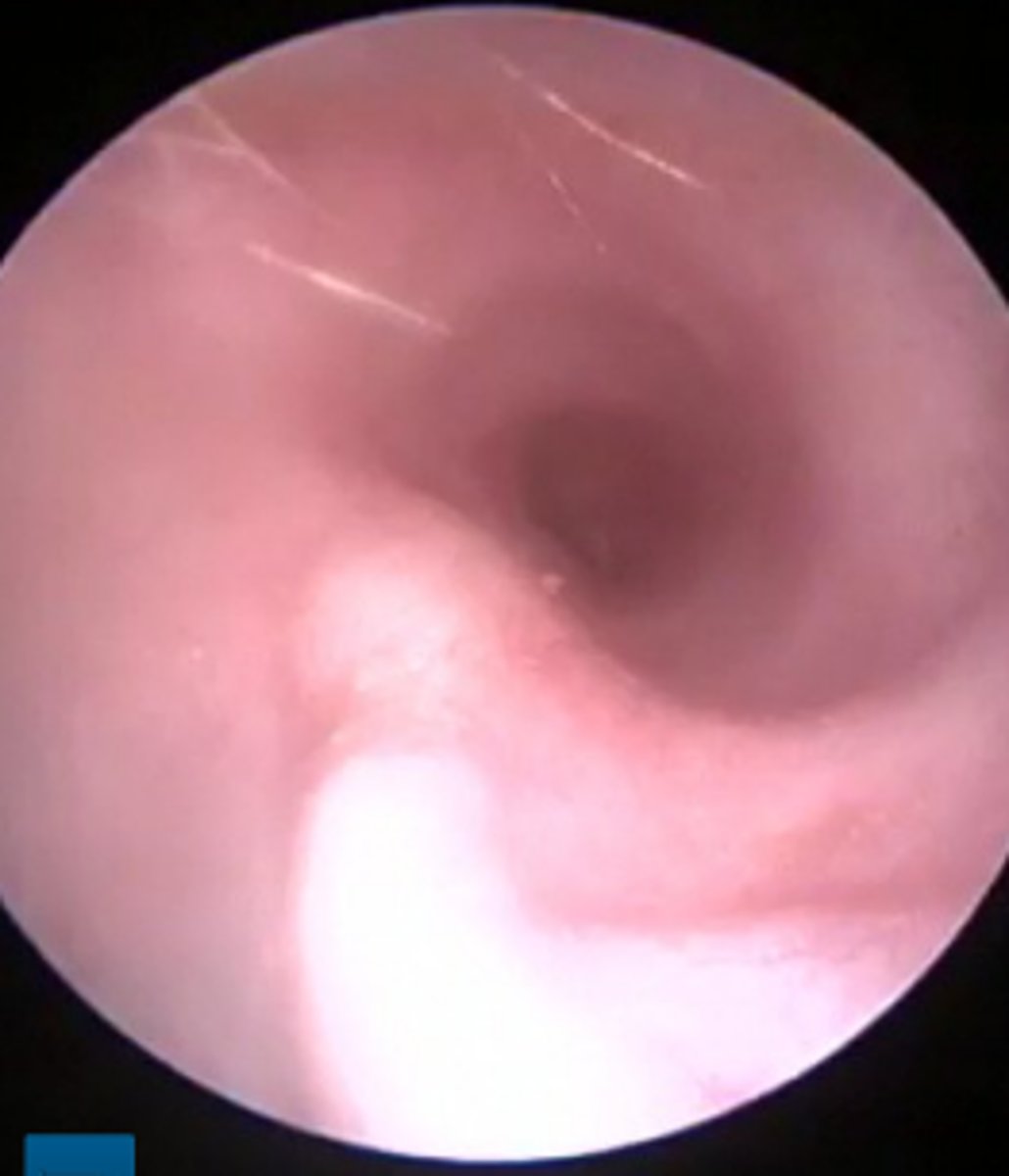
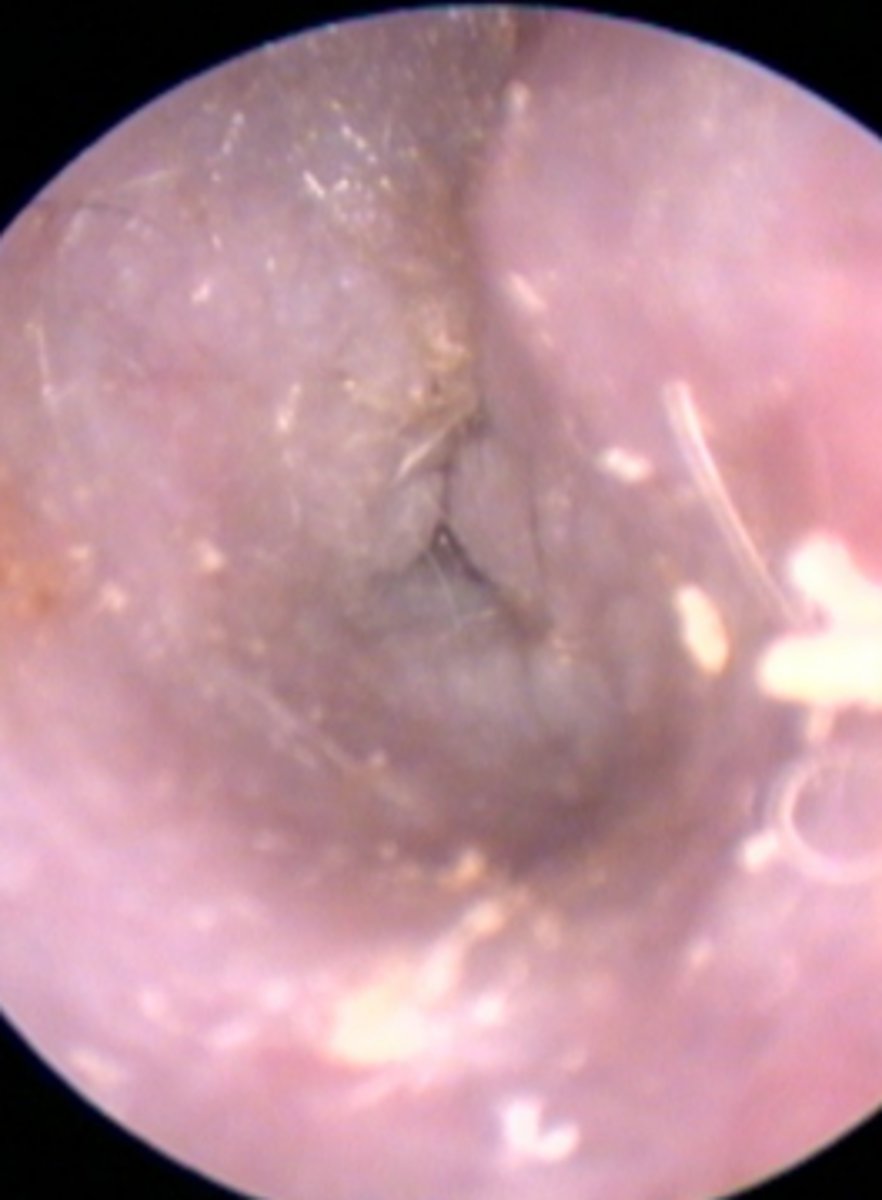
Tympanic membrane (normal)
1. Pars tensa
2. Edge of the pars flaccida
3. Blood vessel
4. Head of Malleus
5. Promontory
6. Cerumen and hair
Tympanic membrane
Small amount of cerumen and hair adjacent
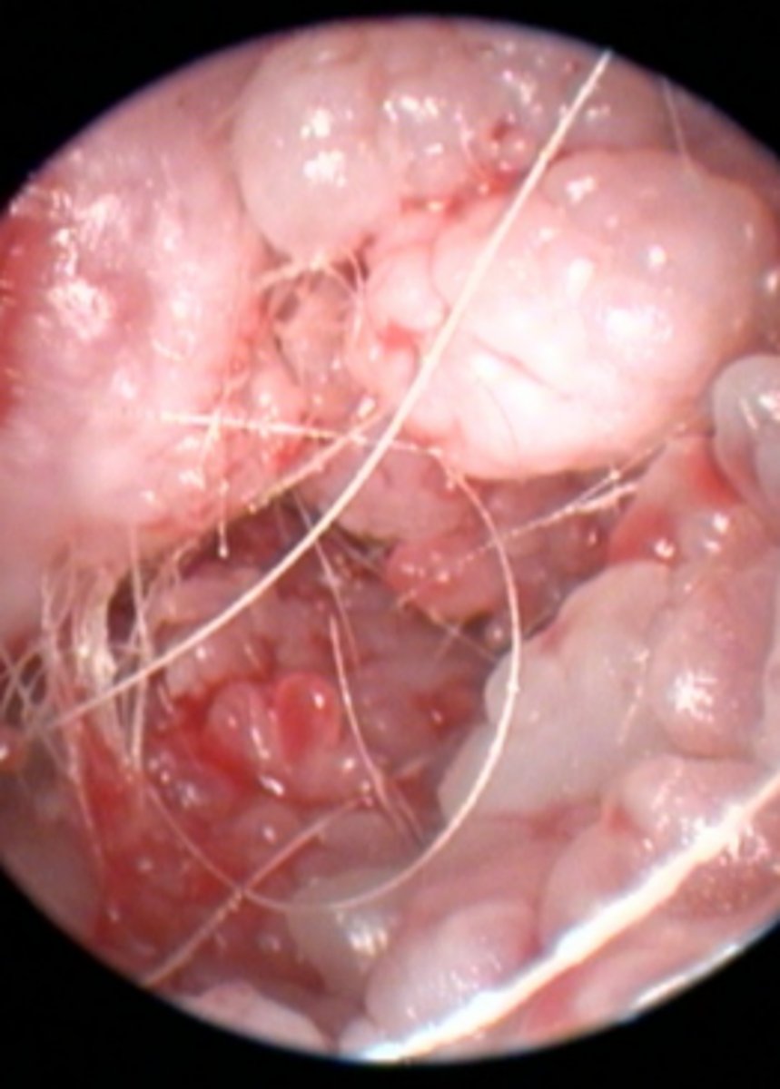
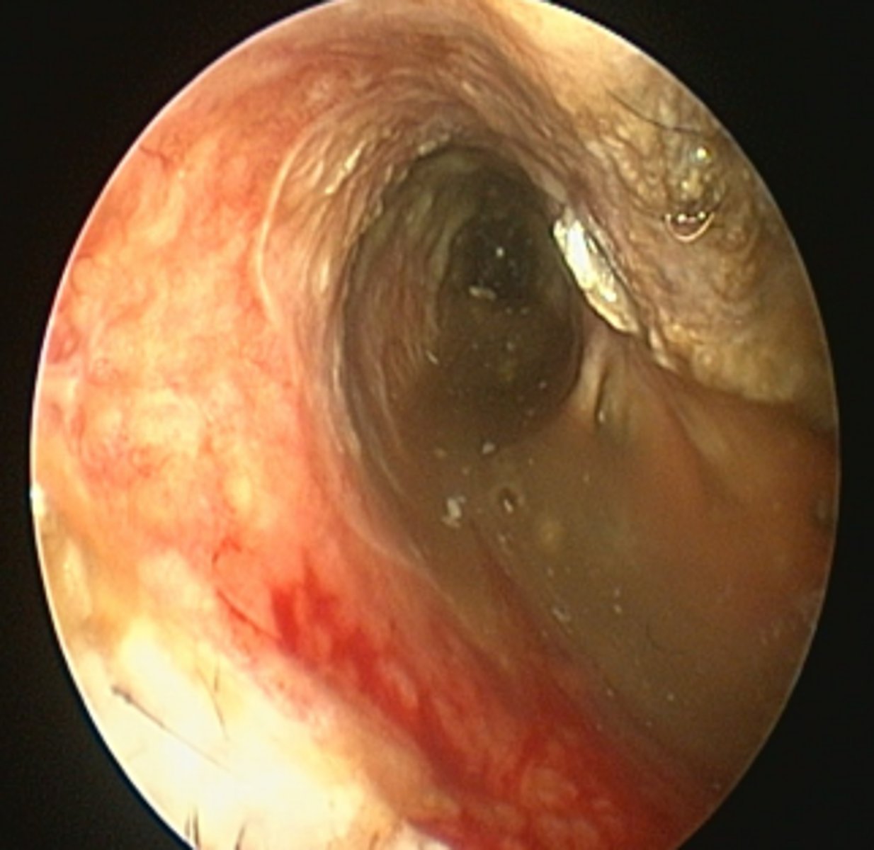
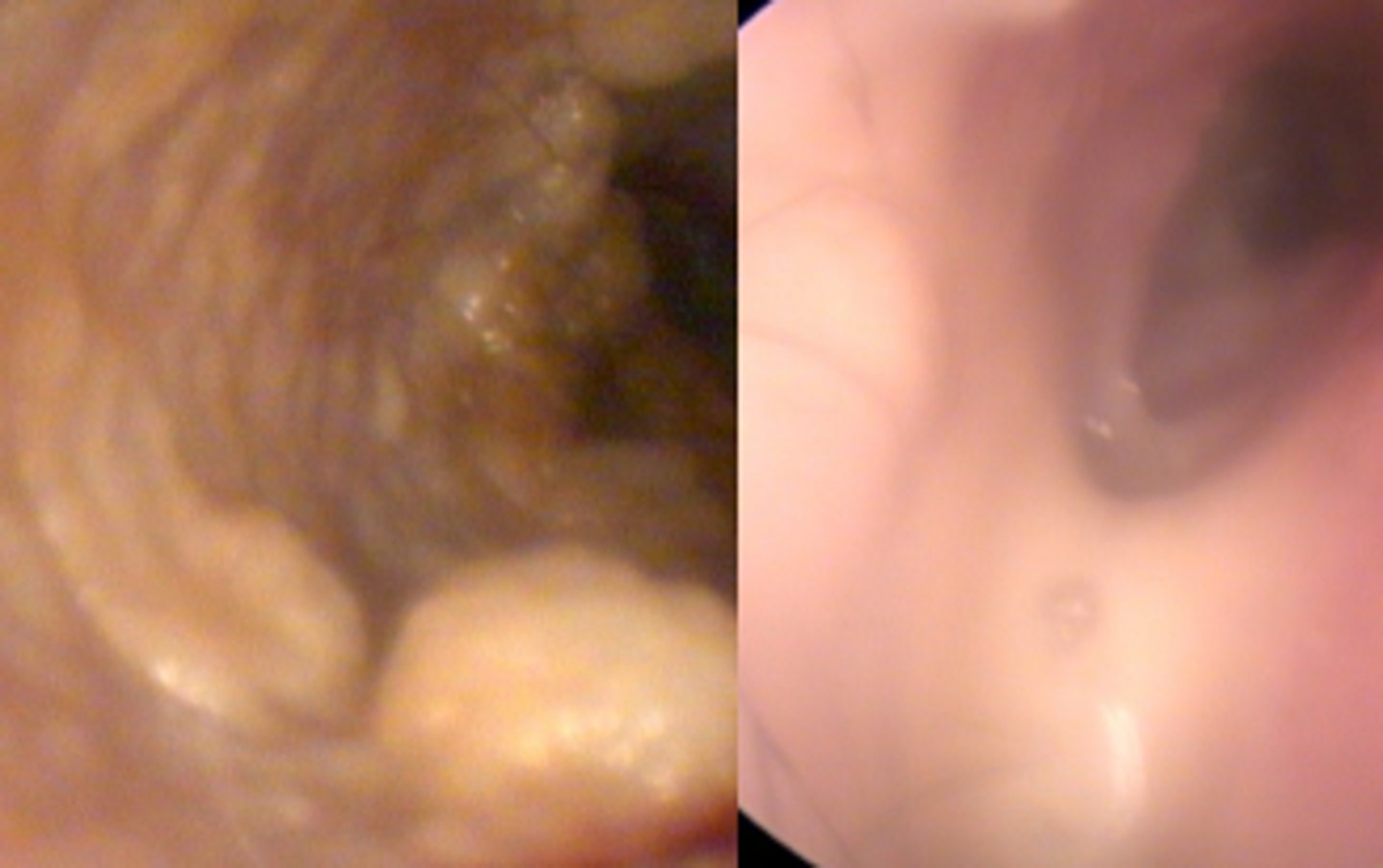
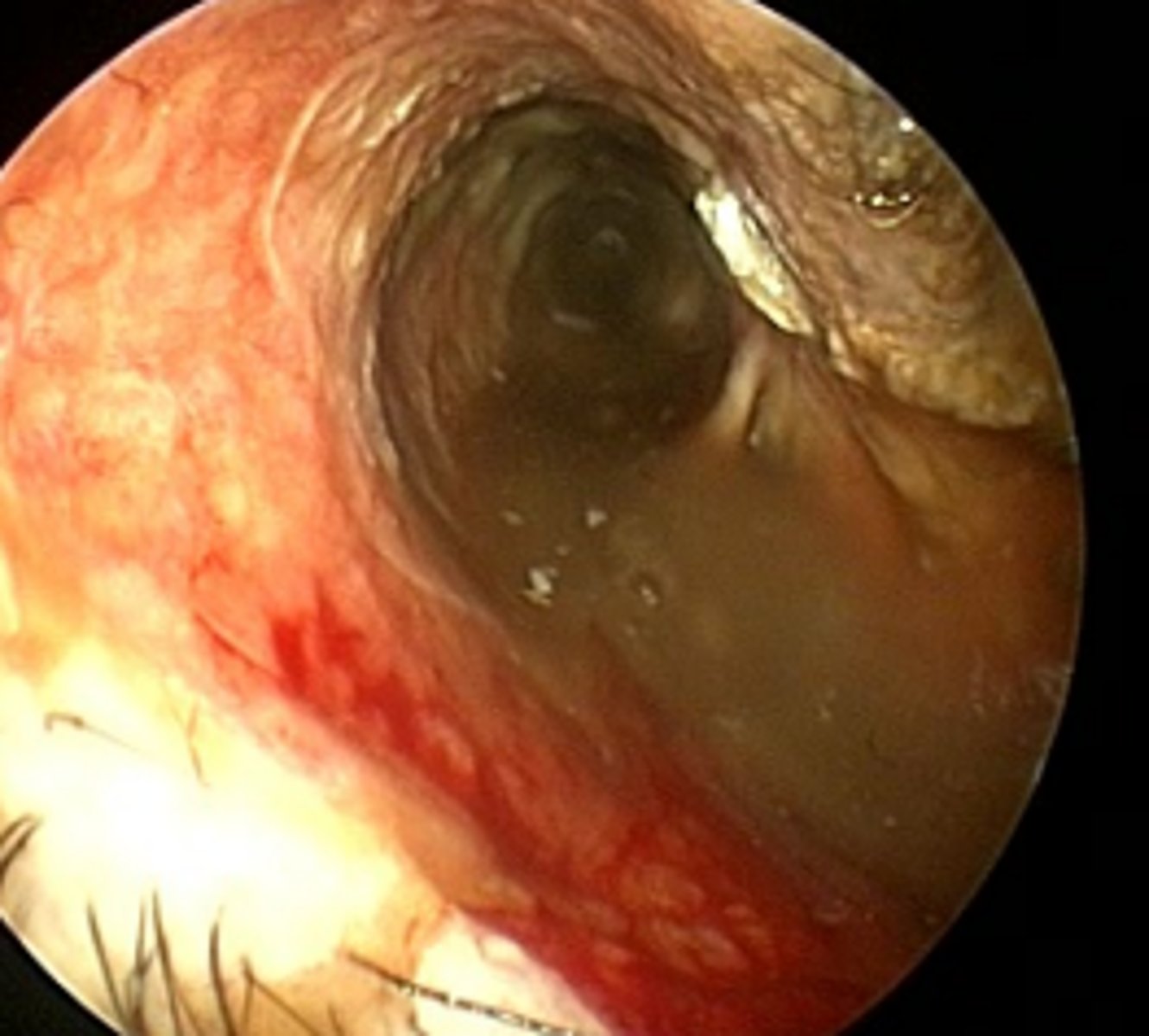
Ear canal
Epithelial fold formation, stenosis, ceruminous gland hyperplasia causing polypoid appearance, ulceration (bleeding)
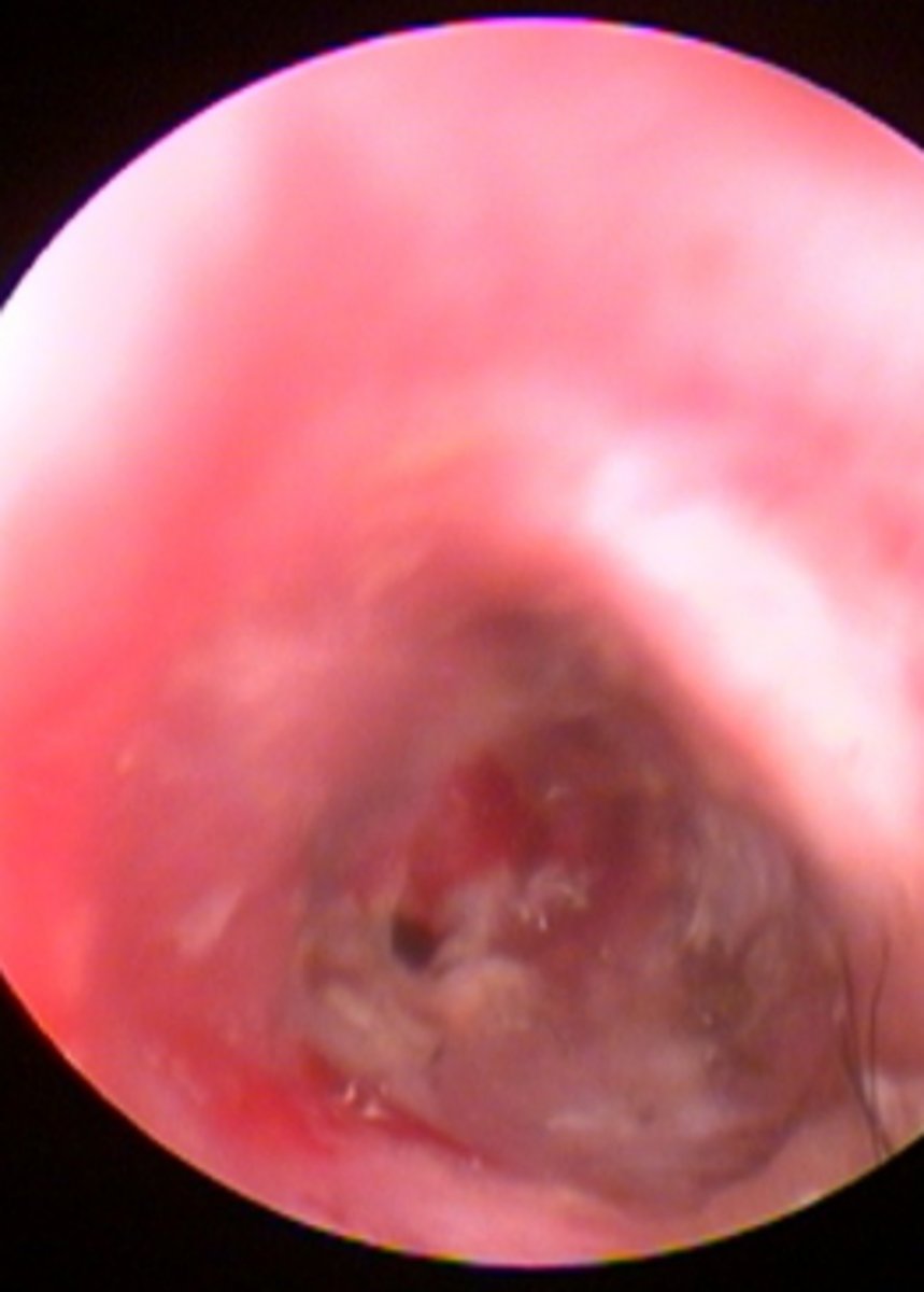
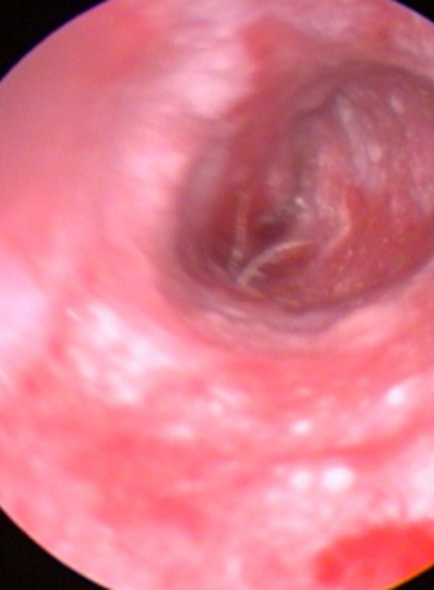
Ear canal
Erythema and mild ulceration of horizontal canal, abnormal opaque tympanic membrane
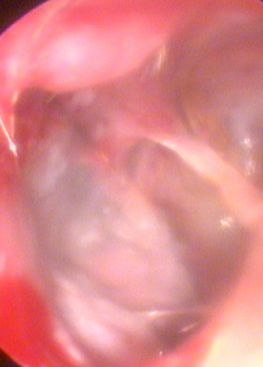
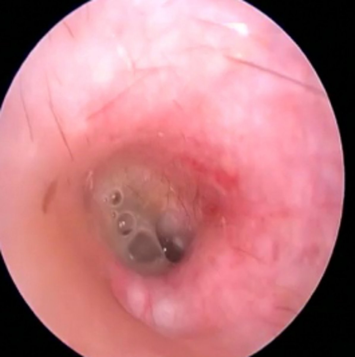
Ear canal
Opaque tympanic membrane, erythema and ulceration, purulent discharge
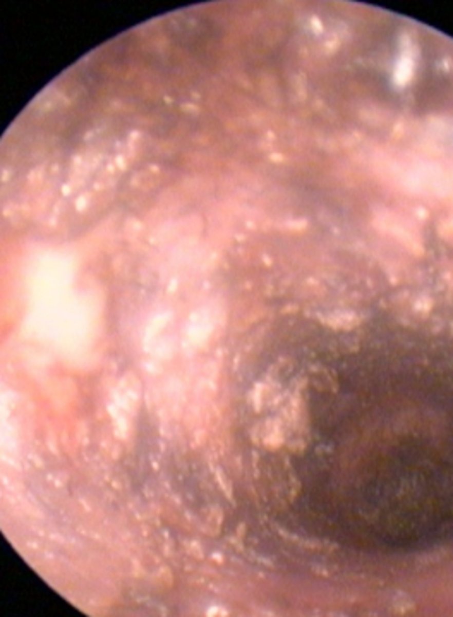
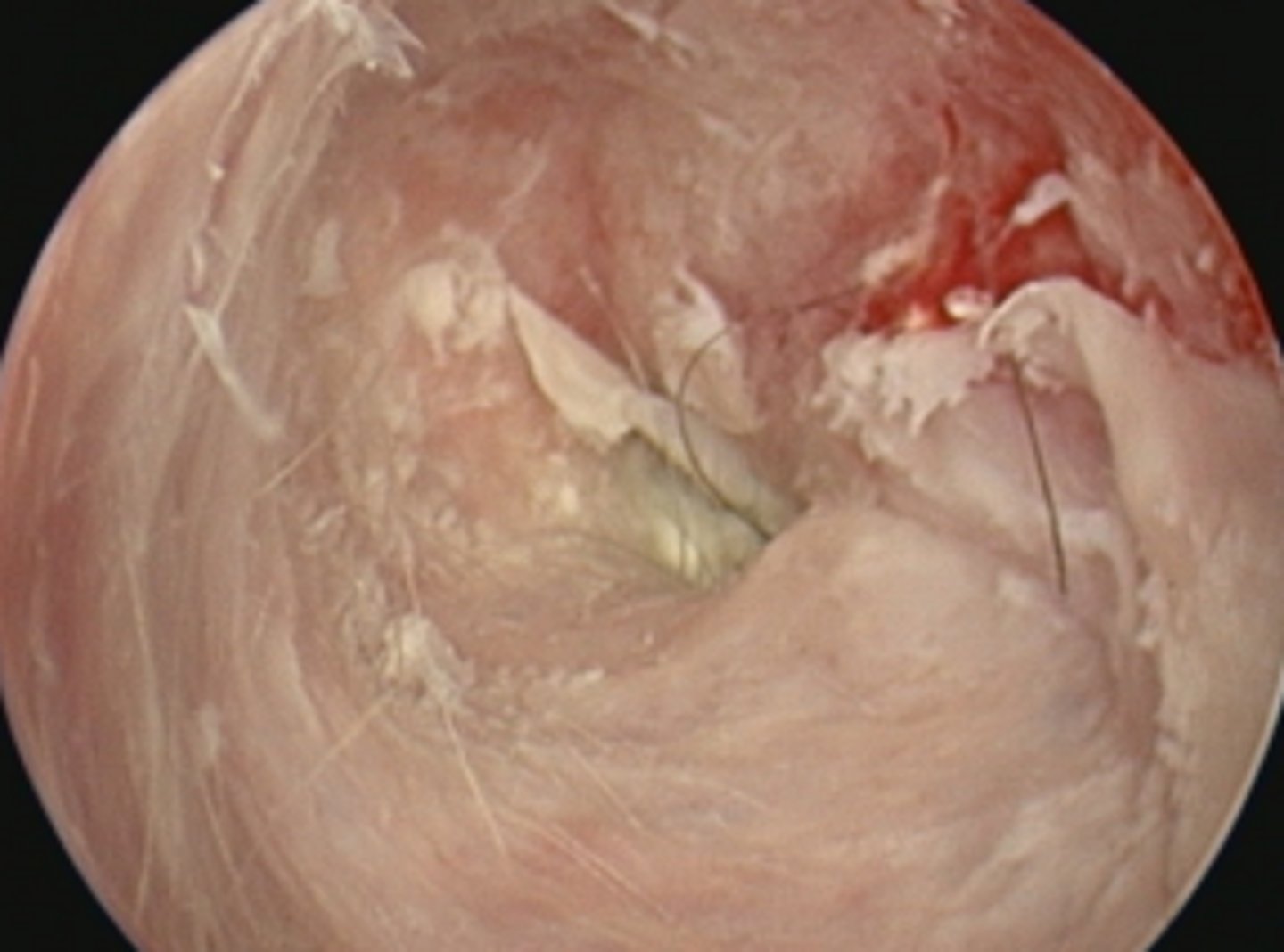
Ear canal
Erythema, ulceration, macerated epithelium (white areas), brown mucopurulent discharge
Ear canal
Erythema, stenosis, ulceration, sebaceous gland hyperplasia (small white spots)
Severe stenosis of the ear canal
Ear canal
Brown ceruminous discharge caused by Malassezia pachydermatis
Ear canal
Purulent discharge due to bacterial infection
Ear canal macerated epithelium
Ear canal - hirsuitism (excess hair)
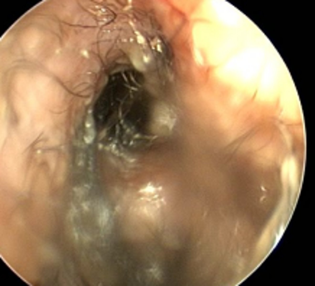
Mucopurulent discharge from the middle ear
Ear canal - profuse slimy purulent discharge due to biofilm production
Grass seed adjacent to the tympanic membrane
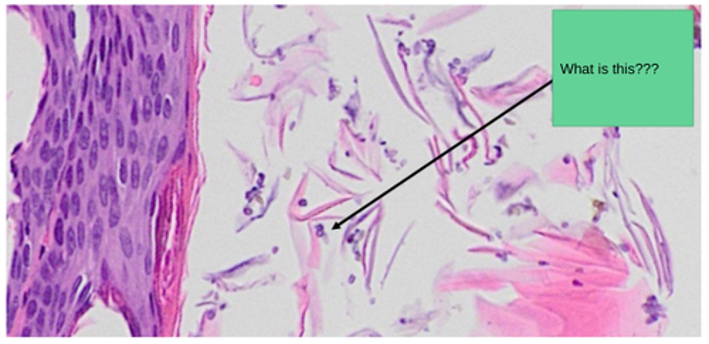
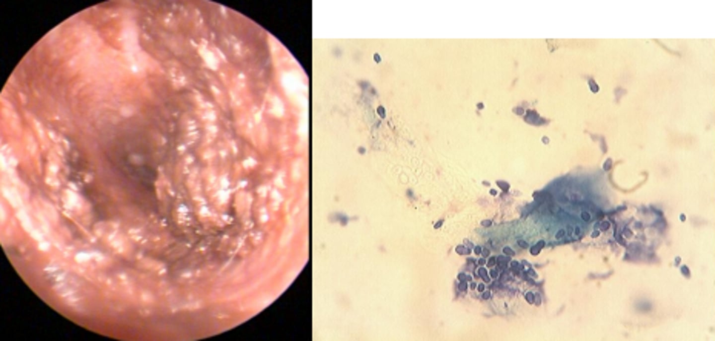
Tan colour otic discharge and otitis
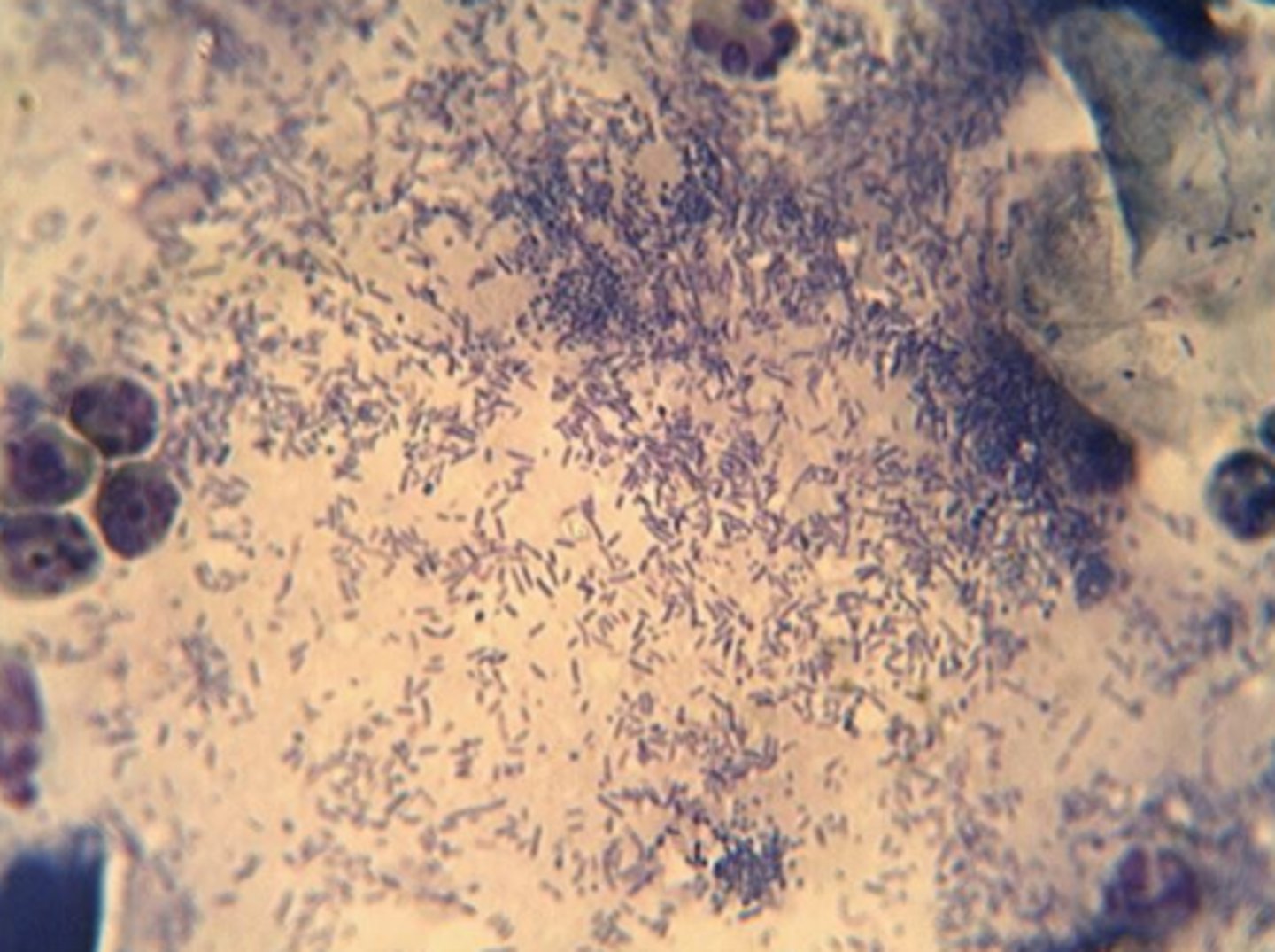
Many neutrophils (hypersegmented nuclei)
Phagocytosed bacteria (cocci) --> Staphylococcus or Streptococcus
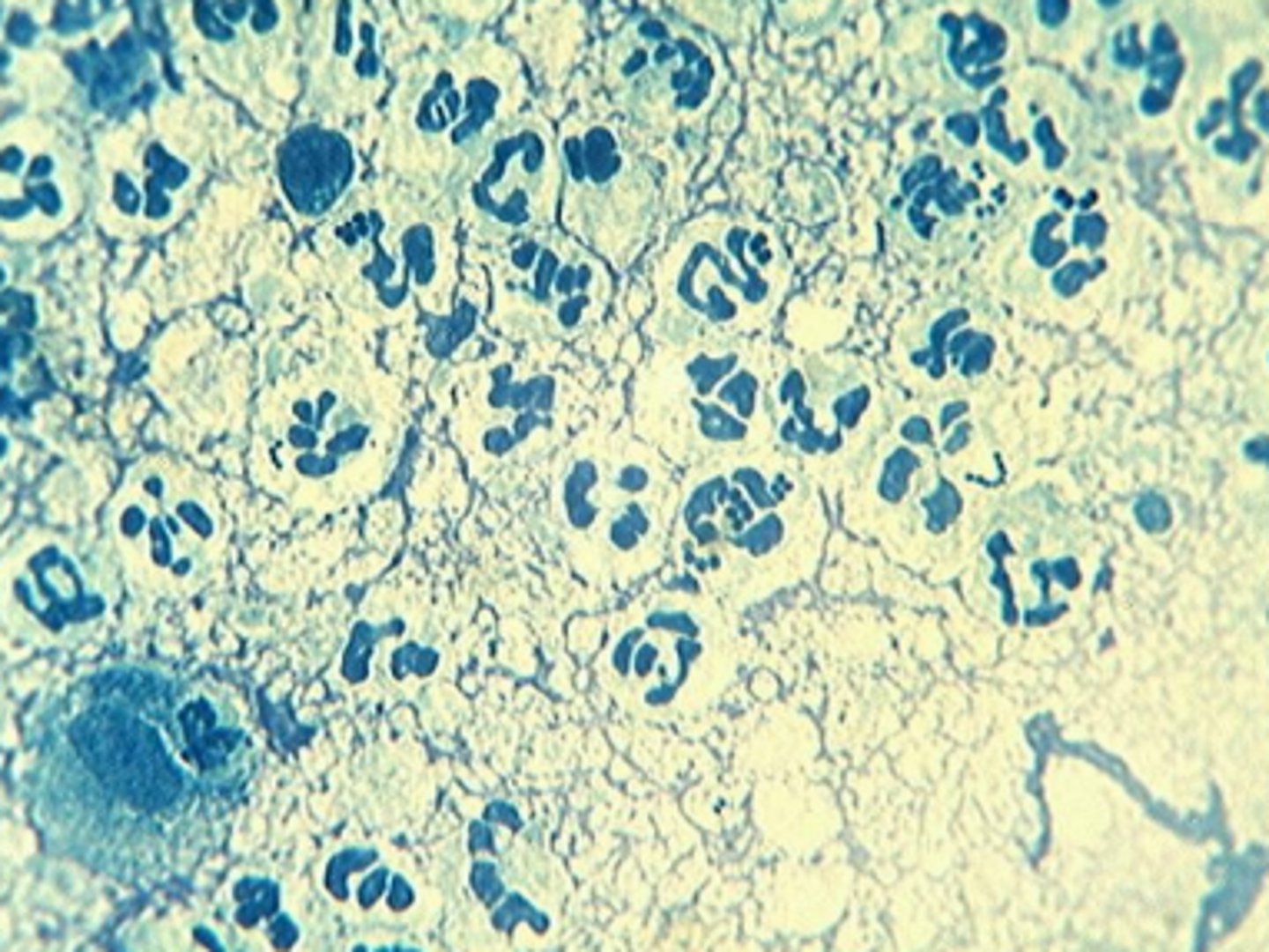
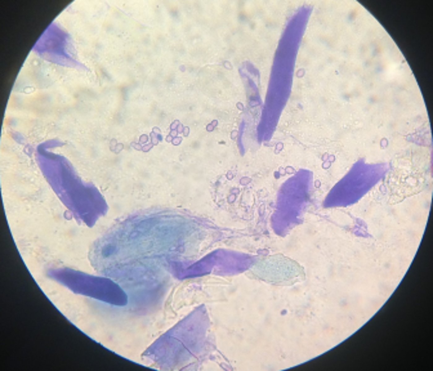
Ceruminous otitis
Unipolar budding yeat --> Malassezia pachydermatis
Purulent, painful and ulcerated malodourous otitis
Small numbers of neutrophuls (hypersegmented nuclei)
Rod shaped bacteria --> Pseudomonas, Proteus, E.coli, Klebsiella
Small number of cocci also present
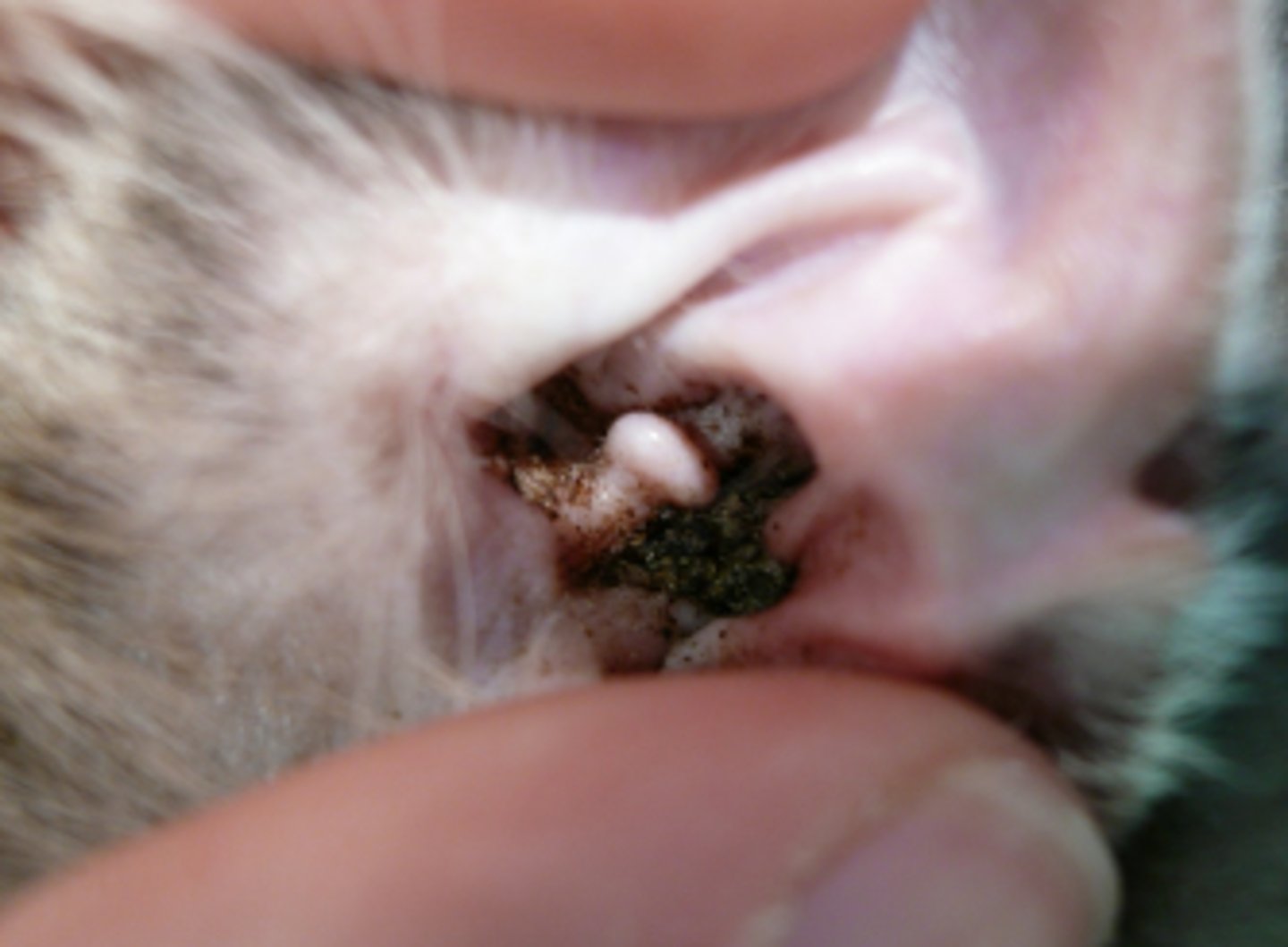
6 month old F cat with pruritic waxy ears - ear mites (otodectes cynotis)
Mild erthema of the pinna and ear canal entrance, dark brown coffee grounds like ceruminous discharge, white specks within cerumen
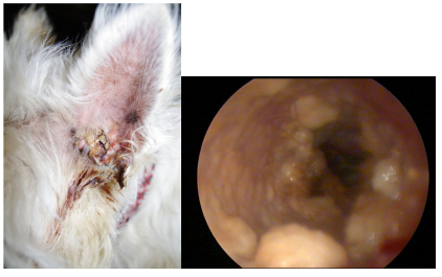
6 year old dog with severely painful and smelly unilateral otitis of 5 months duration
Purulent discharge, small ulcerations on the concave pinna and horizontal canal, tumour on the cranial lateral pinna - covered in crust, cobblestone appearance to the ear canal suggestive of ceruminous gland hyperplasia , inflammed and opaque tympanic membrane
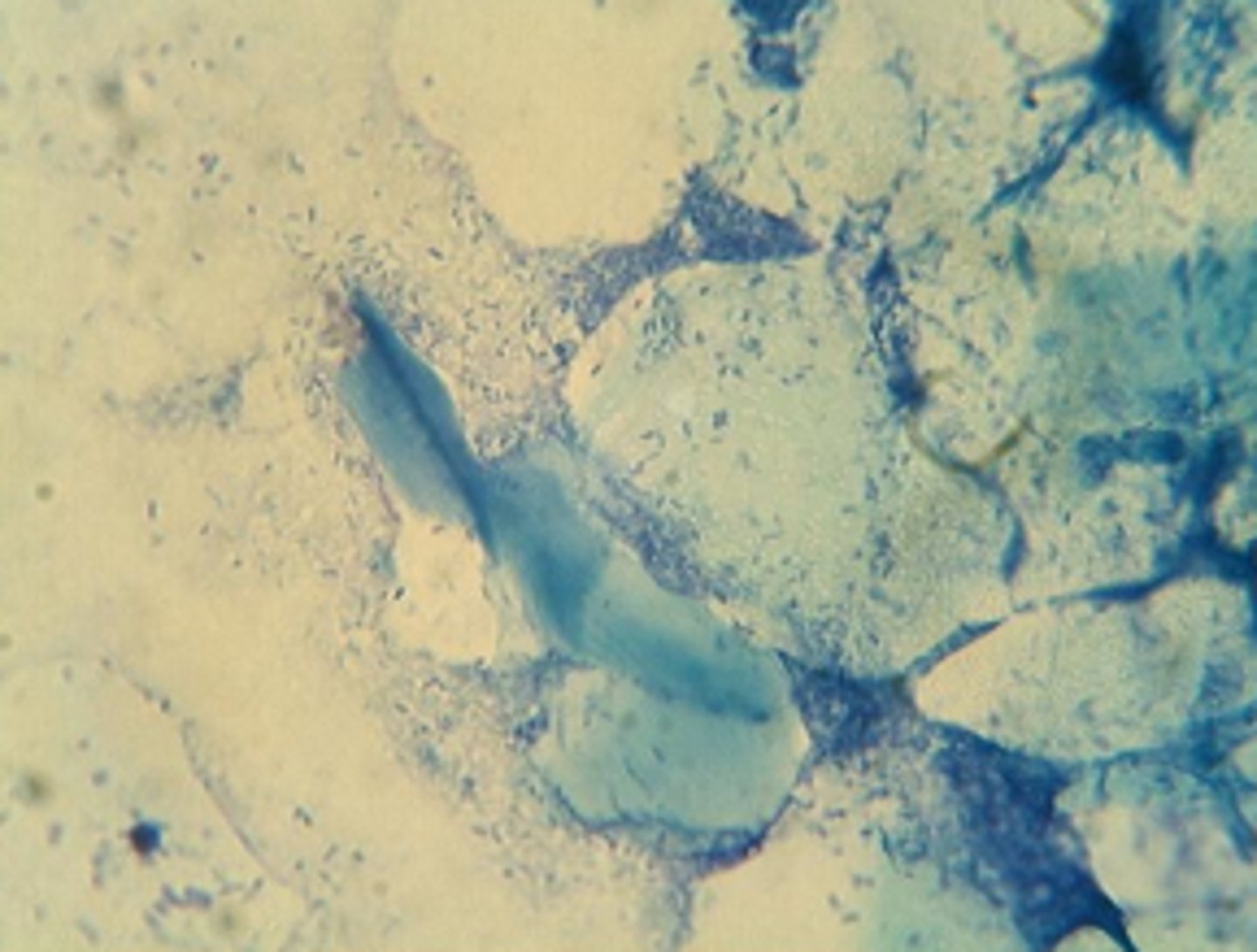
6 year old dog with severely painful and smelly unilateral otitis of 5 months duration - cytology - bacterial infection
Numerous rod bacteria and squames - gram negative --> e Pseudomonas, Klebsiella, Proteus, E.coli
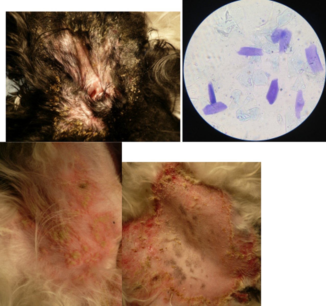
Ceruminous otitis
Head shaking, malodour, ceruminous discharge, erythema and oedema of the ear canal, ceruminous gland hyperplasia (cobblestone appearance), stenosis of the distal horizontal ear canal, numerous yeasts --> Malassezia pachydermatis and squame
Pemphigus foliaceus
Scaling and crusting along the ear pinnal margins that is matting the coat, large pustules in the groin with erythematous margina, axilla is alopecic with a large peripheral epidermal collarette and erosions, hyperpigmented macules, squames on cytology, acanthocytes
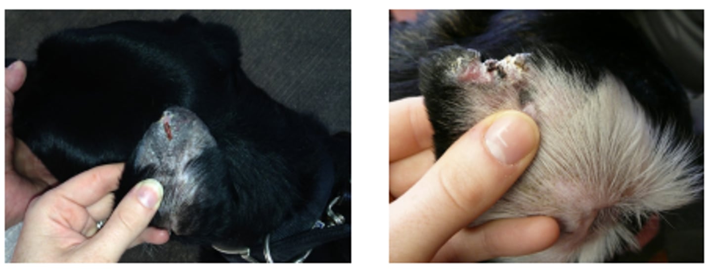
Vasculitis/ vasculopathy
Left pinna - alopecia, hyperpigmentation, linear ulceration covered with crust and eroded pinnal edge
Right pinna - extensive erosion, alopecia, crusting
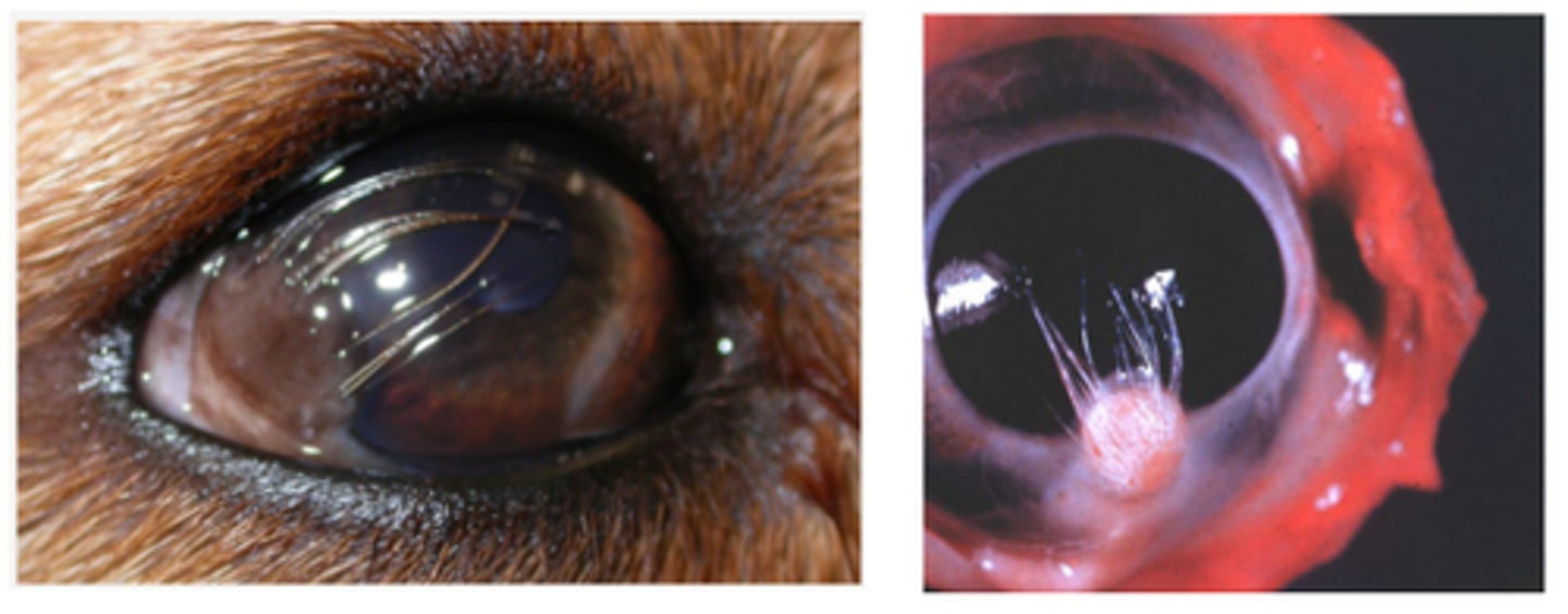
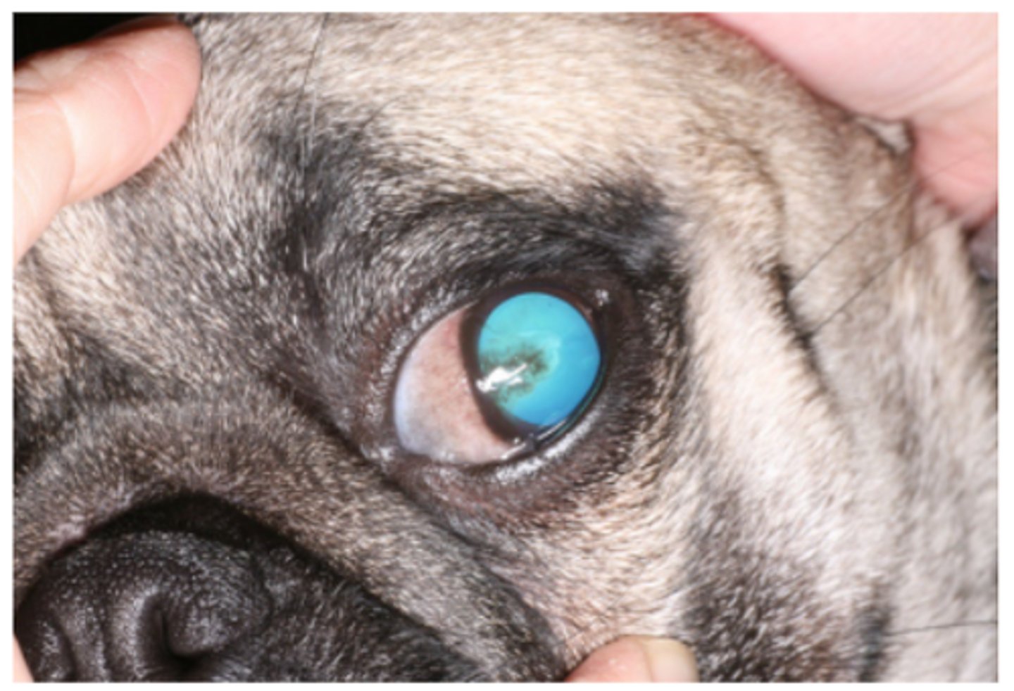
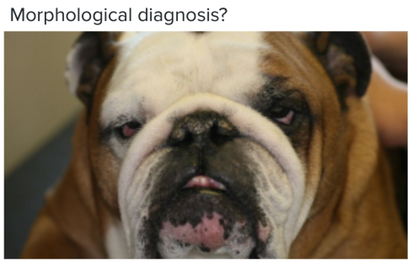
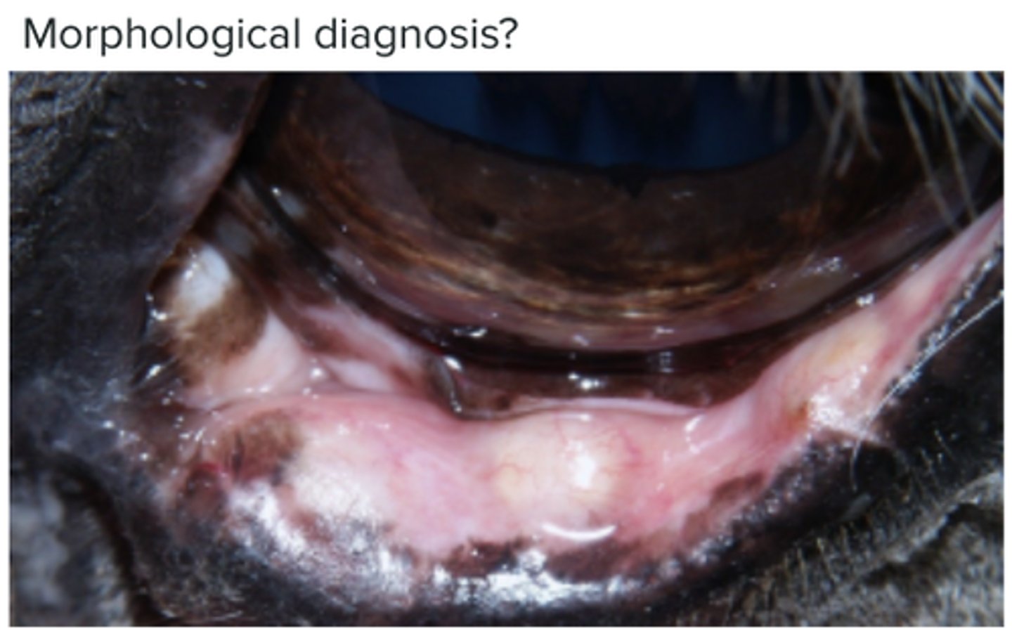
Macropalpebral fissure syndrome
Dogs with a prominent fold of skin over the nose
Corneal ulcer and entropion
Entropion - Shar Pei
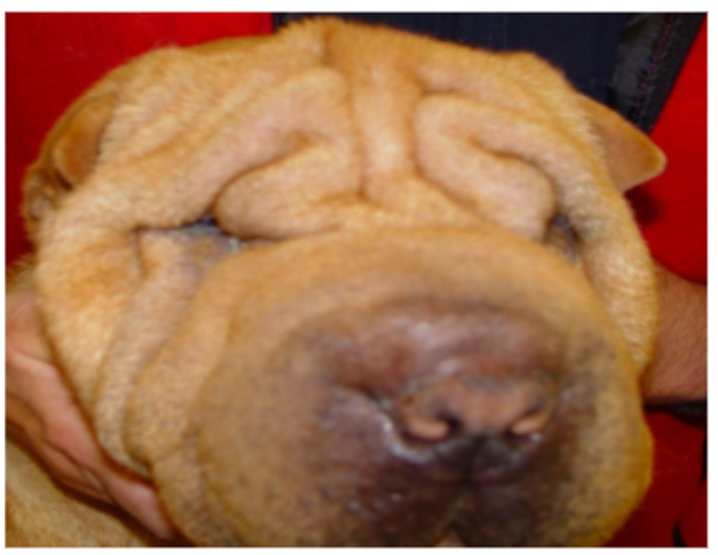
Nasal fold trichiasis (rubbing on the cornea)
Bulbar conjunctiva of labrador - ectopic cilia
Blepharitis with thickened and calcified Meibomian glands
Puppy strangles - Blepharitis
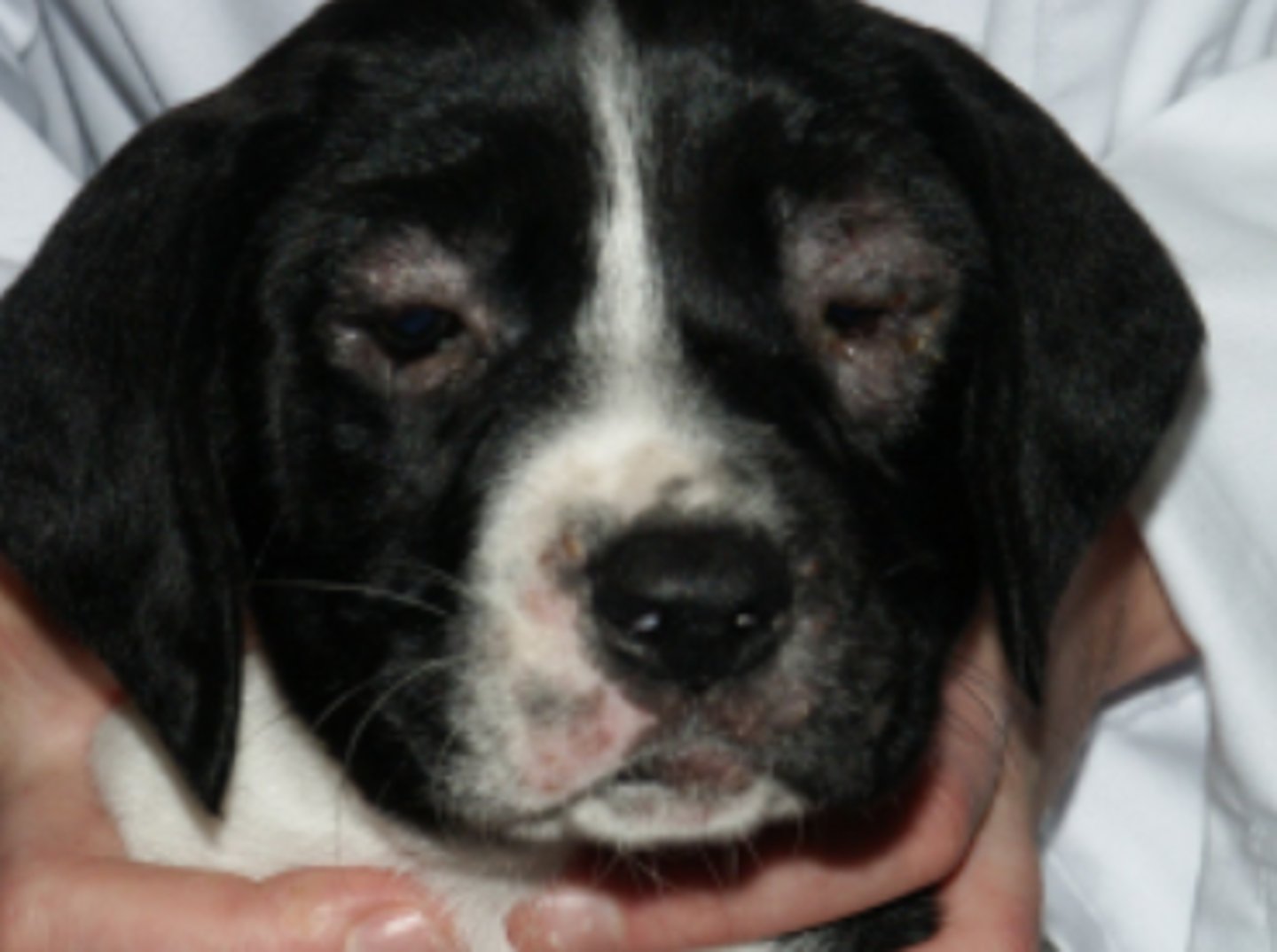
Local blepharitis (inflammation of the meibomian gland)
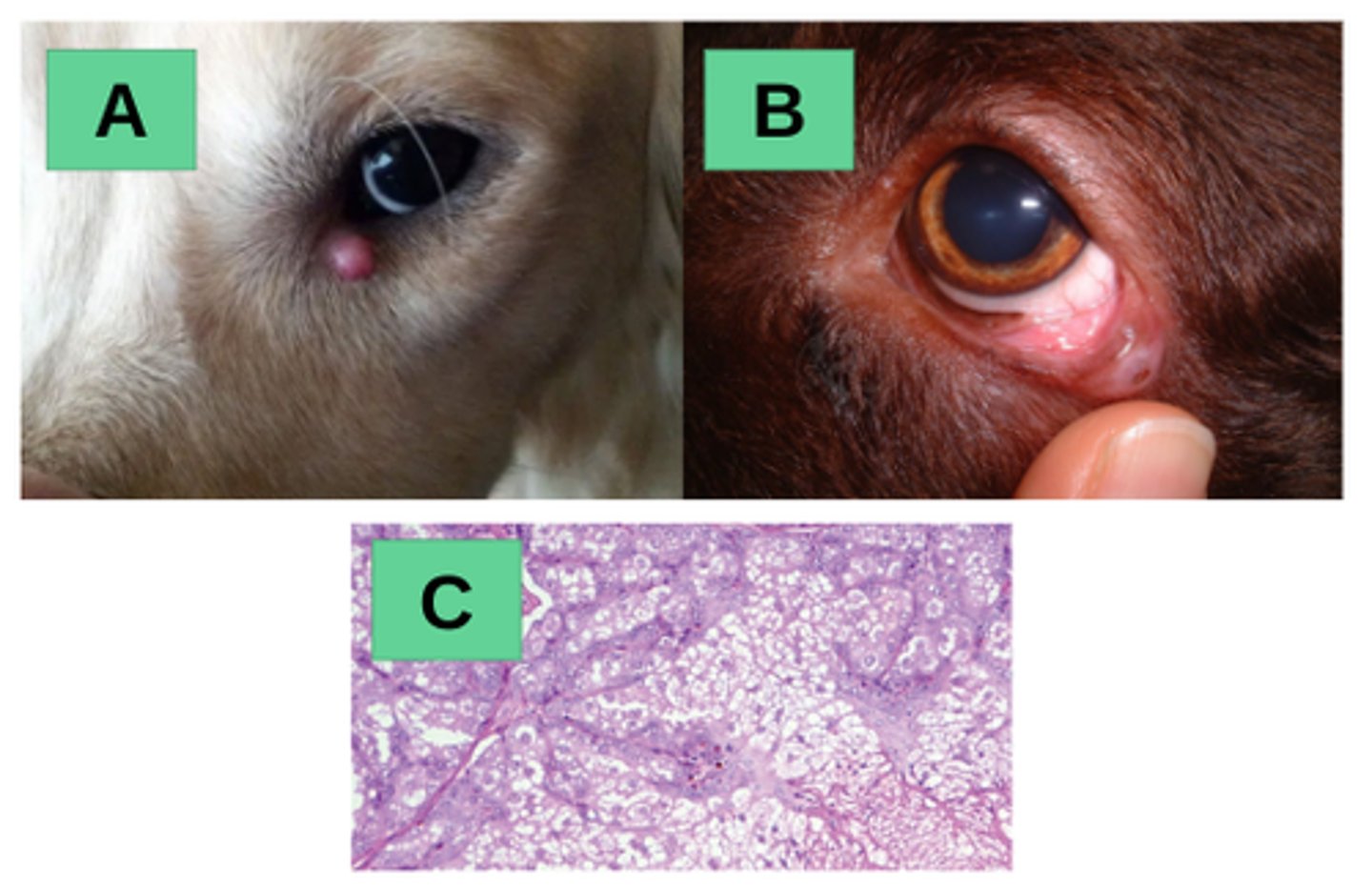
A - chalazion or meibomianitis
B - Chalazion on the left lower eyelid of a 9-year-old male dog. This consists in an inflammatory lesion of the meibomian gland, associated with secretion accumulation
C - Adenoma
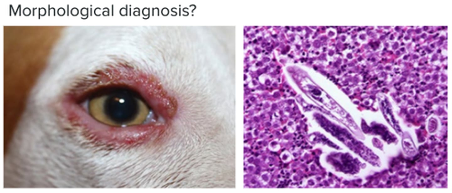
Parasitic blepharitis (Demodex species)
Circumferential alopecia, crusting, discharge and erythema
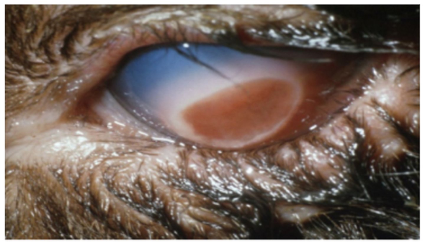
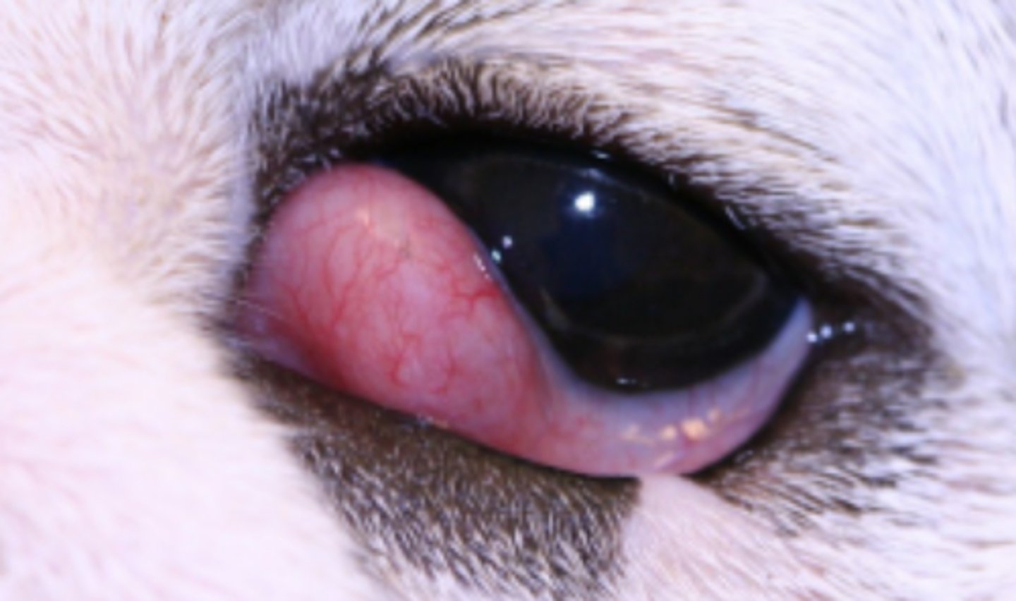
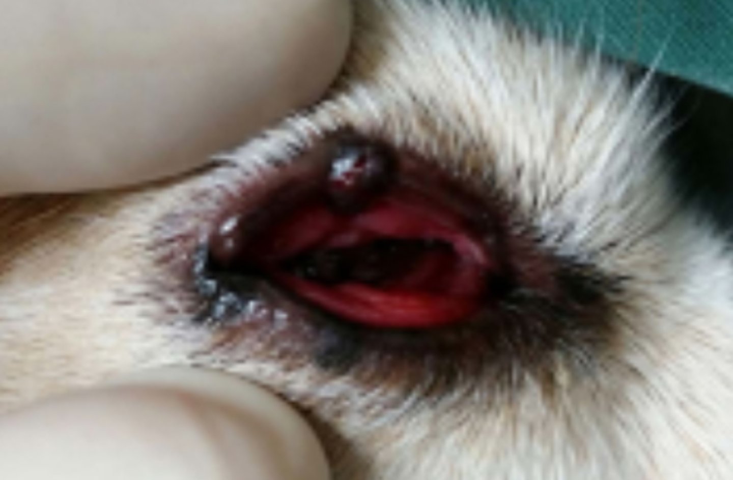
Protrusion of the nictitans gland (cherry eye)
Common in Bulldogs
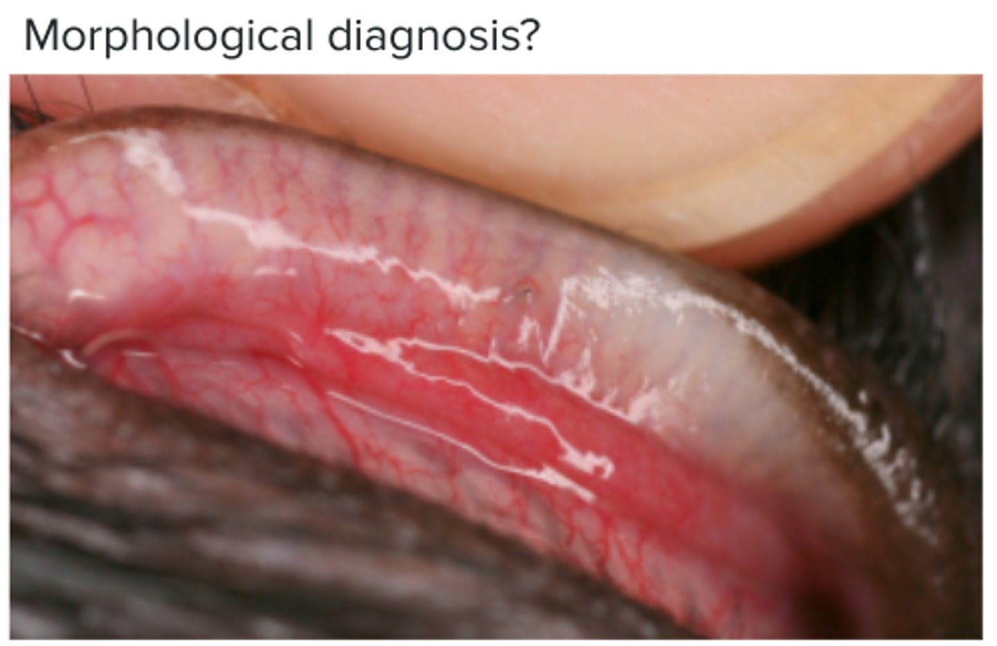
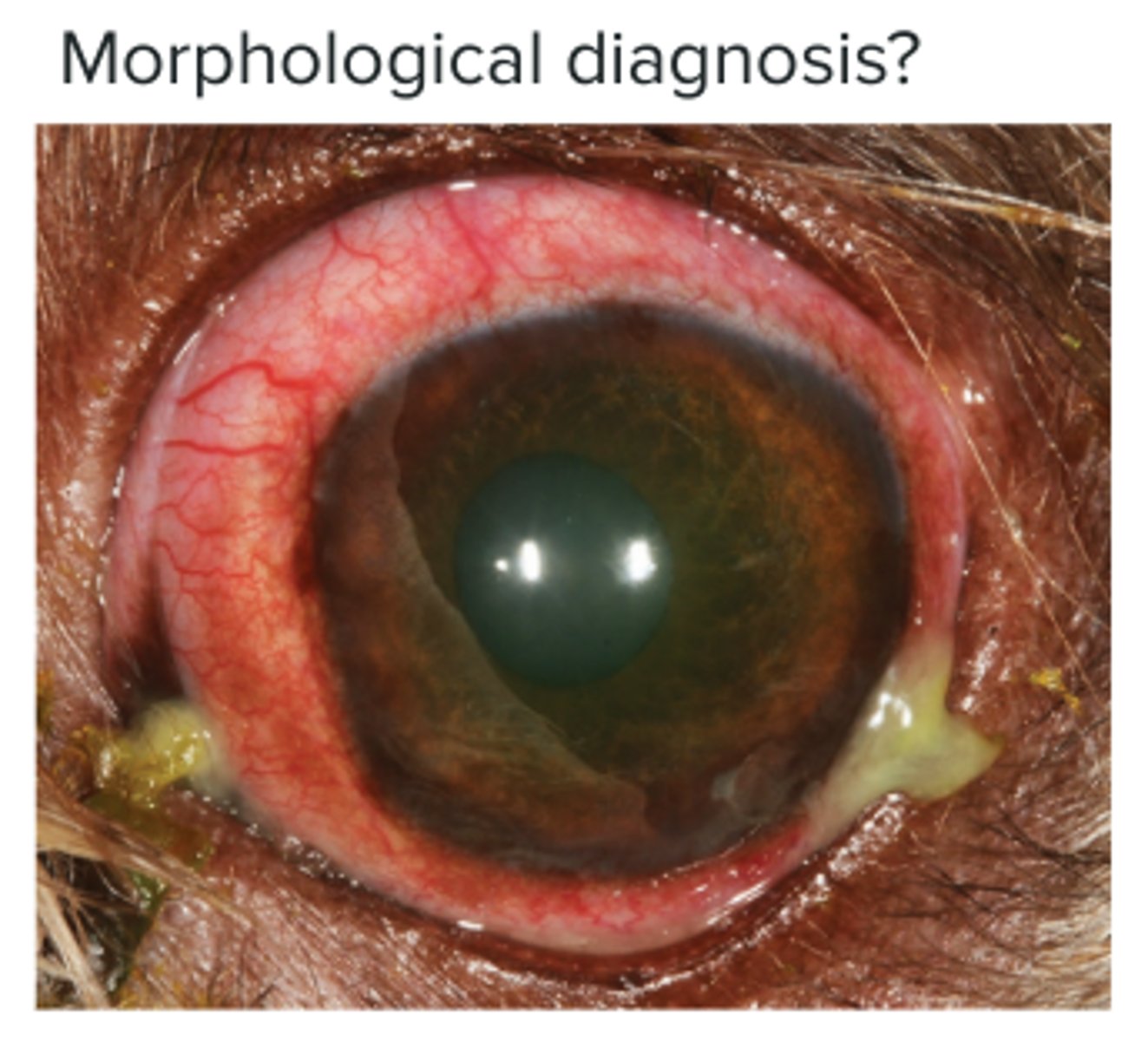
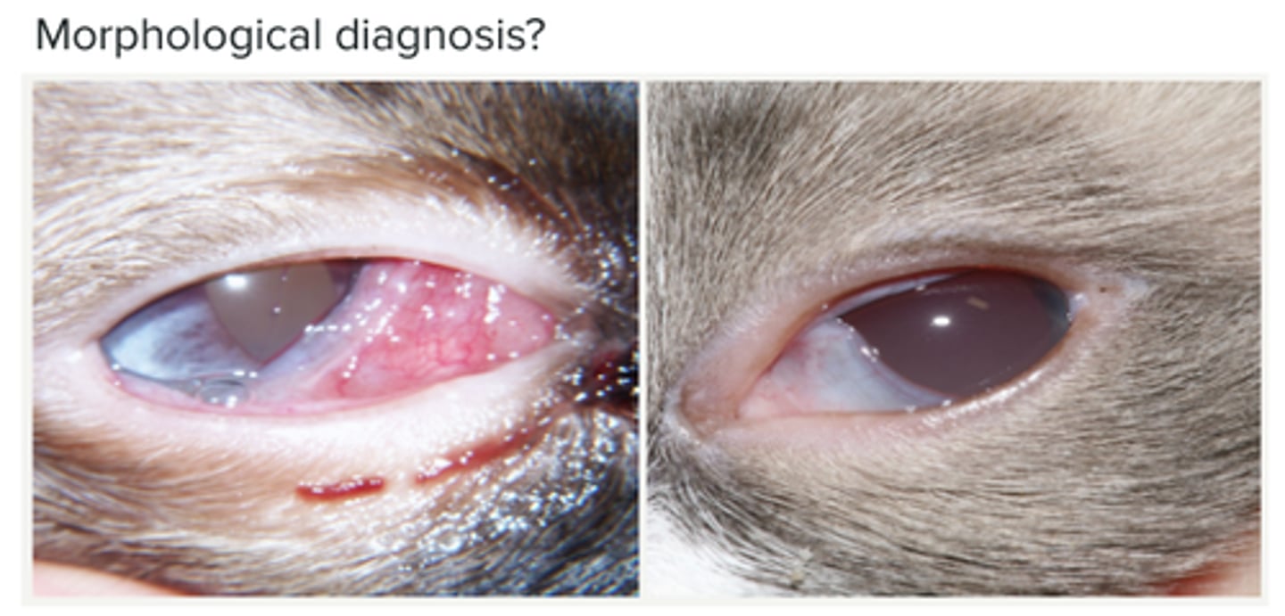
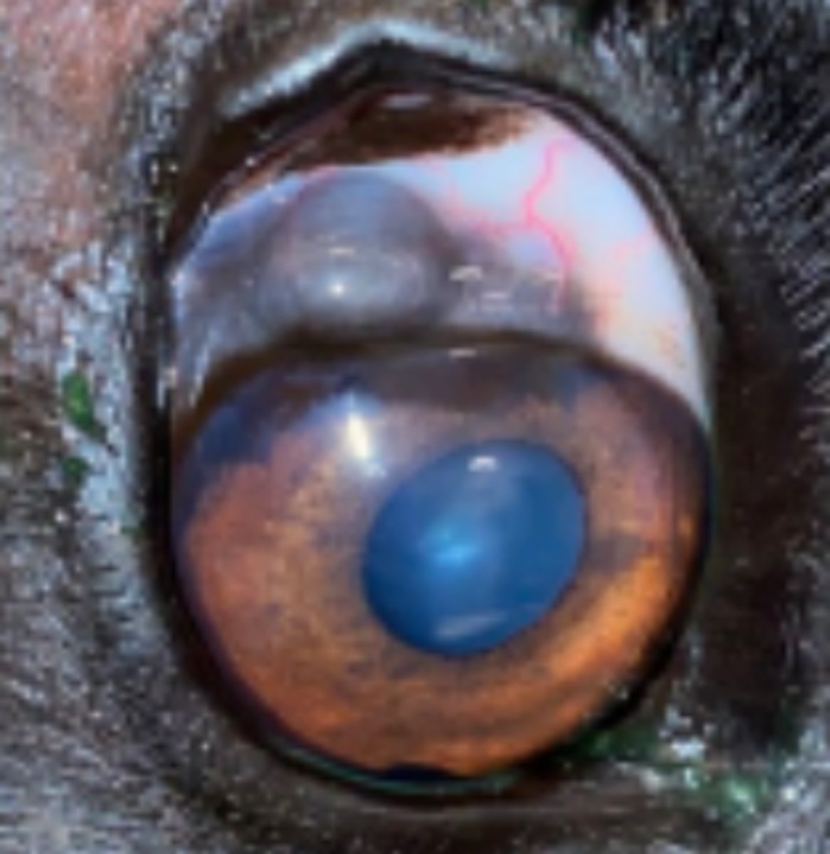
Conjunctivitis
Hyperaemic and oedematous conjunctiva, vasodilation, increased blood flow, lymphoid follicles, ulceration, fibrosis
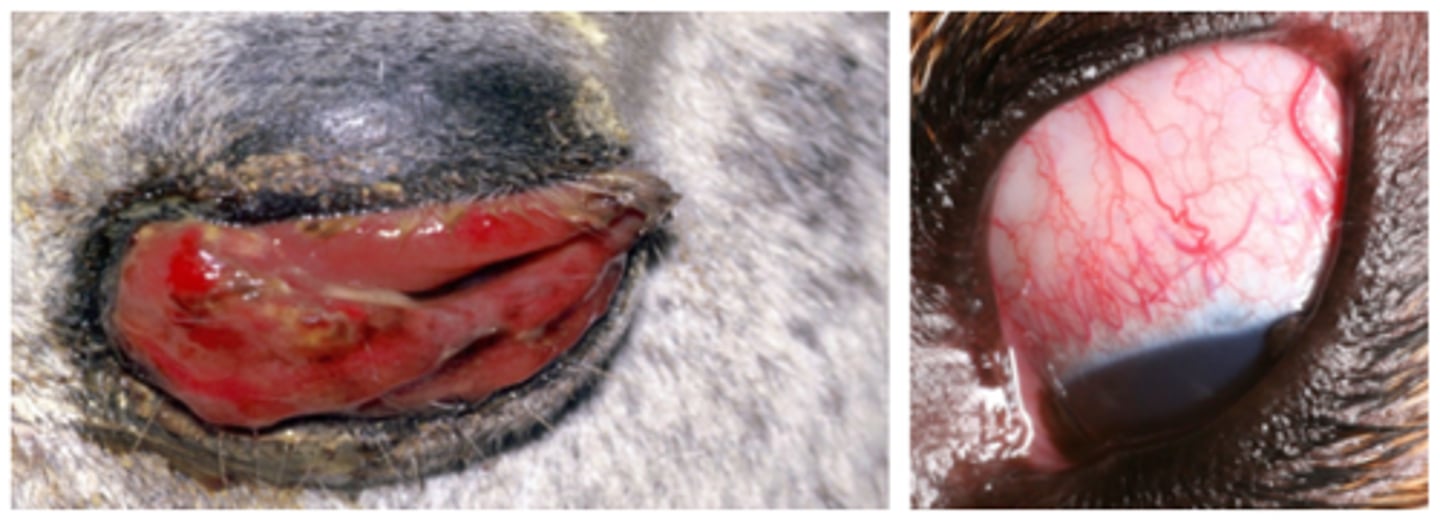
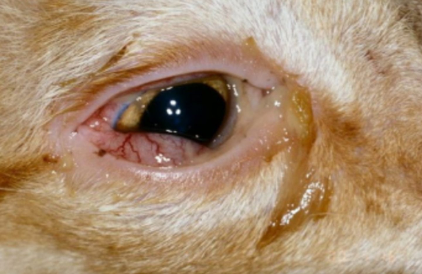
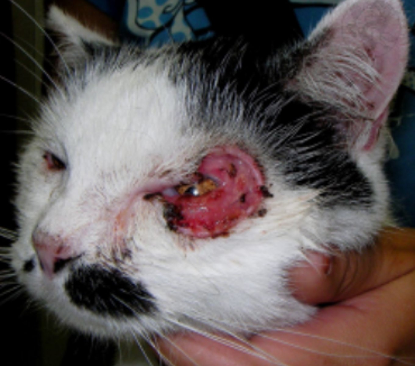
Habronemia conjunctivitis
Conjunctival hyperemia and thick mucopurulent ocular discharge
Chlamydophila felis - unilateral conjunctivitis (infection)
Conjunctivitis in cats associated with feline herpesvirus 1
Infectious Bovine Keratoconjunctivitis (Pink eye) - Moraxella bovis
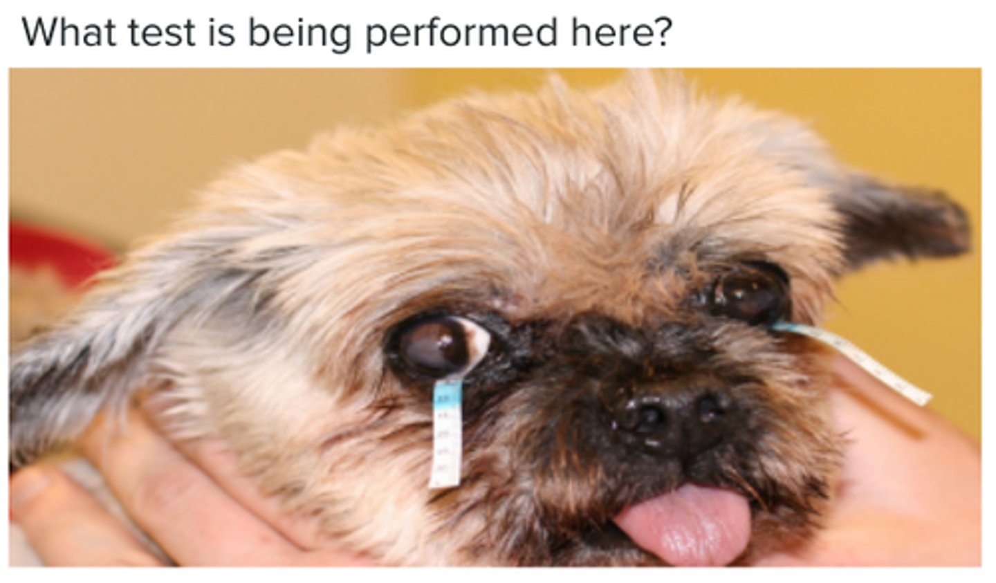
Immune-Mediated Keratoconjunctivitis Sicca (KCS)
Decreased tear film quantity and quality > dry eye
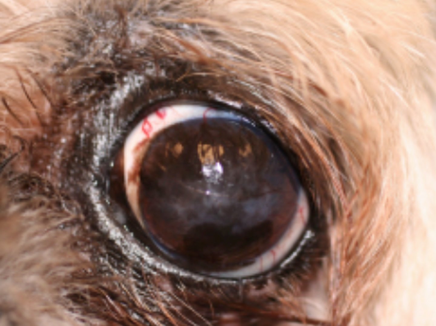
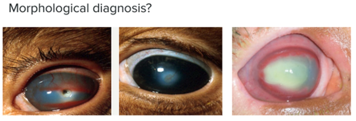
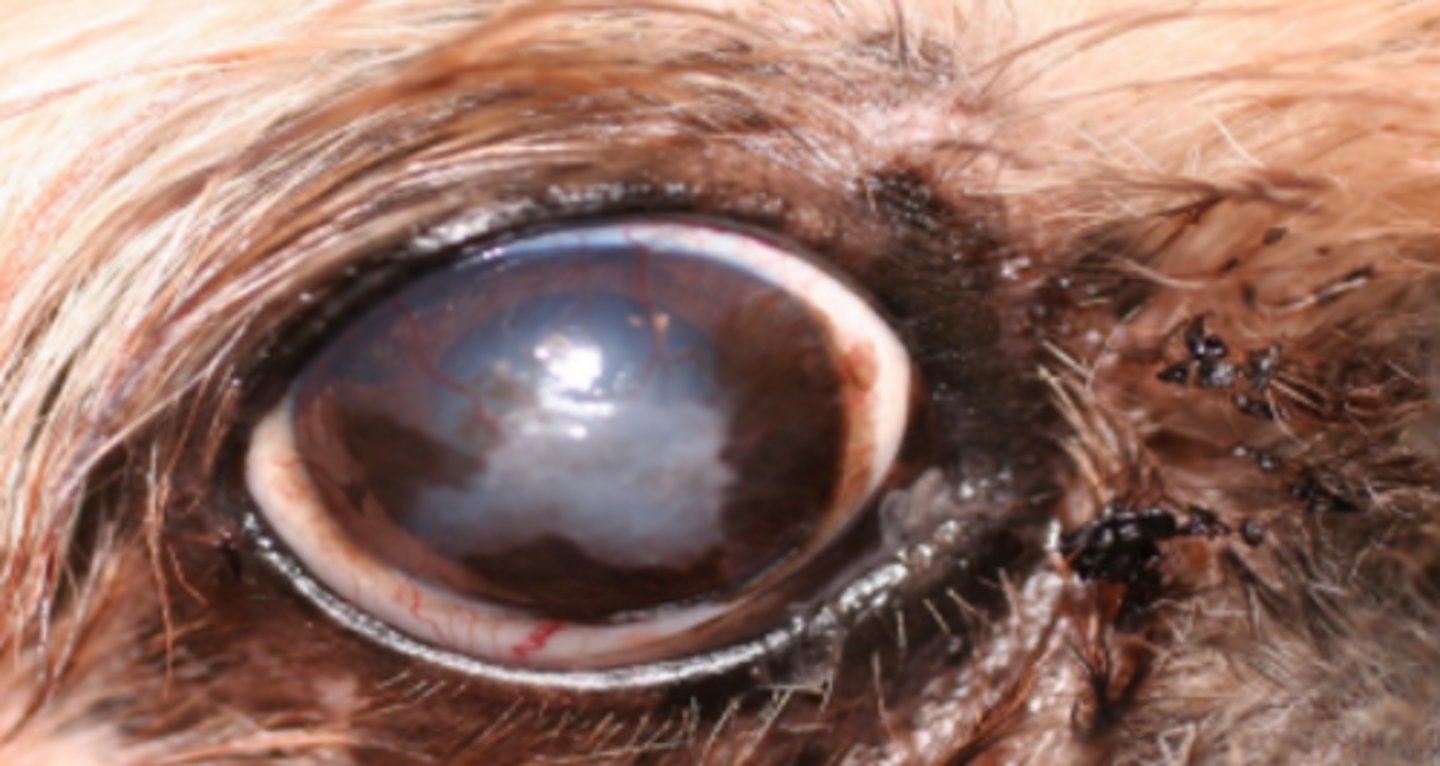
Keratoconjunctivitis
Corneal fibrosis, pigmentation and neovascularisation
Schirmer Tear Test (STT)
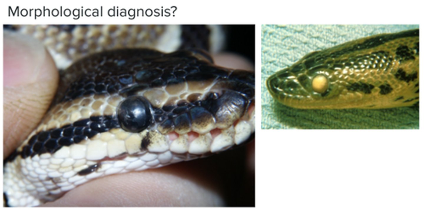
Retained spectacles > leads to abscess formation
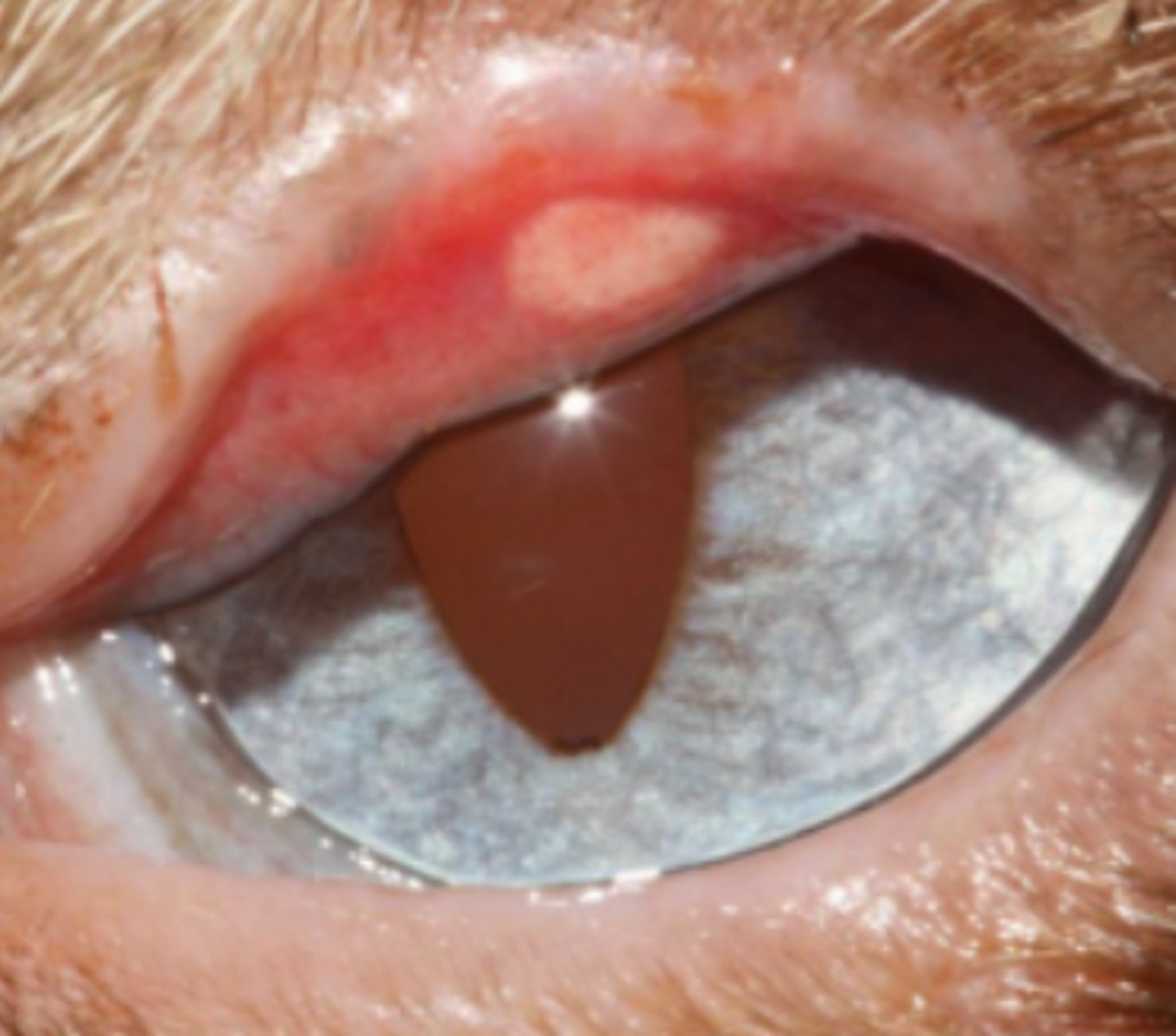
Meibomian adenoma (neoplasm of the eyelid)
Limbal melanocytoma (neoplasm of the eyelid)
Squamous cell carcinoma of the eyelid
Mastocytoma of the eyelid
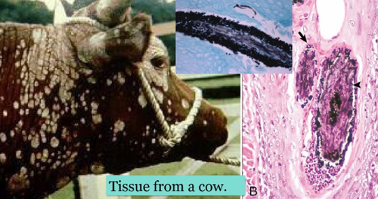
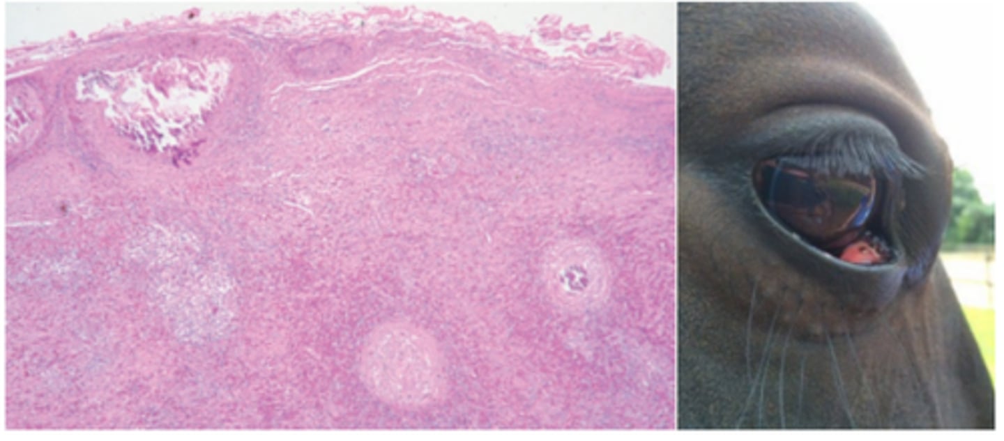
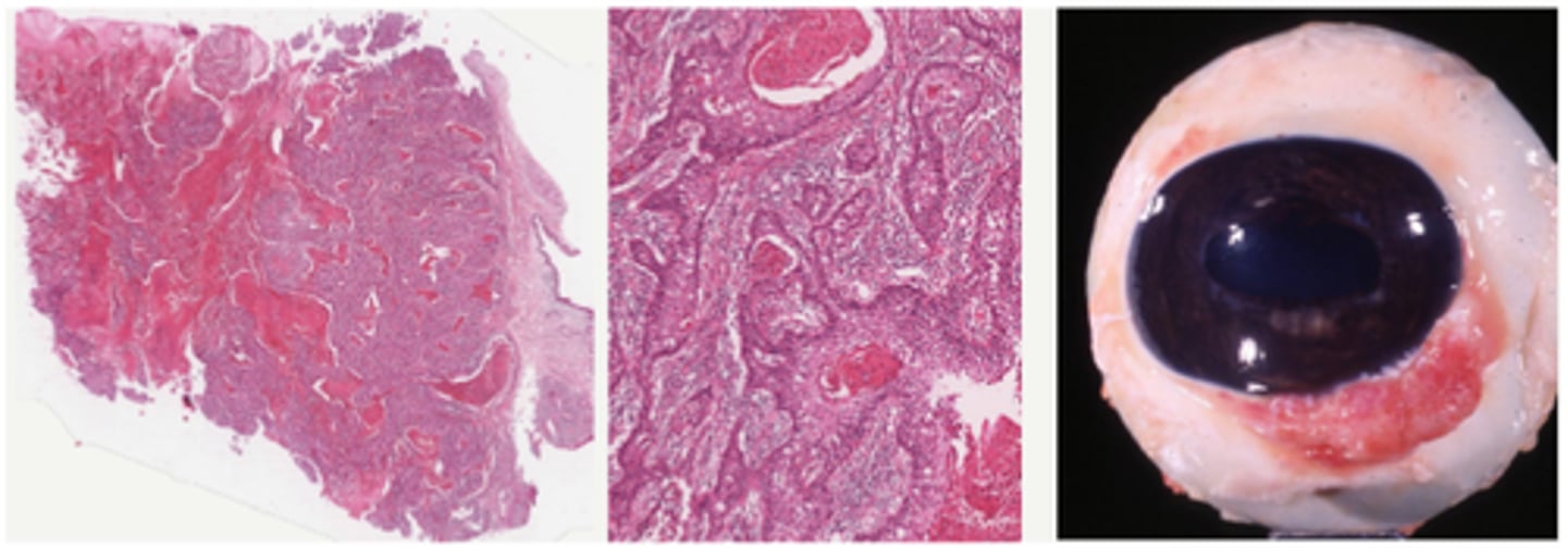
Squamous cell carcinoma, bovine
Polygonal neoplastic cells with scant to moderate amounts of eosinophilic cytoplasm, whorls of lamellar keratin (keratin pearls) within the centers of trabeculae and islands.
Ocular dermoid congenital condition