Unit 4 - Other quizlets combined
1/331
There's no tags or description
Looks like no tags are added yet.
Name | Mastery | Learn | Test | Matching | Spaced | Call with Kai |
|---|
No analytics yet
Send a link to your students to track their progress
332 Terms
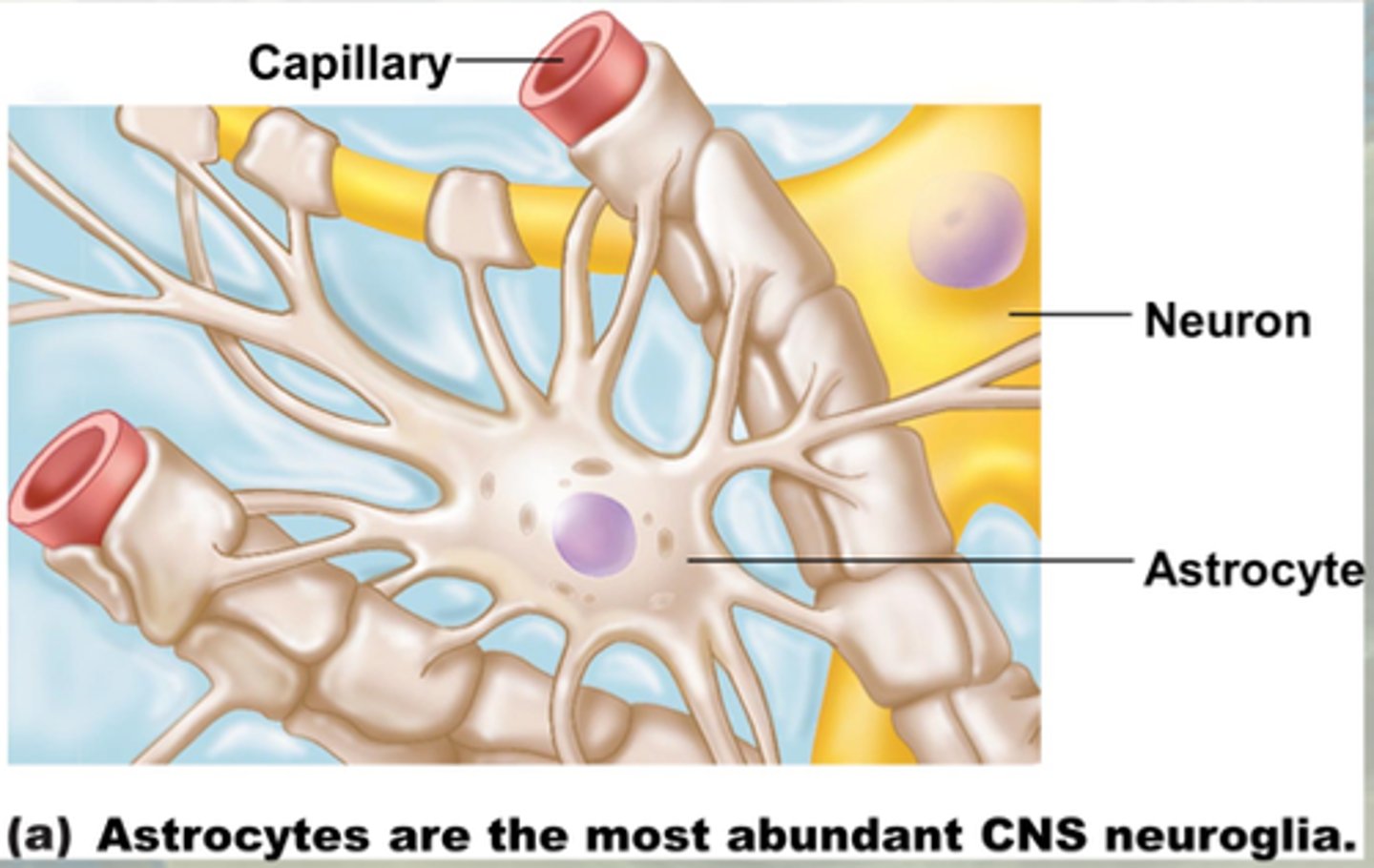
Astrocytes location
CNS
Astrocytes physical appearance
Astrocytes functions
-Most abundant, versatile, and highly branched glial cells
-Cling to neurons, synaptic endings, and capillaries
-Support and brace neurons
-Play a role in exchanges between capillaries and neurons
-Guide migration of young neurons
-Control the chemical environment around neurons
-Respond to nerve impulses and neurotransmitters
-Influence neuronal functioning
-Participate in information processing in the brain
Microglial cells location
CNS
Microglial cells physical appearance
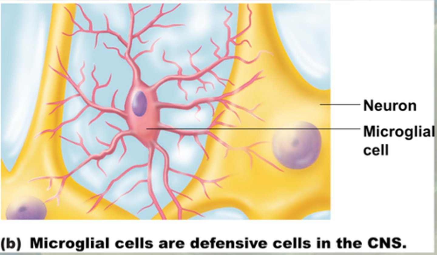
Microglial functions
-Small, ovoid cells with thorny processes that touch and monitor neurons
-Migrate toward injured neurons
-Can transform to phagocytize microorganisms and neuronal debris
Ependymal cells location
CNS
Ependymal cells physical appearance
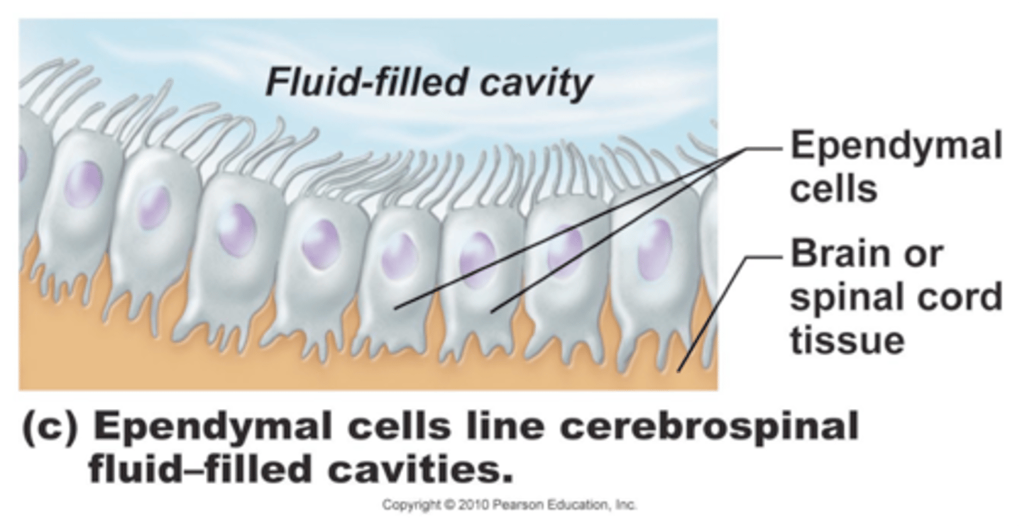
Ependymal cells functions
-Range in shape from squamous to columnar
-May be ciliated
-Cilia beat to circulate CSF
-Line the central cavities of the brain and spinal column
-Form a permeable barrier between cerebrospinal fluid (CSF) in cavities and tissue fluid bathing CNS cells
Oligodendrocytes location
CNS
Oligodendrocytes physical appearance
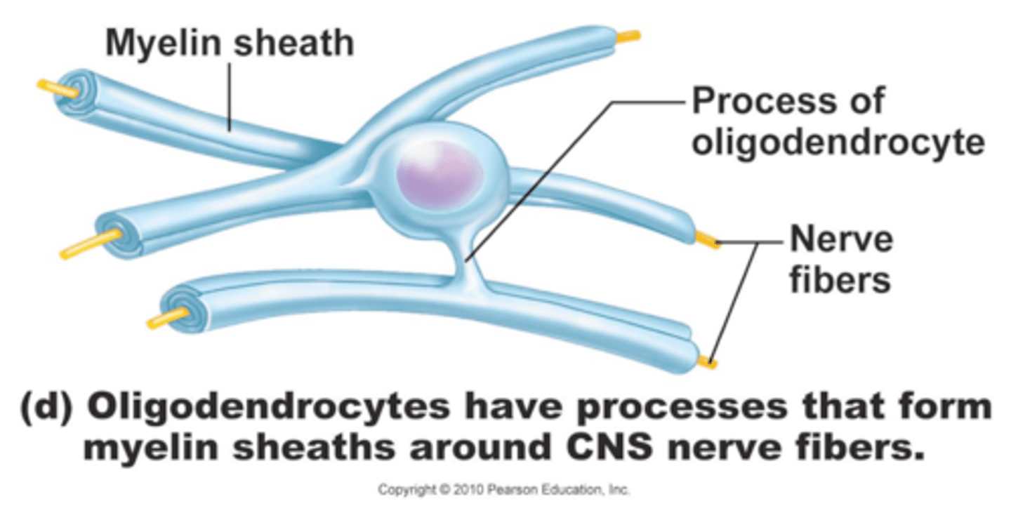
Oligodendrocytes functions
-Branched cells
-Processes wrap CNS nerve fibers, forming insulating myelin sheaths thicker nerve fibers
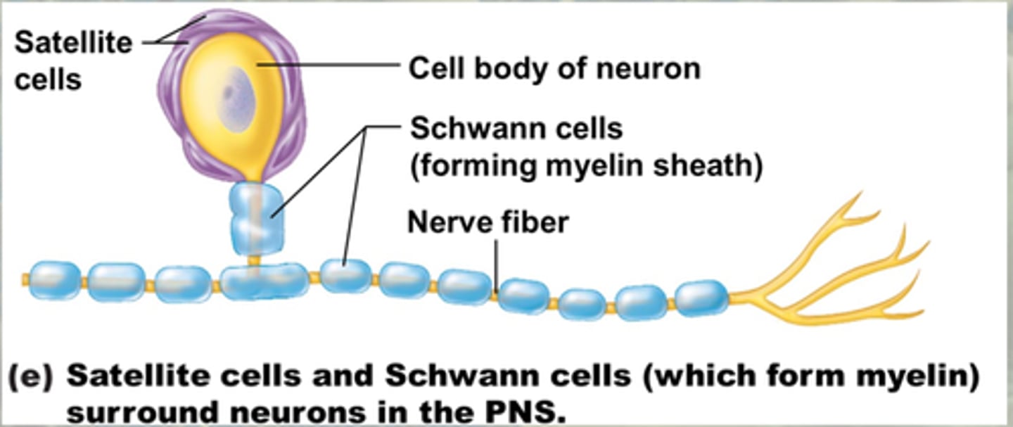
Satellite cells location
PNS
Satellite cells physical appearance

Satellite cells functions
protects and regulates nutrients for cell bodies in ganglia
Schwann cells location
PNS
Schwann cells physical appearance
Neurons - ganglia location
PNS
Neurons location
CNS
Ganglia physical appearance
Neurons physical appearance
Neurons functions
-maintains charge across the membrane
-communication via specialized chemicals
-neurotransmitters
-unipolar, bipolar, multipolar
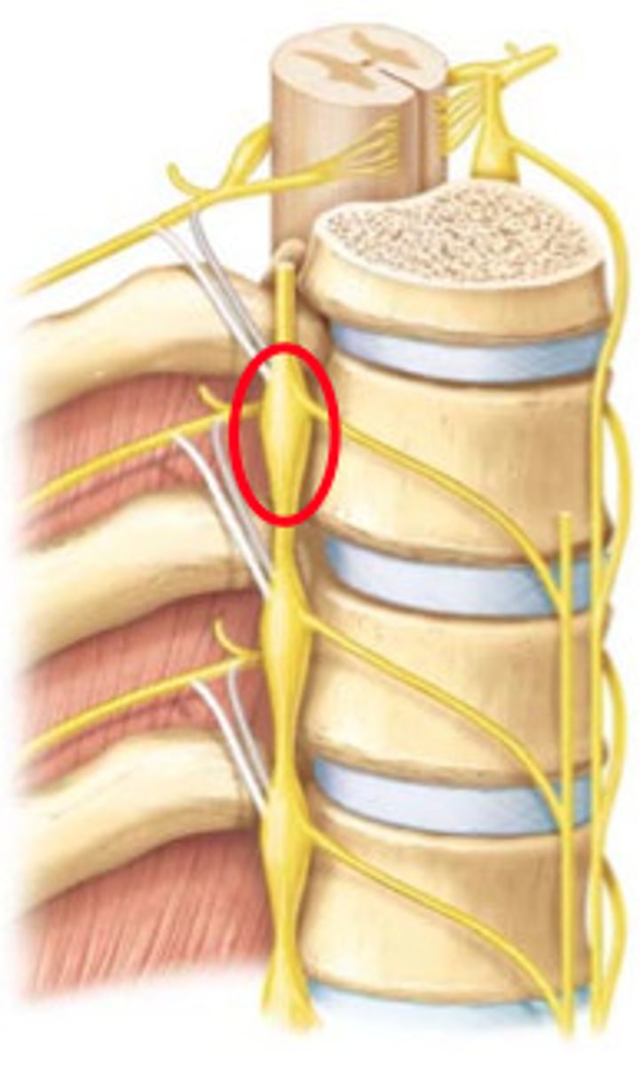
Ganglia function
-receive sensory fibers coming from receptors in the body
-where a first synapse is made prior to entry of the information into the CNS
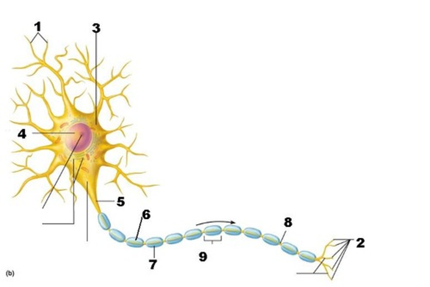
Dendrites
Branchlike parts of a neuron that are specialized to receive information. (A)

Nucleus
Control center of the cell (C)

Nucleolus of neuron
inside the nucleus (K)
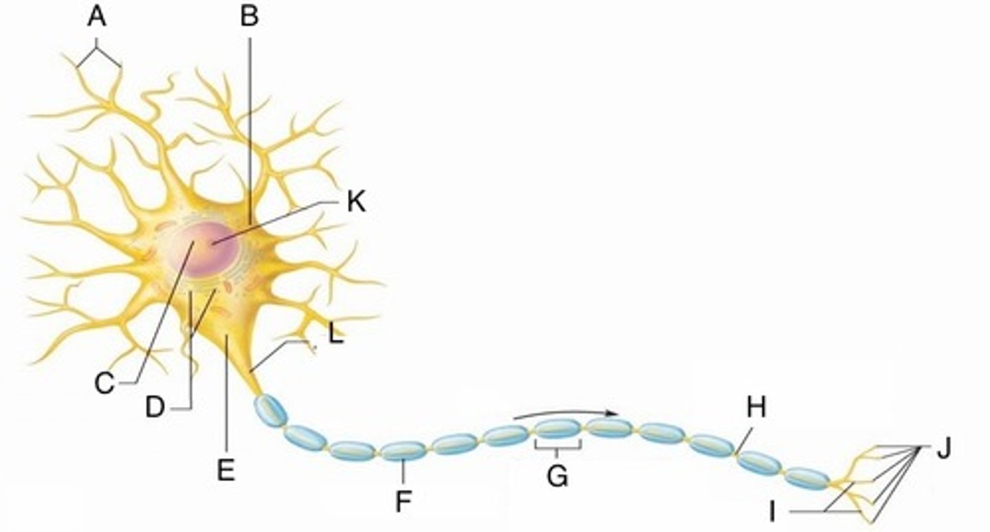
chromatophilic substance
essentially rough endoplasmic reticulum, important metabolically (D)

axon hillock
Cone shaped region of an axon where it joins the cell body. (E)

Axon
-the extension of a neuron, ending in branching terminal fibers, through which messages pass to other neurons or to muscles or glands
-impulse-generating and conducting region (L)

Impulse direction
electrical current is flowing in that direction

Schwann cell of neuron
Surrounds the axon and forms the myelin sheath (G)

Myelin sheath gap
space between adjacent neurolemmocytes (node of ranvier) (H)

Axon terminals (secretory region)
J

Terminal Branches
Branched endings of an axon that transmit messages to other neurons (I)

Axon (Nervous Tissue)

Dendrites (nervous tissue)
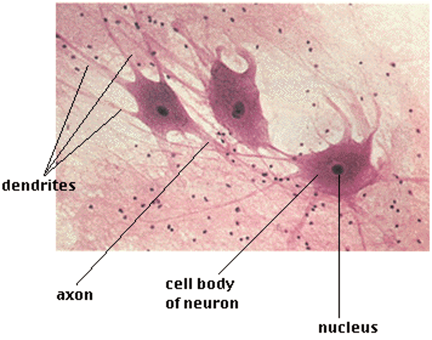
Cell body (nervous tissue)

Myelin sheath/schwann cells (nervous tissue)
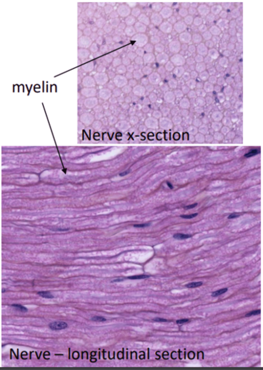
If Schwann cells are damaged, what might occur?
They will lose function or repair themselves
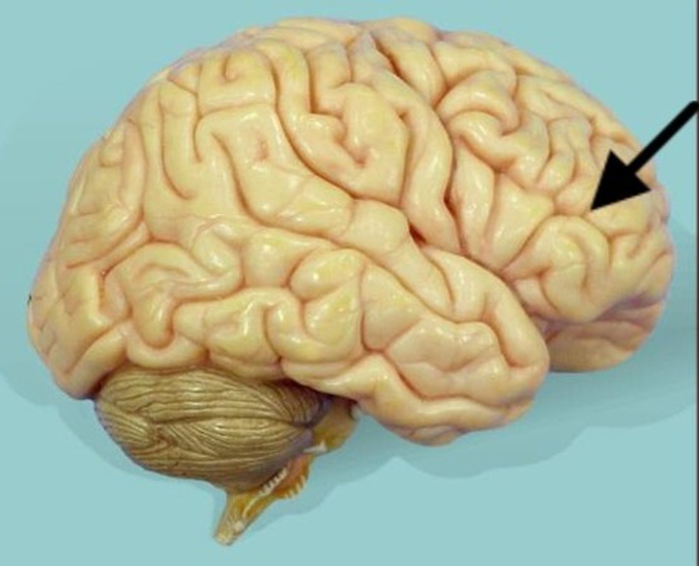
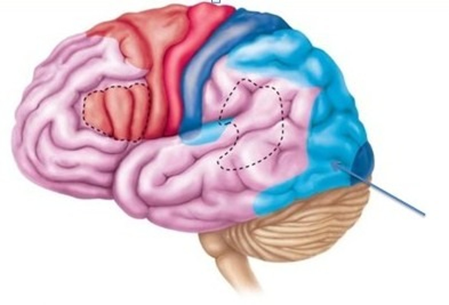
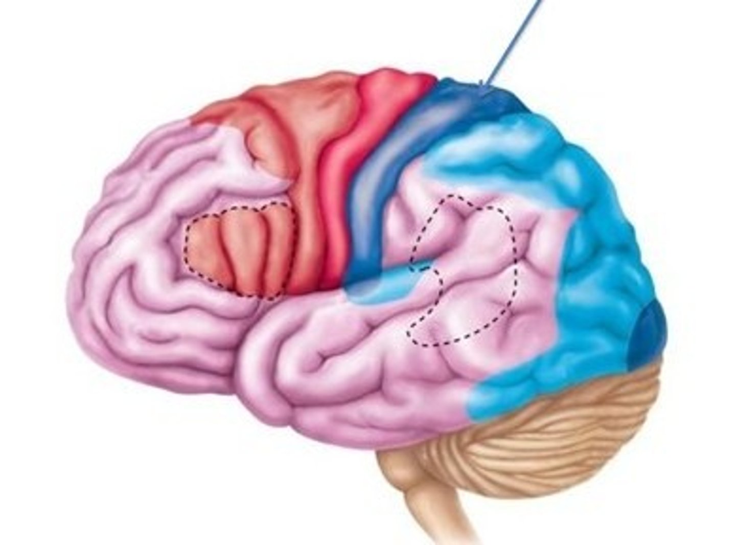
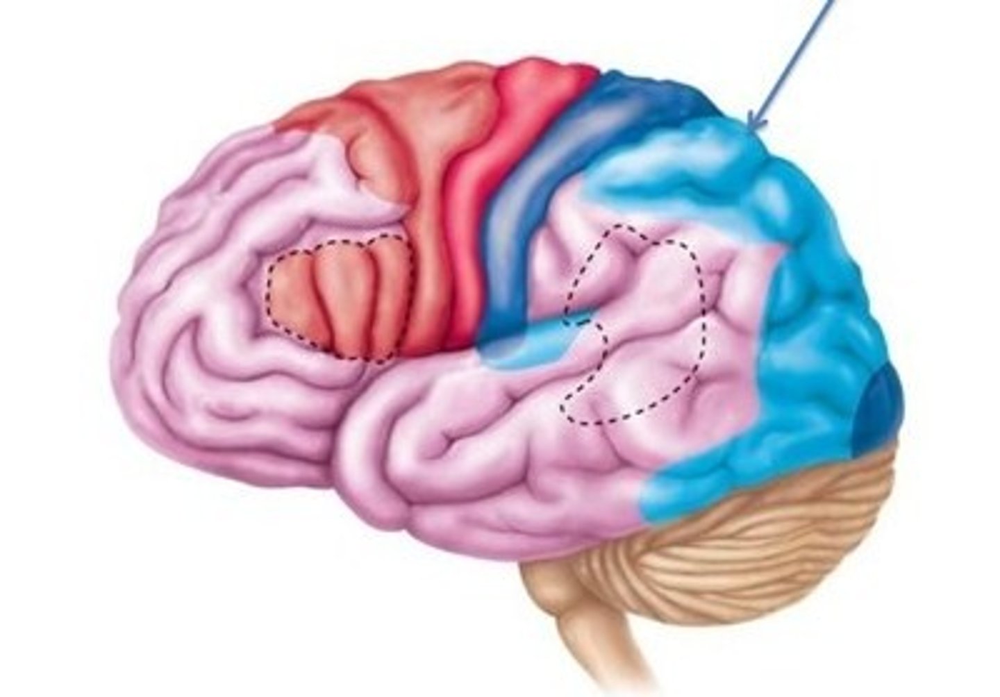
Frontal Lobe
A region of the cerebral cortex that has specialized areas for movement, abstract thinking, planning, memory, and judgment
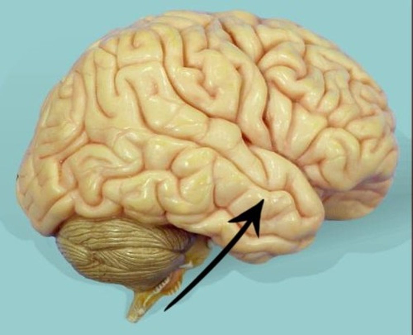
Temporal Lobe
A region of the cerebral cortex responsible for hearing and language.
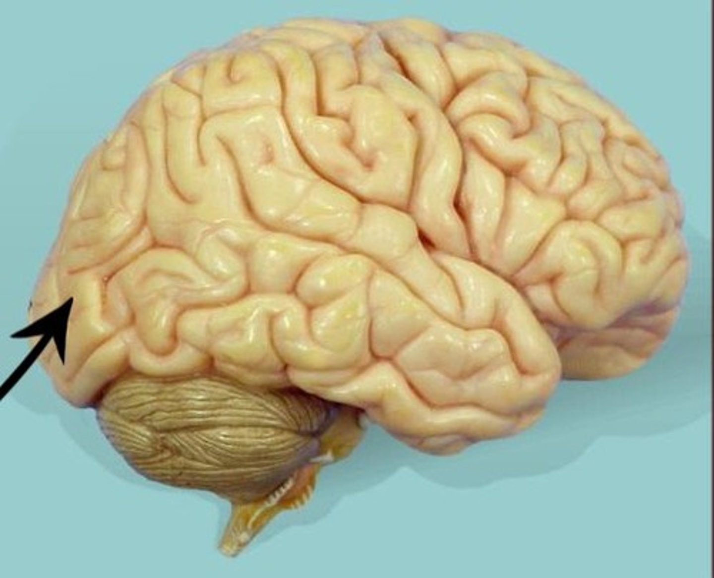
Occipital Lobe
A region of the cerebral cortex that processes visual information
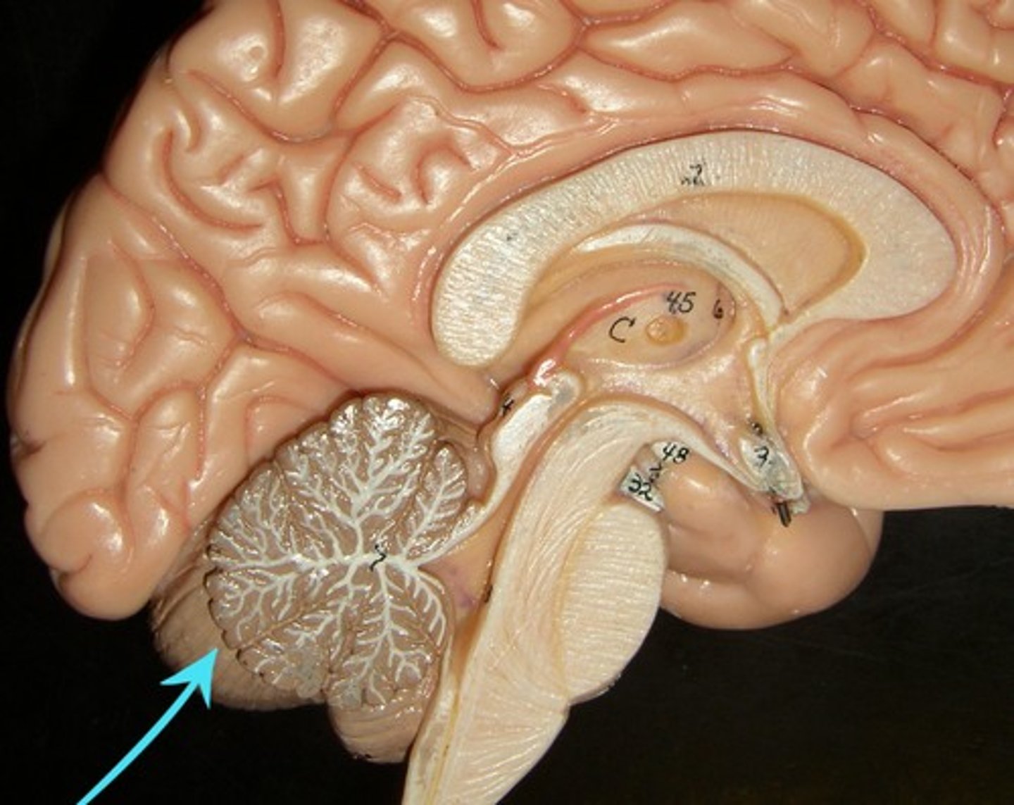
Cerebellum
the "little brain" at the rear of the brainstem; functions include processing sensory input and coordinating movement output and balance
Pons
A brain structure that relays information from the cerebellum to the rest of the brain
Medulla Oblongata
Part of the brainstem that controls vital life-sustaining functions such as heartbeat, breathing, blood pressure, and digestion.
Gyrus
A ridged or raised portion of a convoluted brain surface.
Cortex (gray matter)
The outer layer (approximately one-fourth to one-half inch) of brain tissue containing nerve cell bodies (neurons)
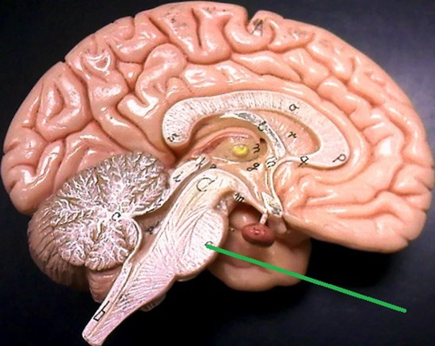
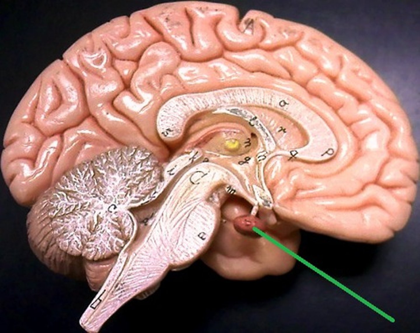
Pituitary Gland
The endocrine system's most influential gland. Under the influence of the hypothalamus, the pituitary regulates growth and controls other endocrine glands.
Optic Chiasma
The crossing of the optic nerves from the two eyes at the base of the brain
Optic Nerve
The nerve that carries neural impulses from the eye to the brain
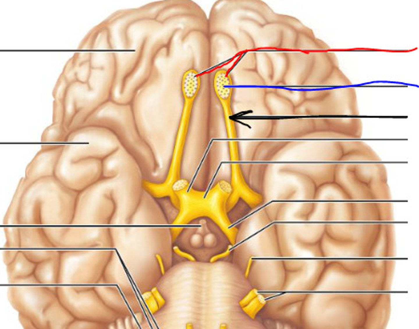
Olfactory Bulb and Tract
Sense of smell
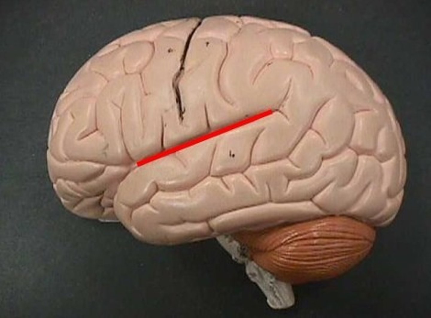
Lateral Sulcus
Separates temporal lobe from parietal and frontal lobes

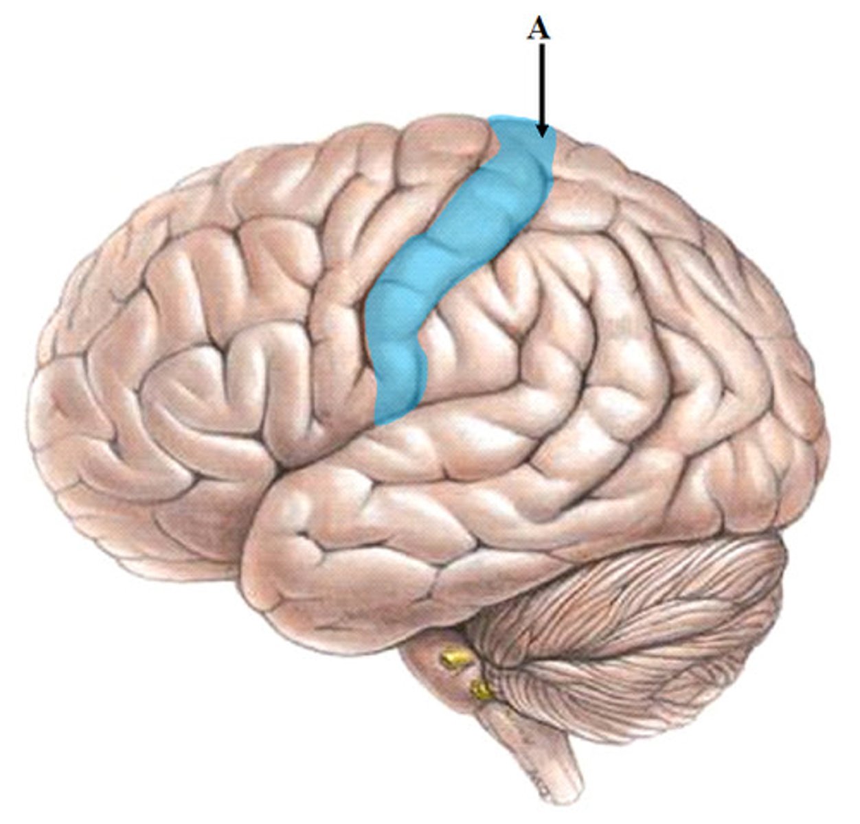
Central Sulcus
Separates frontal lobe from parietal lobe
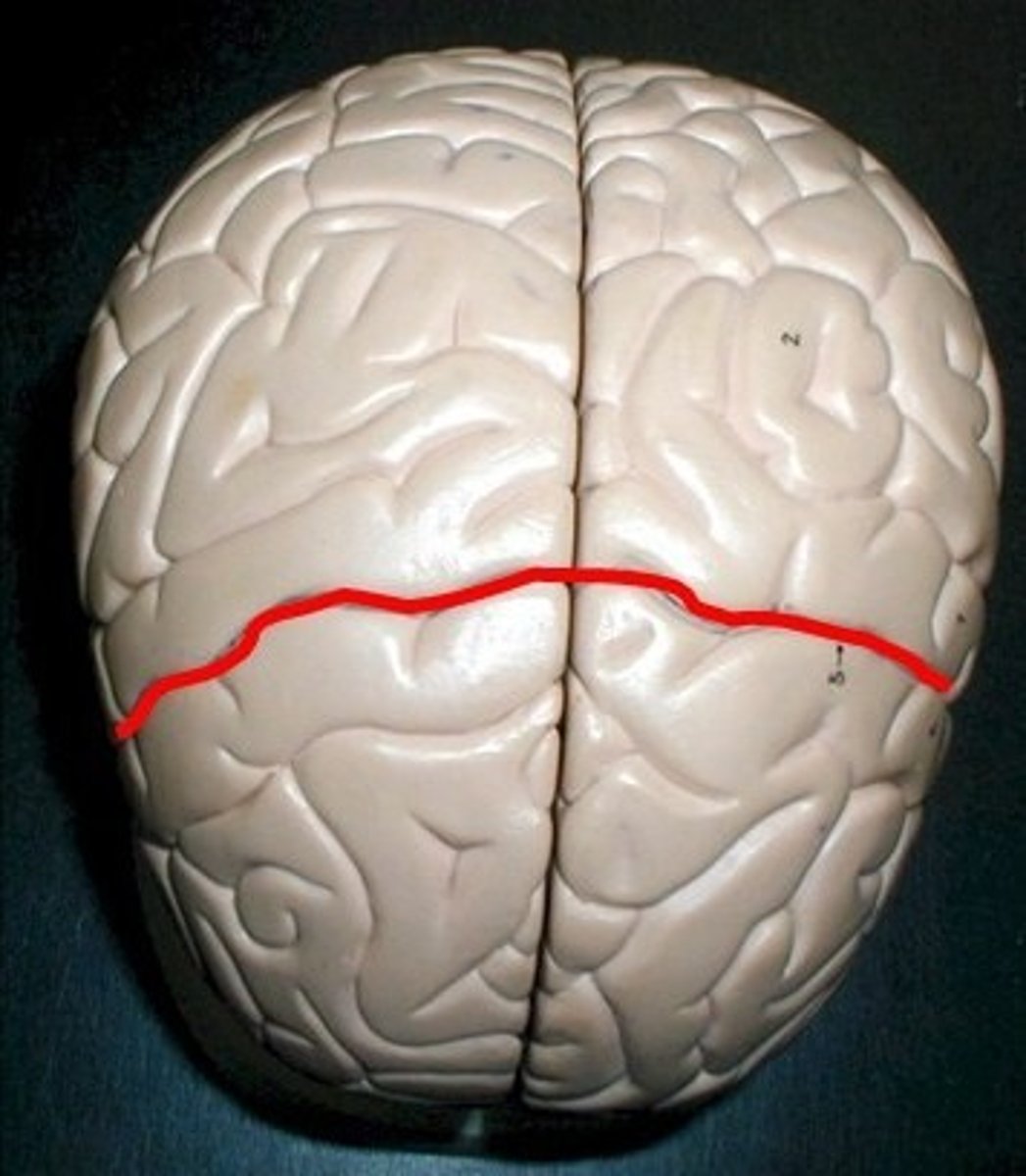
Sulcus
Narrow groove
White Matter
Whitish nervous tissue of the CNS consisting of neurons and their myelin sheaths.
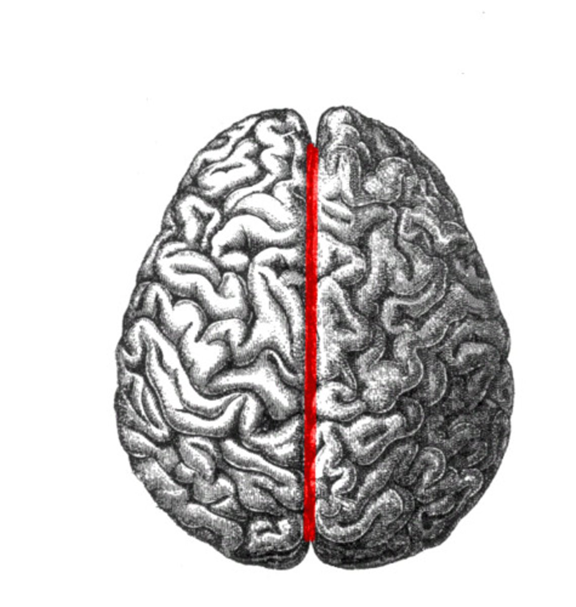
longitudinal fissure
separates left and right hemispheres
precentral gyrus
the strip of frontal cortex, just in front of the central sulcus, that is crucial for motor control
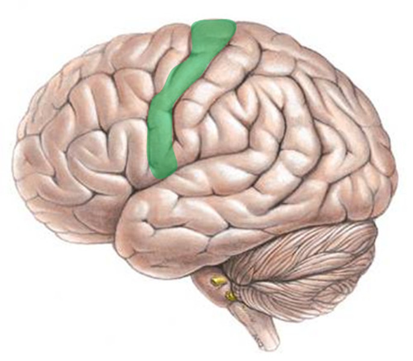
postcentral gyrus
the strip of parietal cortex, just behind the central sulcus, that receives somatosensory information from the entire body
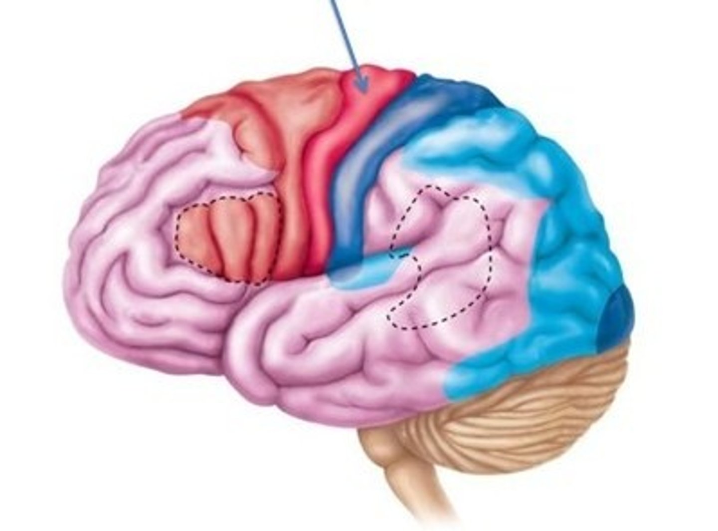
Primary motor cortex
Allows conscious control of precise, skilled, skeletal muscle movements
Premotor cortex
-Helps plan movements; staging area for skilled motor activities
-Controls learned, repetitious, or patterned motor skills
-Coordinates simultaneous or sequential actions
-Controls voluntary actions that depend on sensory feedback
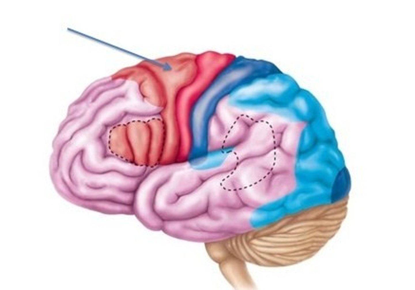
Frontal Eye Field
Controls voluntary eye movements

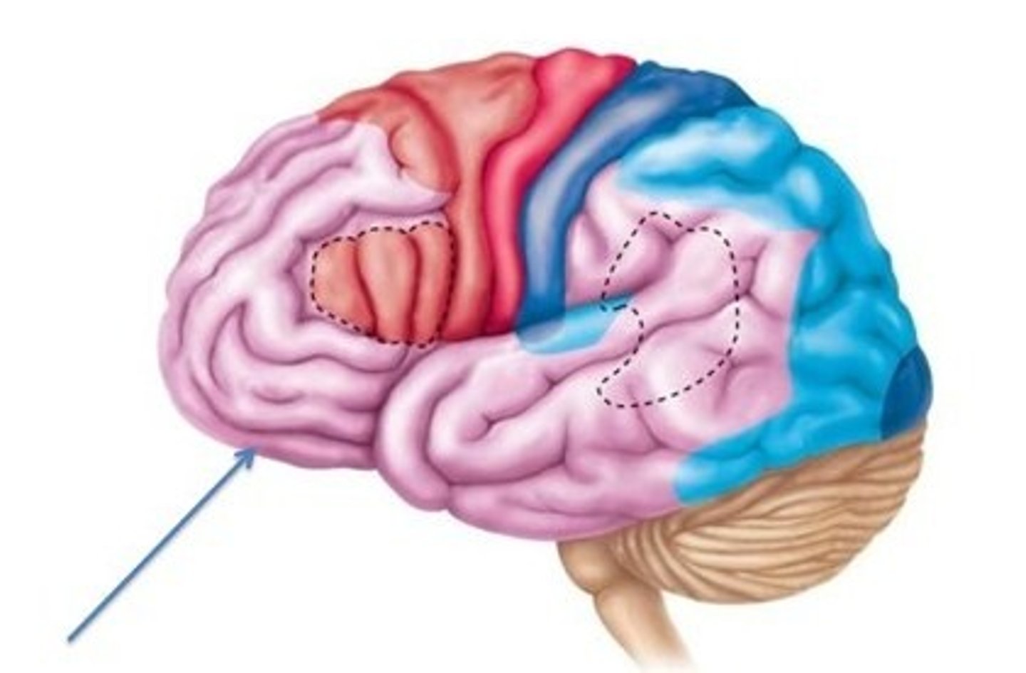
Prefrontal Cortex
part of frontal lobe responsible for thinking, planning, and language
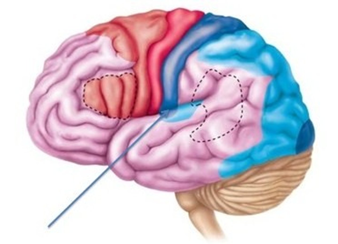
Broca's Area
-Present in one hemisphere (usually the left)
-Motor speech area that directs muscles of speech production
-Active in planning speech and voluntary motor activities
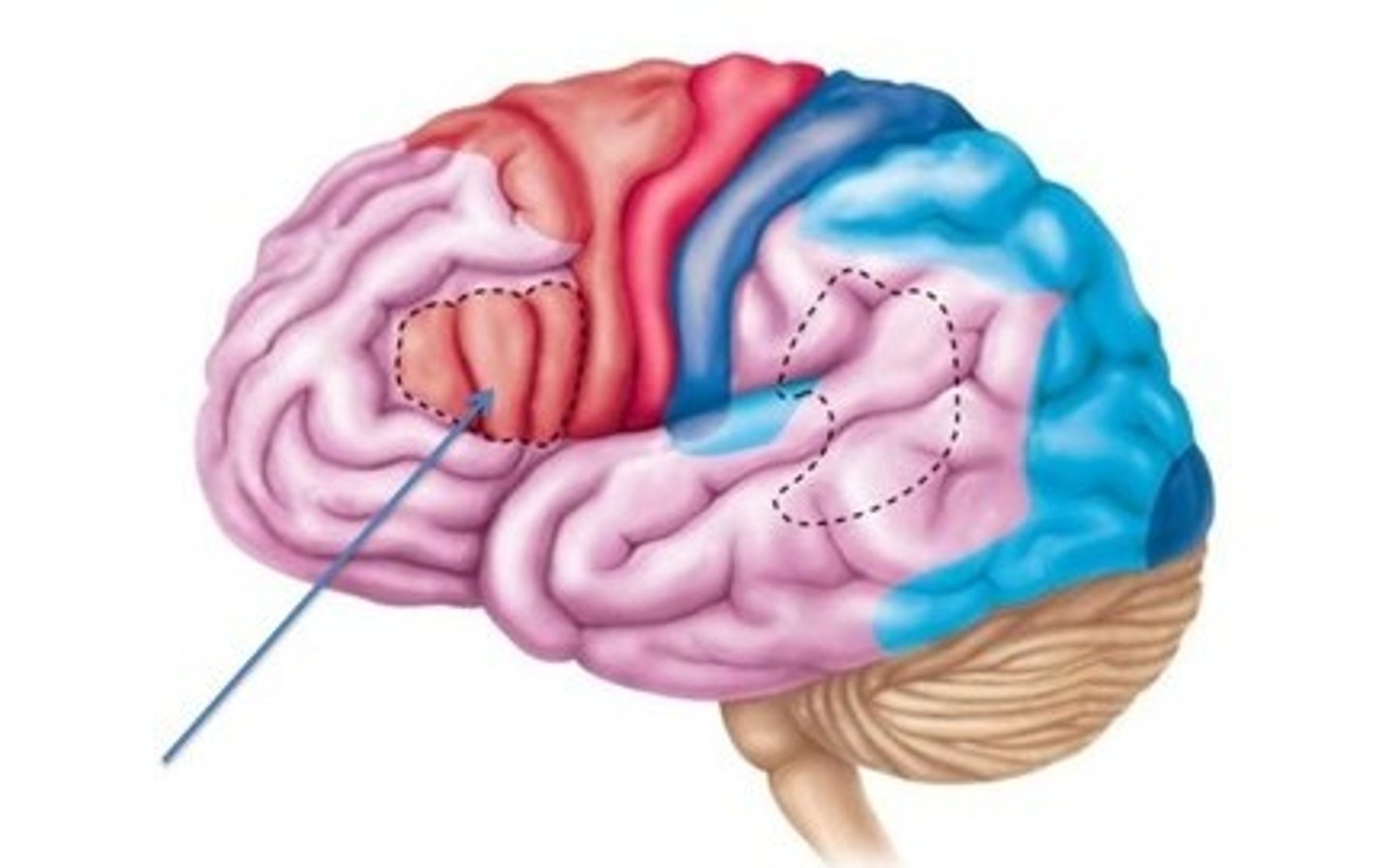
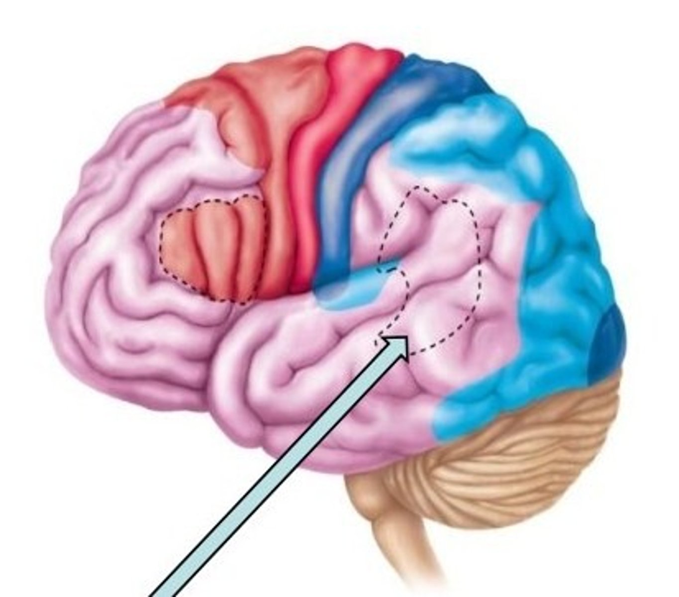
Wernicke's area
controls language reception - a brain area involved in language comprehension and expression; usually in the left temporal lobe
primary auditory area
-Superior margin of temporal lobes
-Interprets information from inner ear as pitch, loudness, and location
auditory association area
-Located posterior to primary auditory cortex
-Stores memories of sounds and permits perception of sound stimulus

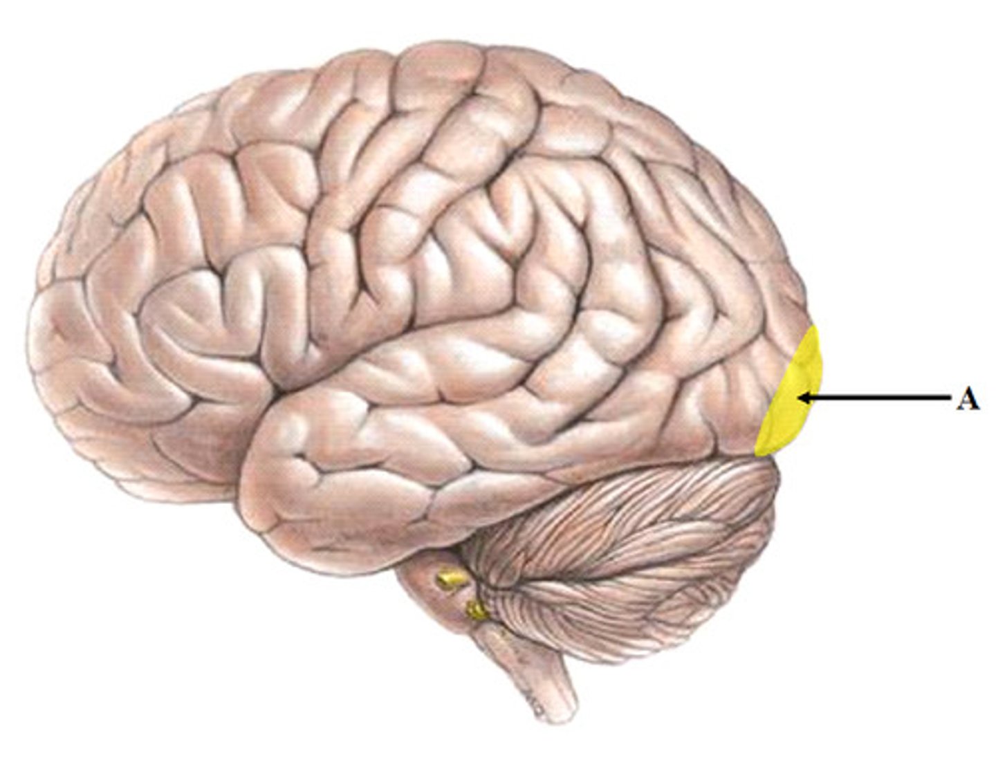
primary visual (striae) cortex
-Extreme posterior tip of occipital lobe
-Most buried in calcarine sulcus of occipital lobe
-Receives visual information from retinas
visual association area
-Surrounds primary visual cortex
-Uses past visual experiences to interpret visual stimuli (e.g., color, form, and movement)
E.g., ability to recognize faces
-Complex processing involves entire posterior half of cerebral hemispheres
primary somatosensory cortex
-In postcentral gyri of parietal lobe
-Receives general sensory information from skin, and proprioceptors of skeletal muscle, joints, and tendons
-Capable of spatial discrimination: identification of body region being stimulated
somatosensory association
-Posterior to primary somatosensory cortex
-Integrates sensory input from primary somatosensory cortex for understanding of object
-Determines size, texture, and relationship of parts of objects being felt
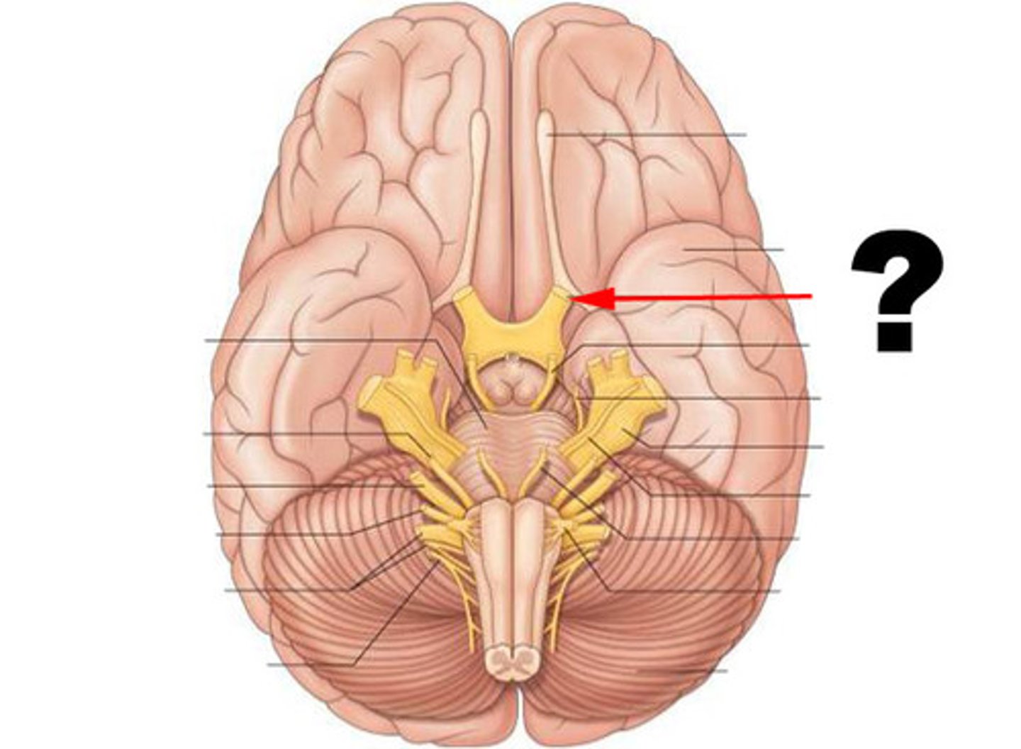
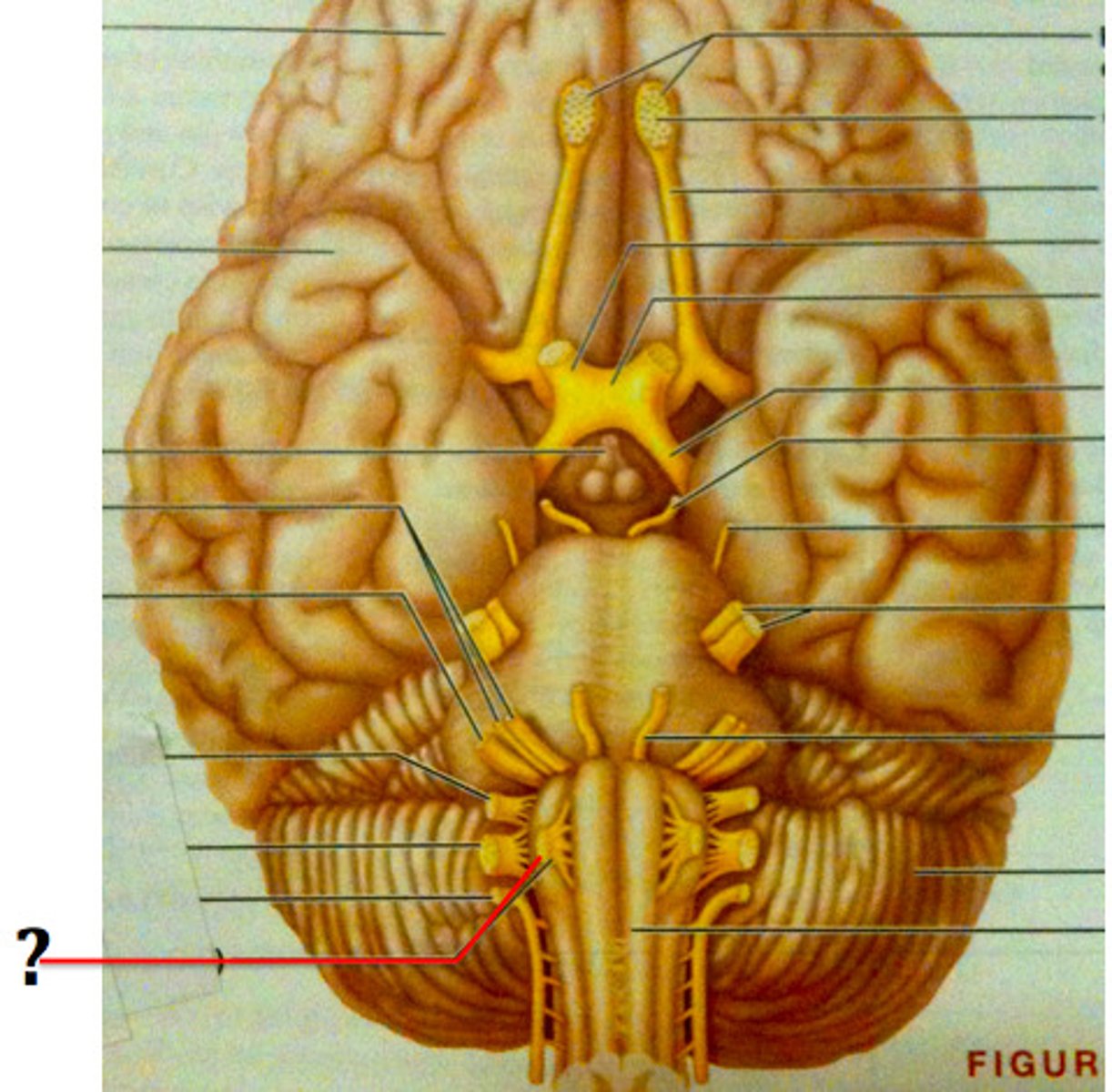
Olfactory Nerves (I)
-Sensory nerves of smell
-Run from nasal mucosa to olfactory bulbs
-Pass through cribriform plate of ethmoid bone
-Fibers synapse in olfactory bulbs
-Pathway terminates in primary olfactory cortex
-Purely sensory (olfactory) function
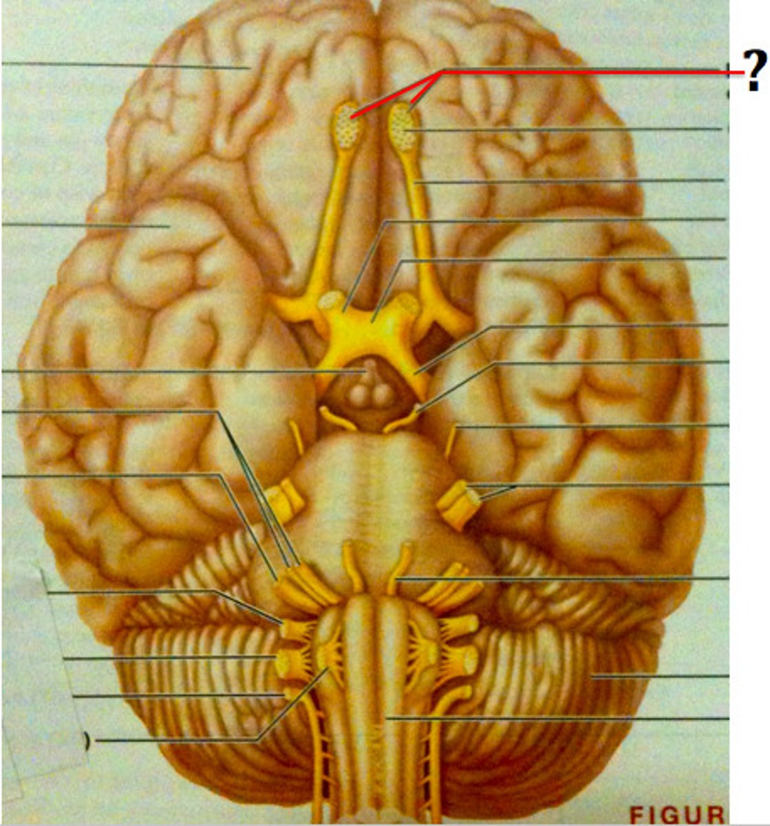
Optic Nerves (II)
-Arise from retinas; really a brain tract
-Pass through optic canals, converge and partially cross over at optic chiasma
-Optic tracts continue to thalamus, where they synapse
-Optic radiation fibers run to occipital (visual) cortex
-Purely sensory (visual) function
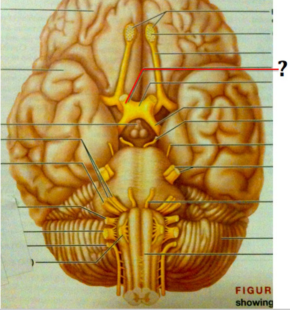
Oculomotor Nerves (III)
-Fibers extend from ventral midbrain through superior orbital fissures to four of six extrinsic eye muscles
-Function in raising eyelid, directing eyeball, constricting iris (parasympathetic), and controlling lens shape
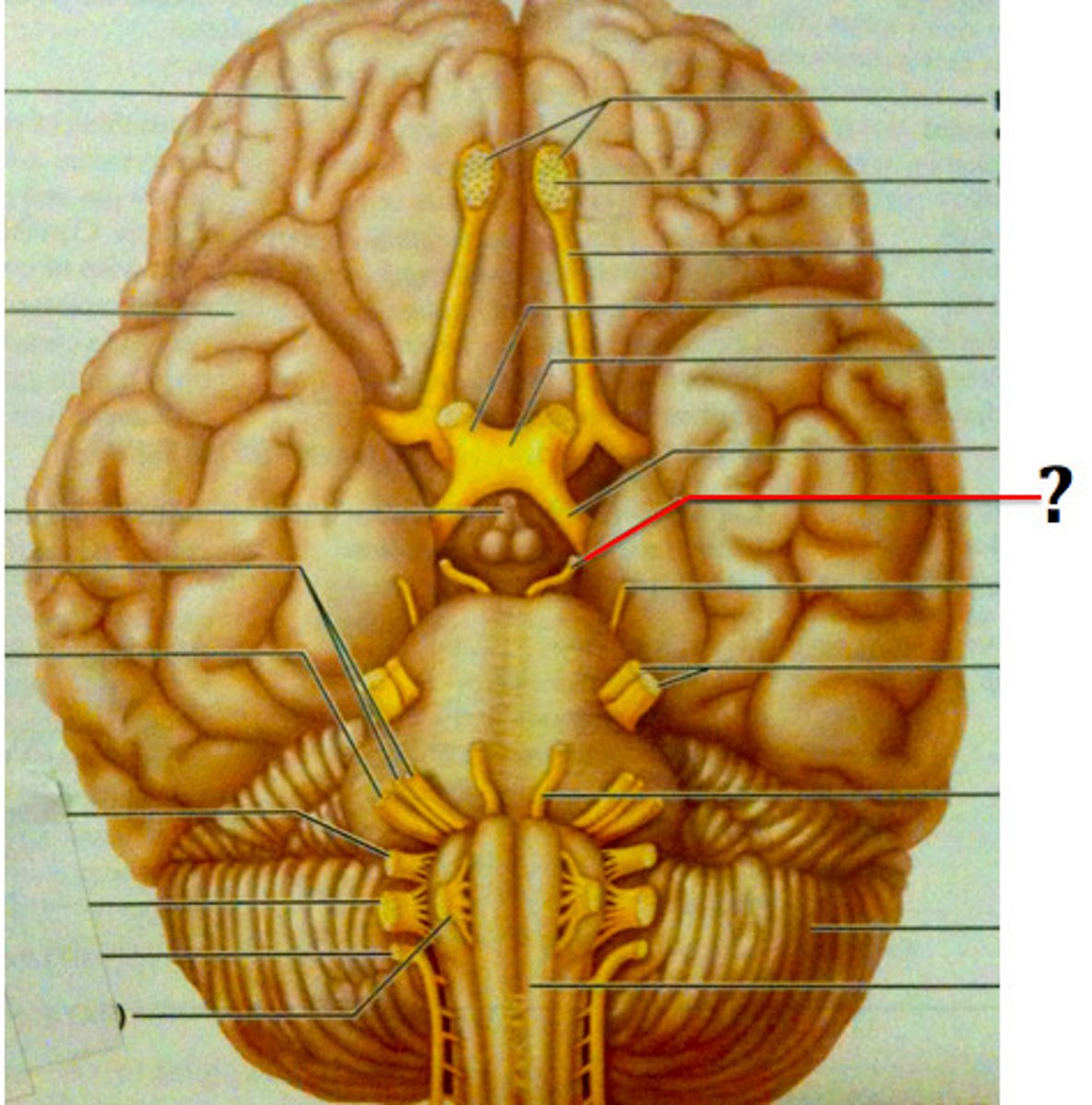
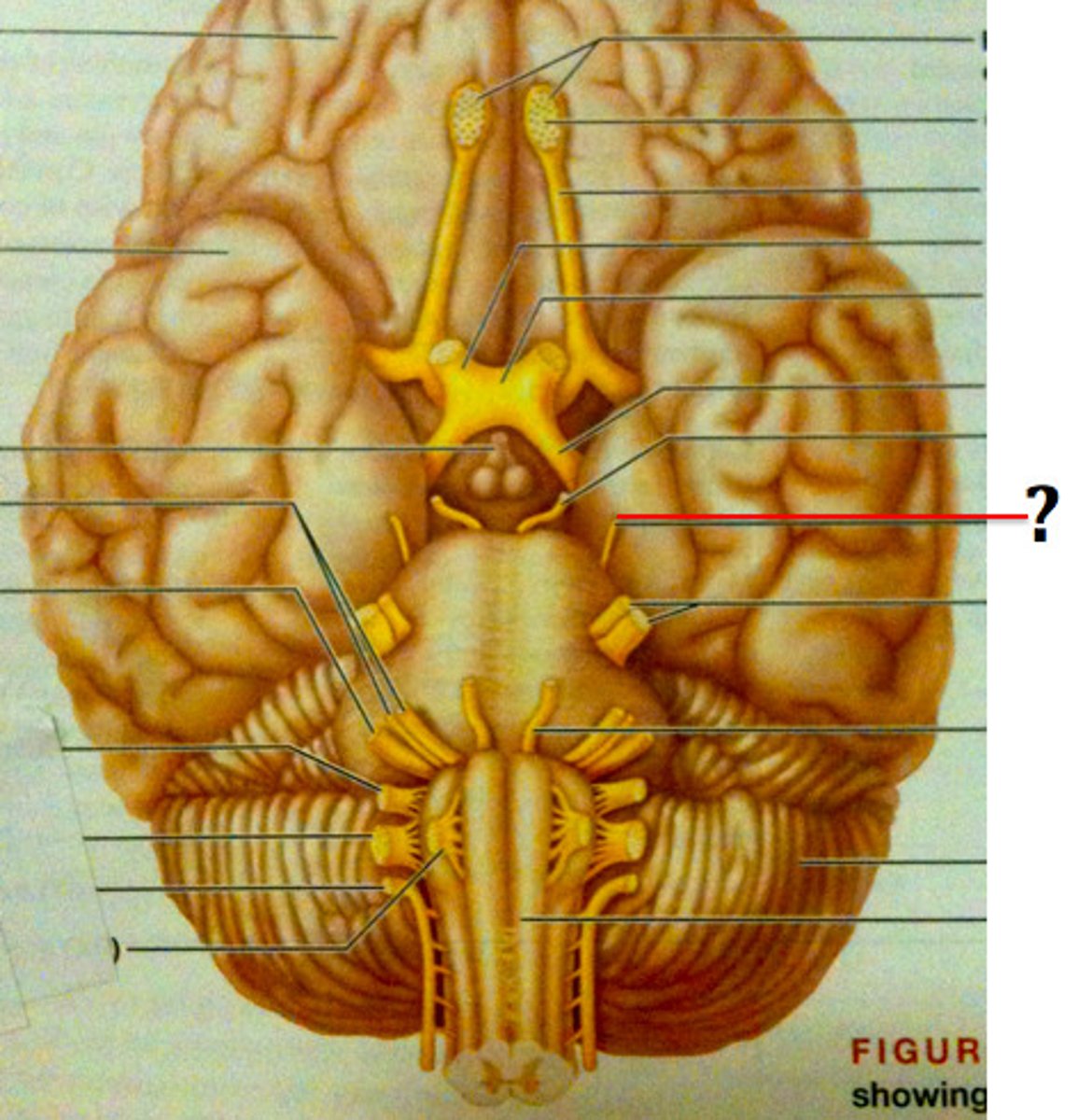
Trochlear Nerves (IV)
-motor, eye movements
-Fibers from dorsal midbrain enter orbits via superior orbital fissures to innervate superior oblique muscle
-Primarily motor nerve that directs eyeball
Trigeminal Nerves (V)
-The fifth pair of cranial nerves, each of which has three major branches; they conduct motor signals from the brain to the muscles involved in chewing, and sensory signals from the same muscles and from other parts of the face to the brain.
-Largest cranial nerves; fibers extend from pons to face
-Three divisions:
-Ophthalmic (V1) passes through superior orbital fissure
-Maxillary (V2) passes through foramen rotundum
-Mandibular (V3) passes through the foramen ovale
-Convey sensory impulses from various areas of face (V1) and (V2)
-Supply motor fibers (V3) for mastication
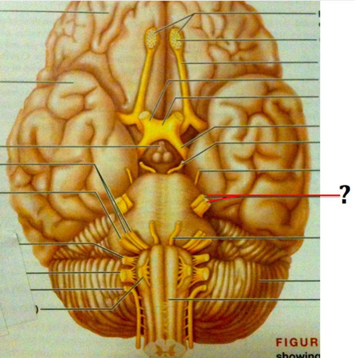
Abducens Nerves (VI)
-lateral eye movements
-Fibers from inferior pons enter orbits via superior orbital fissures
-Primarily a motor, innervating lateral rectus muscle
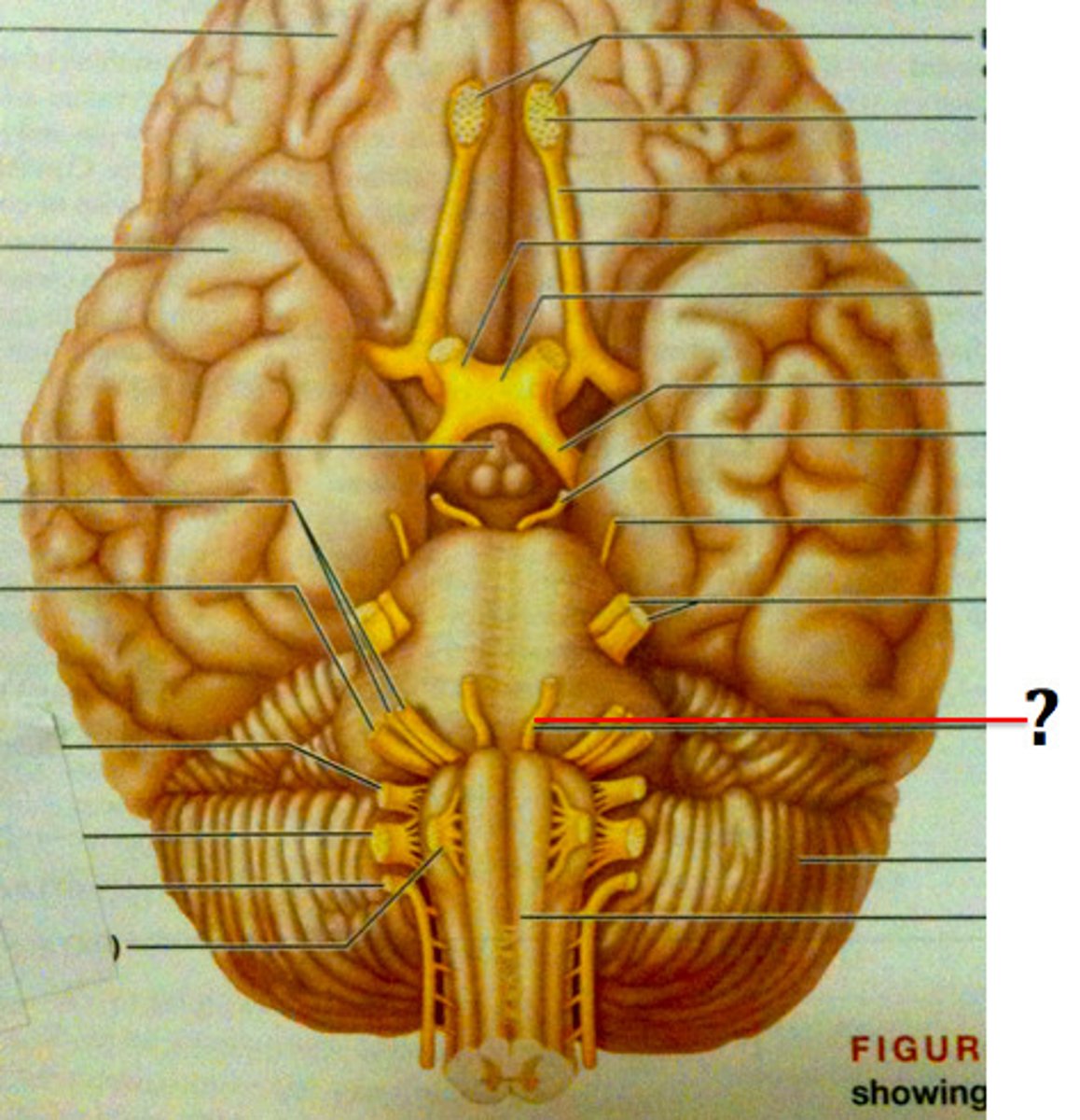
Facial Nerves (VII)
-sensory (taste) and motor function (facial expressions)
-Chief motor nerves of face with 5 major branches
-Motor functions include facial expression, parasympathetic impulses to lacrimal and salivary glands
-Sensory function (taste) from anterior two-thirds of tongue
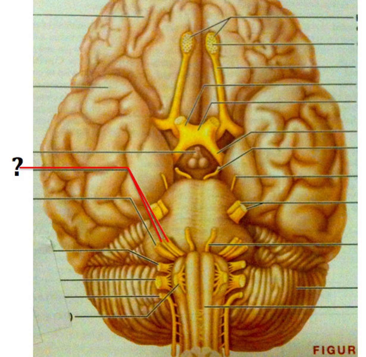
Vestibulocochlear Nerves (VIII)
-special sensory
-vestibular branch = balance and equilibrium
-cochlear branch = hearing
-Afferent fibers from hearing receptors (cochlear division) and equilibrium receptors (vestibular division) pass from inner ear through internal acoustic meatuses, and enter brain stem at pons-medulla border
-Mostly sensory function; small motor component for adjustment of sensitivity of receptors
-Formerly auditory nerve
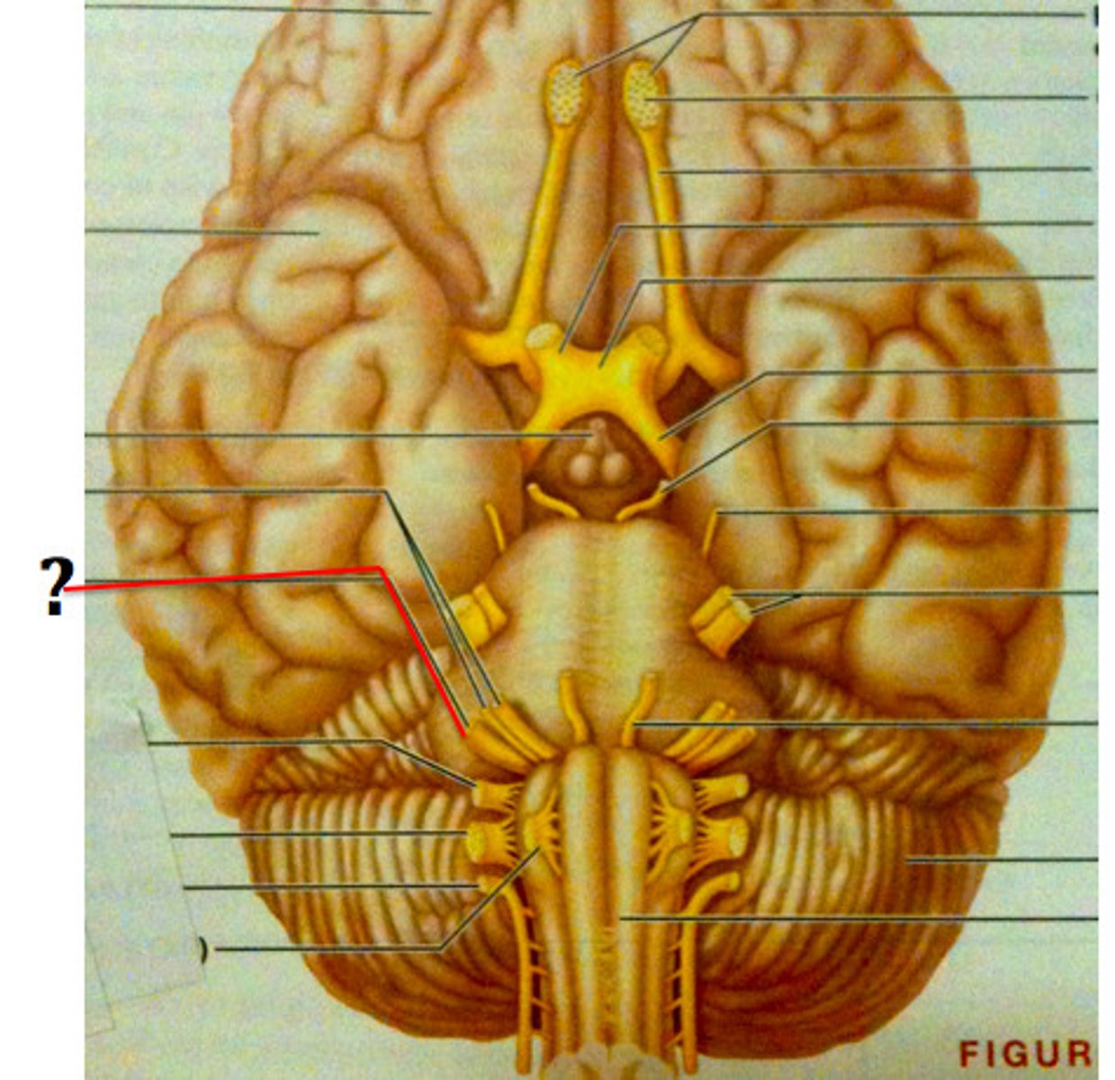
Glossopharyngeal Nerves (IX)
-Fibers from medulla leave skull via jugular foramen and run to throat
-Motor functions - innervate part of tongue and pharynx for swallowing, and provide parasympathetic fibers to parotid salivary glands
-Sensory functions - fibers conduct taste and general sensory impulses from pharynx and posterior tongue, and impulses from carotid chemoreceptors and baroreceptors
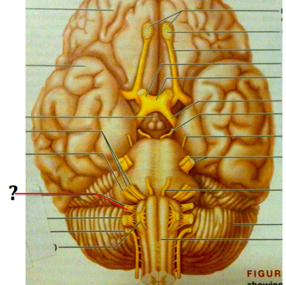
Vagus Nerves (X)
-sensory (viscera) and motor (digestive, respiratory)
-Only cranial nerves that extend beyond head and neck region
-Fibers from medulla exit skull via jugular foramen
-Most motor fibers are parasympathetic fibers that help regulate activities of heart, lungs, and abdominal viscera
-Sensory fibers carry impulses from thoracic and abdominal viscera, baroreceptors, chemoreceptors, and taste buds of posterior tongue and pharynx
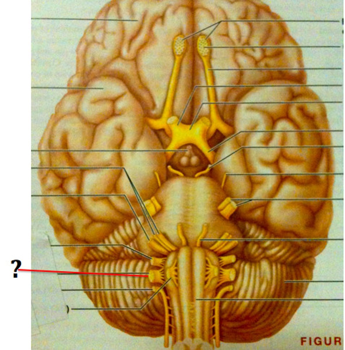
Accessory Nerves (XI)
-Formed from ventral rootlets from C1–C5 region of spinal cord (not brain)
-Rootlets pass into cranium via each foramen magnum
-Accessory motor and sensory nerves exit skull via jugular foramina to innervate trapezius and sternocleidomastoid muscles
-Formerly spinal accessory nerve
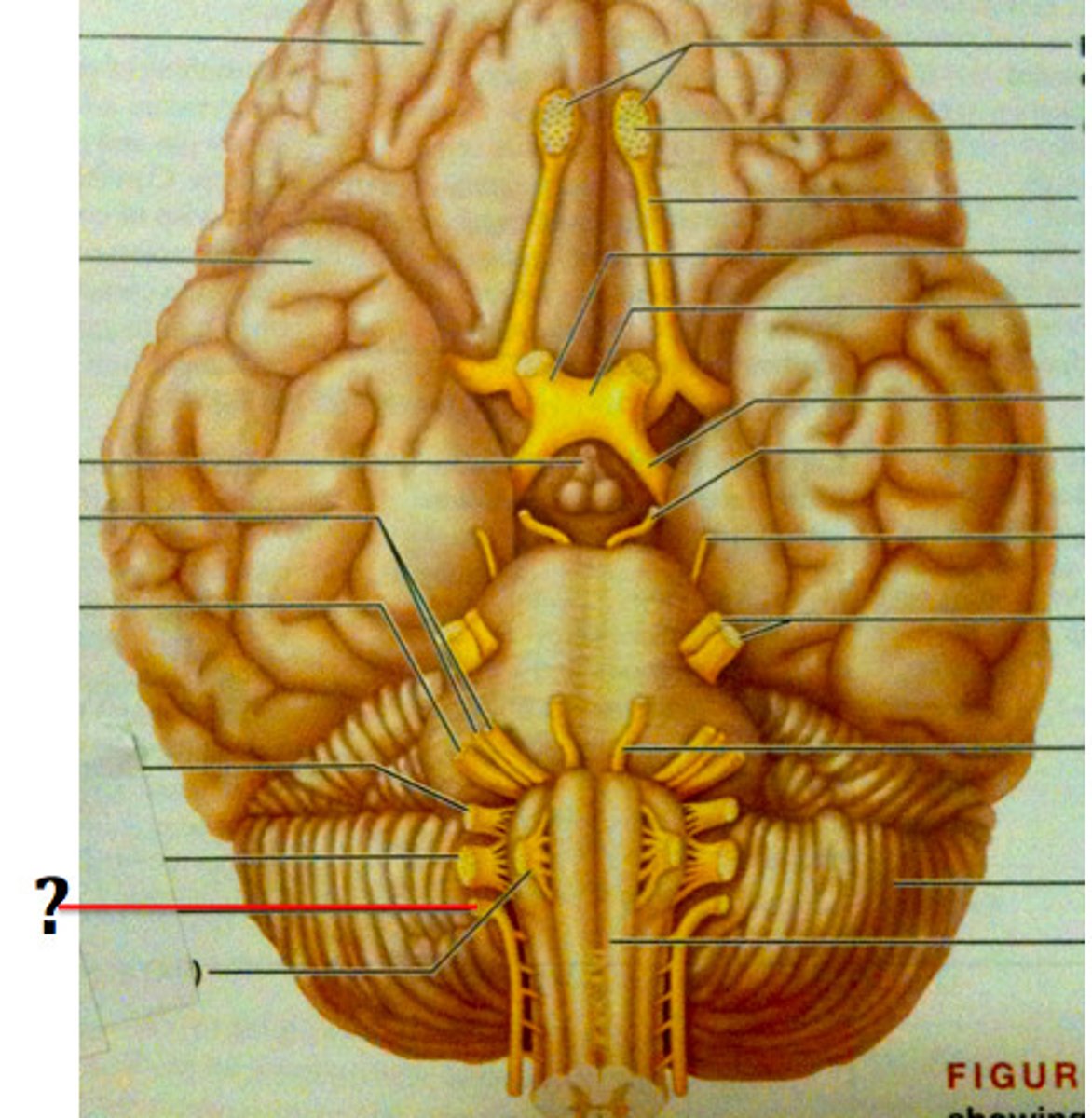
Hypoglossal Nerves (XII)
-Fibers from medulla exit skull via hypoglossal canal
-Innervate extrinsic and intrinsic muscles of tongue that contribute to swallowing and speech
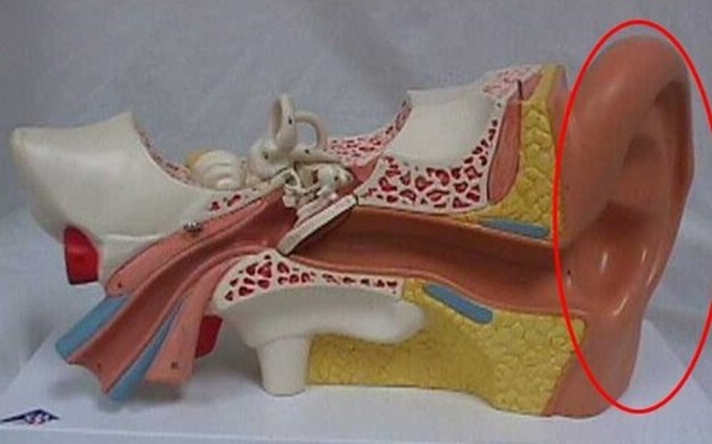
Lobule
Earlobe
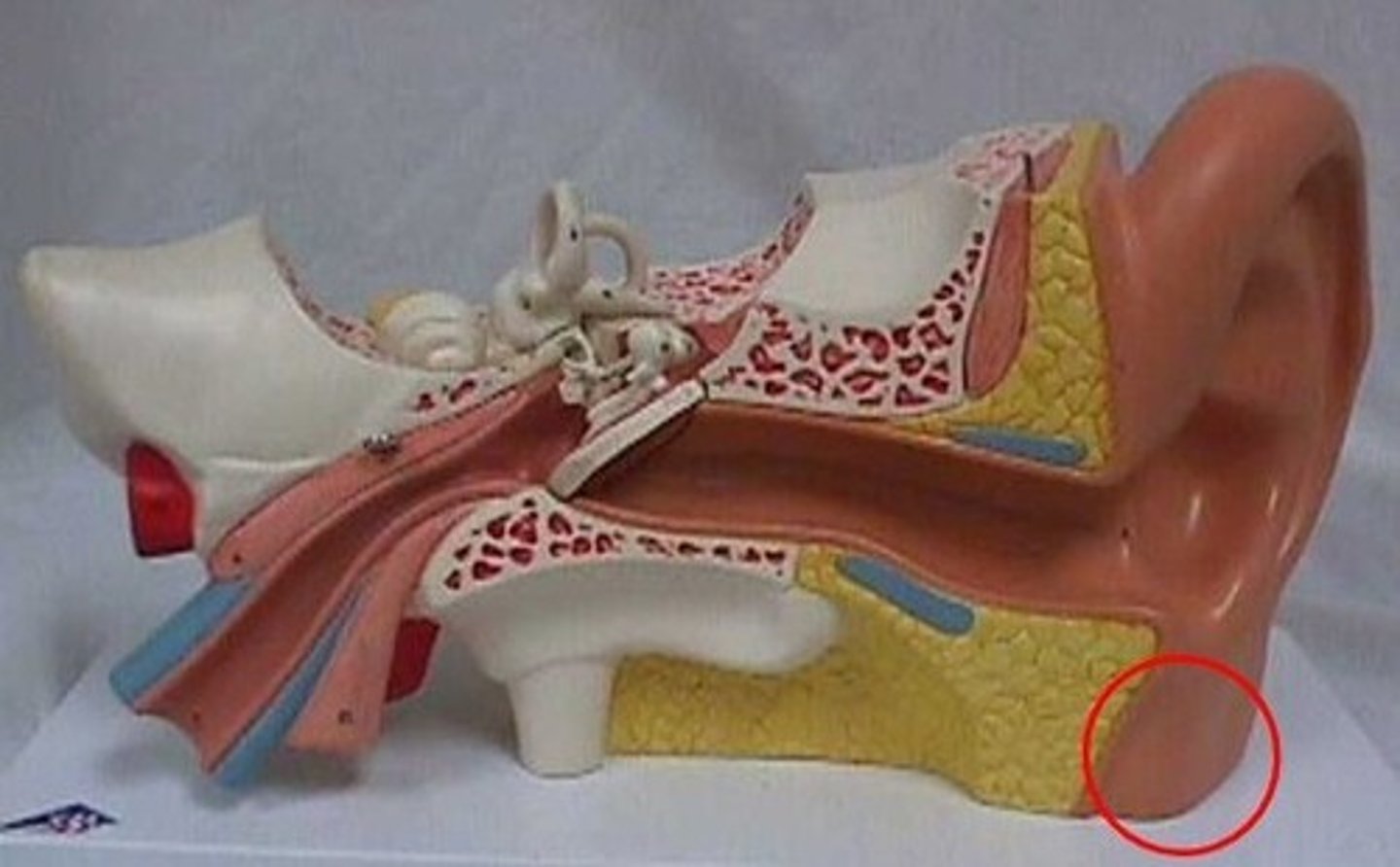
Auricle
External portion of the ear
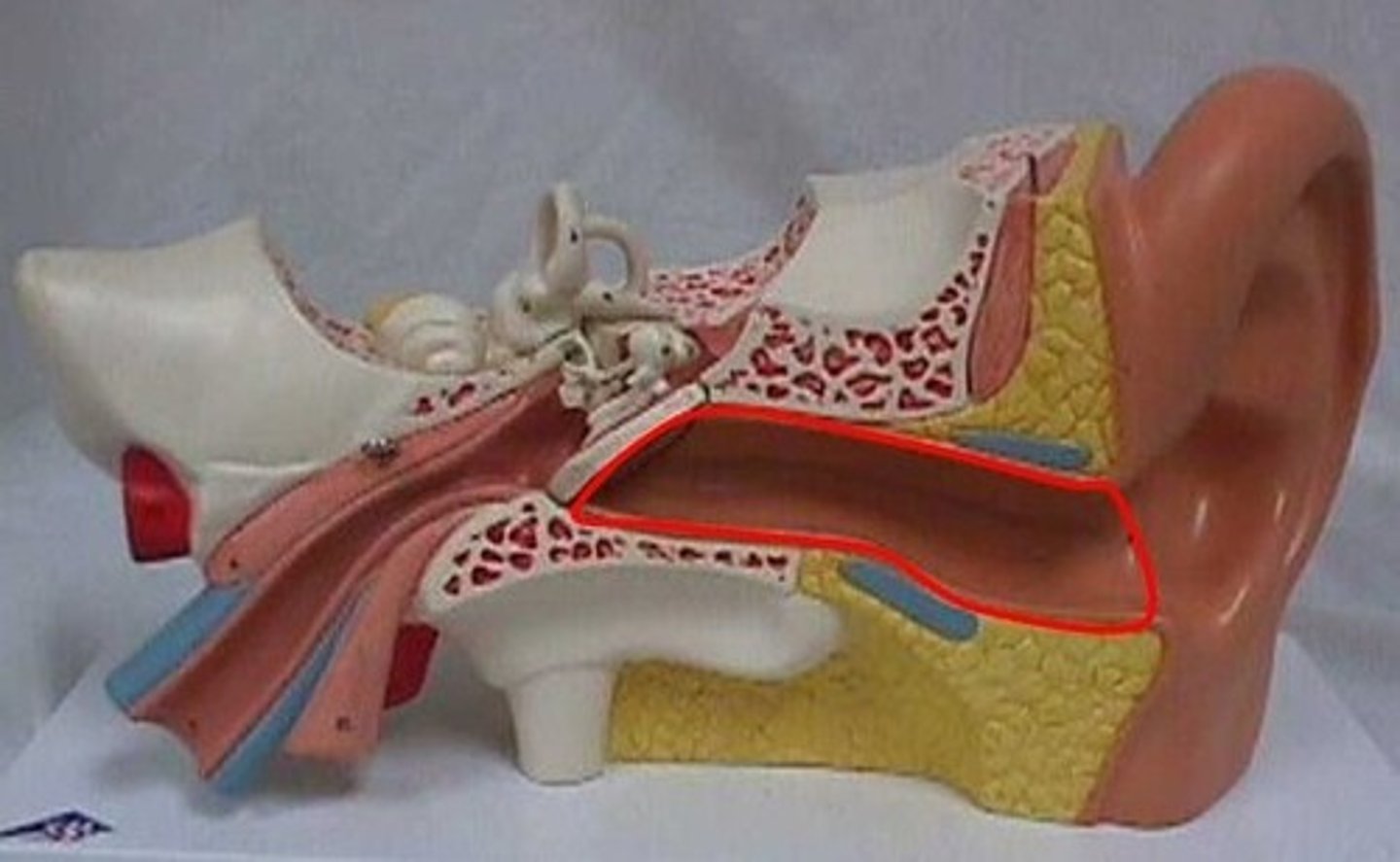
External Acoustic Meatus
Canal leading to eardrum and middle ear
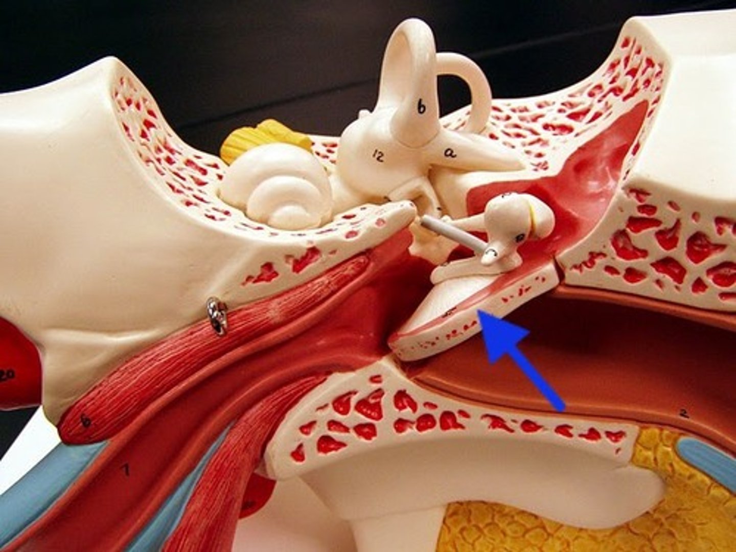
Tympanic Membrane
The eardrum. A structure that separates the outer ear from the middle ear and vibrates in response to sound waves.
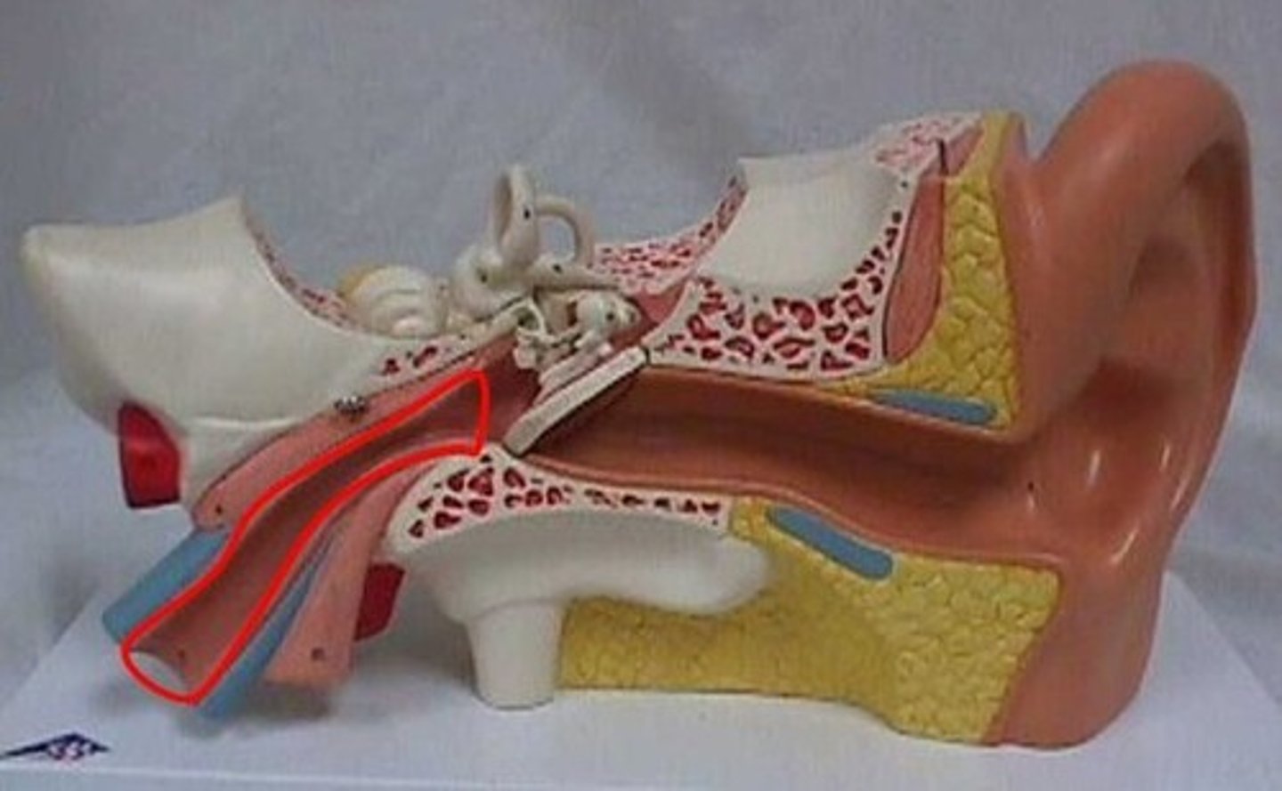
Pharyngotympanic tube
Connects the middle ear with the nasopharynx.
Malleus
hammer; first of the three auditory ossicles of the middle ear
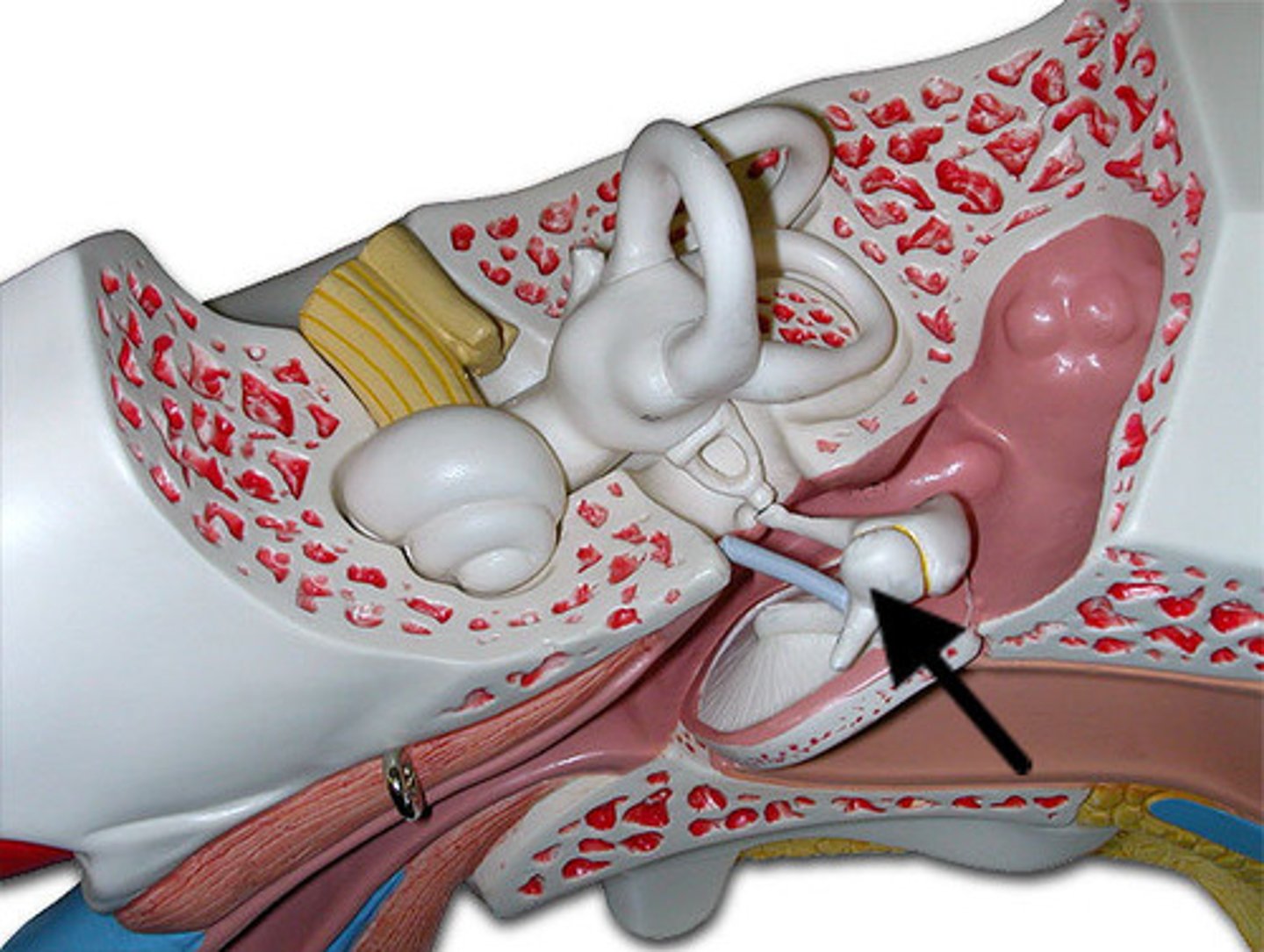
Incus
anvil; middle of the three auditory ossicles of the middle ear
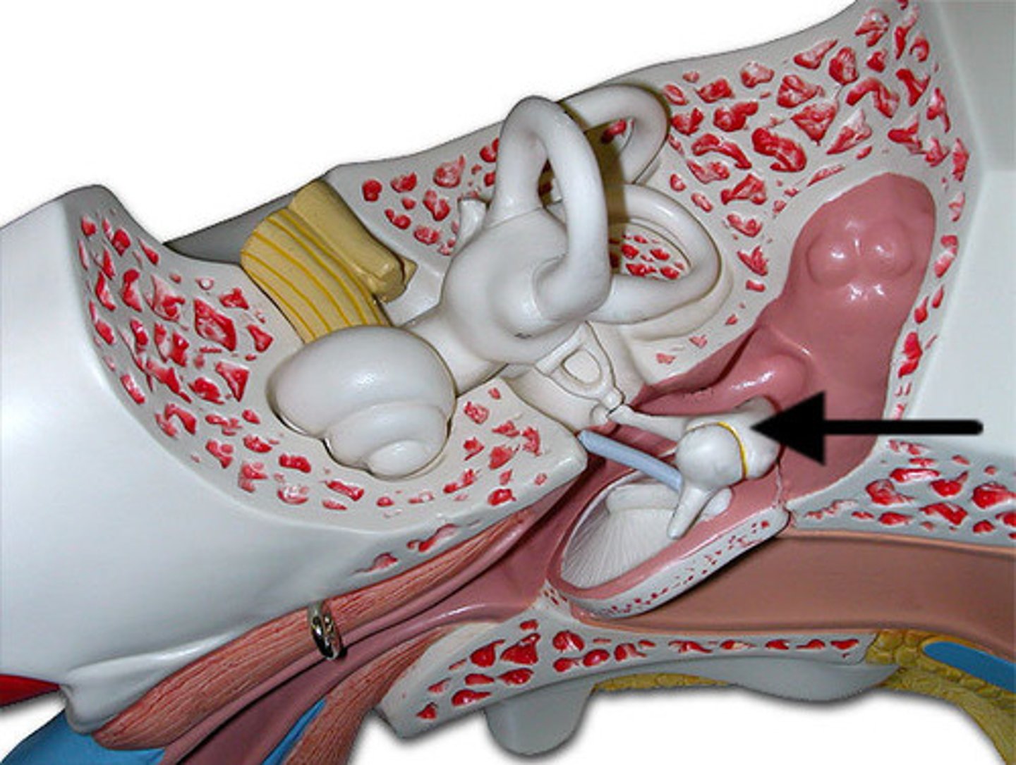
Stapes
stirrup; last of the three auditory ossicles of the middle ear
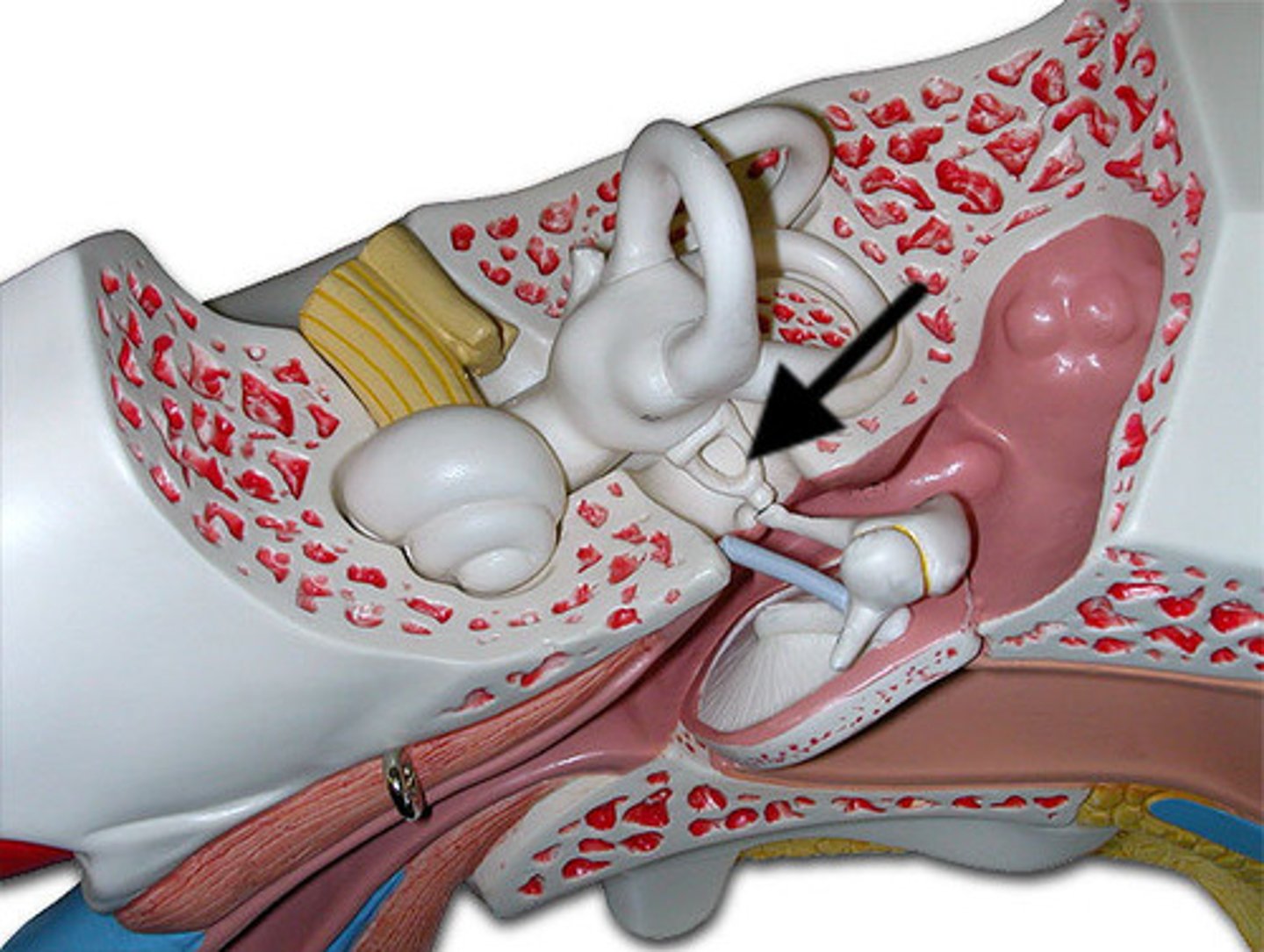
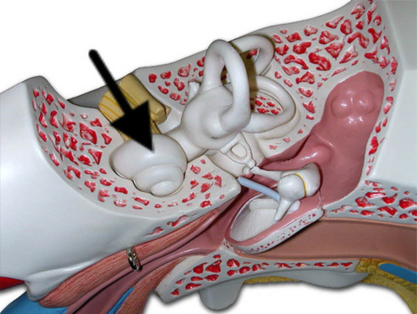
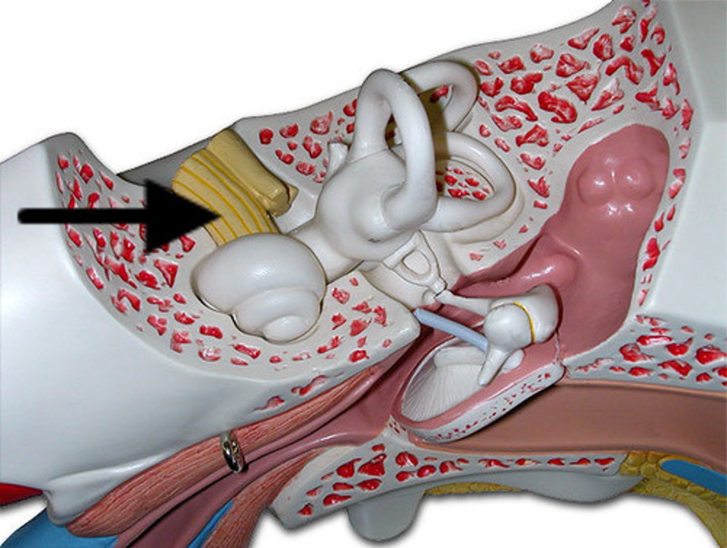
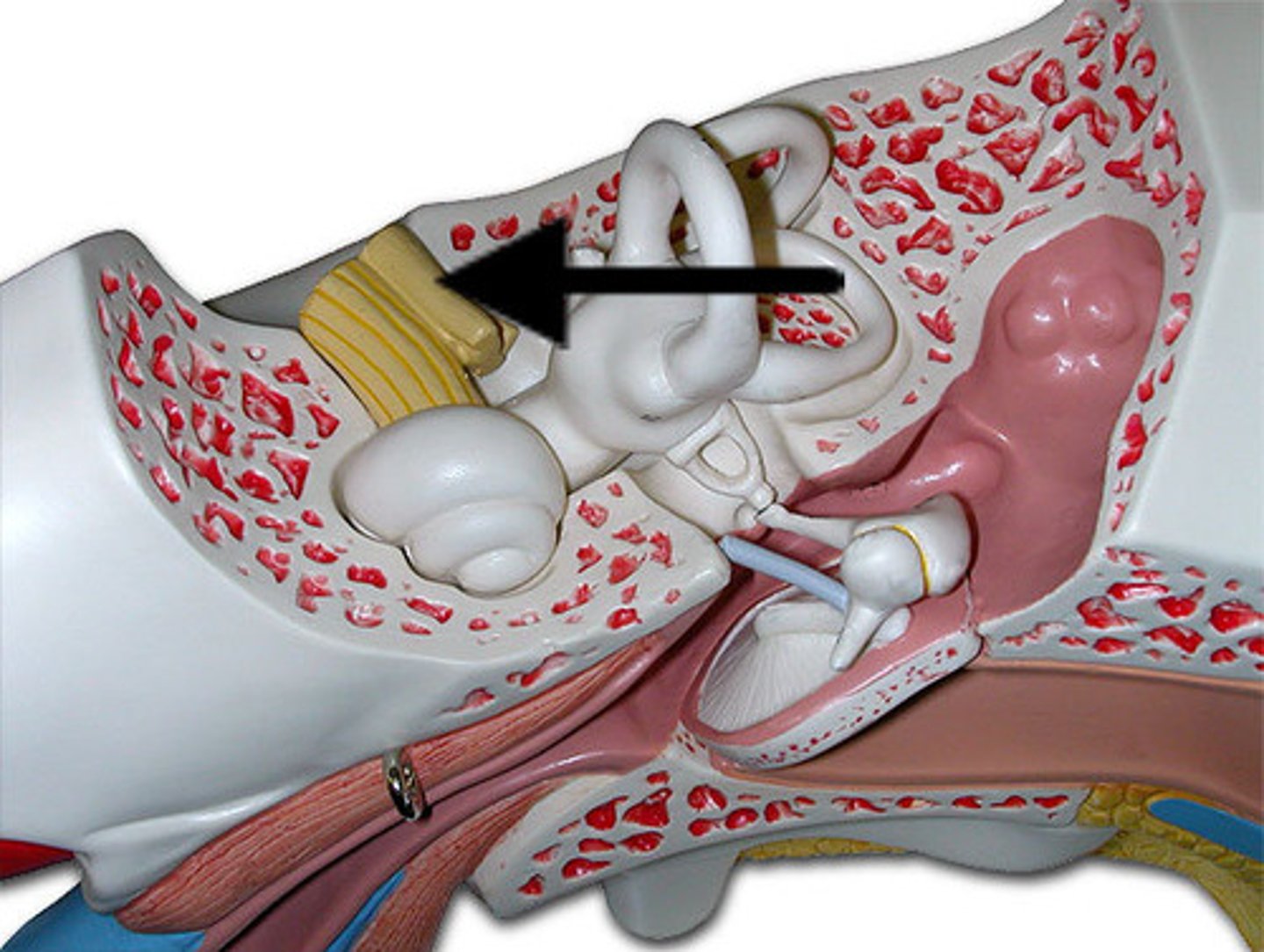
Semicircular canals
three canals within the inner ear that contain specialized receptor cells that generate nerve impulses with body movement
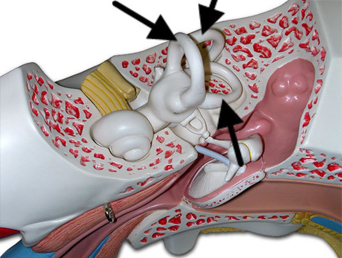
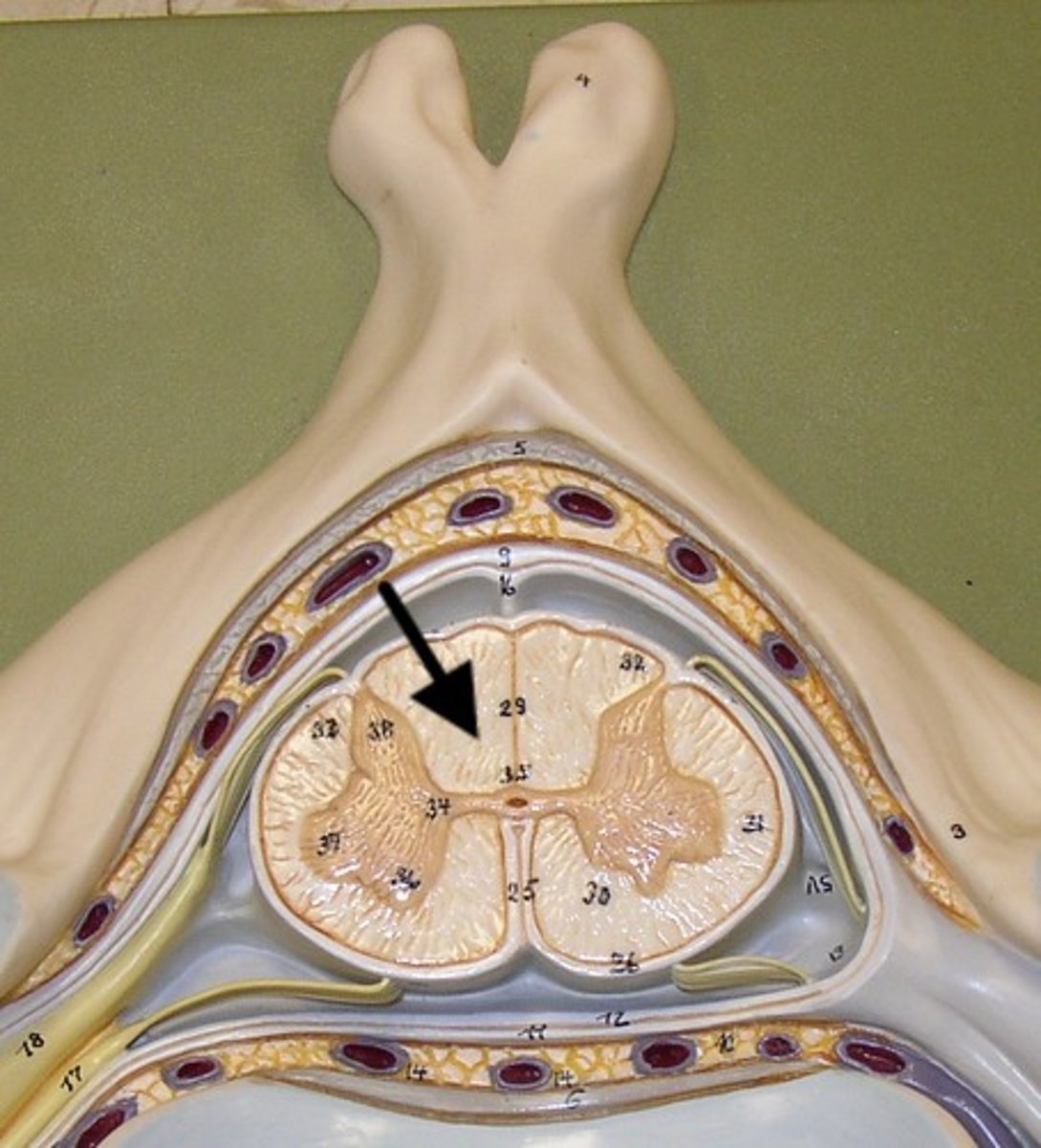
Cochlea
a coiled, bony, fluid-filled tube in the inner ear through which sound waves trigger nerve impulses
Cochlear Nerve
the branch of the auditory nerve that transmits auditory information from the cochlea to the brain
vestibocochlear nerve (VIII)
hearing and balance
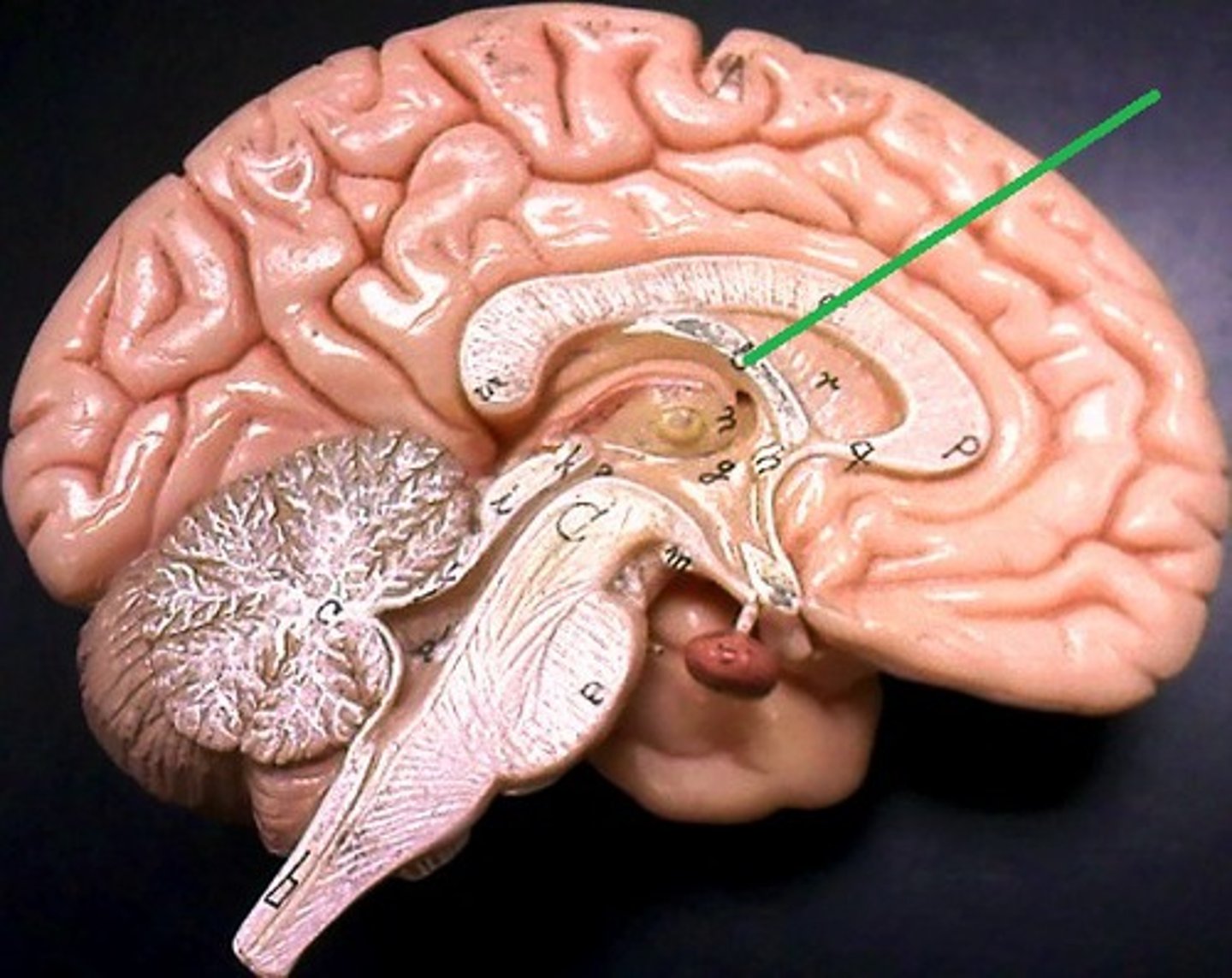
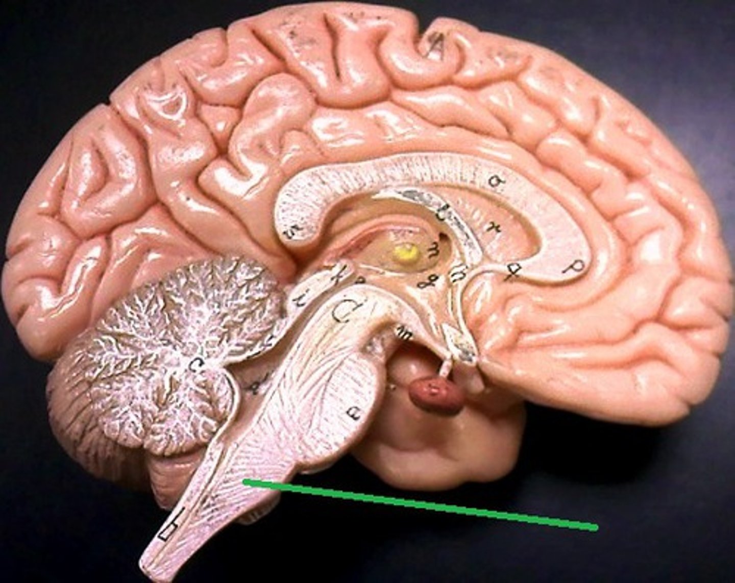
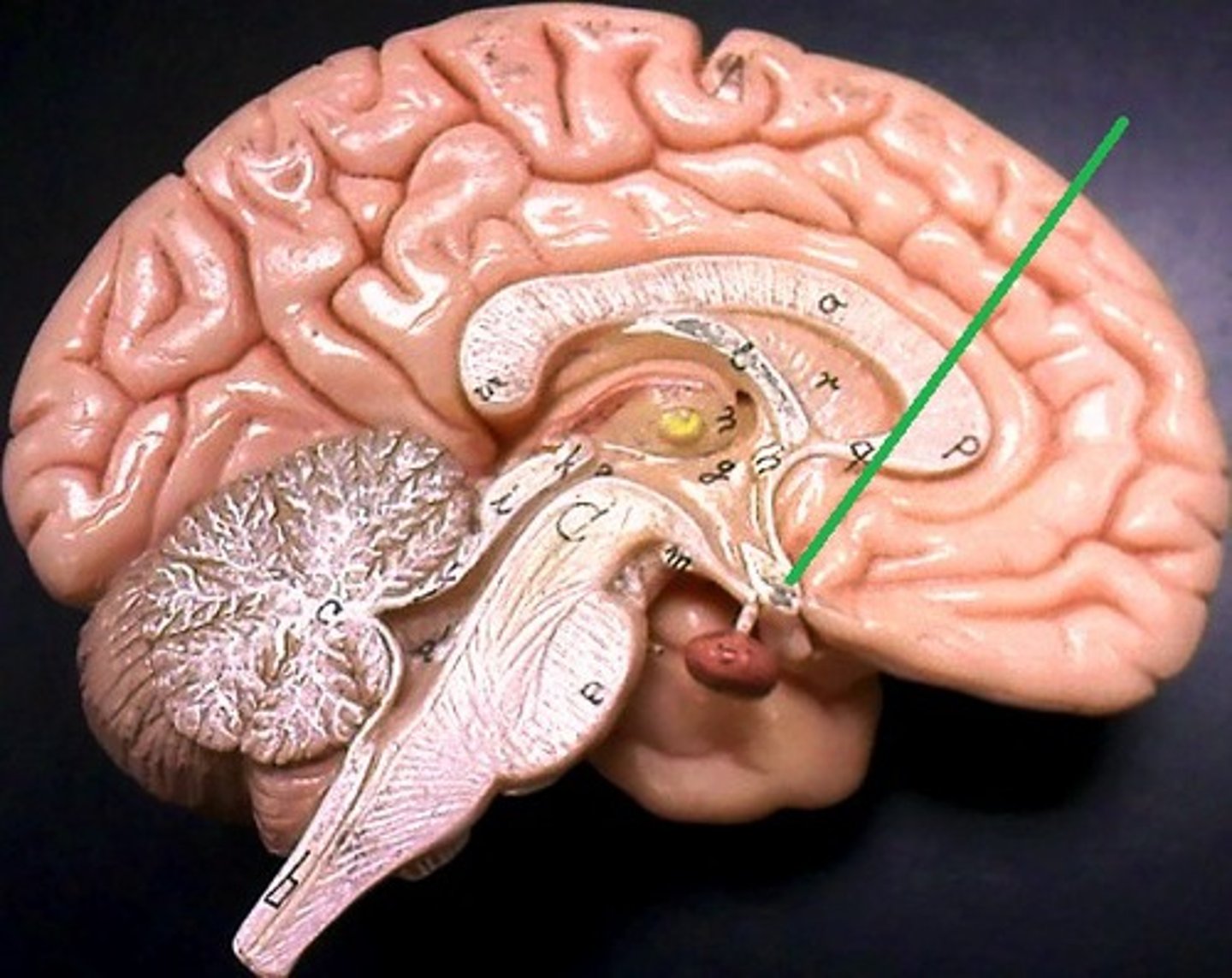
Fornix
a fiber tract that extends from the hippocampus to the mammillary body