Pictures - Histo (C2)
1/43
Earn XP
Description and Tags
Mainly pictures for practice - credit
Name | Mastery | Learn | Test | Matching | Spaced |
|---|
No study sessions yet.
44 Terms
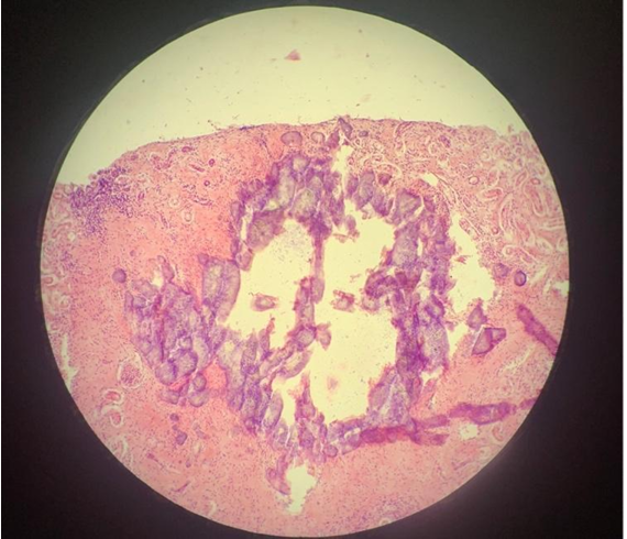
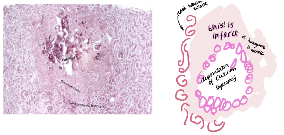
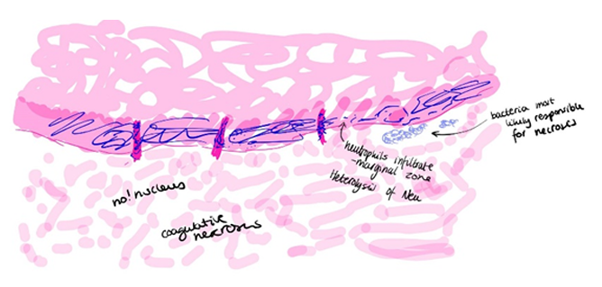
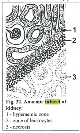
This is?
Dystrophic calcification (infarctus renis anemicus)
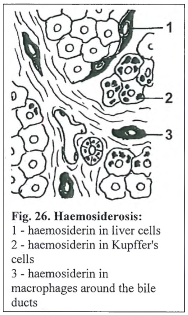
this is?
Hemosiderosis of spleen (Liesegang)
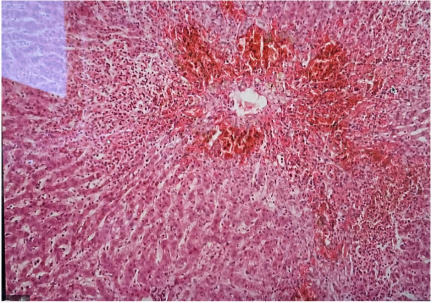
this is?
Necrosis hepatis centrolobularis
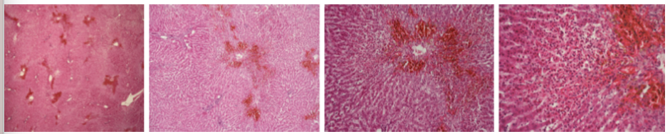
this is?
Necrosis hepatis focalis
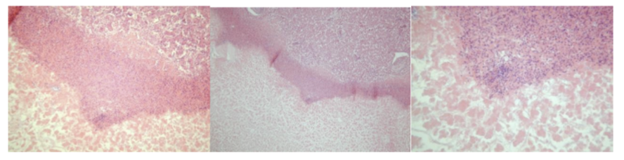
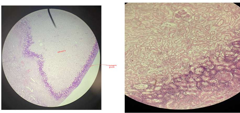
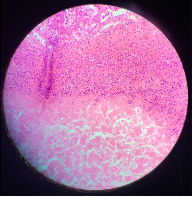
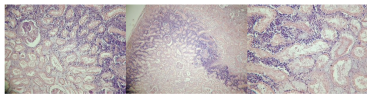
This is?
Infarctus renis anemicus (anaemic infarct of kidney)
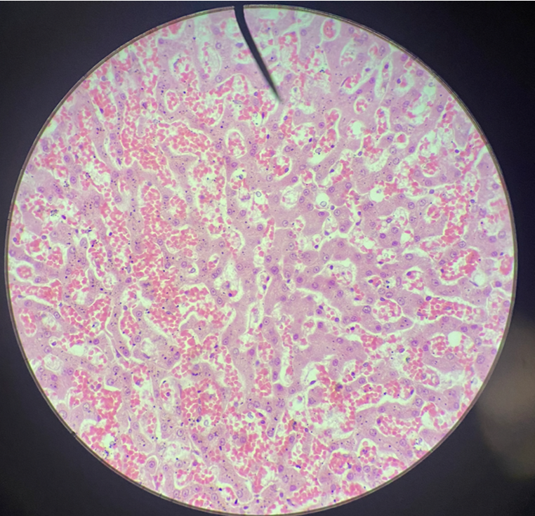
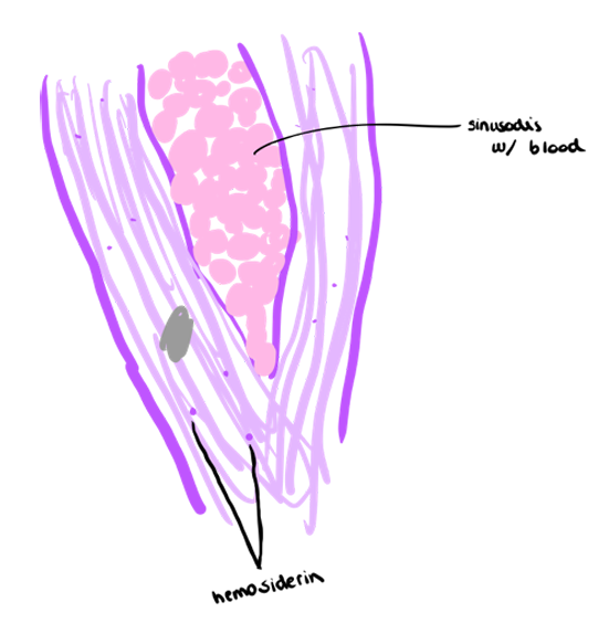
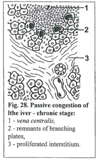
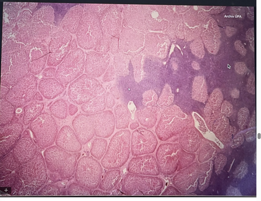
This is?
Chronic passive hyperemia of liver
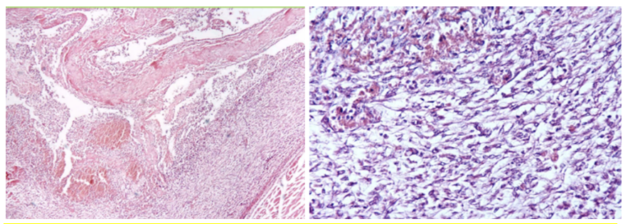
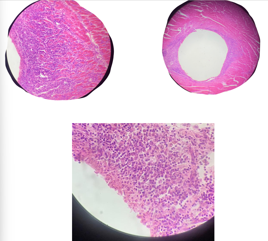
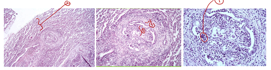
This is?
Pleuritis fibrosa (Repair by organization)
Extra note: Importance of this mechanism (repair by organization) is such that the area does not return to normal, but remains in a scarred state, so that the organ or tissue involved is permanently damaged and may be functionally impaired (defect).
Granulation tissue grows into the fibrous exudate to liquify and engulf it (repair process). This pathological process leads to adherence of the parietal and visceral layers. From the bottom: mature fibrous CT → granulation tissue → fibrous exudate (neutrophils) in picture.
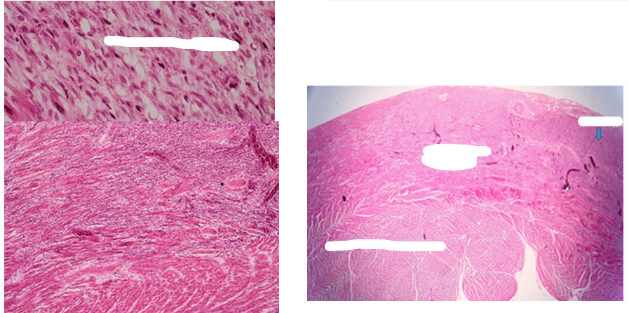
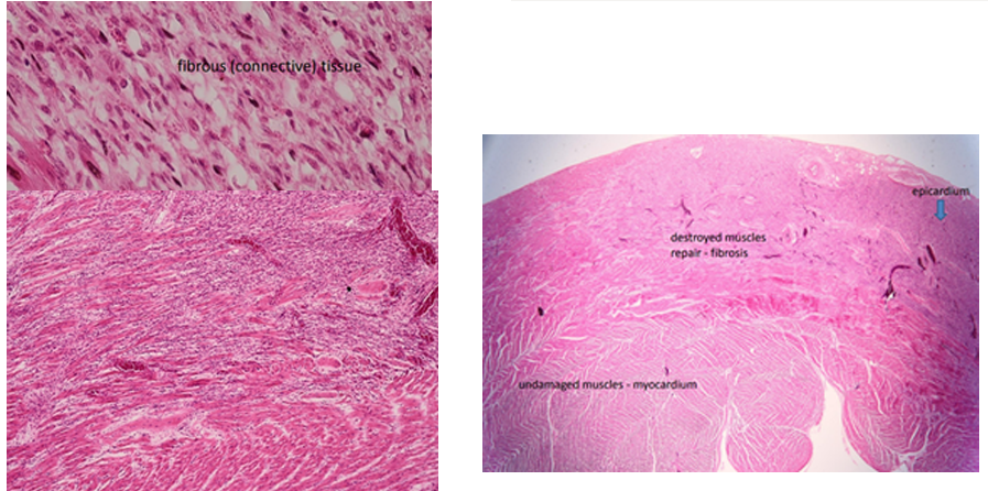
this is?
Fibrosis myocardii
Note: myocardium of heart is locally replaced by mature fibrosis tissue with bright staining. Pink area is normal myocardial tissue while the pale is fibrous tissue. Arises secondary to cardiac stress/damage, cardiovasc. disease. The stress → substances activate fibroblasts + transdifferentiate into myofibroblasts → incr. production of proteins that are dep. in EC matrix → incr. prod. of collagenous scar tissue → myocardial fibrosis.
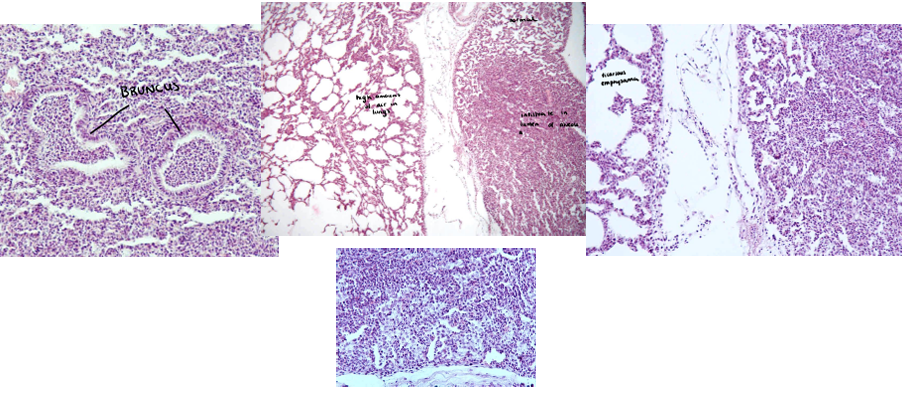
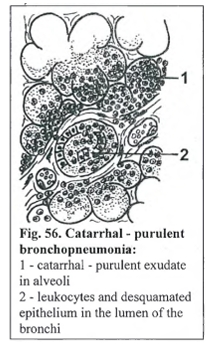
This is?
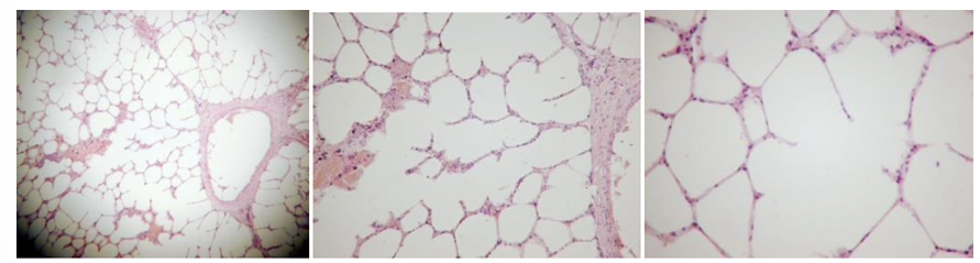
Bronchopneumonia purulenta
note: from left: bruncus (round strunctures), high amount of air in the lungs are seen in middle picture, infiltrate in lumen of alveoli while normal at the top, at right: vicarious emphysema
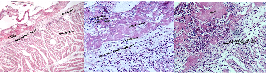
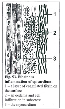
This is?
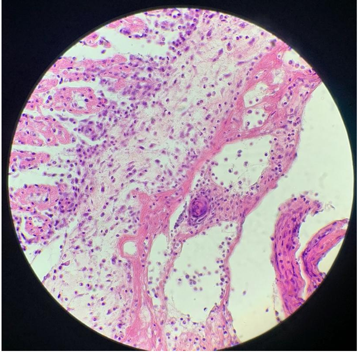
Pericarditis fibrinosa
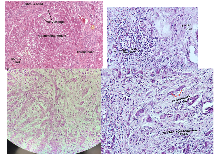
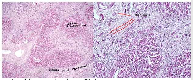
this is?
Cirrhosis hepatis (hepatitis interstitialis chronica)
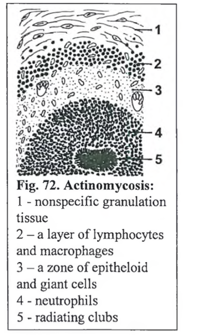
this is?
Actinomycosis (most often mandibular, less frequent maxilla) - Bildet e mest sannsynlig maxilla
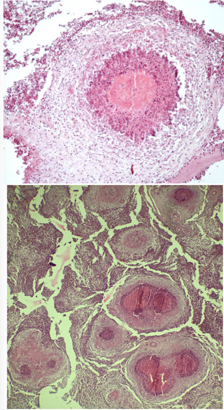
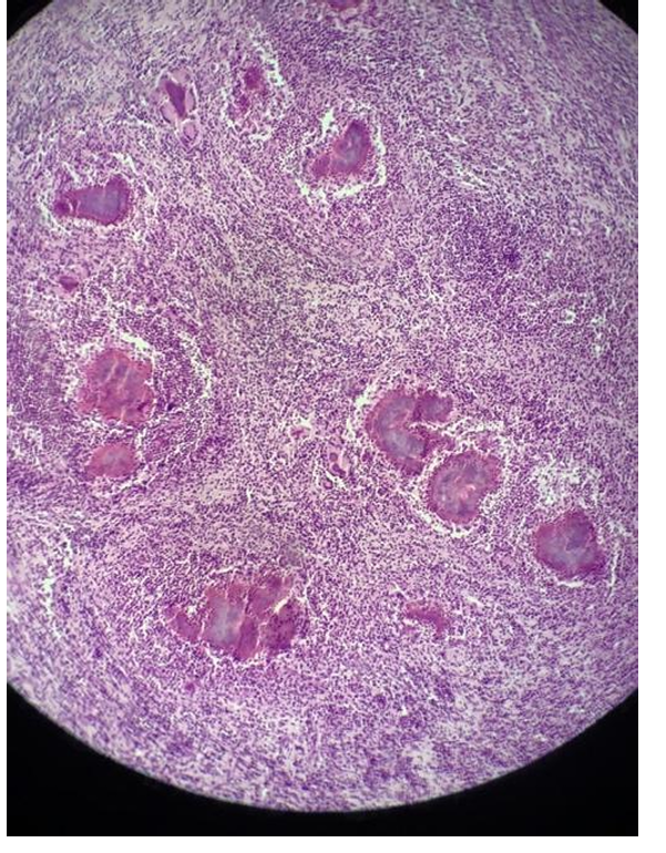
This is?
Pneumonia tuberculosa miliaris– poultry
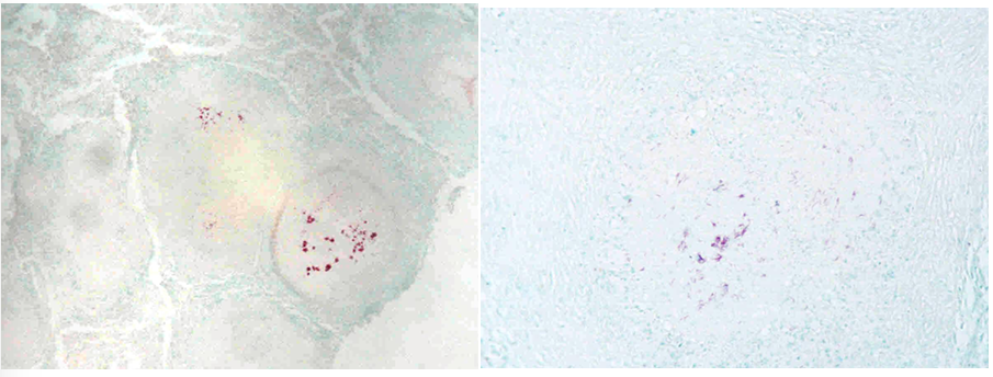
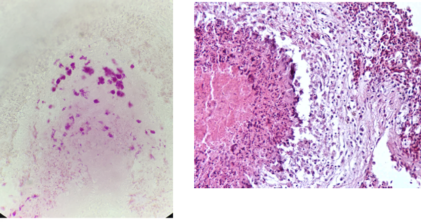
this is?
Pneumonia tuberculosa miliaris– hydina/poultry– ZN
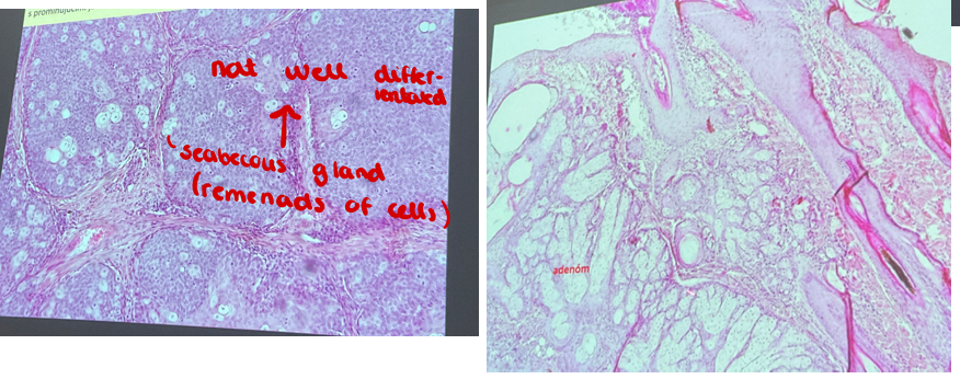
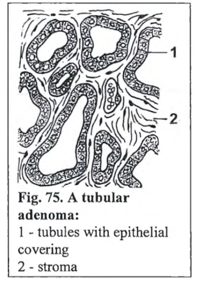
this is?
Adenoma et adenocarcinoma sebaceous gland

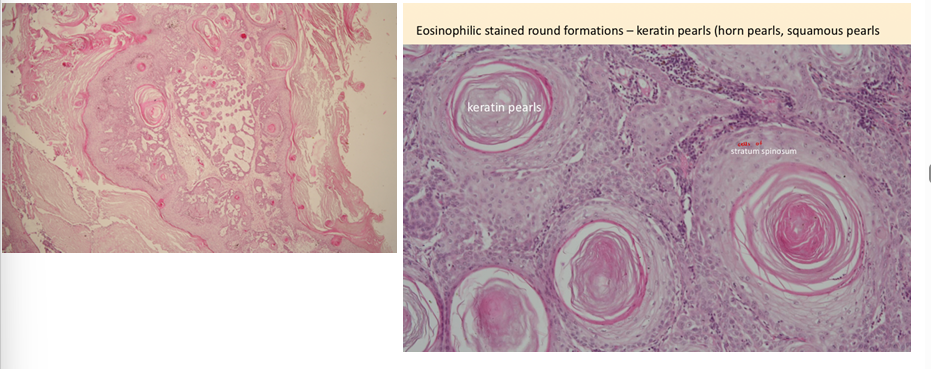
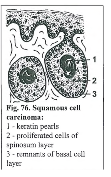
this is?
squamous cell carcinoma (horse)
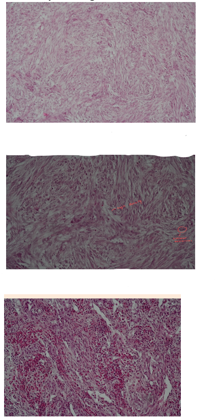
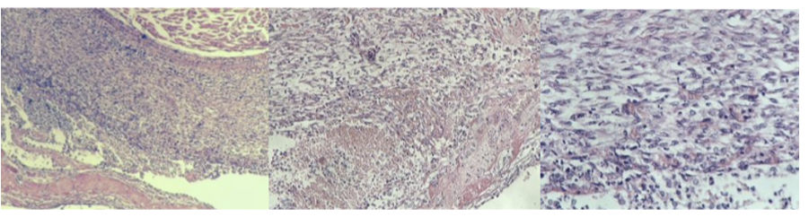
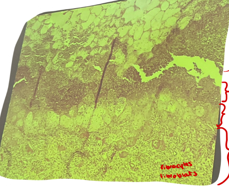
This is?
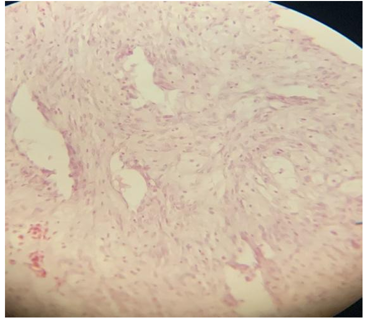
Fibroma mole - tumor
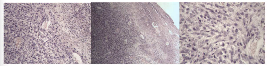
picture 1: aimlessly running of fibrous fibers, in many directions - felt structure.
pic. 2: miscellaneous differentiation of cells - fibroblast and fibrocytes - small amount of collagen
pic 3: cell infiltrations - heterophils, lymphocytes and macrophages and focal edema
this is?
post traumatic focal infalmmatory reaction of myocardium
in myocardium → round hole surrounded by expressive reaction tissue (neutrophils, macrophages + lymphocytes). Process locally → deeper areas, result of sticking the bullet in heard (foreign body).
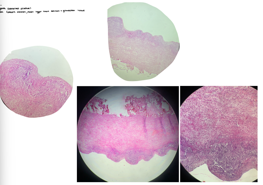
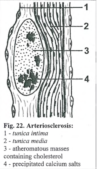
this is?
Vasculitis chronica or mediocalcinosis
tunica intigema (abnormal structure)
t. media (smooth muscles, much bigger than normal + granulation tissue)
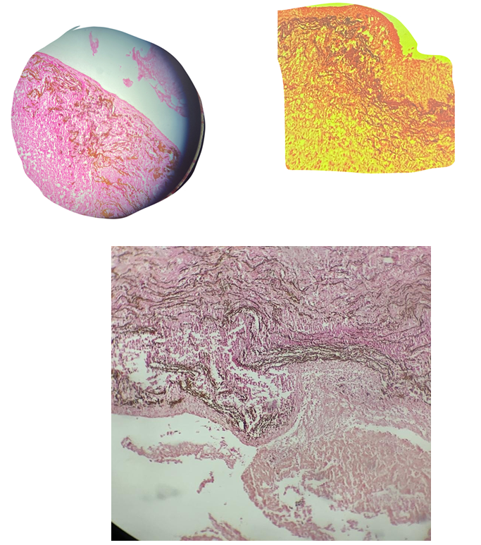
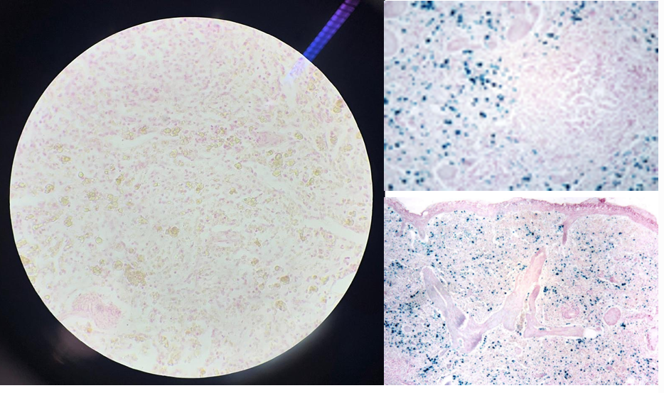
this is?
Mediocalcinosis (van Kossa)
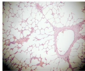
this is?
emphysema alveolare chronic
septa is broken and air area increased
this is?
endobronchiolitis obliterans and peribronchitis nodosa
bronchioles that have been infiltrated with fibroblastic granulation tissue (1)
alveoli compressed by inflamed bronchi (2)
outside is filled with inflammatory cells
the wall of bronchi is thickened (3)
inside filled with inflammatory cells (4)
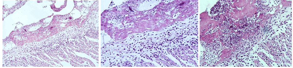
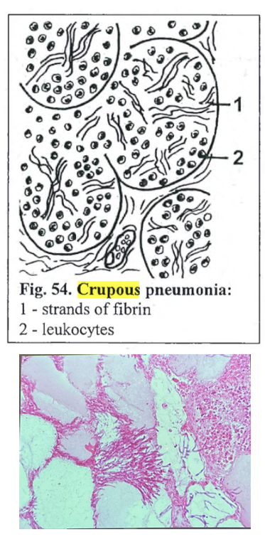
this is?
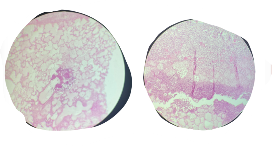
crupous (fibrinous lobar) pneumonia and chronic interstitial pneumonia
notes:
1st layer: chronic intestinal pneumonia (nederst)
2nd layer: respond to secondary infection (exudate)
3rd layer: severe accumulation neutrophils
4th layer: fibrous (upper with all the white spaces)
Bacterial origin, or fungi chyle formation, fibrin (blood protein) escapes from bv to extravascular space - formation of mass
cant see alveoli.
Nederst på bildet: fibroblasts and fibrocytes replace lung tissue. Exudation above this tissue. Line of neutrophils above exudation. Øverst: lung tissue (alveoli) with infiltration of neutrophils and exudation.

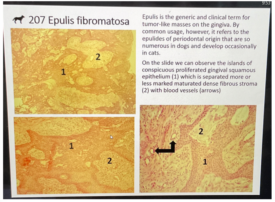
this is?
epulis fibromatosa
this is?
Colitis fibrinosa (Levaditi)
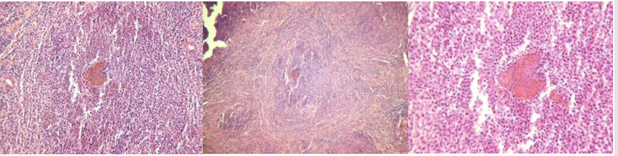
this is?
Lymphadenitis acuta simplex
this is?
infectious bursal disease - changes in bursa (side of B-lymph production) of fabricius during infectious bursal disease (gumboro, birnavirus)
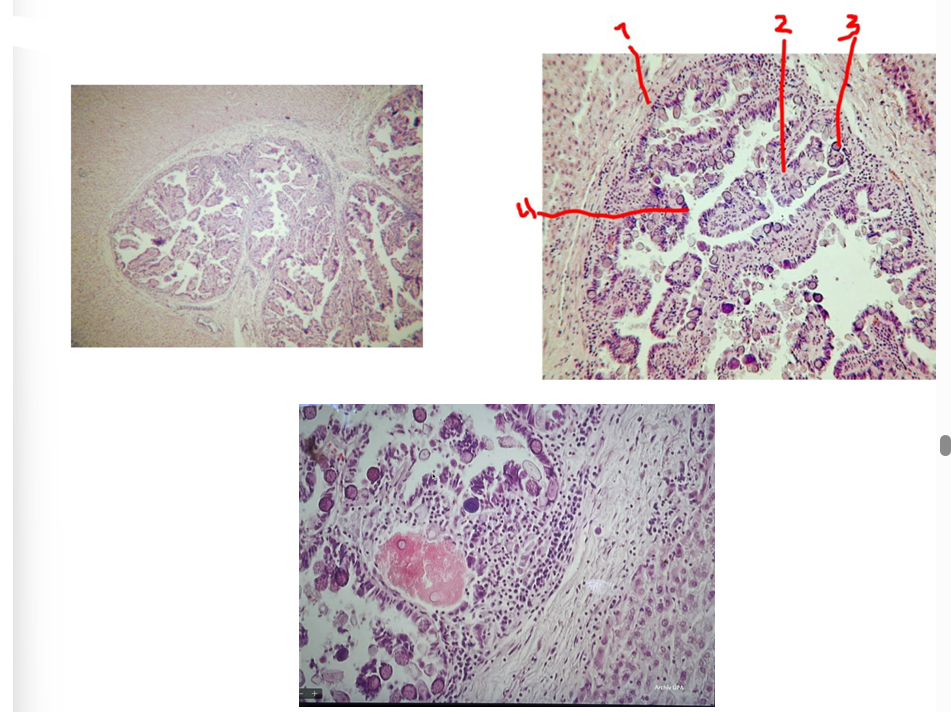
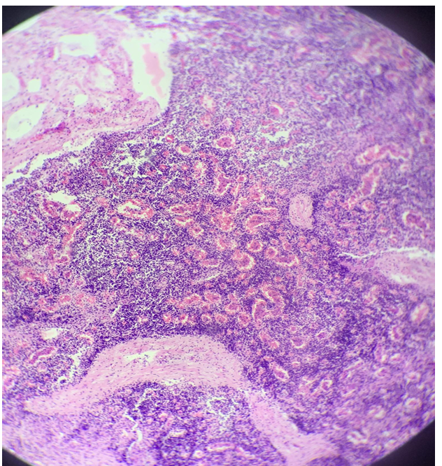
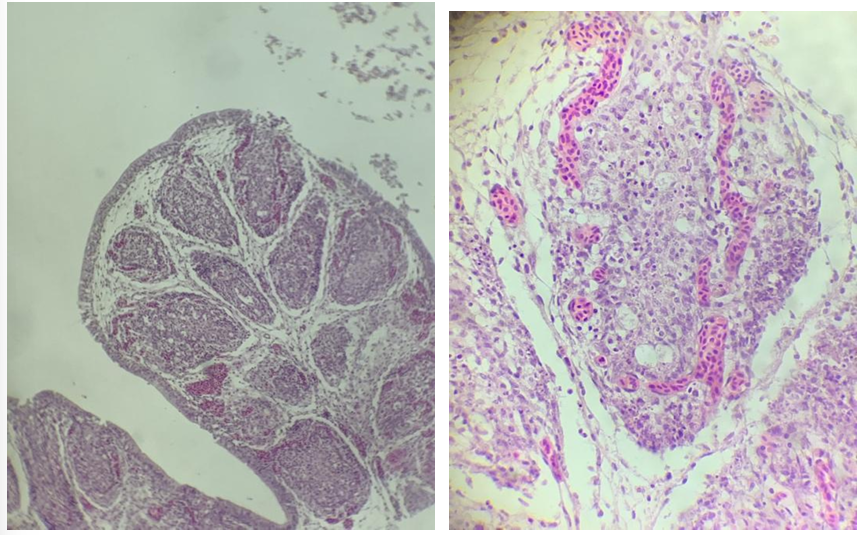
this is?
cholangitis et pericholangitis chronica hyperplastica (coccidiosis hepatis cuniculorum)
bile duct wall
papillary folds
coccidia
epithelial cells
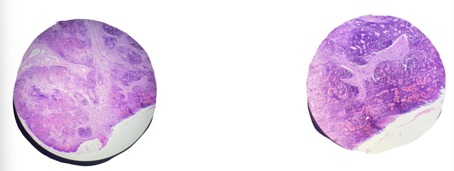
this is?
Hepatitis purulenta acuta parenchymatosa (traumatic disease of forestomach)
on the slide - cross section of cow liver with advanced traumatic reticuloperitonitis
this is?
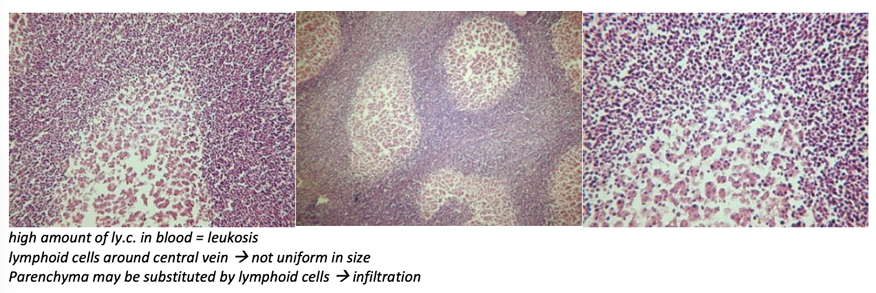
Leucosis lymphadenoidea hepatis - lymphosarcoma
one end of liver → basophilic stained tumor tissue formed by slightly differentiated lymphocytes
this is?
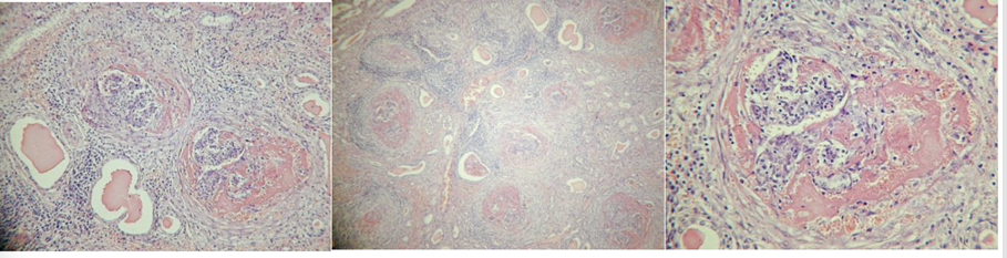
Glomerulonephritis chronica
Bilde 1: hyaline in glomeruli and tubules
bilde 2: periglomerulitis chronica, gradual fibrotization of glomeruli
bilde 3: higher magn. of bilde 1, glomeruli with neutrophils and hyaline
this is?
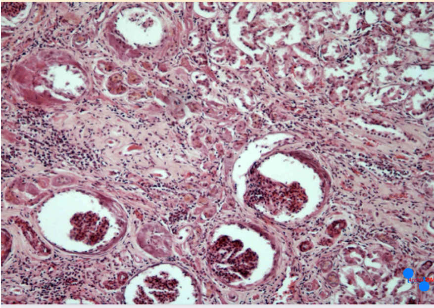
Nephrocirrhosis (nephritis interstitialis chronica)
Ca deposition in tissue of kidney - HE staining
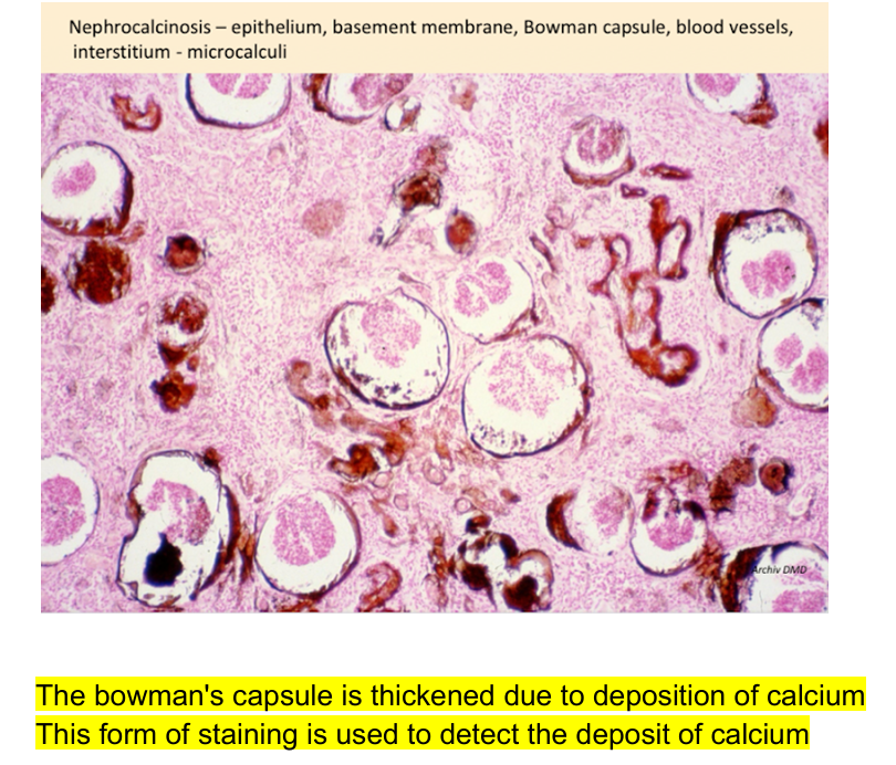
this is?
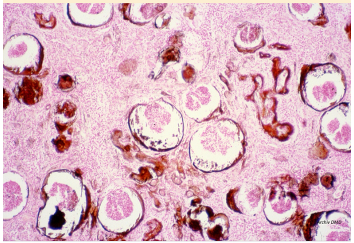
Nephrocirrhosis (Van Koss)
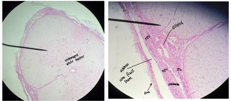
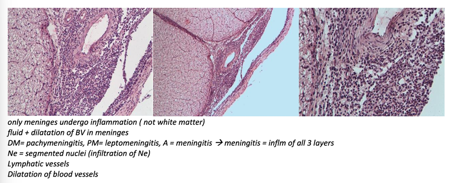
this is?
Meningitis spinalis purulenta
meninges are dilated and infiltrated by cells
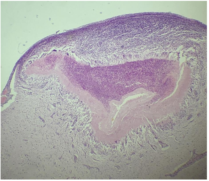
this is?
cysticercus ovis (Mozog/brain)
at periphery of section under the meninges, there is a cysts that intrudes into the brain.

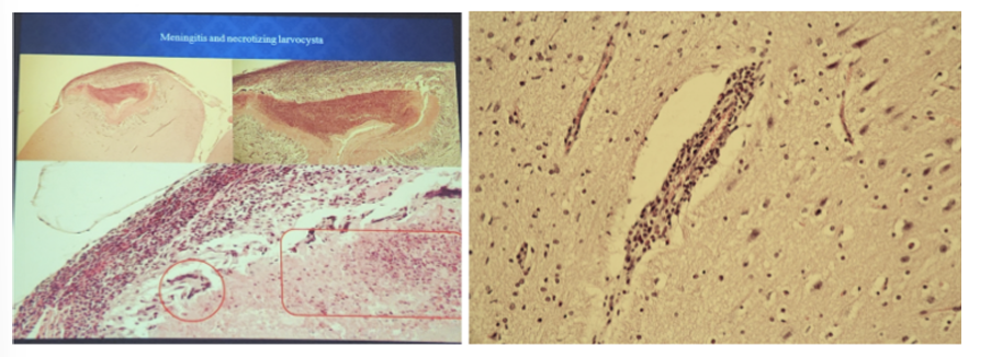
this is?
fibrosarcomna uteri
this is?
hypertrophy of prostata
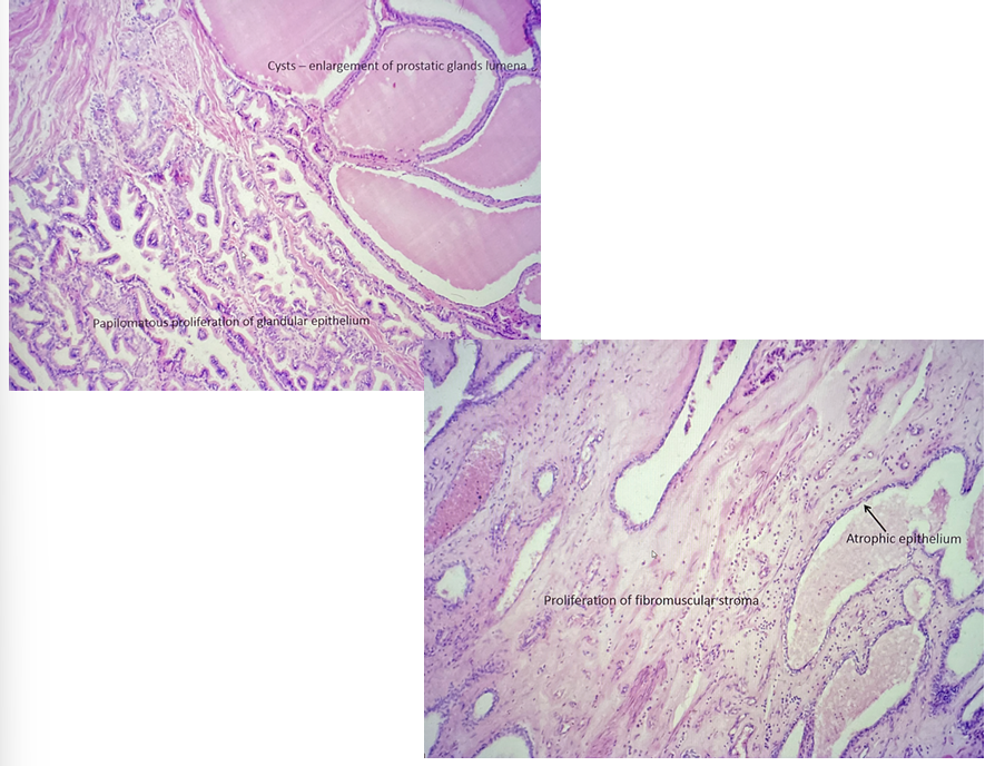
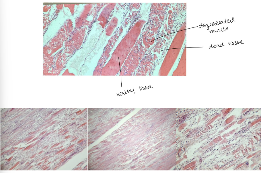
this is?
Dystrophia musculorum
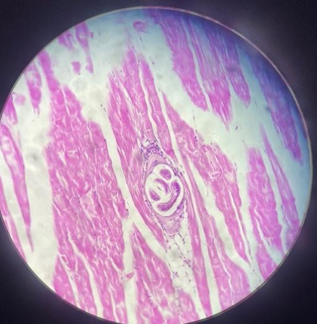
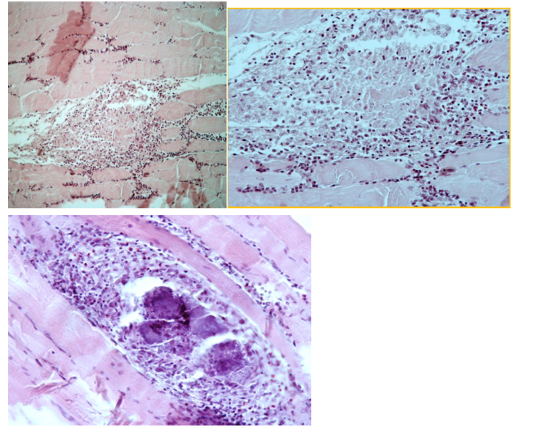
this is?
trichinellosis musculorum
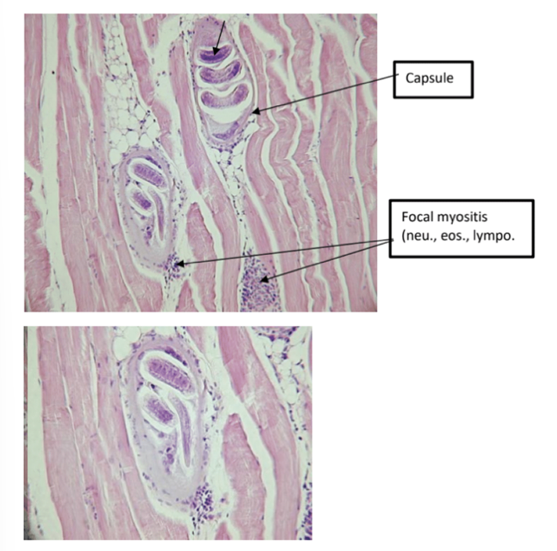
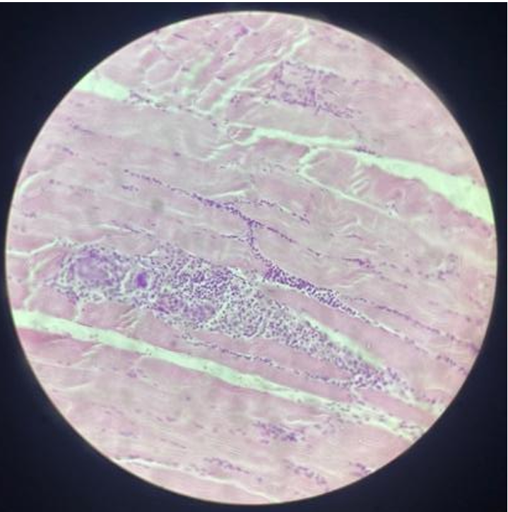
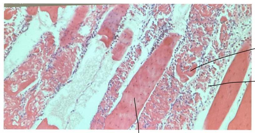
this is?
Myositis sarcosporidica
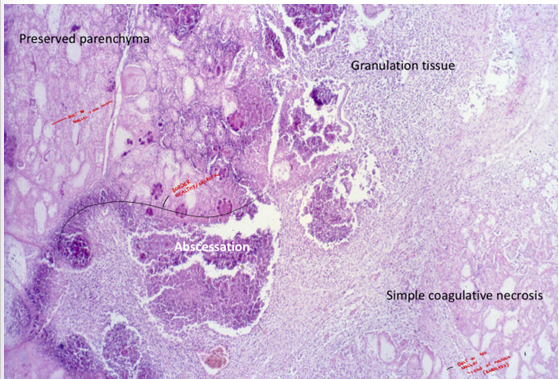
this is?
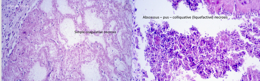
Mastitis apostematosa
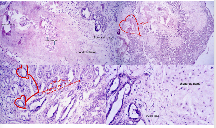
this is?
tumor mixtus mammae
this is?
Dermatitis Eosinophilica
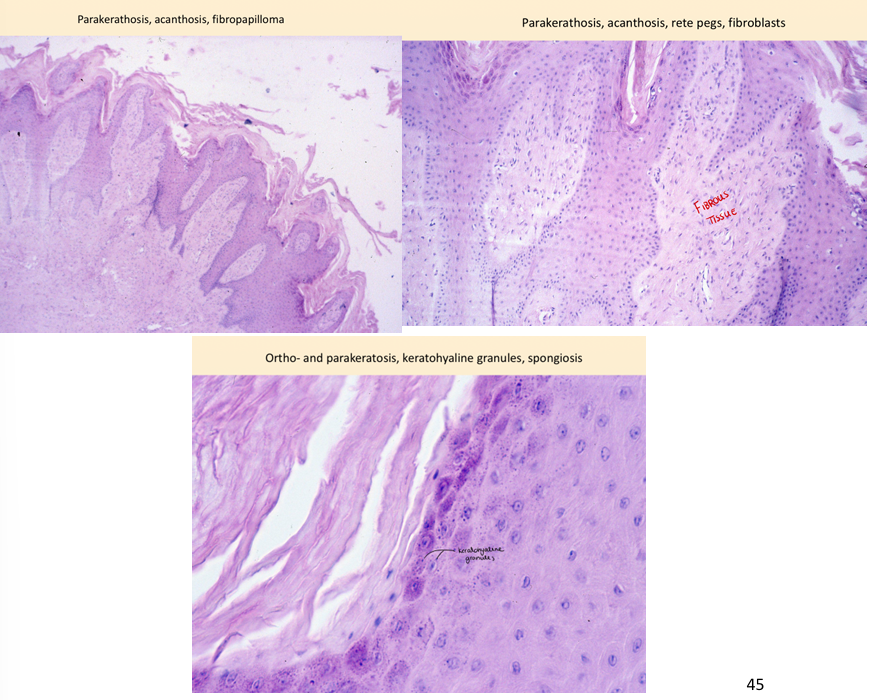
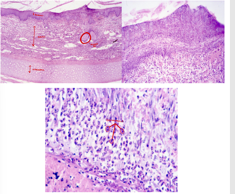
this is?
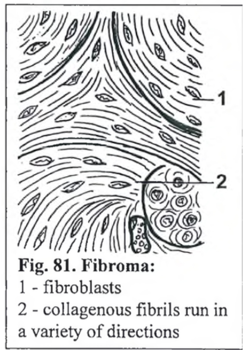
Fibropapilloma mixture of fibro and papilloma