Harp GU Images
5.0(1)
Card Sorting
1/110
There's no tags or description
Looks like no tags are added yet.
Study Analytics
Name | Mastery | Learn | Test | Matching | Spaced |
|---|
No study sessions yet.
111 Terms
1
New cards
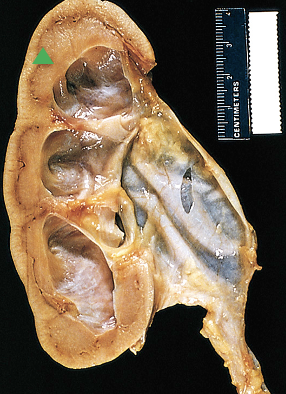
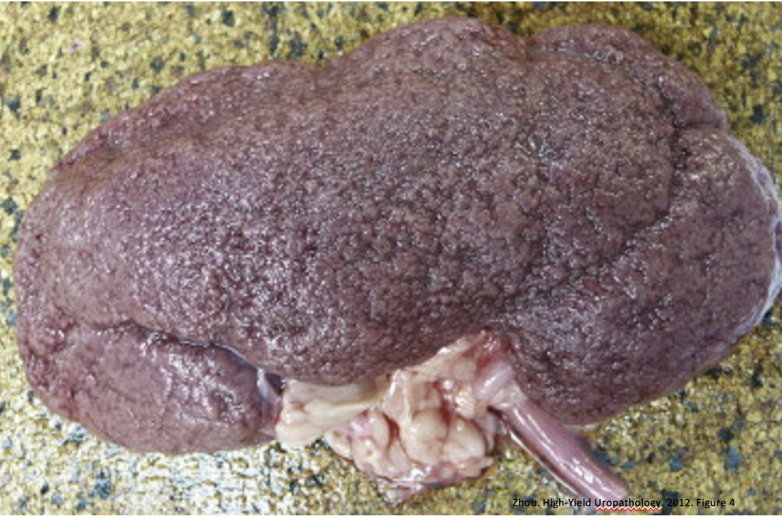
What pathology is indicated by this image?
Hydronephrosis
2
New cards
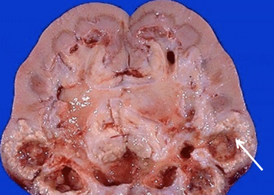
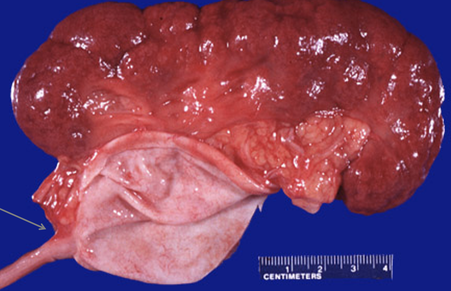
What pathology is indicated by this image? What is indicated at the tip of the white arrow?
Hydronephrosis & inflammation from an infection
3
New cards
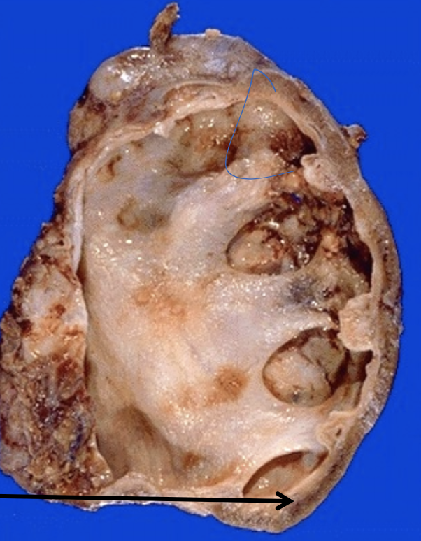
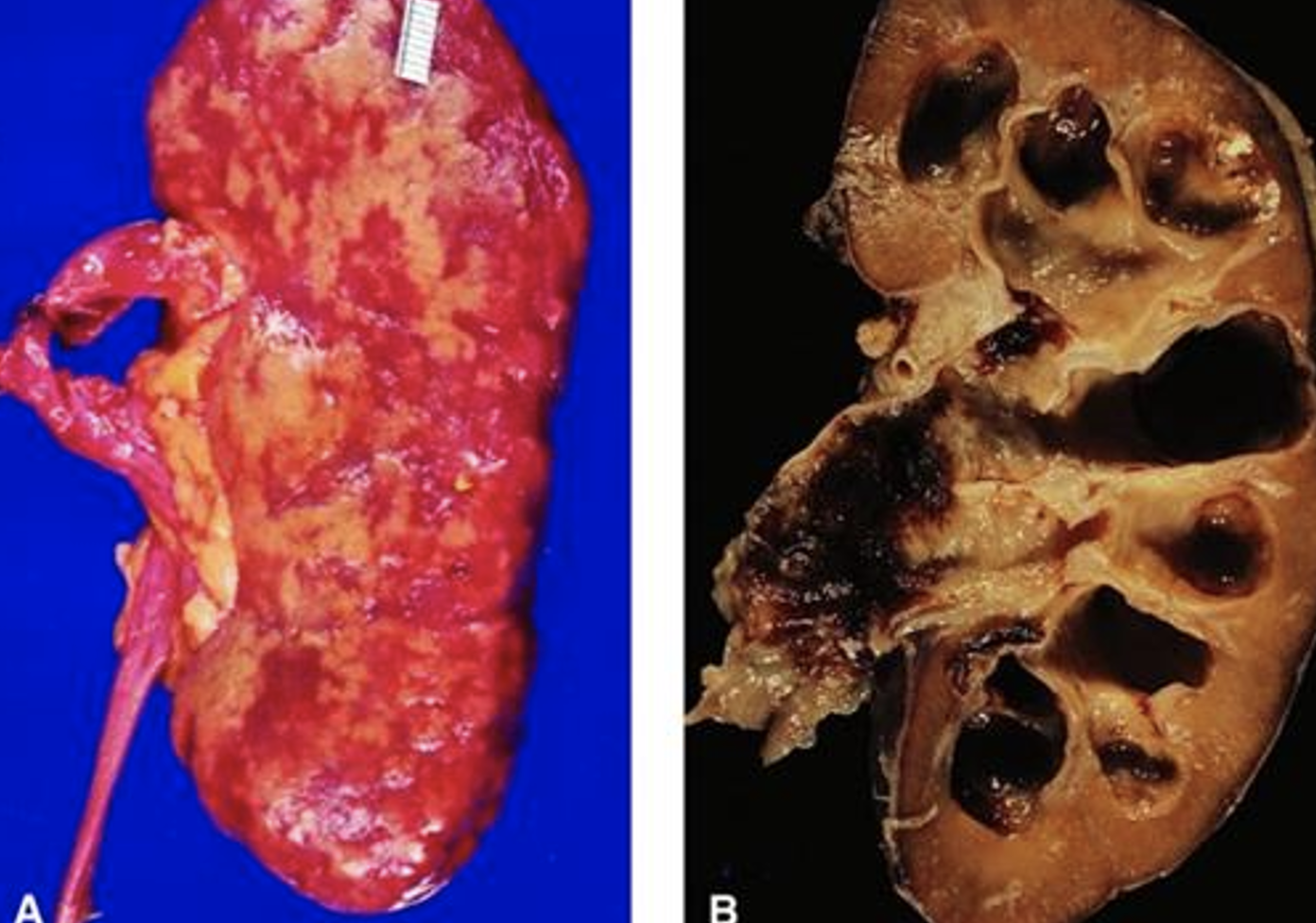
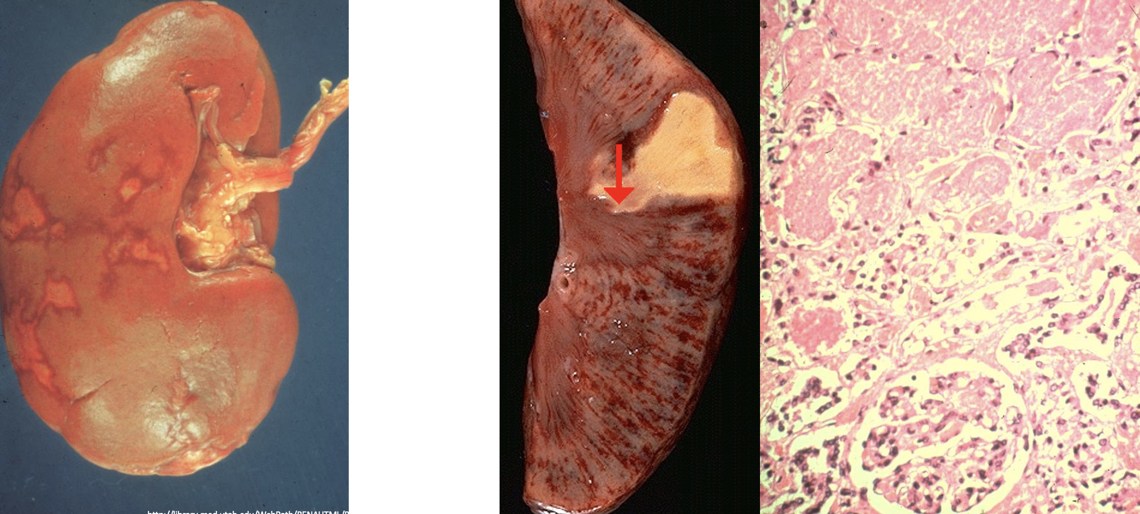
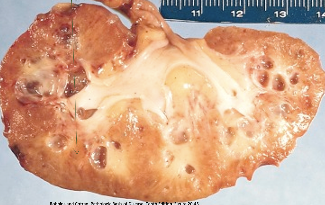
What pathology is indicated by this image? What is indicated at the arrow?
Hydronephrosis & loss of renal parenchyma meaning loss of kindey function and a source of ongoing infection
4
New cards
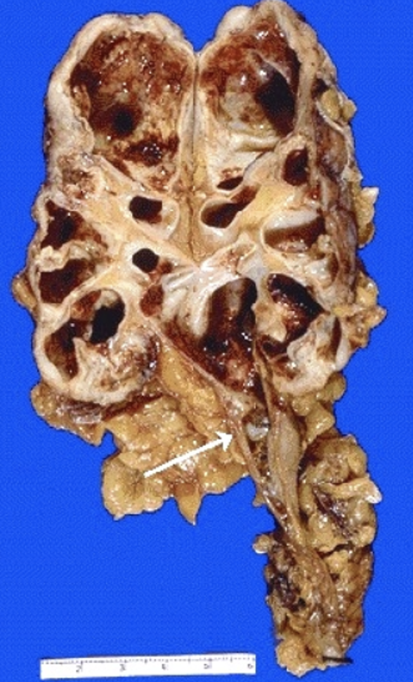
What pathology is indicated by this image? What is indicated at the arrow?
Hydronephrosis & ureteral calculus
5
New cards
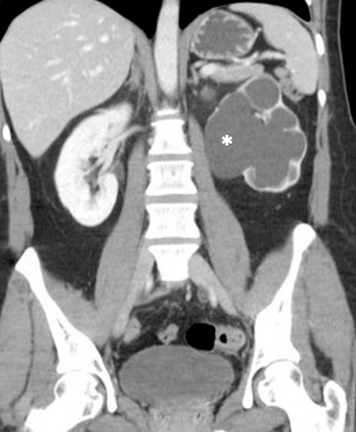
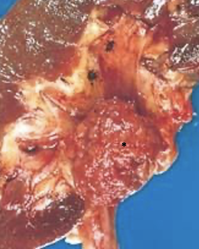
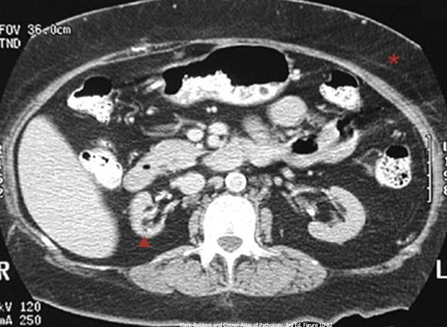
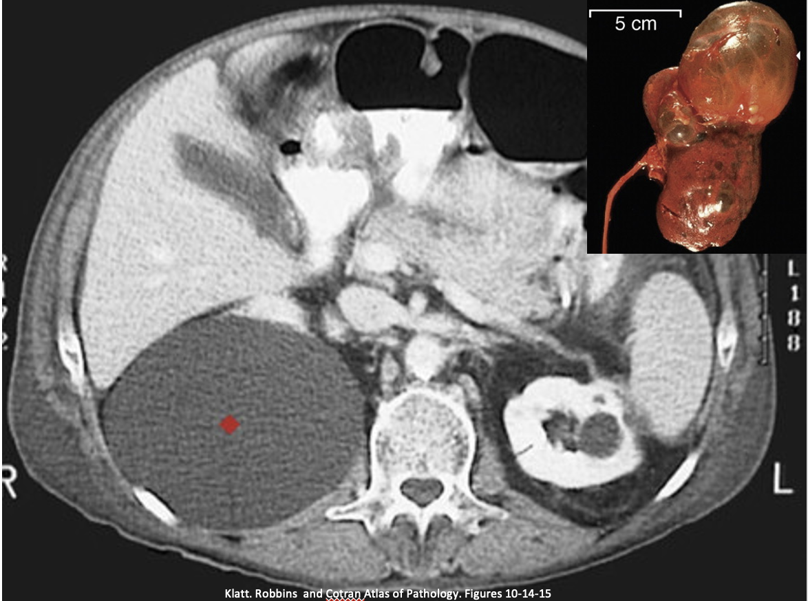
What pathology is indicated at the asterisk?
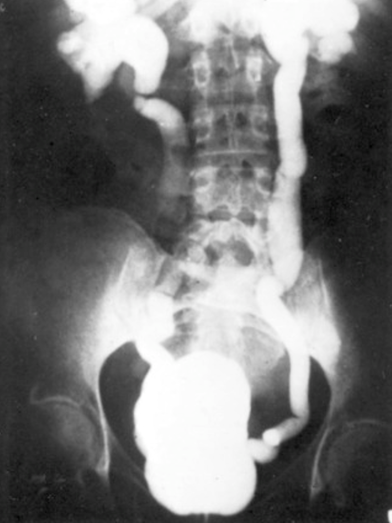
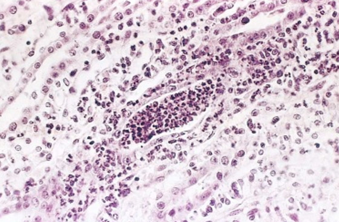
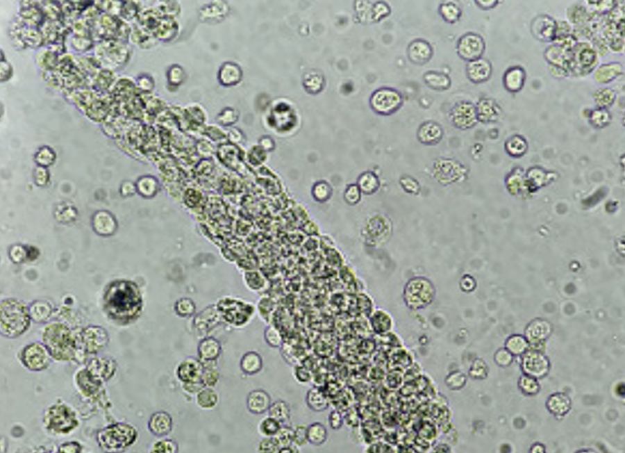
Hydronephrosis
6
New cards
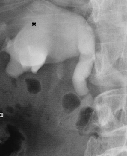
What pathology is indicated at the asterisk?
hydroureter & hydronephrosis
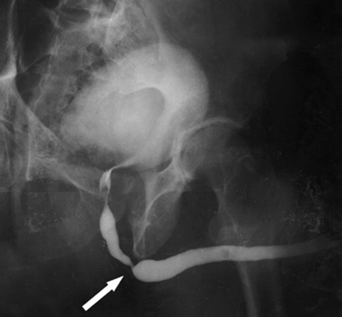
7
New cards
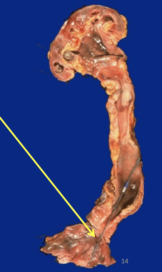
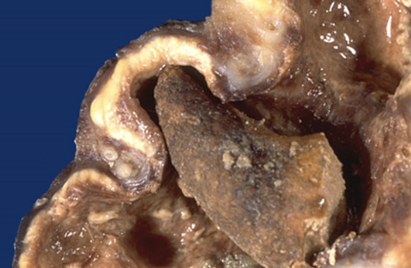
What is indicated at the arrow?
Long-standing obstruction at junction between ureter and bladder
8
New cards
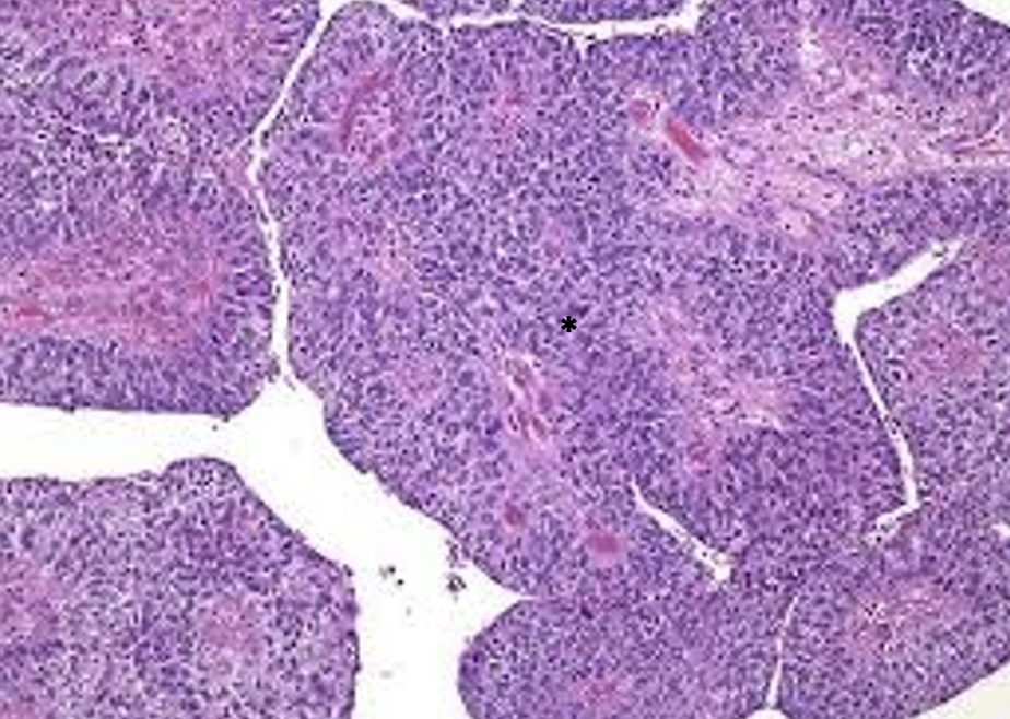
What pathology is indicated at the asterisk?
Urothelial carcinoma
9
New cards
What pathology is indicated at the asterisk? What exam finding is commonly associated with this pathology?
Urothelial carcinoma & hematuria
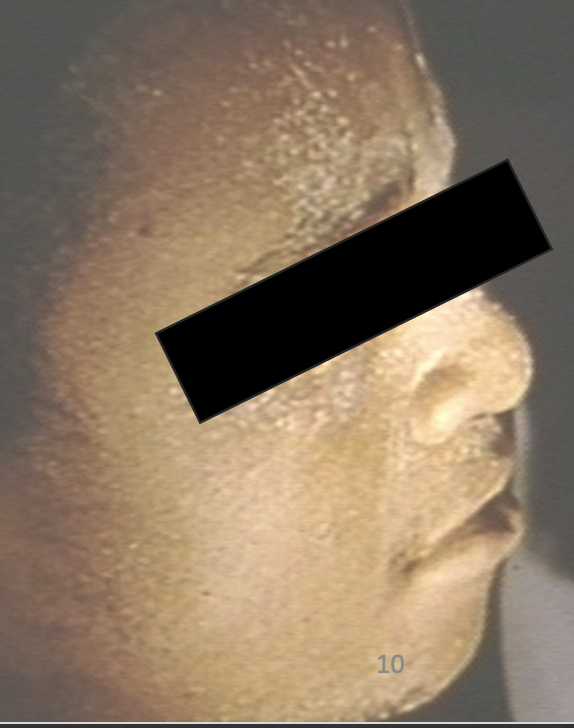
10
New cards
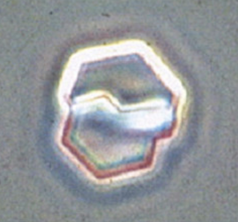
What kind of obstructive stone is this?
Calcium oxalate stone
11
New cards
What pathology is indicated by this image?
Prostatic enlargement causing urethral compression shown at the arrow
12
New cards
What kind of obstructive pathology is shown at the arrow?
Urethral stricture
13
New cards
What pathology is indicated by this image?
Urolithiasis - specifically primary oxalosis/oxaluria
14
New cards
What pathology is shown at the arrow? What is the name of the mechanism causing it?
Urolithiasis - supersaturation (increased urinary concentration)
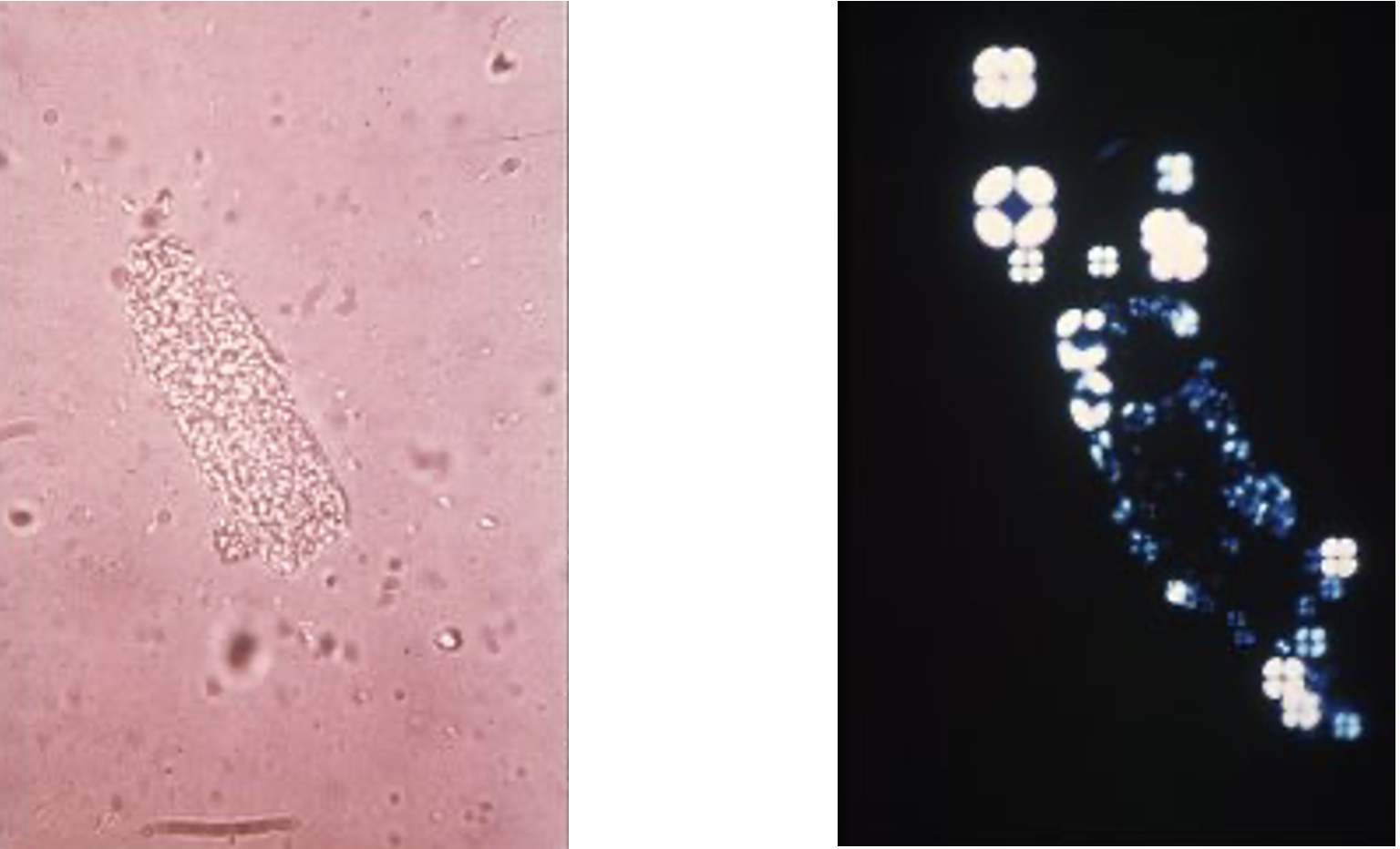
15
New cards
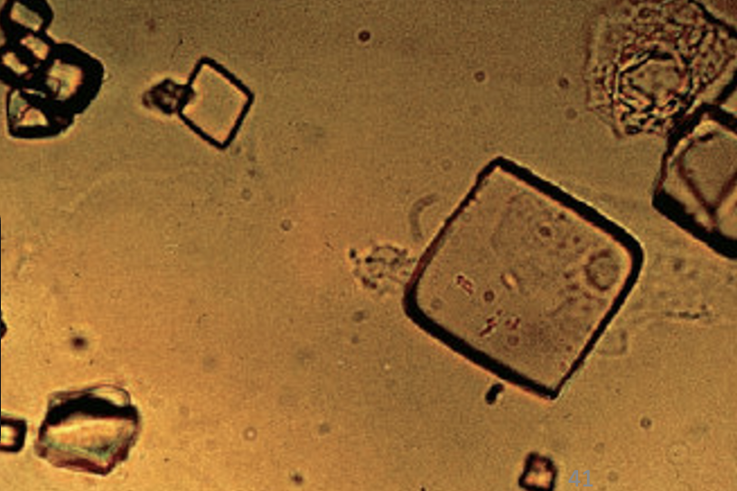
What kind of crystals are shown? What kind of urine is it found in?
Calcium oxalate crystals
Acidic urine
Acidic urine
16
New cards
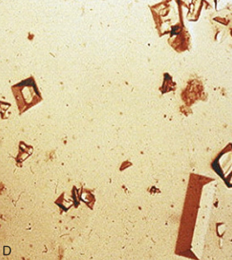
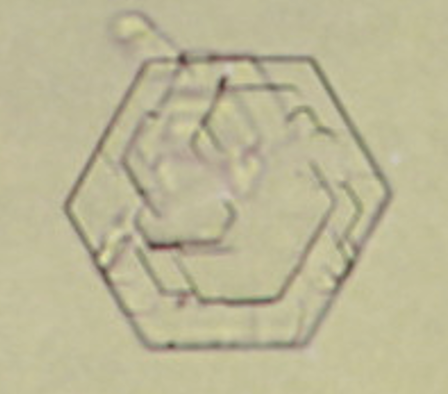
What kind of crystals are shown? What kind of urine is it found in?
Uric acid (urate) crystals
Acidic urine
Acidic urine
17
New cards
What kind of crystal is shown?
Cysteine
18
New cards
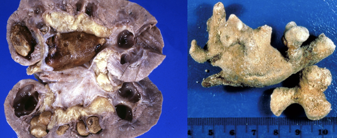
What kind of crystals are shown?
Ammonium-magnesium phosphate *aka* struvite stones or staghorn
19
New cards
What kind of crystals are shown?
Ammonium-magnesium phosphate *aka* struvite stones or staghorn
20
New cards
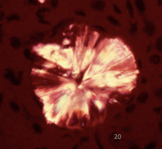
What kind of crystals are shown?
Ammonium-magnesium phosphate *aka* struvite stones or staghorn
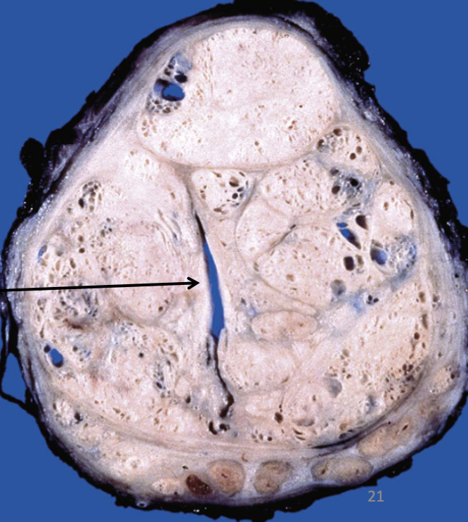
21
New cards
What kind of crystals are shown?
Uric acid (urate) crystals
22
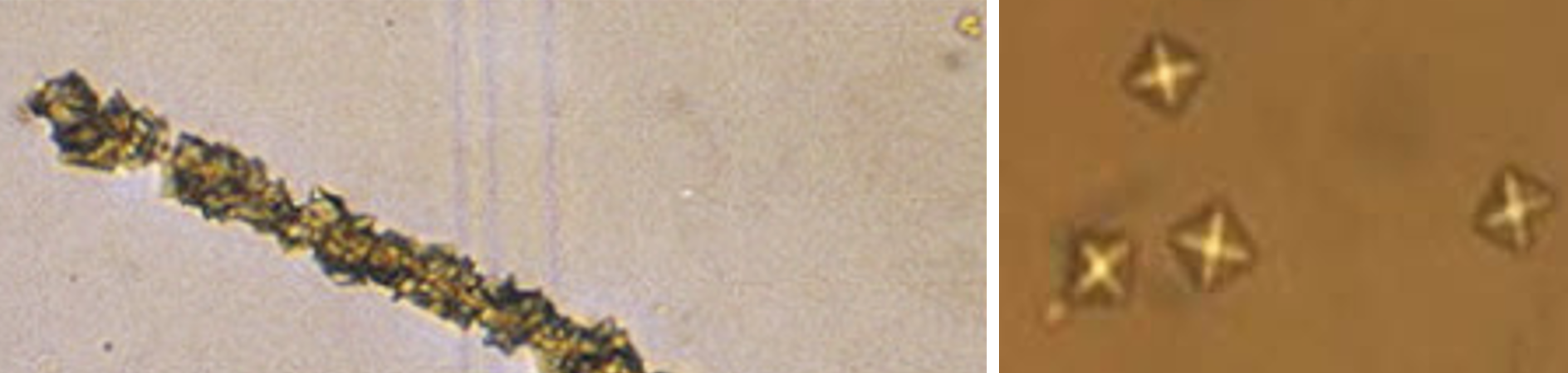
New cards
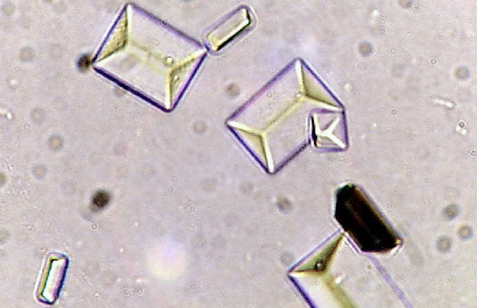
What kind of crystals are shown? What kind of urine are they found in?
Triple phosphate
Alkaline urine
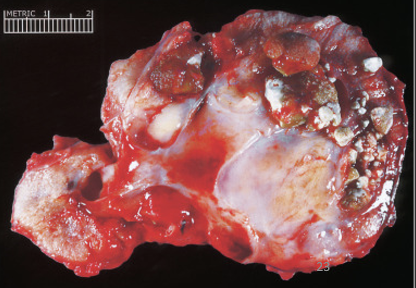
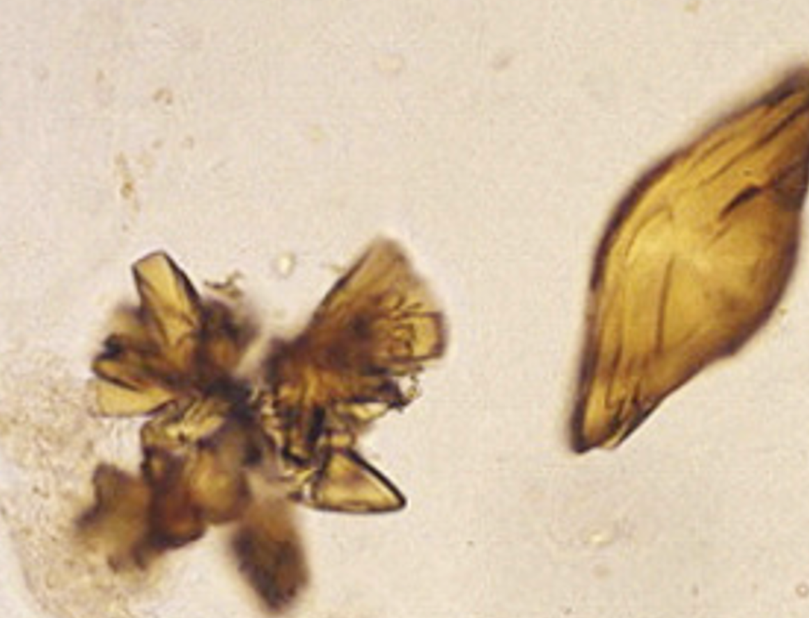
23
New cards
What kind of crystals are shown?
Cysteine
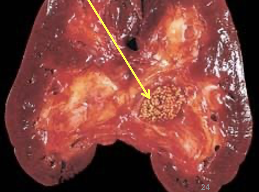
24
New cards
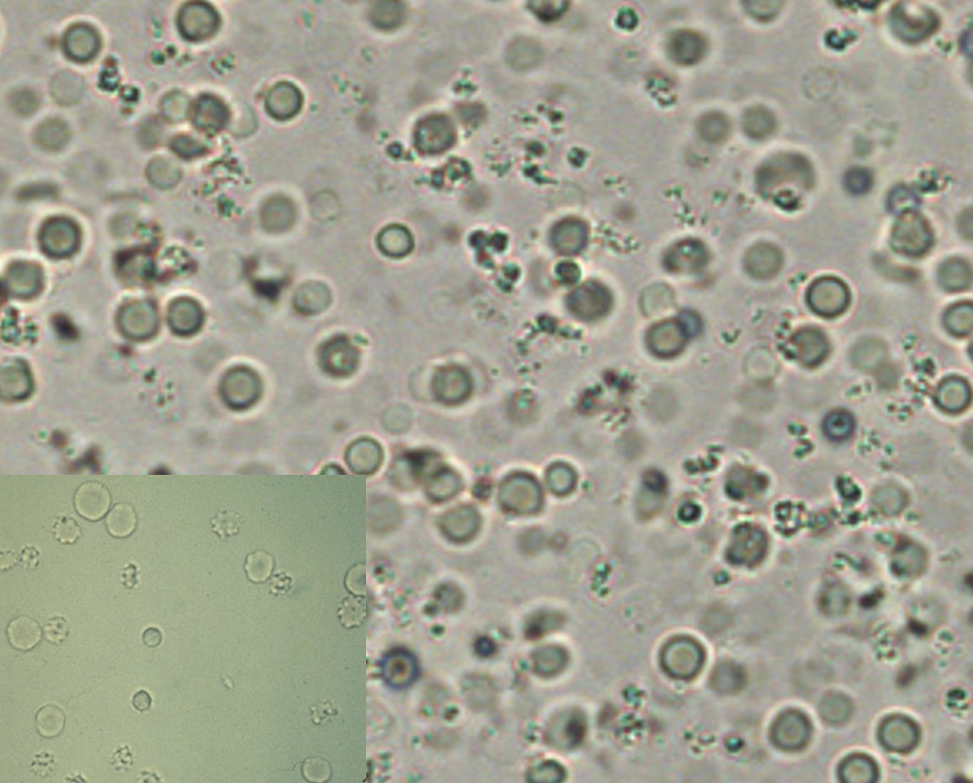
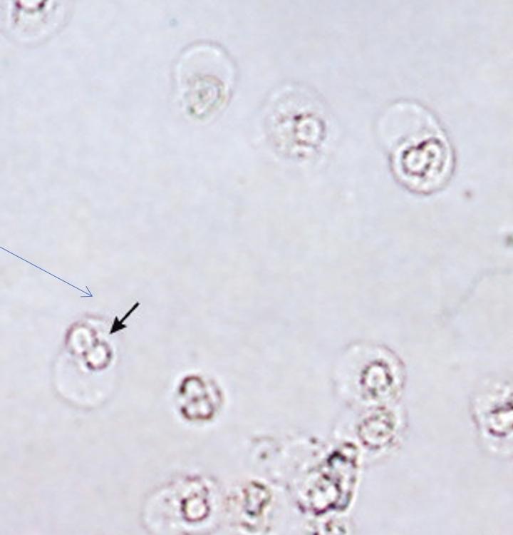
What type of cell is shown from this urinalysis? What does it indicate?
Red blood cells → hematuria
25
New cards
What type of cell is shown from this urinalysis? What does it indicate?
Neutrophils (pyuria) → UTI
26
New cards
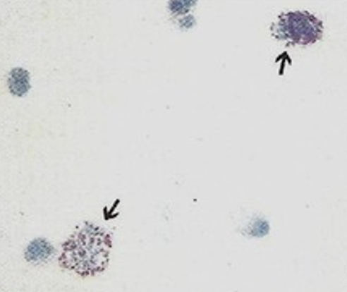
What type of cell is shown from this urinalysis? What does it indicate?
Eosinophils → Drug induced nephritis, transplant rejection
27
New cards
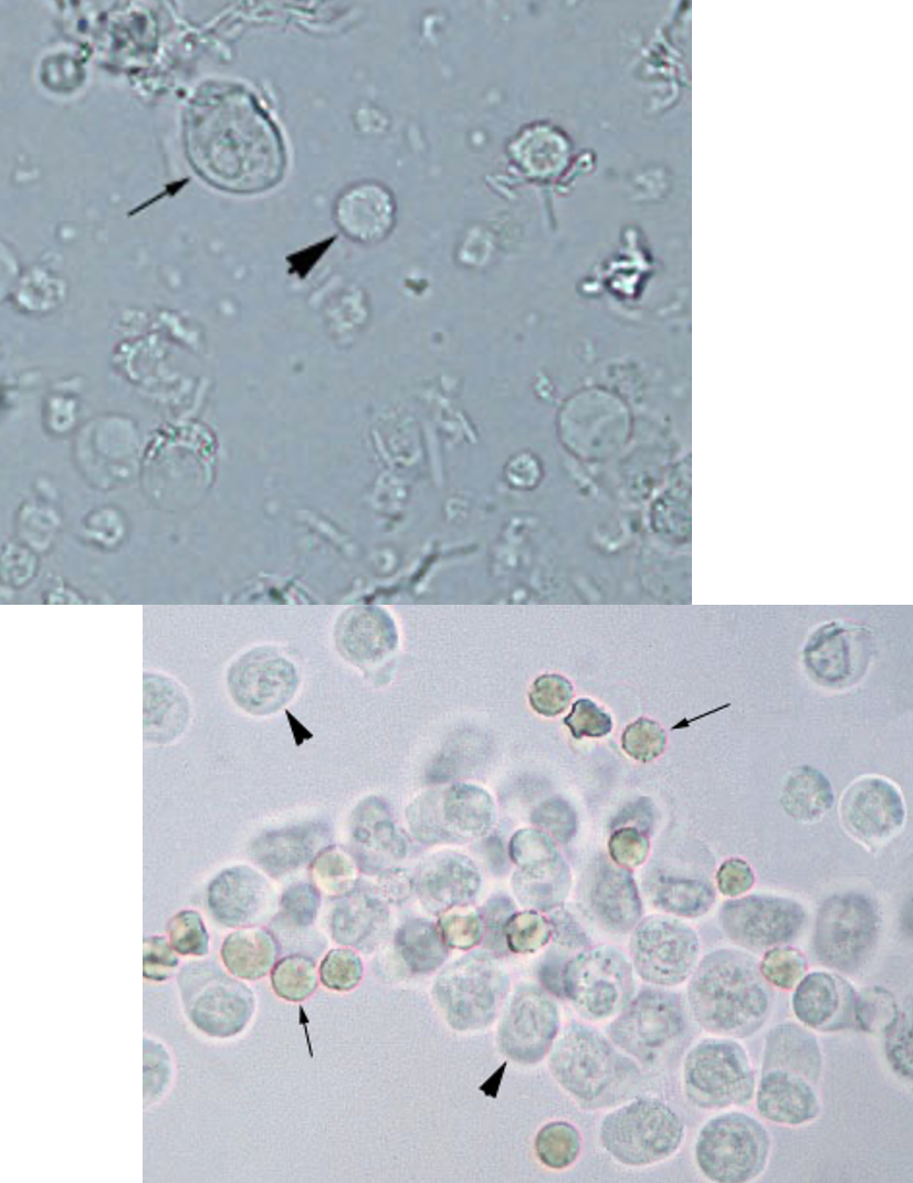
What broad type of cell is shown in these images of urine?
Leukocytes
28
New cards
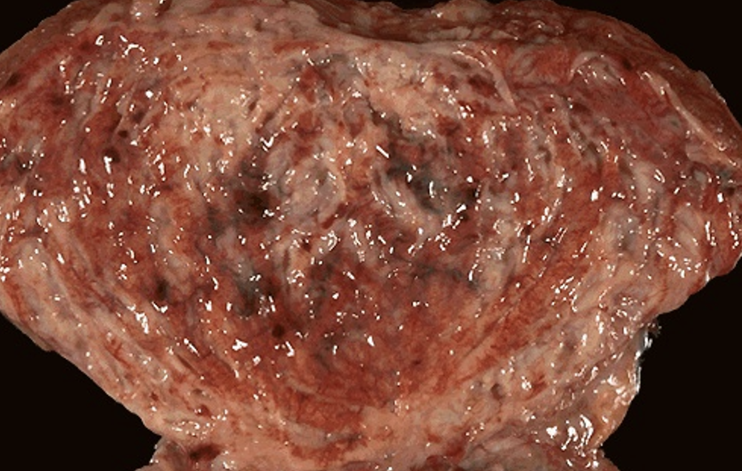
What pathology is shown from this image?
Hyperemic mucosa indicating a UTI aka acute cystitis
29
New cards
What pathology is shown from this image and what type of study is used to detect it?
Vesicoureteral reflux evaluated with voiding cystourethrogram
30
New cards
What type of cast is this and what pathology does it indicate? What are the primary clinical signs? What reflux is it associated with?
Leukocyte cast → Acute pyelonephritis
Sudden pain at costovertebral angle, fever, dysuria, frequency, urgency
vesicoureteral reflux
Sudden pain at costovertebral angle, fever, dysuria, frequency, urgency
vesicoureteral reflux
31
New cards
What type of cast is this?
Leukocyte cast
32
New cards
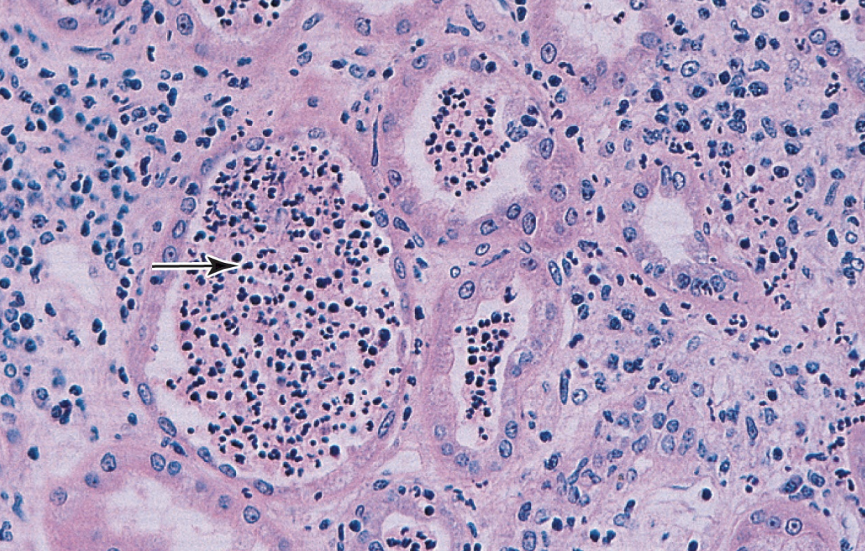
What type of cell is very prominent in this image? What does it indicate?
Neutrophils indicating acute pyelonephritis
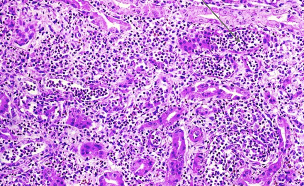
33
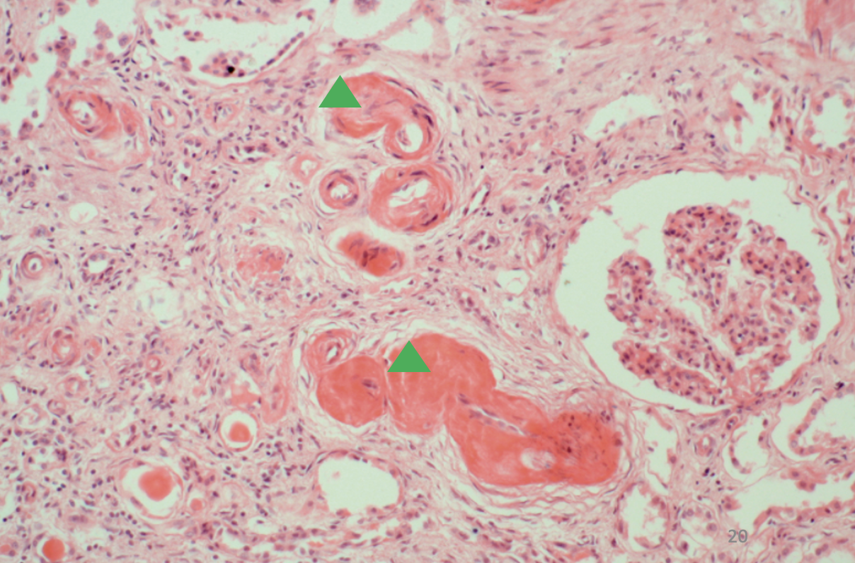
New cards
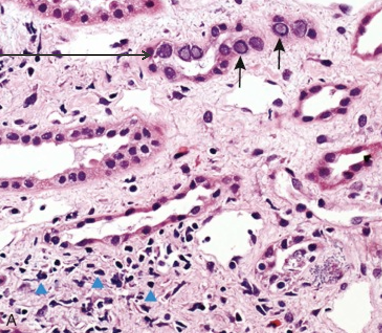
This image indicates Polyoma virus at the black arrows…..what clinical diagnosis would this indicate? What is found at the blue triangles?
Pyelonephritis
T lymphocytes
T lymphocytes
34
New cards
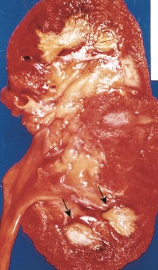
What complication of an acute pyelonephritis is shown at the arrows?
Abscess
35
New cards
What complication of an acute pyelonephritis is shown at the arrow?
abscess formation *within* kidney tubules
36
New cards
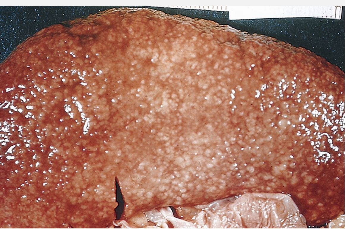
What complication of an acute pyelonephritis is shown?
Cortical abscesses
37
New cards
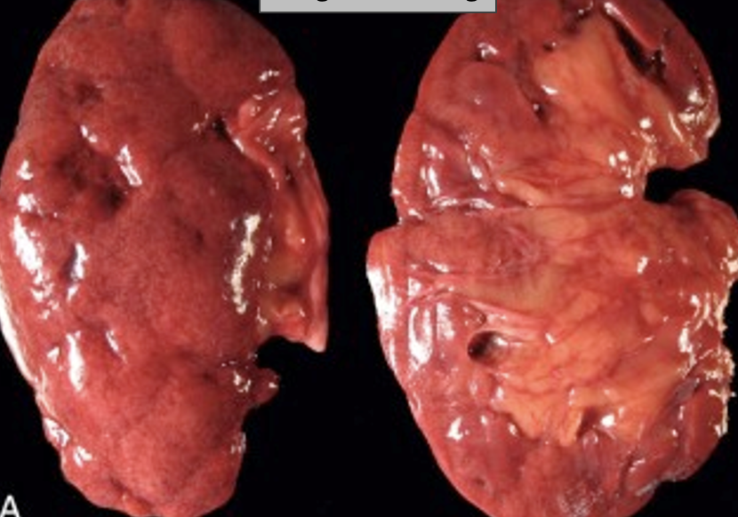
Small abscess hematogenous spread shown in the image is from what underlying pathology?
acute pyelonephritis
38
New cards
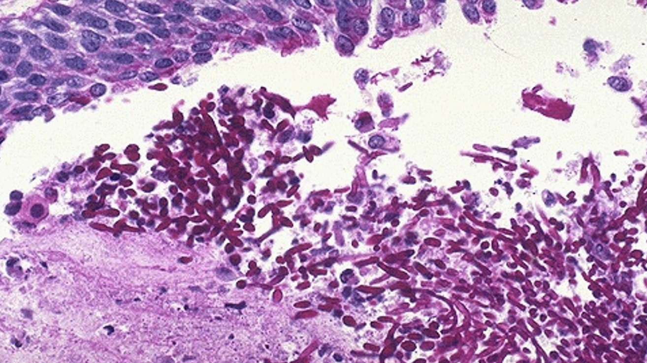
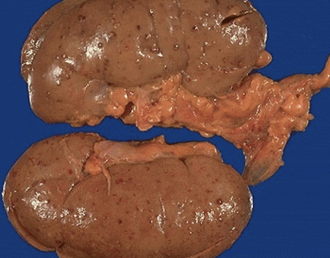
What bug is causing this acute pyelonephritis?
candida
39
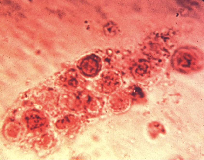
New cards
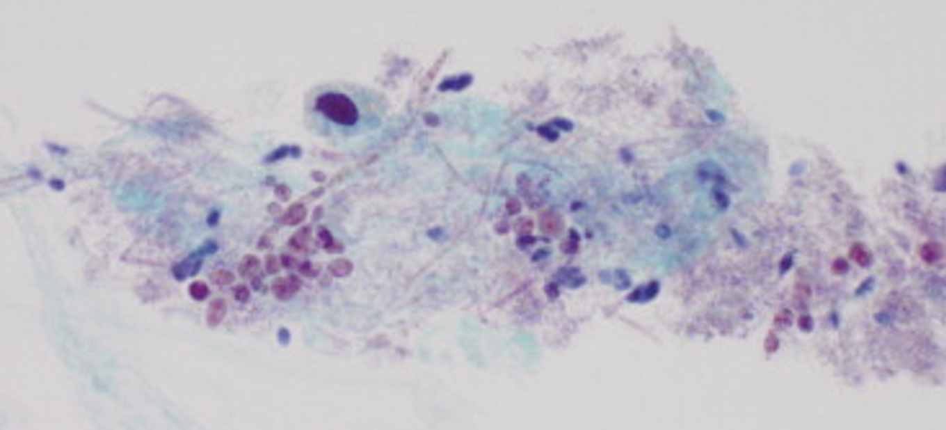
What bug is shown in this cytology sample?
candida
40
New cards
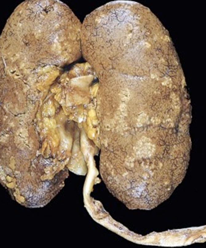
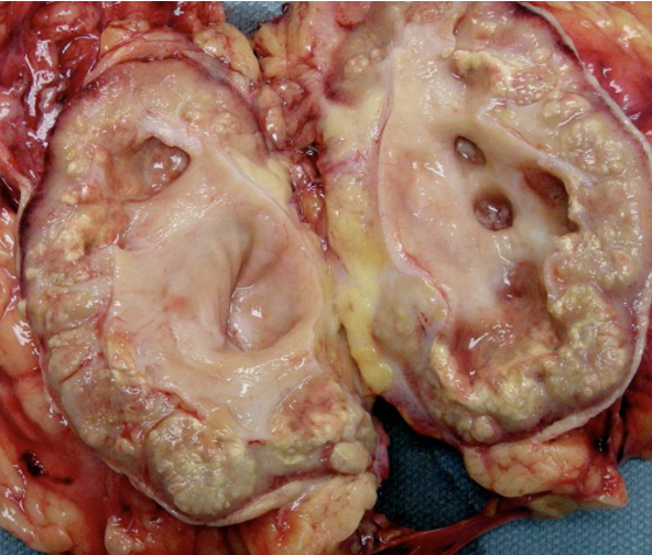
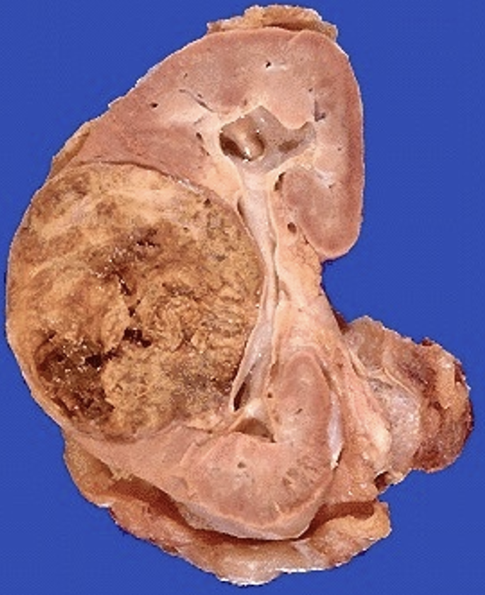
What pathology is shown here?
Chronic pyelonephritis “irregular scarring & blunting of calyces & pelvis”
41
New cards
What pathology is shown here?
Chronic pyelonephritis
42
New cards
What pathology is shown here?
Chronic pyelonephritis showing inflammatory destruction of papillae
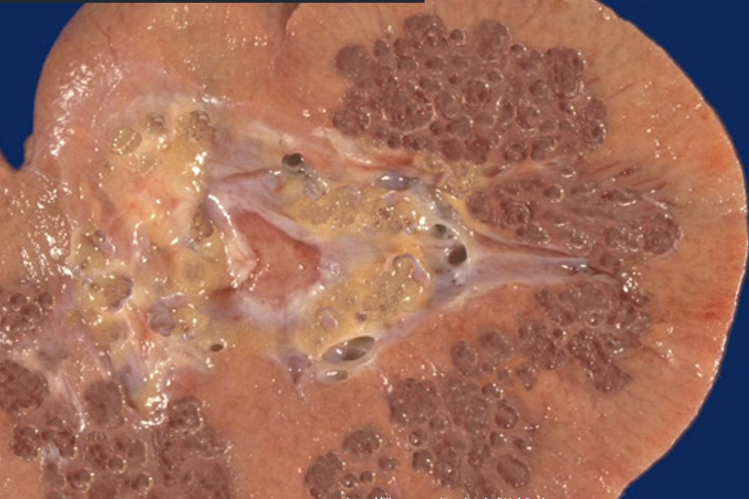
43
New cards
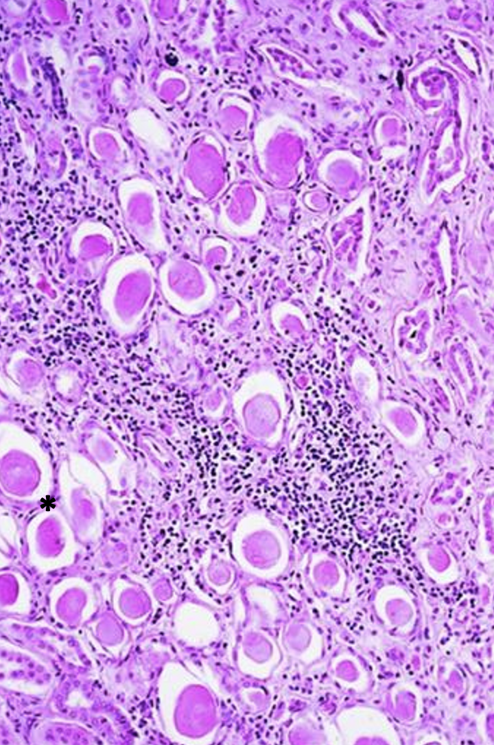
What pathology is shown here? What is indicated at the asterisk?
Chronic pyelonephritis
thyroidization
thyroidization
44
New cards
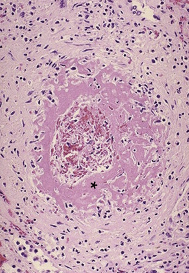
What pathology is shown here?
Xanthogranulomatous pyelonephritis (chronic)
45
New cards
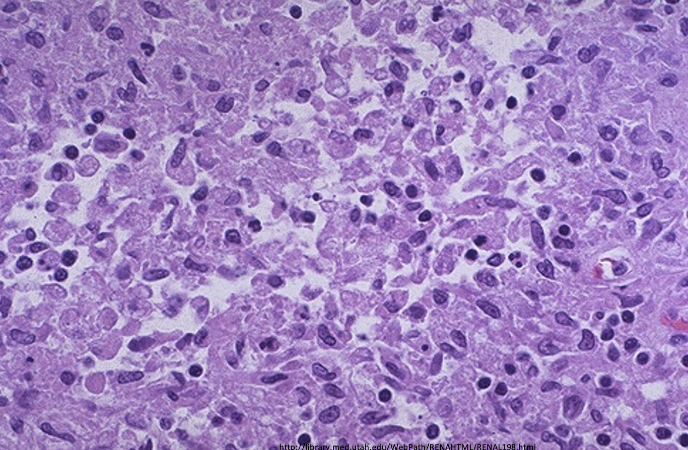
What from this histology slides indicates a Xanthogranulomatous pyelonephritis?
foamy macrophages
46
New cards
What pathology is shown here?
Xanthogranulomatous pyelonephritis (chronic)
47
New cards
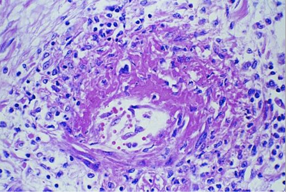
What pathology is shown here?
Fibrinoid necrosis in an ANCA related vasculitis
48
New cards
What pathology is shown here?
Nephrosclerosis
49
New cards
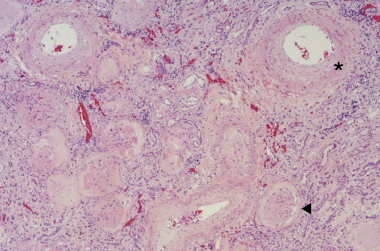
What is shown at the asterisk & triangle of this histology of nephrosclerosis?
Asterisk: medial thickening of the small arteries
Triangle: Glomerulosclerosis
Triangle: Glomerulosclerosis
50
New cards
What pathology is shown here?
hyaline sclerosis - nephrosclerosis
51
New cards
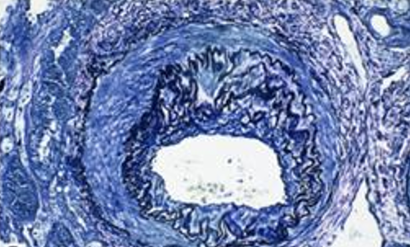
What pathology is shown here?
fibrotic intimal thickening causing narrowing of the lumen - nephrosclerosis
52
New cards
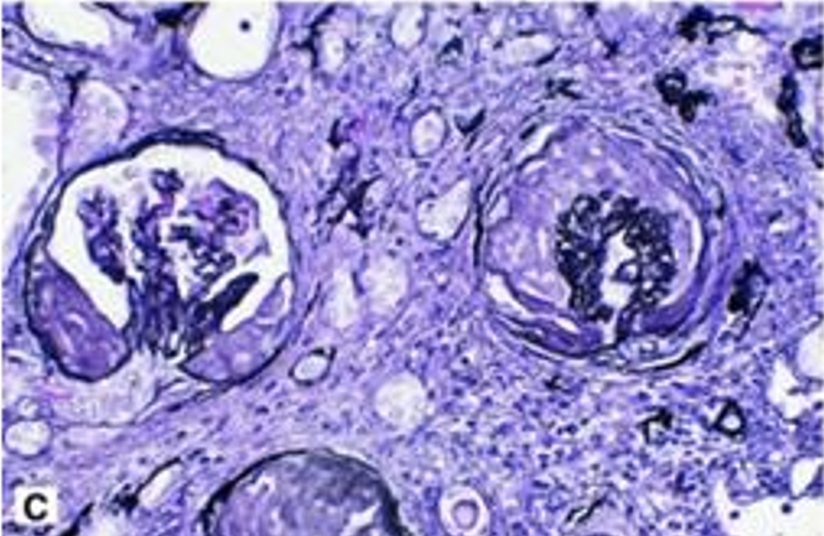
What pathology is shown here?
one glomerulus with global sclerosis and one with segmental sclerosis (glomerulosclerosis tubular atrophy, interstitial fibrosis and chronic inflammation)
53
New cards
What is the pathology shown here mostly associated with?
hypertension & aging - nephrosclerosis
54
New cards
What pathology is shown here?
nephrosclerosis
55
New cards
What pathology is indicated with the left kidney?
Nephrosclerosis
56
New cards
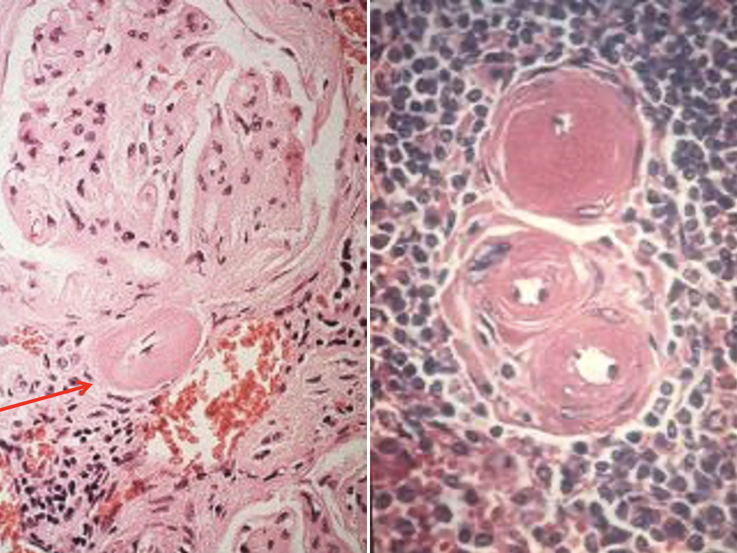
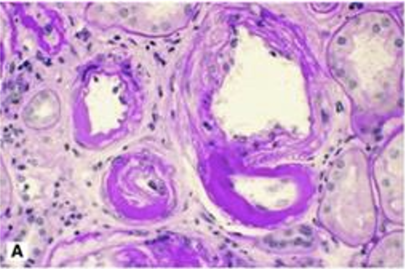
What pathology is shown in these images?
Hyaline arteriolosclerosis
57
New cards
From this image, what type of injury is indicated with this hyaline arteriolosclerosis? noted from the yellow triangles
type 2 diabetes injury
58
New cards
What is shown here and what is it indicative of?
Fibrinoid necrosis indicating accelerated (malignant) hypertension
59
New cards
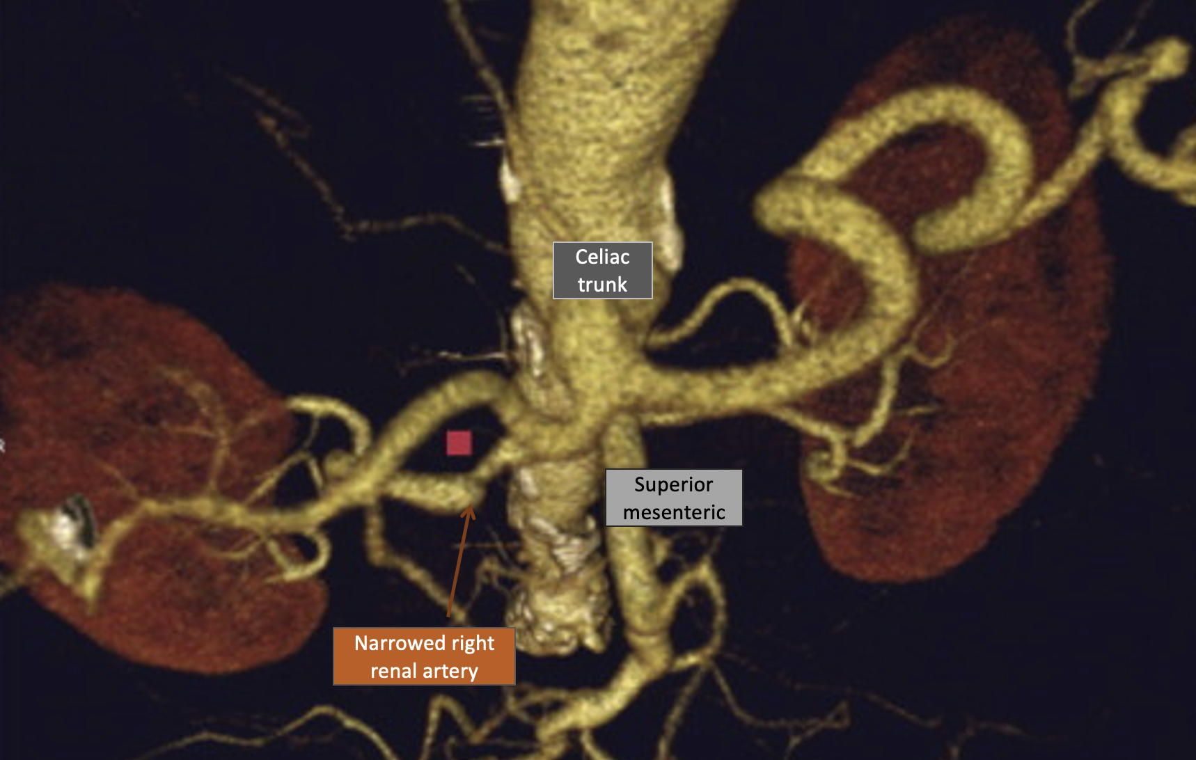
What does this image indicate?
fibromuscular dysplasia
60
New cards
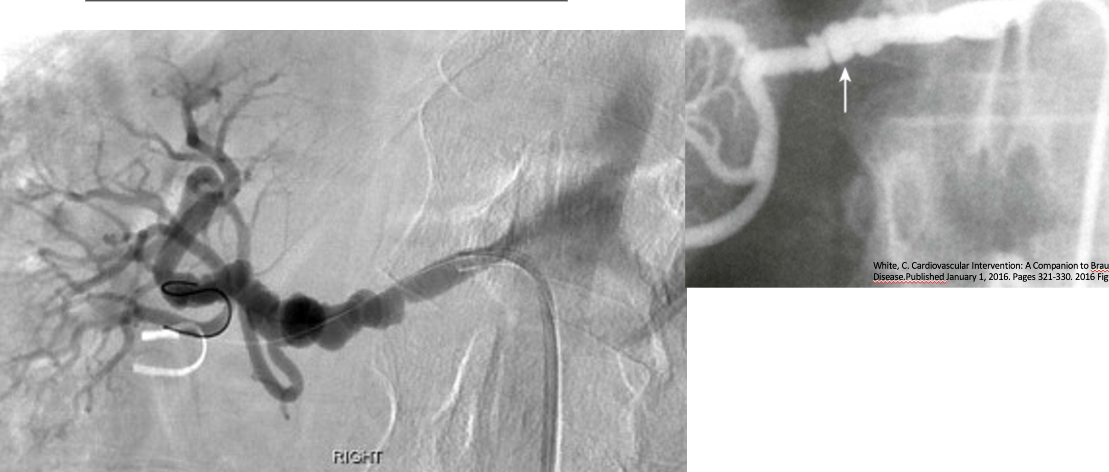
What do these “string of beads” indicate?
fibromuscular dysplasia
61
New cards
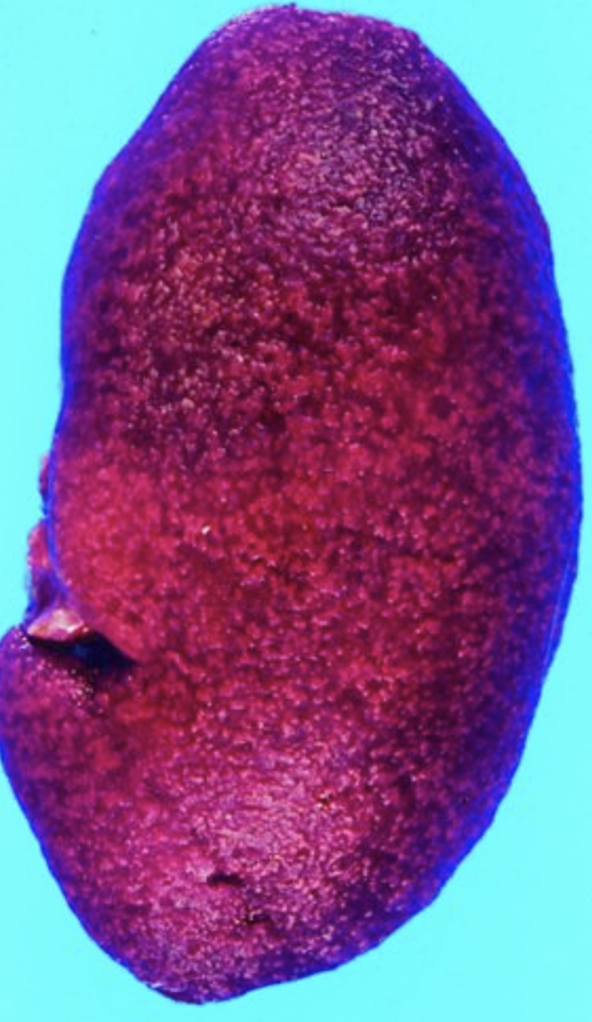
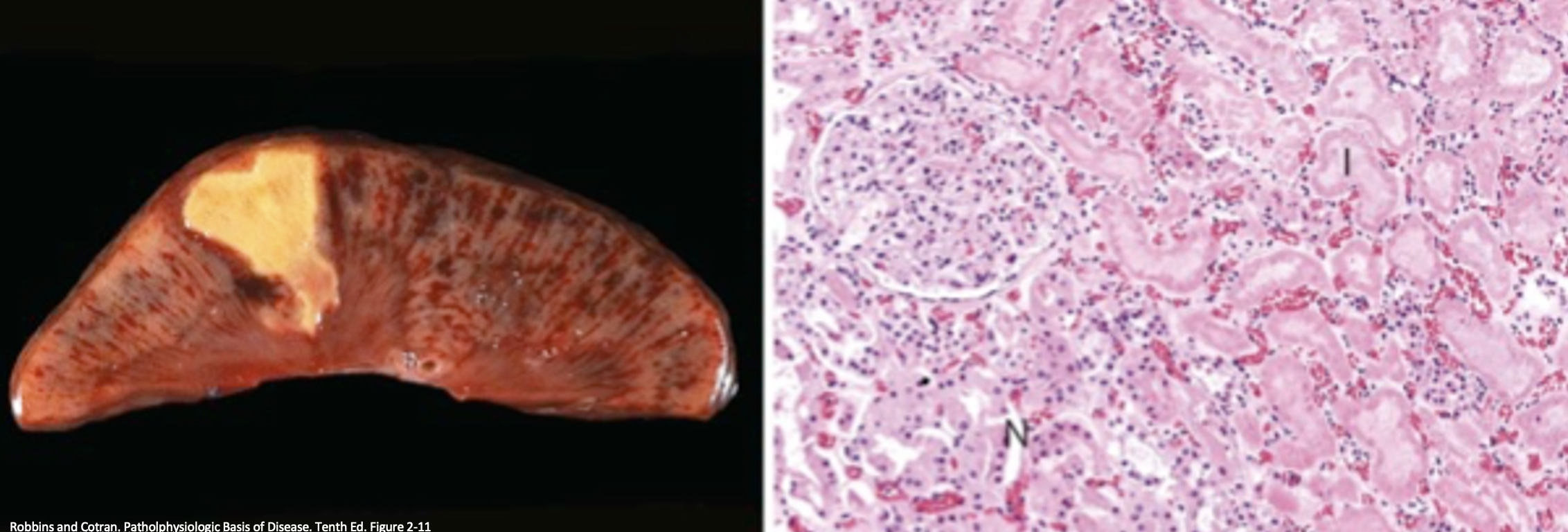
What do these images show?
Renal infarct
Eosinophillic necrotic tissue (coagulation necrosis)
Eosinophillic necrotic tissue (coagulation necrosis)
62
New cards
What do these images show?
Renal infarct
63
New cards
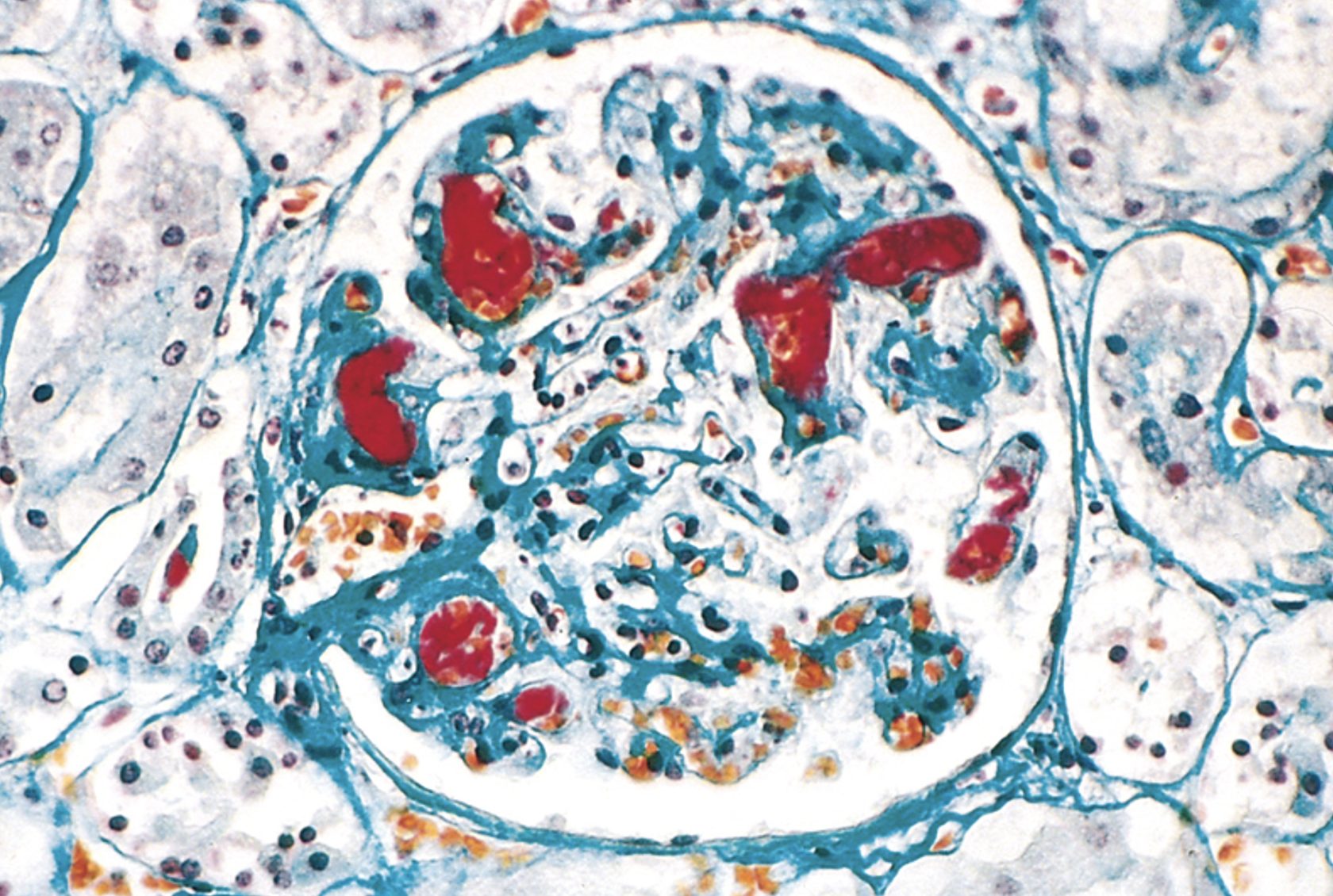
What is indicated by the red in this image?
platelet-fibrin thrombi
64
New cards
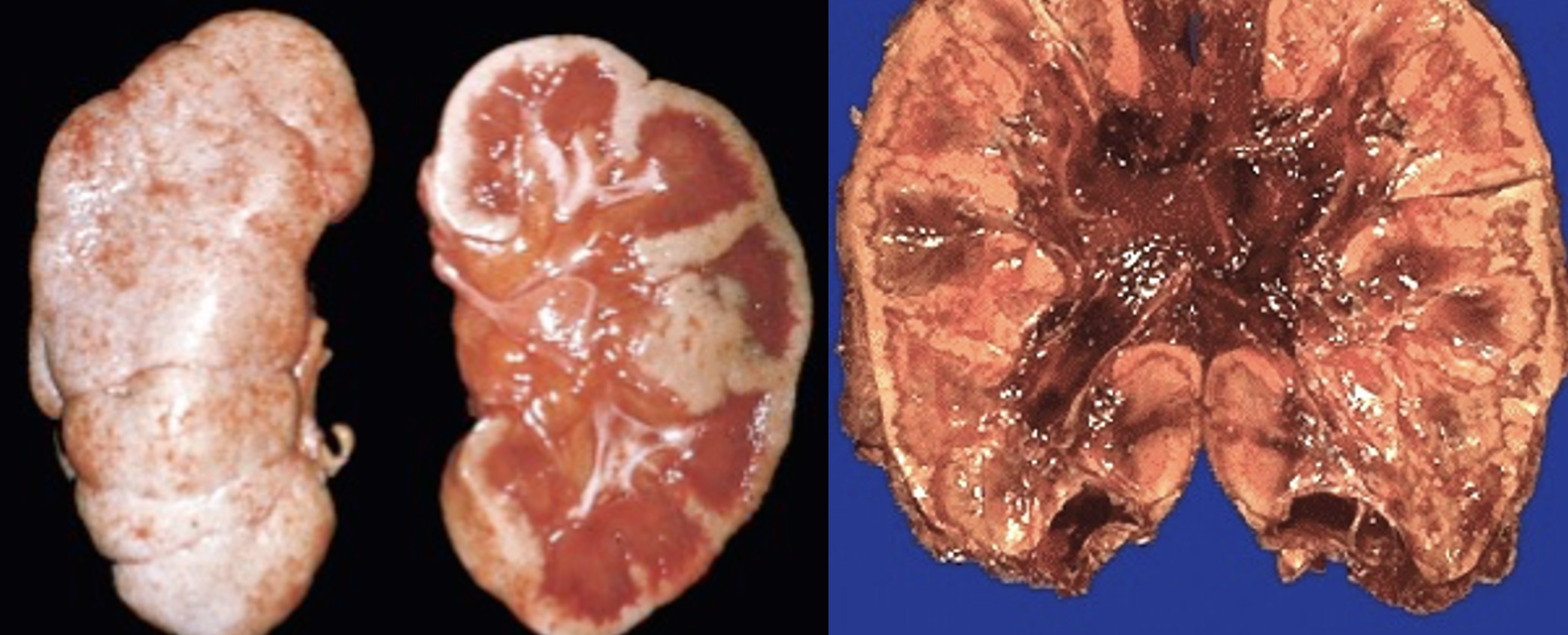
What necrosis is shown in these images?
Diffuse cortical necrosis
65
New cards
What is indicated by this photo?
uremic frost - renal failure
66
New cards
What is shown at the green triangles and what is causing it?
hyalinzation (hyalinosis) and sclerosis from CKD
67
New cards
What pathology is indicated?
glomerular disease from an immune mechanism
68
New cards
What pathology is indicated?
Nephrotic syndrome
69
New cards
What pathology is indicated?
Focal segmental\* glomerulosclerosis
70
New cards
What is indicated at the red triangle?
HIV-associated and idiopathic collapsing FSGS have the *worst* prognoses
71
New cards
What is indicated at the arrows? What disease does it indicate?
Effacement of foot processes - minimal change disease
72
New cards
What pathology is indicated?
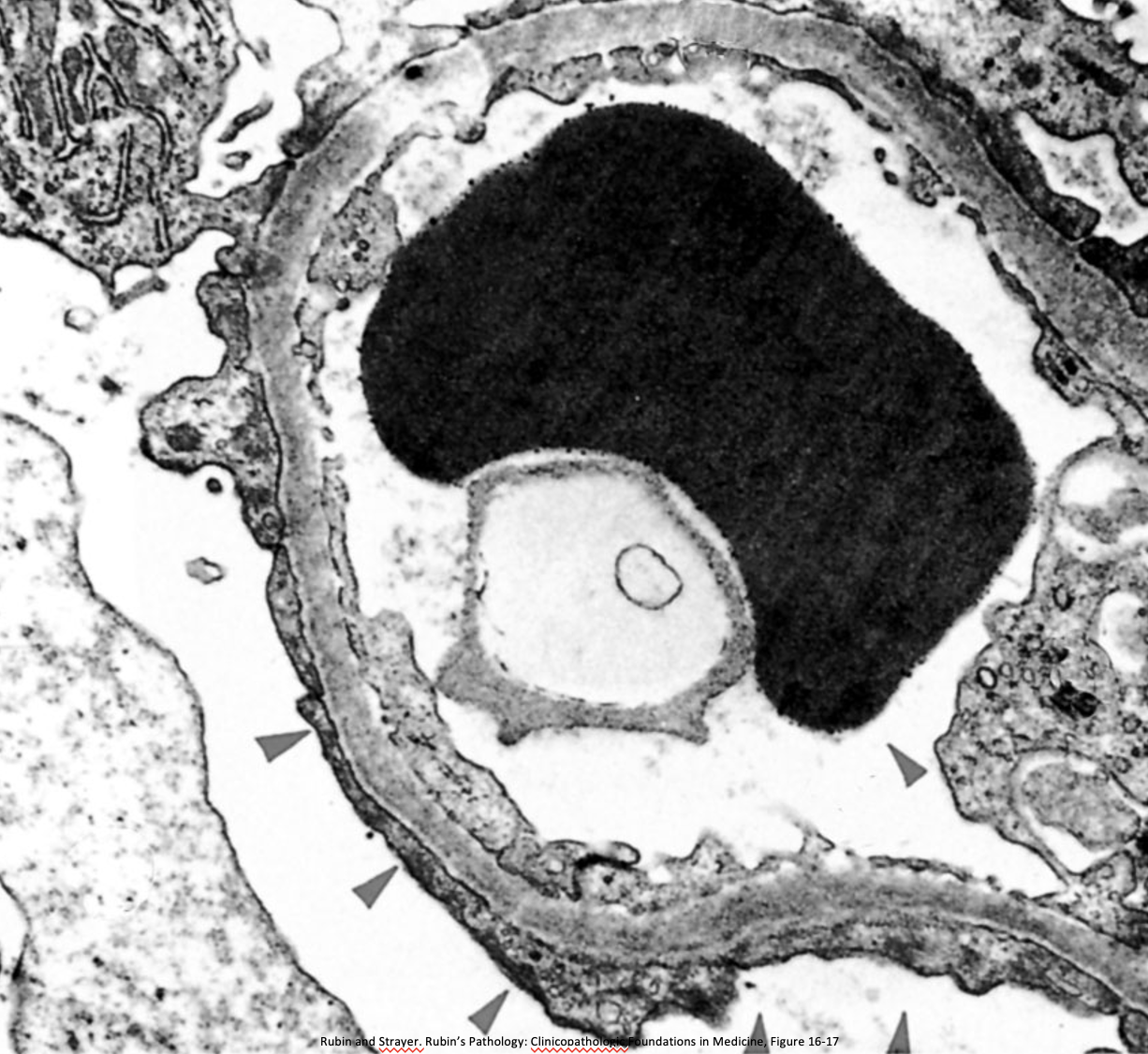
Membranous nephropathy
73
New cards
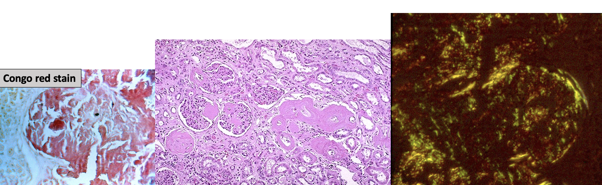
What is indicated by these images?
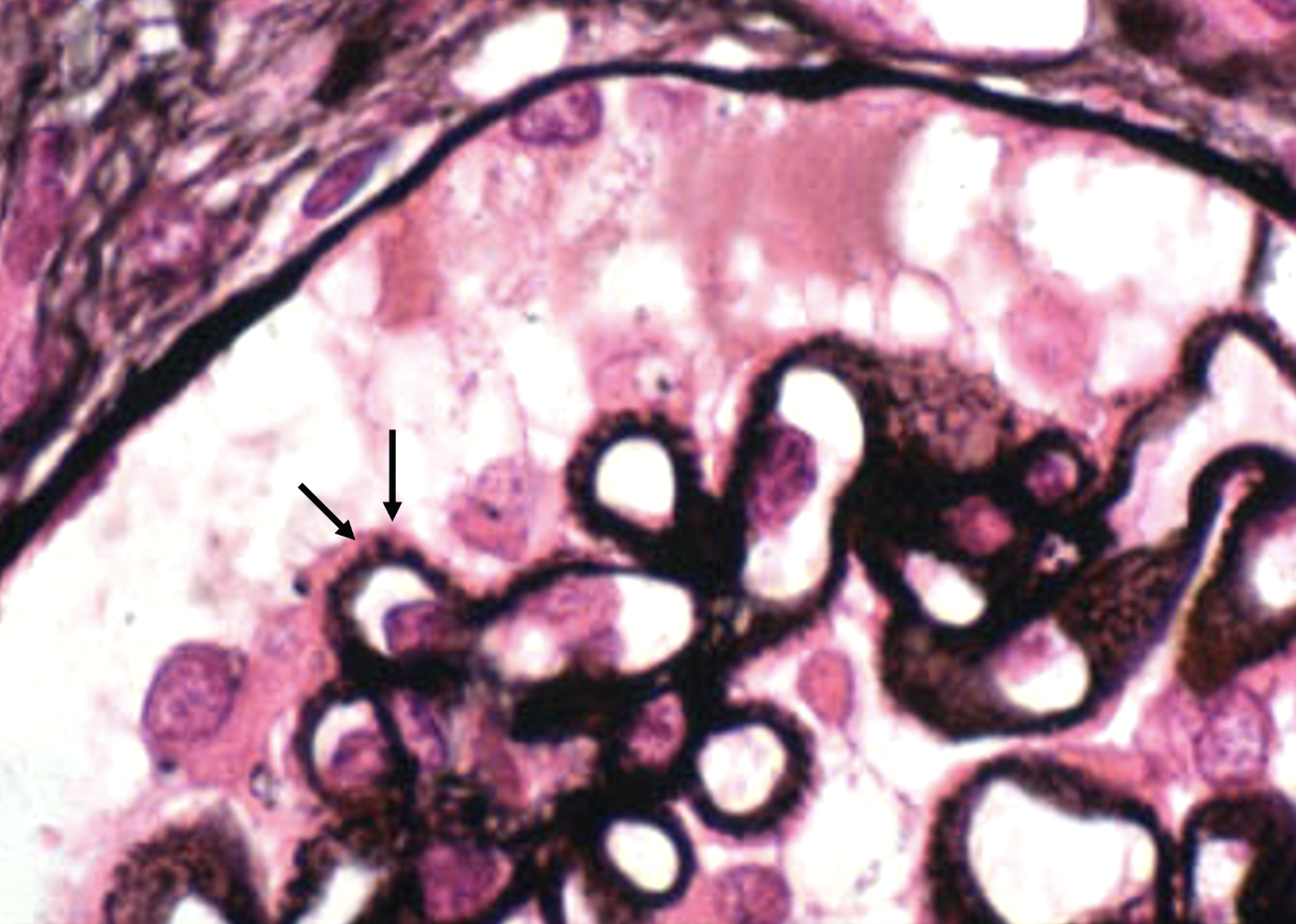
Amyloidosis
74
New cards
What is indicated by these images?
Nephrotic syndrome-amyloidosis
75
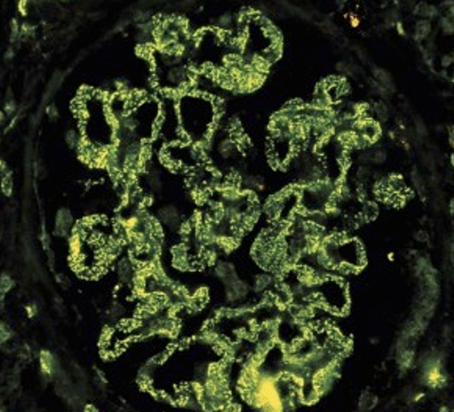
New cards
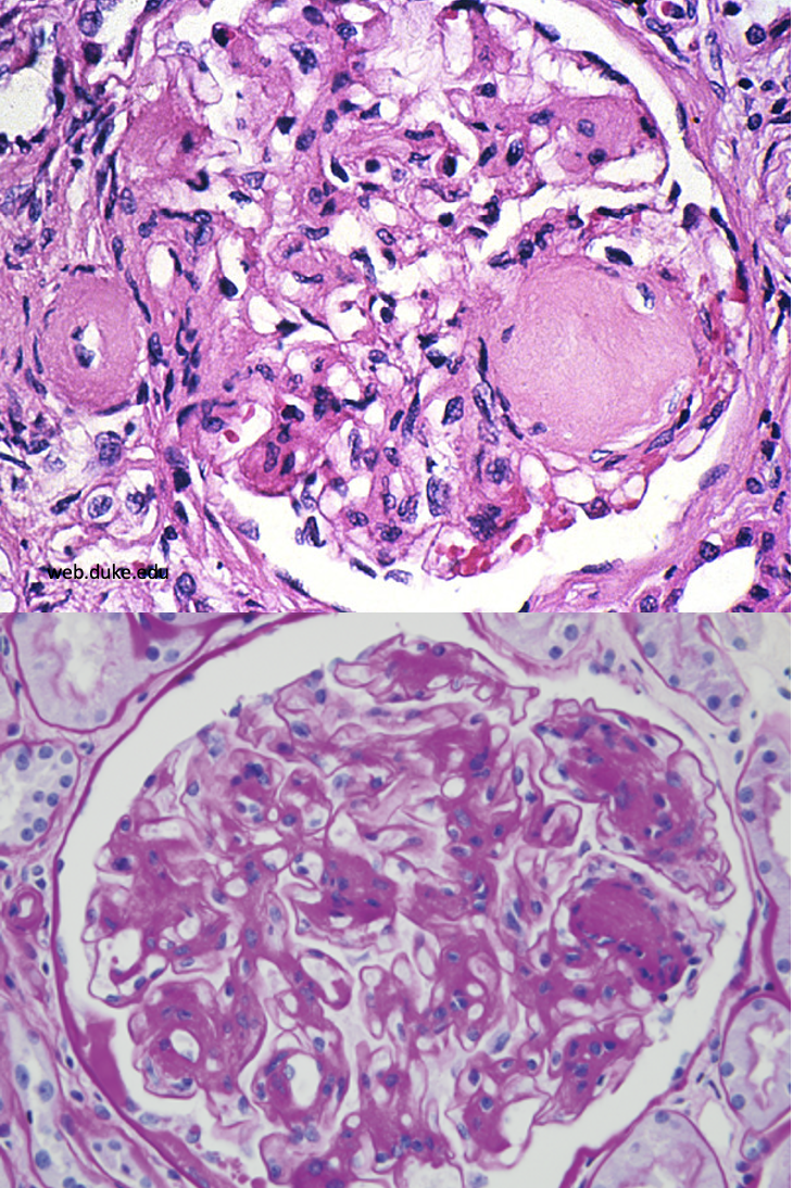
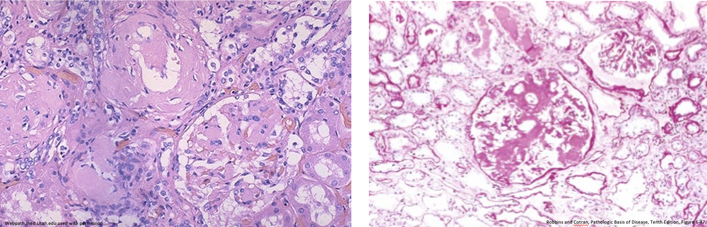
What is indicated by these images?
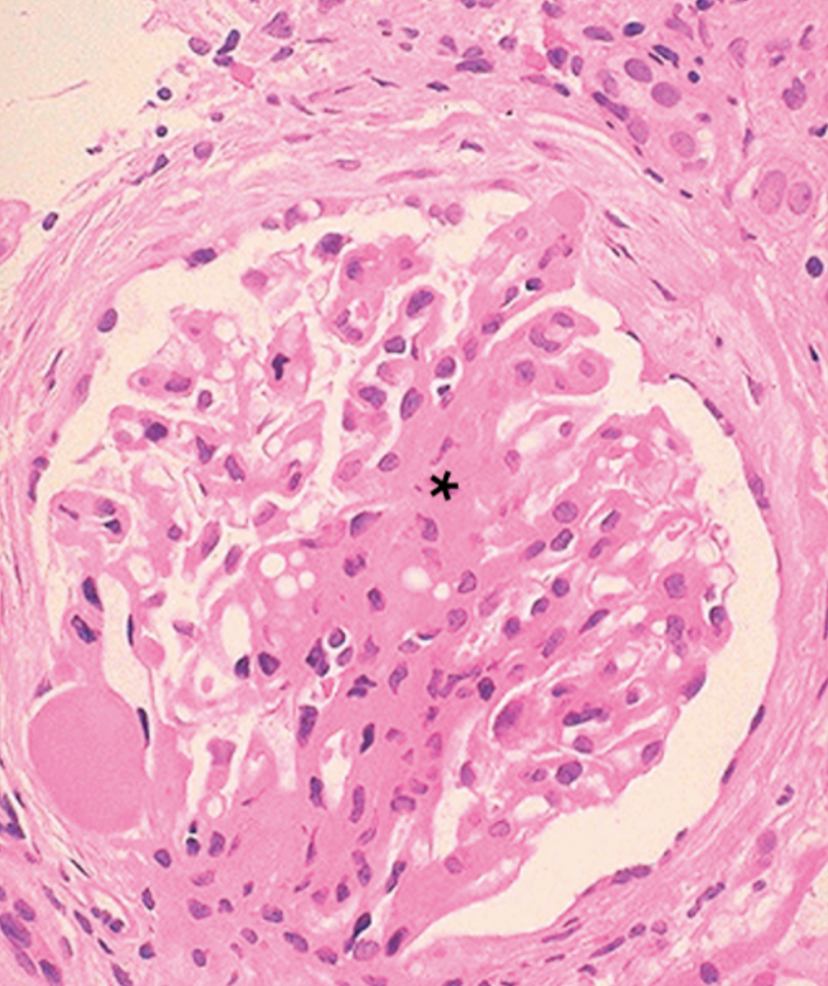
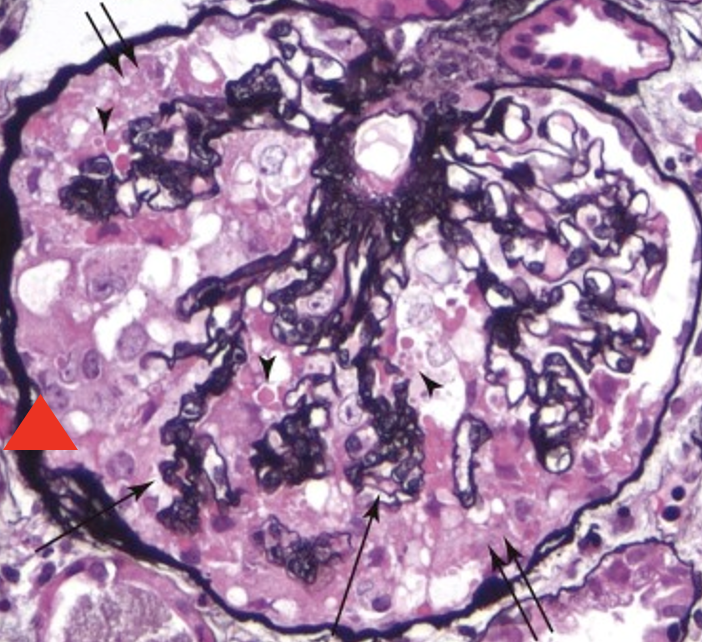
Diabetic glomerulosclerosis
76
New cards
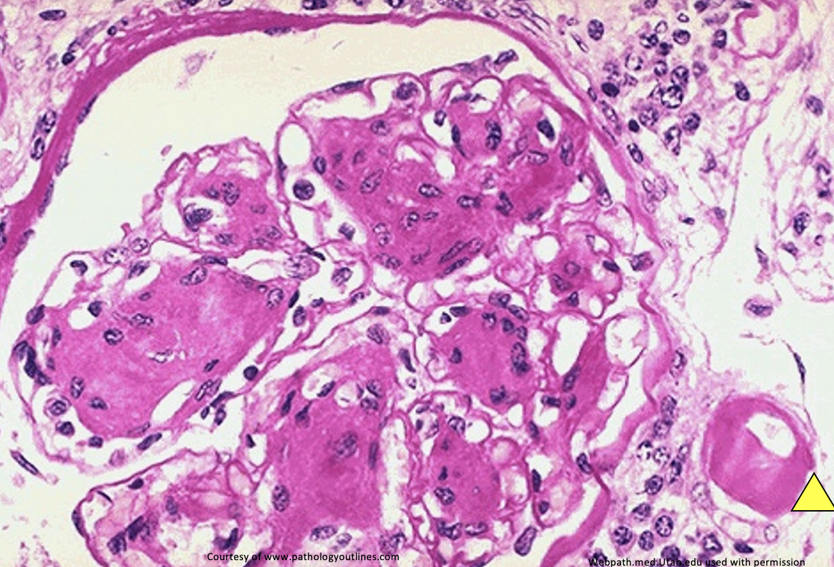
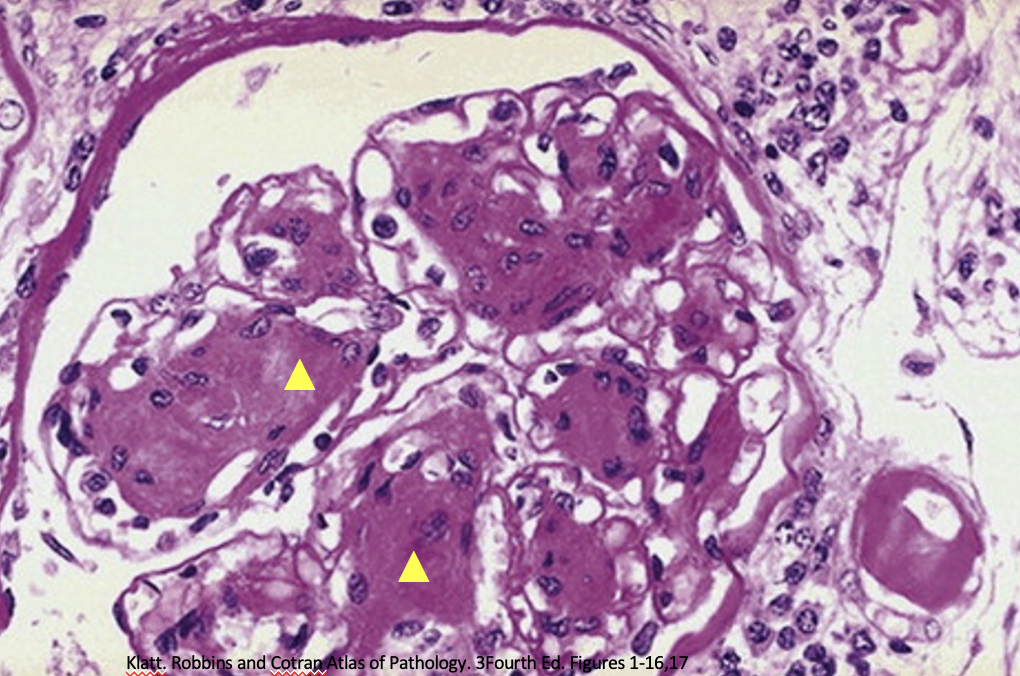
What is indicated by this image? What is shown at the yellow triangle?
Diabetic glomerulosclerosis
hyaline arteriosclerosis
hyaline arteriosclerosis
77
New cards
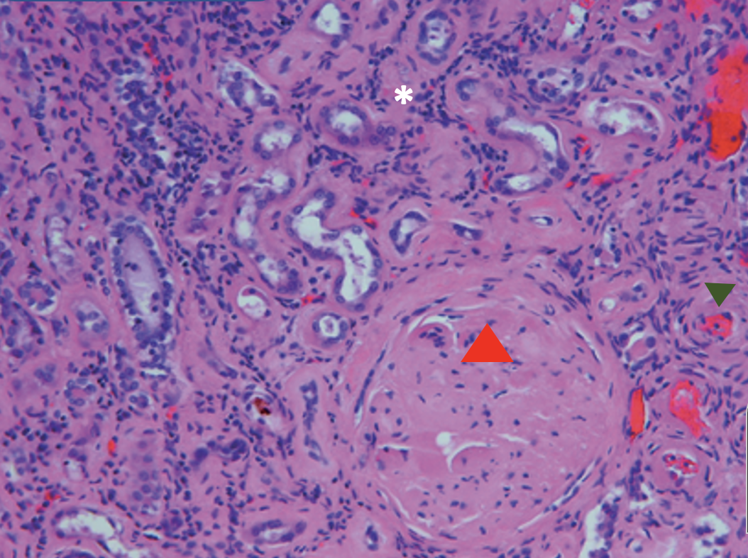
What is the diagnosis?
What is shown at the green triangle?
What is shown at the red triangle?
What is shown at the white asterisk?
What is shown at the green triangle?
What is shown at the red triangle?
What is shown at the white asterisk?
Chronic glomerulonephritis
Hyaline arteriolosclerosis
sclerotic glomeruli
Thinned cortex and tubular atrophy
Hyaline arteriolosclerosis
sclerotic glomeruli
Thinned cortex and tubular atrophy
78
New cards
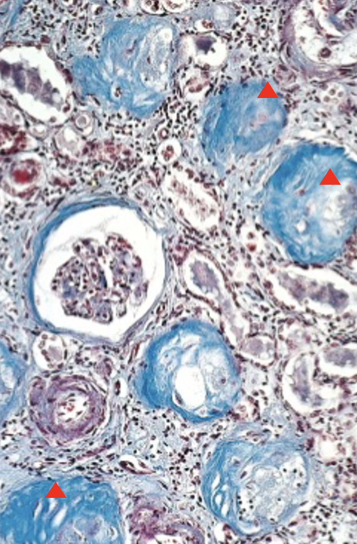
What is the diagnosis?
What is shown at the red triangle?
What is shown at the red triangle?
Chronic glomerulonephritis
sclerotic glomeruli
sclerotic glomeruli
79
New cards
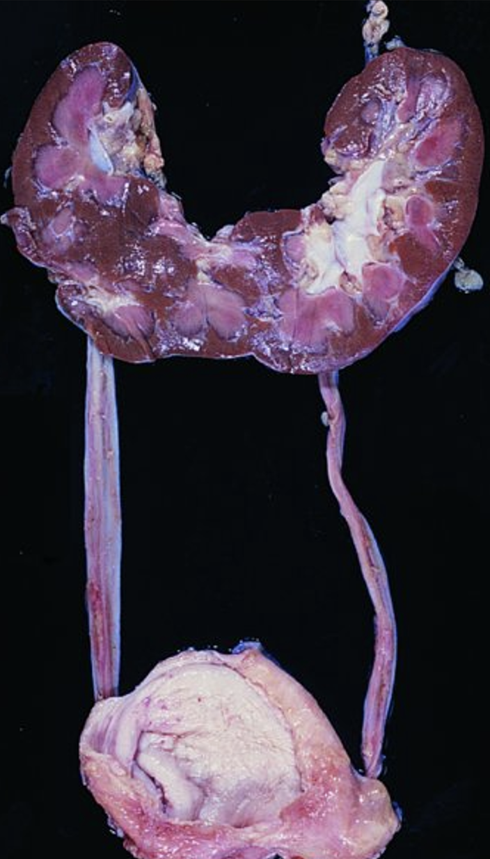
What abnormality is this? What are those with this more at risk of?
congenital horseshoe kidney
infections & renal calculi
infections & renal calculi
80
New cards
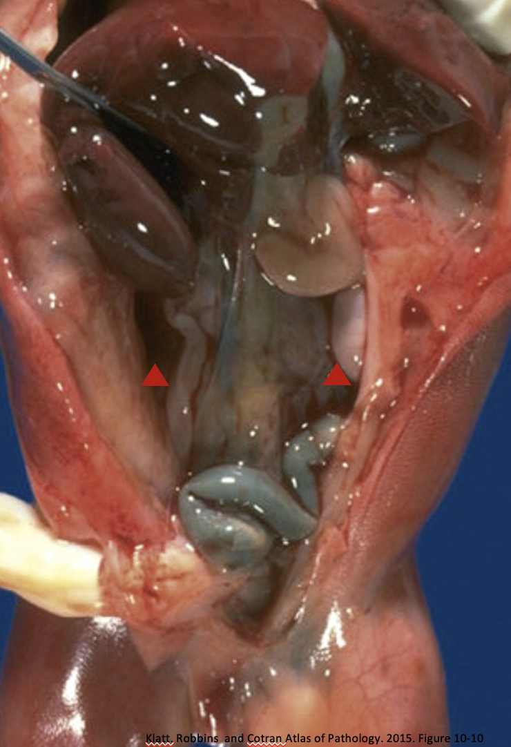
What is missing at the red arrows?
KIDNEYS!! - renal agenesis
81
New cards
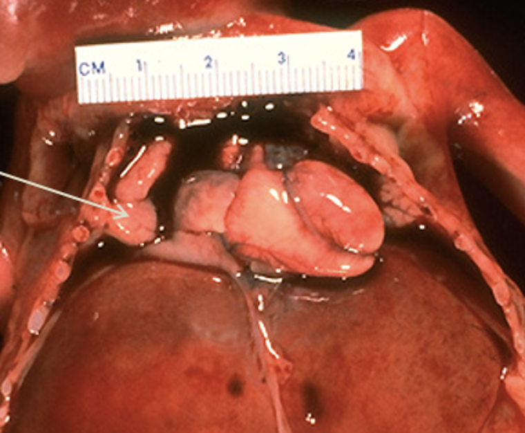
What is indicated by this image?
Renal agenesis-oligohydramnios
82
New cards
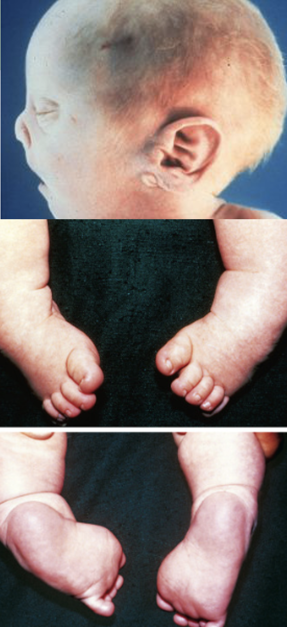
What is the name for these physcial exam findings? What condition causes this?
potter facies & talipes equinovarus caused from Renal agenesis-oligohydramnios
83
New cards
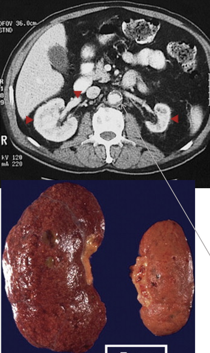
What is indicated by these images?
What is wrong with the right kidney?
What can happen from the issue with the left kidney?
What is wrong with the right kidney?
What can happen from the issue with the left kidney?
(acquired) renal hypoplasia
right: nephrosclerosis
left: increased renin secretion causing HTN
right: nephrosclerosis
left: increased renin secretion causing HTN
84
New cards
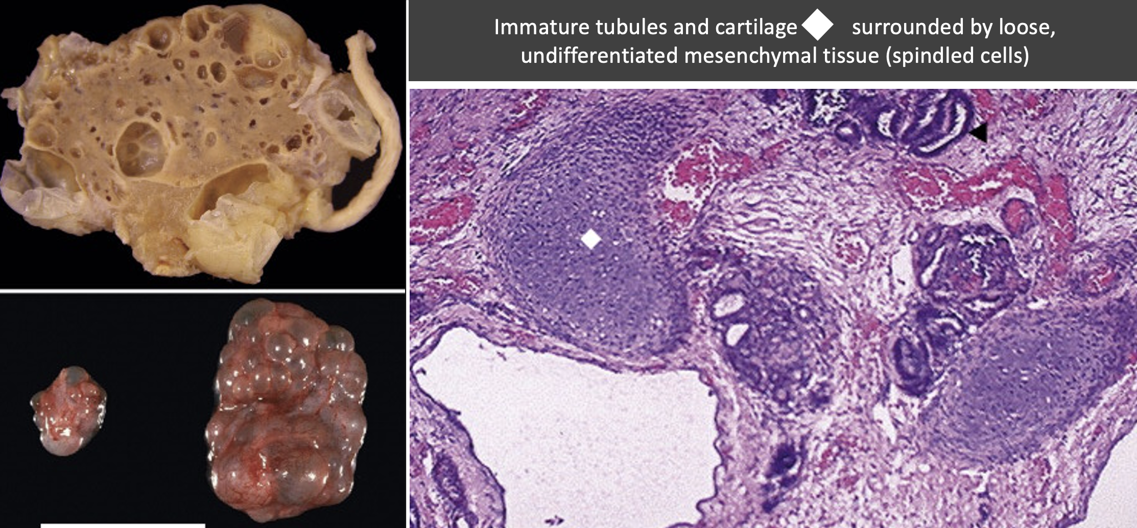
What is going on here?
Renal dysplasia (multicystic renal dysplasia) from
abnormal metanephric differentiation
abnormal metanephric differentiation
85
New cards
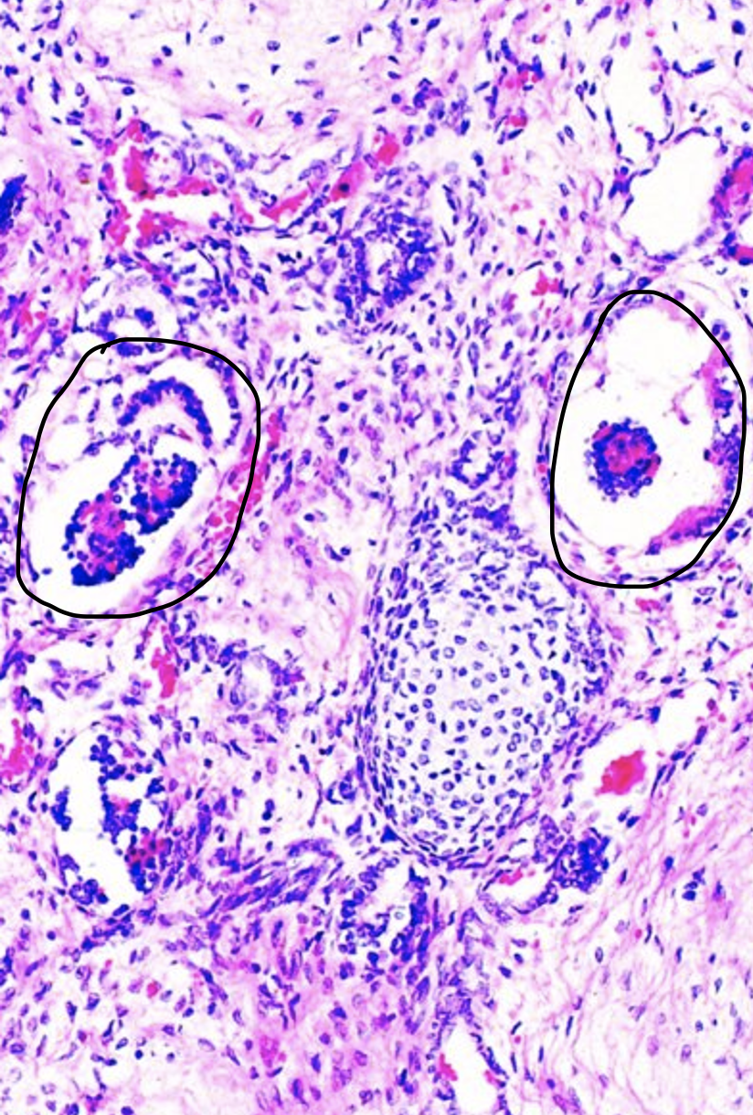
Explain this histology image of renal dysplasia.
Immature glomeruli, tubules and cartilage are surrounded by loose, undifferentiated mesenchymal tissue (spindled cells) {glomeruli are circled in black}
86
New cards
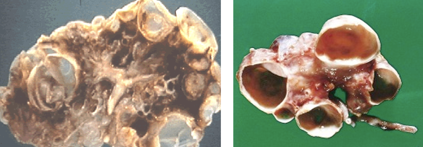
What do these gross findings indicated?
Renal dysplasia
87
New cards
What do these findings indicate?
Renal dysplasia
88
New cards
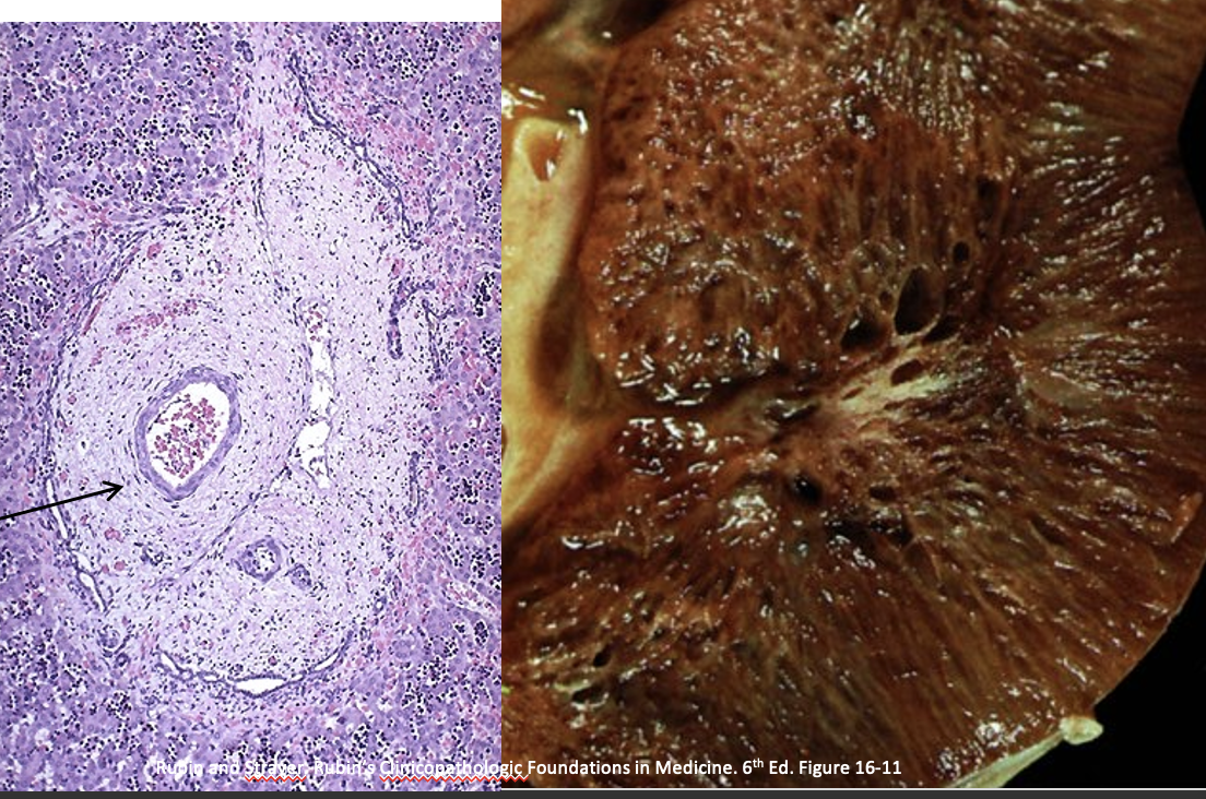
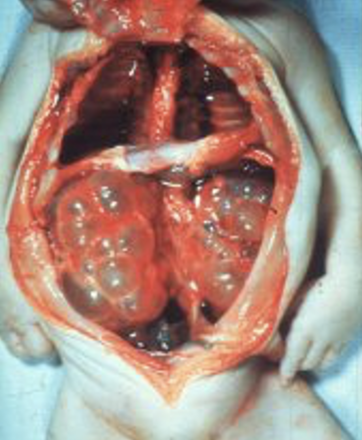
What do these findings indicate?
Childhood (juvenile) polycystic kidney disease
89
New cards
What is wrong with this picture?
Childhood polycystic kidney disease \n *aka*-autosomal recessive polycystic kidney disease (ARPKD)
90
New cards
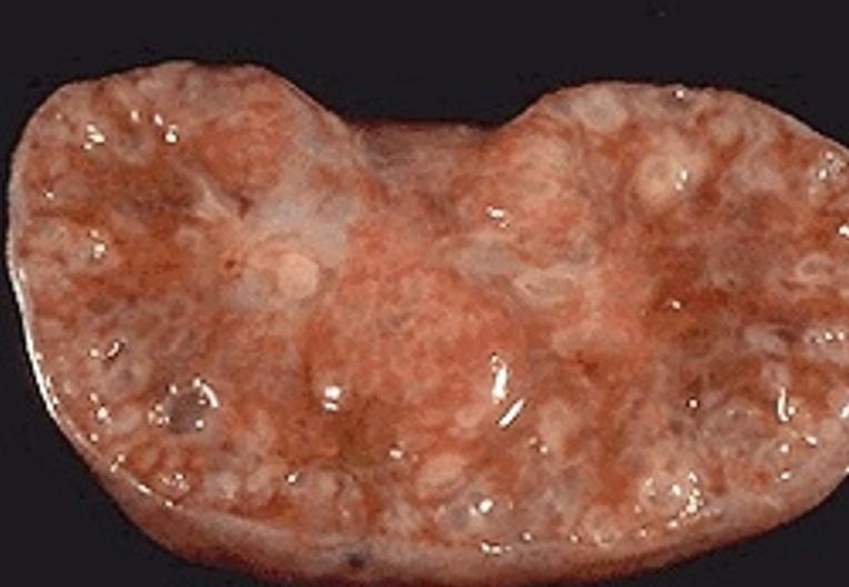
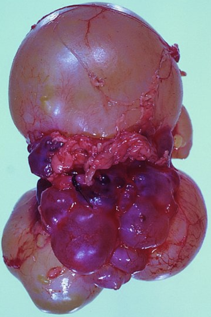
What caused this child’s kidney to look like this?
Autosomal recessive polycystic kidney disease (ARPKD)
91
New cards
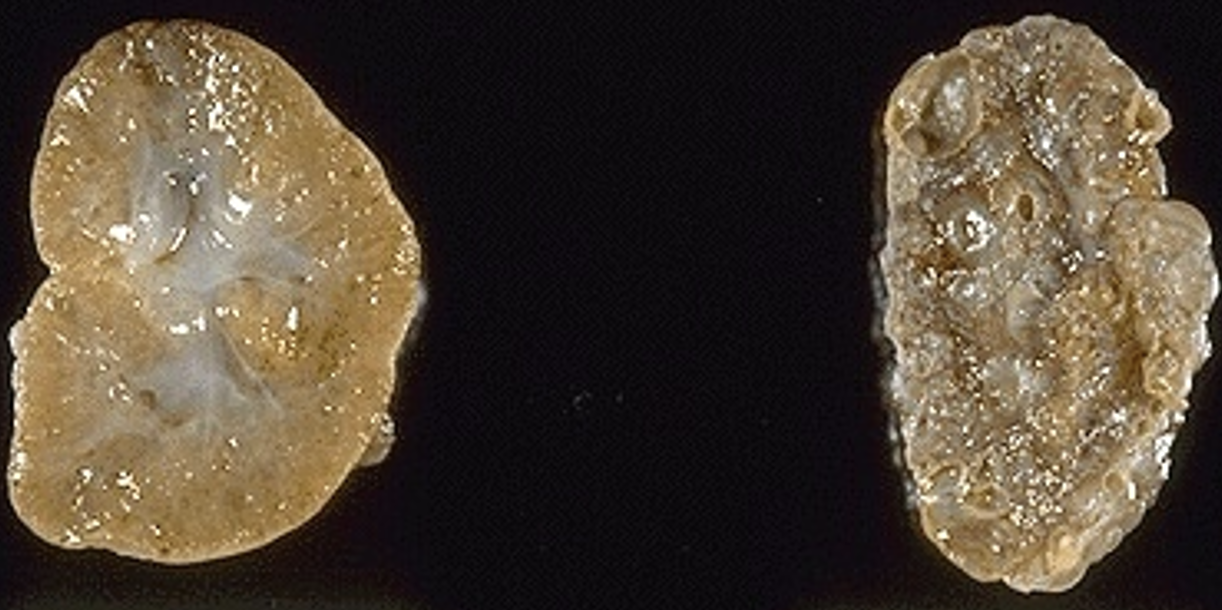
What is indicated by the left kidney? right kidney?
L: ARPKD
R: Multicystic renal dysplasia
R: Multicystic renal dysplasia
92
New cards
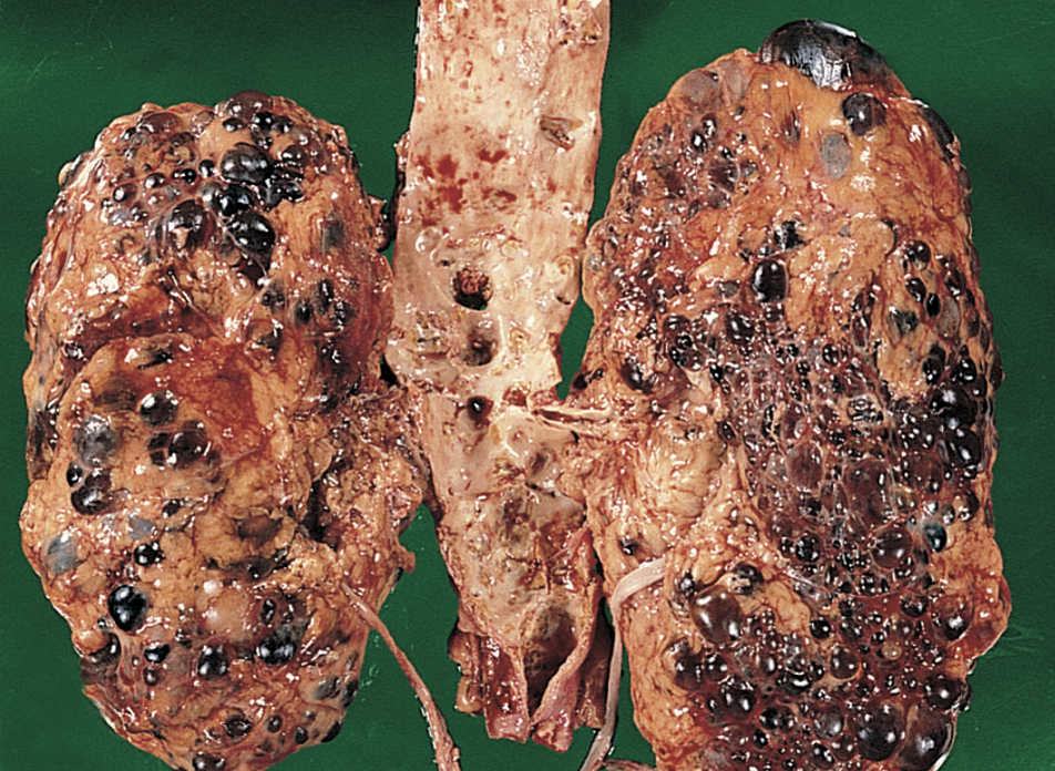
What pathology is shown?
Adult polycystic kidney disease
93
New cards
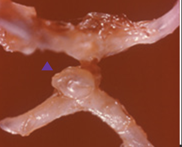
What is shown at the purple arrow? What is the underlying cause?
berry aneurysm from adult PKD
94
New cards
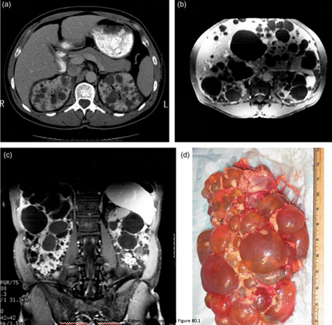
What is shown from these images?
Adult polycystic kidney disease (ADPKD)
95
New cards
What pathology is shown?
Medullary sponge kidney
96
New cards
What pathology is shown?
Medullary cystic kidney disease complex-nephronophthisis
97
New cards
What pathology do these images show?
Simple cysts
98
New cards
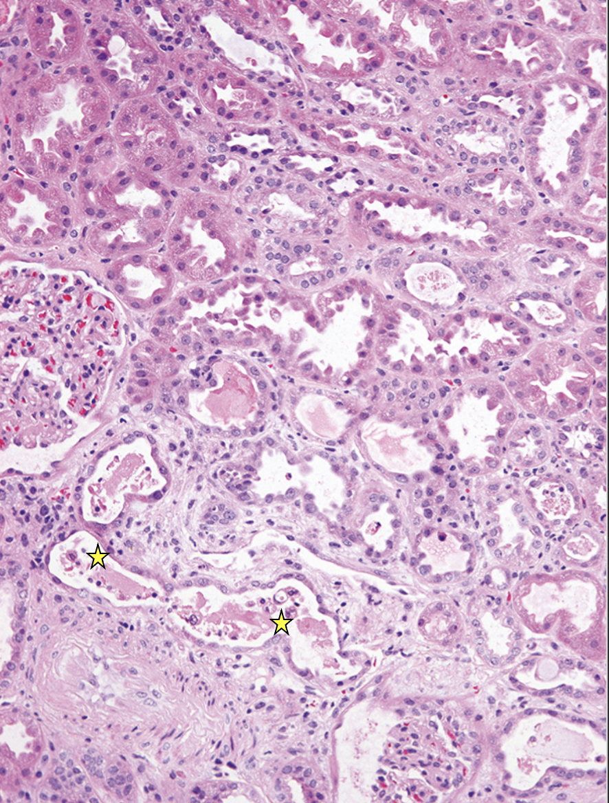
What pathology is indicated? What is shown at the yellow stars?
Acute tubular injury (ATI)
Dead cells have sloughed into lumen
99
New cards
What kind of cast is this? What pathology is it associated with?
tubular cast - acute tubular injury
100
New cards
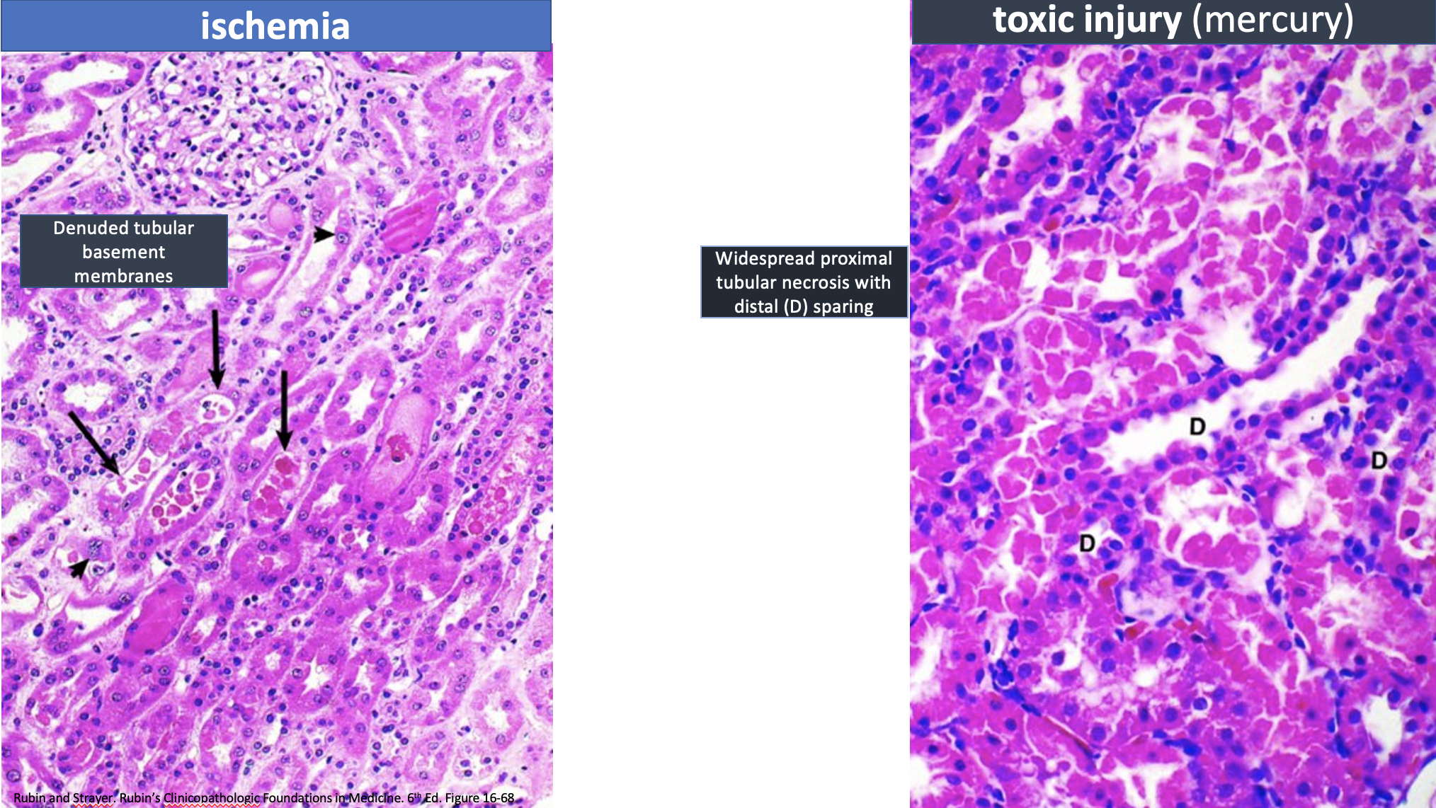
What do these 2 etiologies cause?
acute tubular injury