Osteomyelitis is degenerative disease of bone in close area of joint True False
False
Backscattered radiation represent only 5% of the total impact dose during patient fixation True False
False
On 30 day pregnancy we can count the skulls on the radiograph True False
True
Cats have free-floating clavicle bones True False
True
Myelography is contrast study for identification of problem with muscle tendons True False
False
Pulpitis is inflammation and on the radiograph we read very thin radiopaque zone in the tooth True False
False
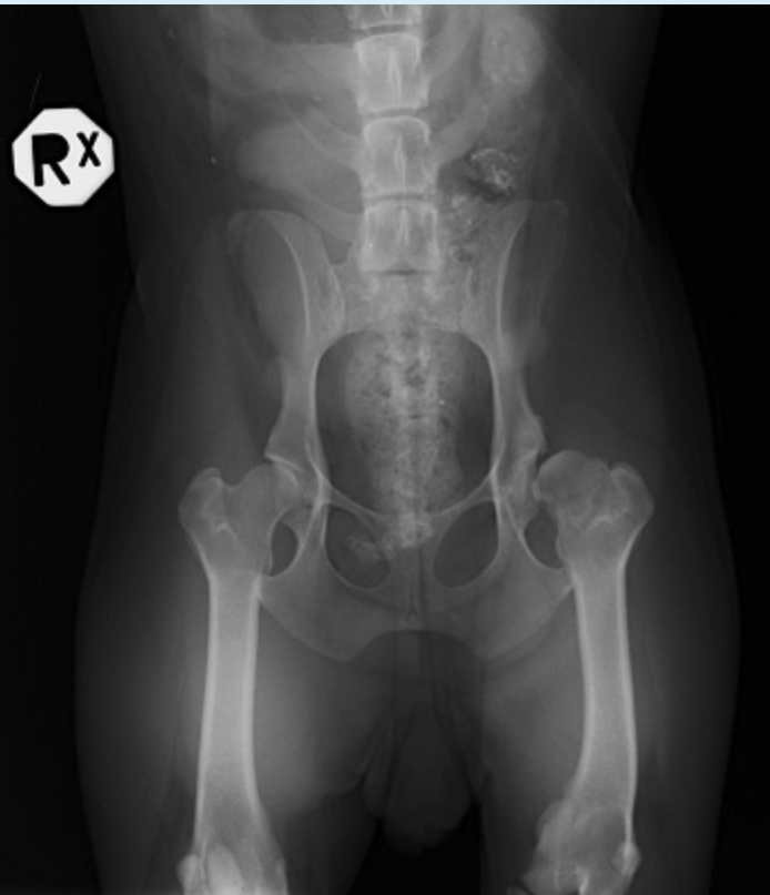
Radiographic report - pathology
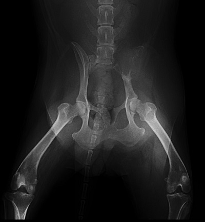
VD projection adult dog pelvis et atriculationes coxarum reduced radiopacity of the left ala ossis illi, possible osteosarcoma
Perthes is a. necrosis of lateral condyle of humerus b. necrosis of head and neck of humerus c. necrosis of lateral condyle of femur d. necrosis of head and neck of femure. necrosis of medial condyle of humerus
necrosis of head and neck of femur
Anticlinal vertebrae for small breed dog is TH12 True False
False
Legg-Calve-Perthes: is a disease of fast-growing dogs of large breeds: necrosis of the digital epiphysis of the humerus True False
False
Hydronephrosis is: accumulation of liquid in the brain True False
False
Brain is most sensitive to radiation? True False
False
CT computed tomography works ionising radiation from protons True False
False (electrons)
Scattering radiation represents a. 30% of the total dose of radiation on the person during fixation of the animal b. 70% of the total dose of radiation on the person during fixation of the animal c. 5% of the total dose of radiation on the person during fixation of the animal d. 50% of the total dose of radiation on the person during fixation of the animal
30% of the total dose of radiation on the person during fixation of the animal
Periosteum is: a. all answers are correct b. surrounding bone except at articular surface c. fibroblastic connective tissue d. inner layer produces bone by intramembranous ossification
All answers are correct
Wobbler's syndrome is: a. cervical spondylitis b. sacral spondylitis c. thoracal spondylitis d. lumbal spondylitis
cervical spondylitis
Shape of ulna growing plate: a. linear b. wavy c. V shape
V shape
2 types of osteophytes
medusa, morgan line
Name angles of elbow dysplasia
Mediolaterally: Neutral – 110° (90°-120°)Mediolaterally: Flexed – 45°Craniocaudally: Extended elbow - 15°
Types of secondary grids
Bucky – in the table Lysholm – movable
Why are secondary grids used?
prevents oblique wavelengths from reaching the film and only allows straight wavelengths through for improved image quality
Where is the end of the spinal canal in dogs?
vertebrae lumbales VI-VII
What is hemivertebra?
failure of the body of a vertebra to develop fully resulting in an abnormally wedge-shaped vertebra
C Grade of hip dysplasia is evaluated by 3 conditions
norberg angle between 100-105 degrees mild join incongruity mild signs of osteoarthritis centre of caput femoris is on the border or immediately lateral to dorsal acetabular rim
What do we examine in hip dysplasia?
luxation, secondary osteochondritis, incongruity, asymmetry of pelvis and femur
Between which vertebrae are there no intervertebral discs?
C1-C2 (atlas and axis)
Until what vertebra is Wobbler's syndome?
C7
What is fat pad syndrome?
increase in synovial fluid in articulatio genus causing depression of the fat pad
Where is fluid inserted in myelography?
into subarachnoid space into the cisterna magna or between L5-L6
4 x-ray characteristics
no charge no mass invisible cannot be felt penetrate all matter to some degree ionising
Vertebral spaces/discs appear as lucent or opaque?
lucent (black)
Write down the 5 materials from opaque to lucent
metal (bright white) bone (light grey) soft tissue/fluid (mid-grey) fat (dark grey) air (black)
Write at least 4 types of periosteal reactions
lamellar (onion skin) smooth sunburst Codman's triangle
3 functions of secondary grids
reduce scattered radiation improve image contrast stop oblique wavelengths from entering the film
What are two negative contrast media?
air, oxygen, carbon dioxide, water
Hypertrophic osteodystrophy is: a. inflammation of metaphysis b. inflammation of epiphysis c. necrosis of epiphysis d. necrosis of metaphysis
necrosis of metaphysis
Foramen magnum is oval shape in frontal bone True False
False
Atlanto-axial instability--we read when is extrusion of intravertebral disc True False
False
Elbow dysplasia grade 2 is: we see on the radiograph a. osteophytes 3-5 mm, fragmented coronoid process, osteochondritis dissecans b. osteochondritis dissecans, fragmented coronoid process, ununited anconeal process c. osteophytes 3-5 mm d. osteophytes more than 3 mm, fragmented coronoid process
osteophytes 3-5 mm
The brain is more resistant to x-rays than the colon True False
True
Protective dress is with a minimal lead equivalent a. 0.35 cm b. 3 cm c. 0.35 mm d. 3.5 cm e. 3.5 mm
0.35 mm
Fat pad syndrome: we read in arthritis of the joint True False
True
Elbow dysplasia 3 grade is a. in the radiograph to see osterochondritis dissecans b. in the radiograph to see osteophytes in size 3 mm c. in the radiograph to see 3 mm joint space
in the radiograph to see osterochondritis dissecans
Periodontal ligament on the radiograph is radiopaque as soft tissue True False
False
Hip dysplasia B a. Norberg angle is 100 with slight osteoarthrotic change b. Norberg angle is 115 with slight osteoarthrotic change c. Norberg angle is 100 and slight incongruity d. Norberg angle is 105 and slight incongruity
Norberg angle is 105 and slight incongruity
Subchondral bone is a. thick radiolucent layer in diaphysis b. thin radiopacity layer in bone diaphysis c. thin radiopacity layer of bone beneath articular cartilage
thin radiopacity layer of bone beneath articular cartilage
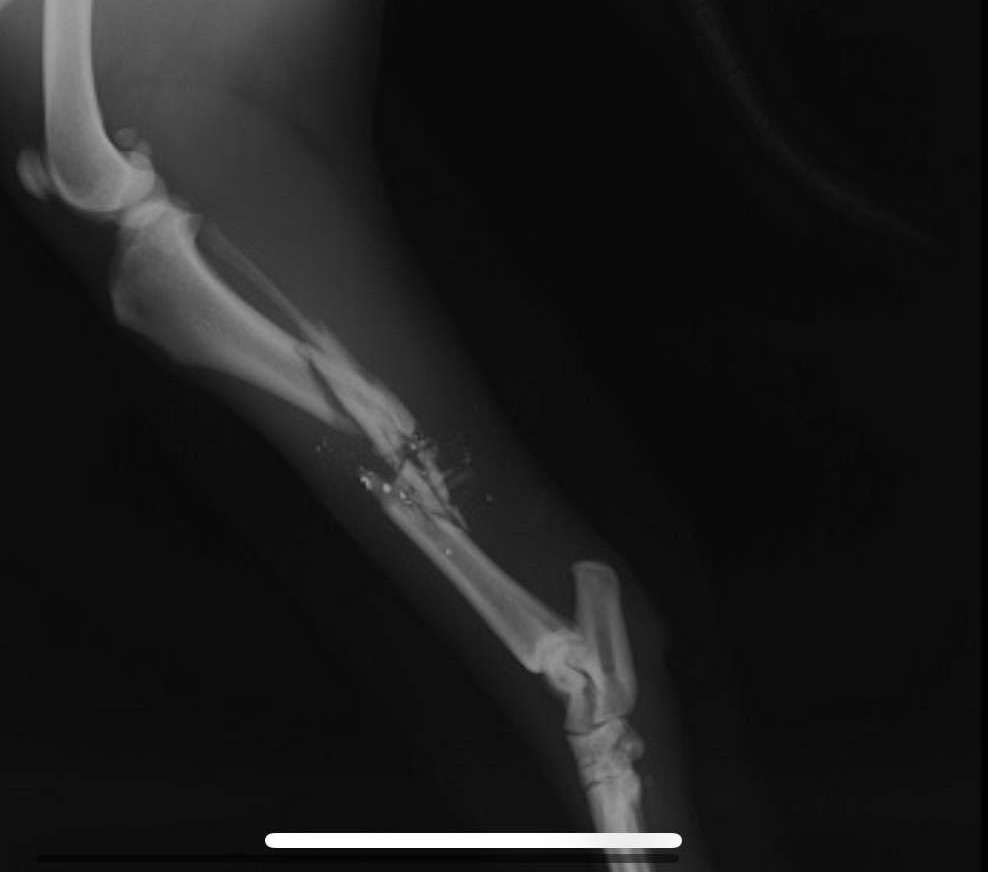
Radiographic report
ML projection juvenile cat articulatio cubiti luxation of the ulna and radius, fracture of the distal area of the humerus (epicondylus medialis, epicondylus lateralis)
Radiographic report - pathology
ML projection adult cat articulatio humeri and articulatio cubiti closed, simple, complete fracture of the humerus
Latin name of joint and full Latin name of bone - red mark
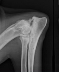
articulatio humeri, tuberculum supraglenoidale
Radiograph is dark -- write me 2 causes why radiograph more dark
presence of air, overexposure (long time, high kV, high mA)
Brain is a very sensitive organ on x-ray radiation vs. bone marrow True False
False
Most massive cat's tooth in maxilla is premolar 1 True False
False (maxilla: P3, mandibula: M1)
SLOB technique is for separation teeth roots in: a. premolaris IV in maxilla b. caninus in mandibula c. premolaris I in maxilla
premolaris IV in maxilla
We need the Standard position II for the screening examination of hip joint for breeding True False
False
Secondary radiation is radiation generated in the focus anode in the x-ray tube True False
False
Write 3 parameters for hip dysplasia evaluation (no position but evaluation)
norberg angle size of joint space shape of cranial margin of acetabulum shape of caput femoris percentage of caput femoris covered by acetabulum
Locality of cisterna magna is between C1-C2 True False
False
Contrast material for evaluation of bulla tympany is Barium sulphate True False
False
a. Latero-lateral position, Articulatio coxae, aggressive lesion in proximal epiphysis on Tibia b. Articulatio Cubiti, aggressive lesion on proximal epiphysis on Radius c. Articulatio Genus, Fractura medial Malleolus Tibiae d. Latero-lateral position, Articulatio Genus, aggressive lesion on Tibia e. Latero-lateral position, Articulatio Cubiti, Dysplasia, Grade 3
Latero-lateral position, Articulatio Genus, aggressive lesion on Tibia
Latero-lateral position, dog: Fractura vertebrae lumbales 6 True False
False
Growth plate closure time: Anconeal process a. 12 months of age b. 6 months of age c. 1 month of age d. 10 months of age e. 24 months of age
6 months of age
Infrapatellar fat pad syndrome on the radiograph is readable in rupture of intra-articular ligaments True False
True
Osteomyelitis is inflammation for all bone structure True False
True
Cortex is: a. compact, lamellar bone, uniformly b. bone uniformly radiopacity thick structure in area epiphysis c. uniformly radiopacity thick cancellous bone d. formed by intramembranous ossification from cartilage on physis
Compact, lamellar bone, uniformly
On the radiograph--Nutrient foramen for blood vessel through the cortex is read as radiolucent line True False
True
Is it pathology Hansen IV? True False
False
Salter-Harris classification is classification for diseases in intervertebral disc True False
False
Oblique fracture is: a fracture when the perpendicular to the longitudinal axis of the bone is less than 30 degrees True False
False
Radiographic anatomy Latin name bone B and A
B: os tibiae, A: calcaneus
Olecranon ulnae is apophysis True False
True
Spondylosis deformans is a. malignant tumour on body of vertebrae and sternebrae b. degradation on vertebrae body and new bone formation c. inflammation on vertebrae body and new bone formation
degradation on vertebrae body and new bone formation
Panosteitis is a. non bacterial inflammation of bone in the marrow and endosteum b. bacterial inflammation of bone in the marrow and endosteum c. bacterial inflammation of bone in the cortex and periosteum d. degenerative disease of the marrow and bone cortex e. non bacterial inflammation of bone in the cortex and periosteum
non bacterial inflammation of bone in the marrow and endosteum
Myelography is contrast study on the spinal canal with negative contrast medium with application between Atlas and Axis True False
False
Fronto-occipital position is position for foramen magnum evaluation True False
True
Legg Calve Perthes is a. necrosis of medial condyle of humerus b. necrosis of head and neck of humerus c. necrosis of lateral condyle of humerus d. necrosis of head and neck of femur e. necrosis of lateral condyle of femur
necrosis of head and neck of femur
Myelography is a. Contrast study for spine cord and spinal canal with iodine and application is between os occipital and atlas b. Contrast study for spine cord and spinal canal with negative contrast medium and application is between os occipital and atlas c. Contrast study for muscle structure with iodine d. Contrast study for spine cord and spinal canal with iodine and application is between atlas and axis e. Contrast study for spine cord and spinal canal with barium sulphate and application is between os occipitale and atlas
Contrast study for spine cord and spinal canal with iodine and application is between os occipital and atlas
Radiographic report - pathology
VD projection Adult female dog Pelvis et articulationes coxarum Hip dysplasia E (severe) of left articulatio coxae
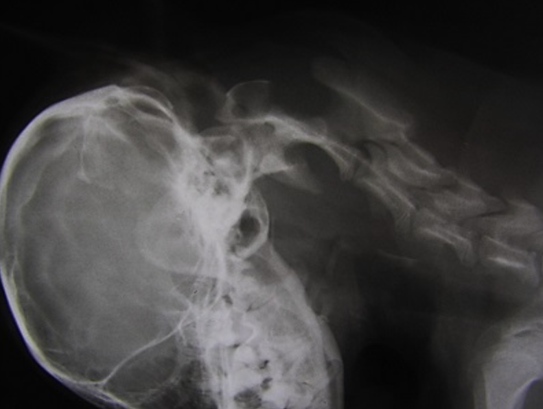
Radiographic report - pathology
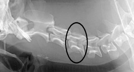
LL projection Adult dog Skull and vertebrae cervicales I to V Pathology: rupture of atlanto-axial ligament causing atlanto-axial instability
Salter-Harris II is a. extrusion of intervertebral disc b. protrusion of intervertebral disc c. fracture through the physis and metaphysis in a 3-old dog d. fracture through the physis and metaphysis in a 7-month-old dog
fracture through the physis and metaphysis in a 7-month-old dog
Codman triangle must be 105 degrees True False
False (Norberg angle)
Radiographic anatomy Latin name of joint (red color) and bone and number - green color
articulatio tarsi os metatarsale V
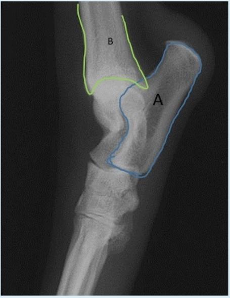
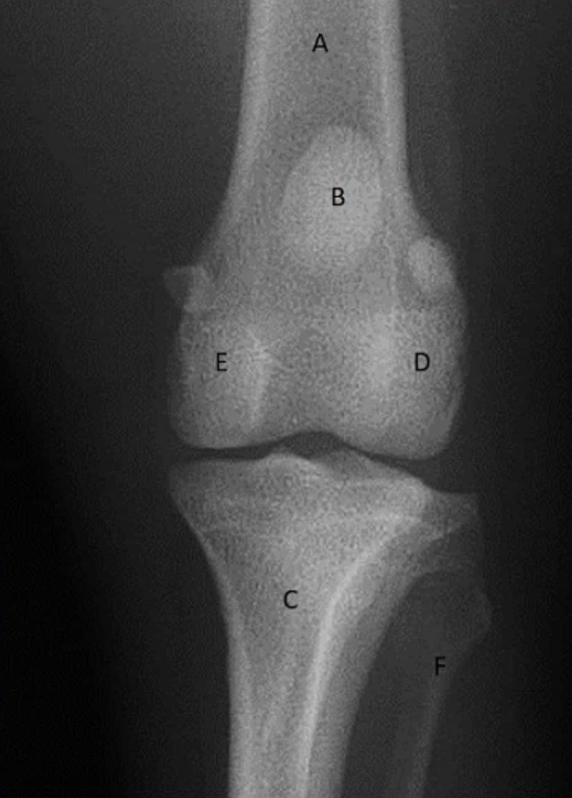
Radiographic anatomy full Latin name
A. os femoris B. patella C. os tibiae D. condylus lateralis E. codylus medialis F. os fibulae
Radiographic report
female dog LL position spondylosis of vertebrae lumbales
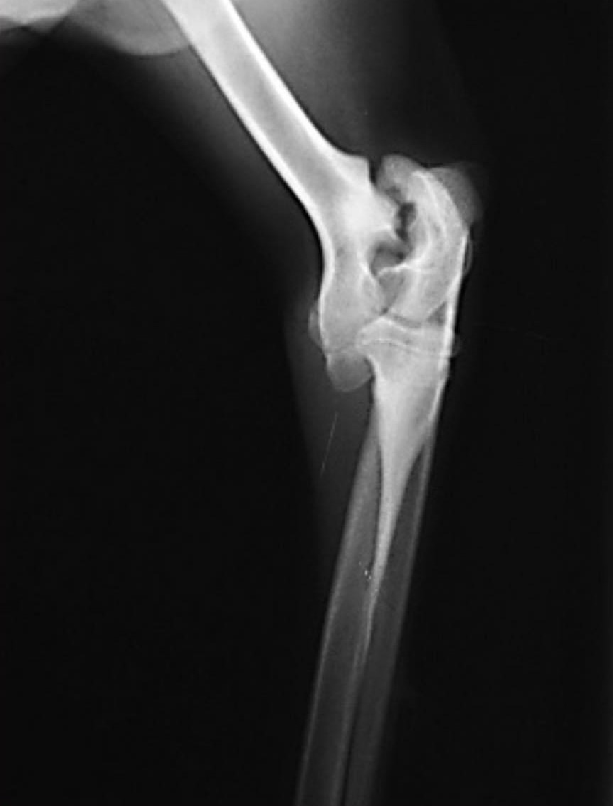
Radiographic report
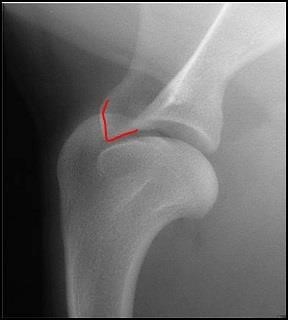
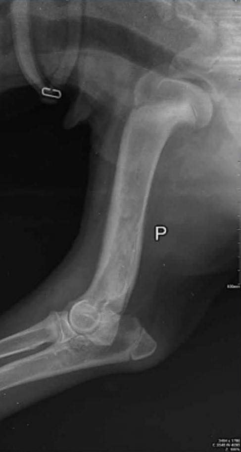
mediolateral view dog young dog (growth plates are still visible) radiogram of os humeri also containing articulatio humeri and articulatio cubiti simple panosteitis of os humeri (medulla is less radiolucent than normal)
Panosteitis is: a. Inflammation diseases with high irregular periosteal reaction and reaction in the cortex structure of bone b. Inflammation diseases with reaction in endost and marrow in bone c. Degenerative diseases with high irregular periosteal reaction and reaction in the cortex structure of bone d. Degenerative diseases with reaction on endost and marrow in bone and reaction in the cortex structure of bone in diaphysis
Inflammation diseases with reaction in endost and marrow in bone
1 year old dog normally has between the sacral vertebrae are radiolucent vertebral disc True False
False
Vertebrae formula for cat is a. C7 TH13 L7 S3 Ca for breed b. C7 TH11 L5 S4 Ca for breed c. C5 TH13 L5 S3 Ca for breed d. C6 TH13 L7 S3 Ca for breed
C7 TH13 L7 S3 Ca for breed
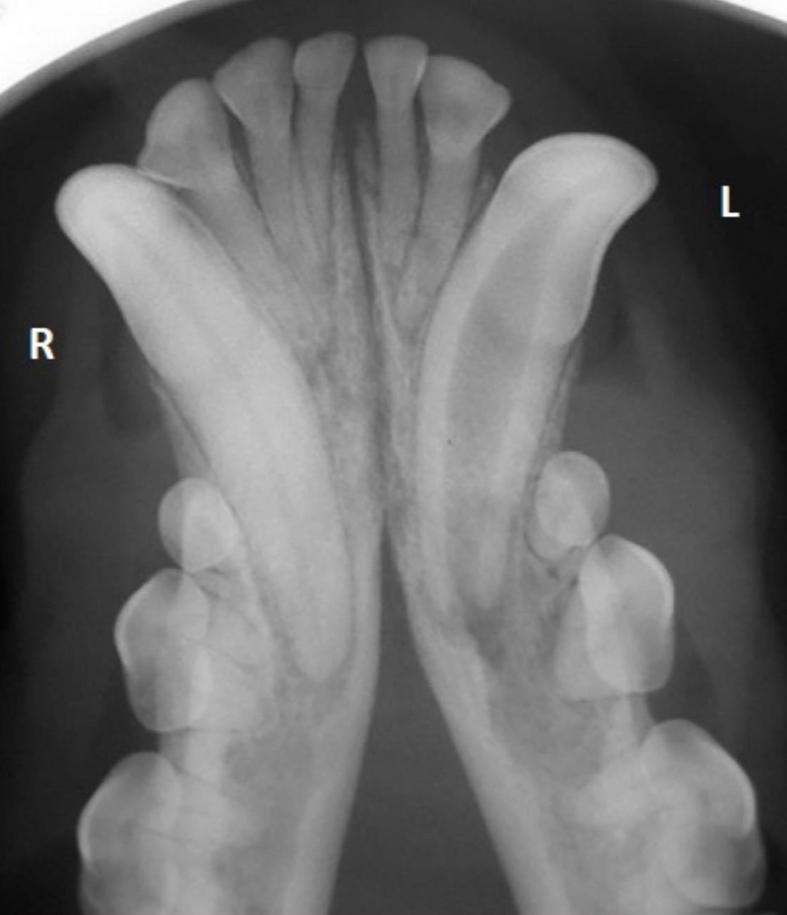
Radiographic Anatomy - full name of hard tissue and number
vertebra cervicalis IV
To length growing ulnae has one growth plate True False
False (distal and proximal growth plates)
Write radiographic report, position, pathology change, and grade of dysplasia
ML projection adult dog articulatio cubiti ununited anconeal process elbow dysplasia grade 3 unclear processus coronoideus medialis, stepping incongruity, osteophytes
Write names for two apophysis
tuberositas tibiae, olecranon ulnae
Write position, which part of body is pathology and name pathology
lateral position mandibula (canalis mandibulae is present) radiolucent periapical abscesses surrounding the apical deltas on premolar 4 radiolucent rudimental gingiva
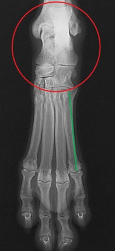
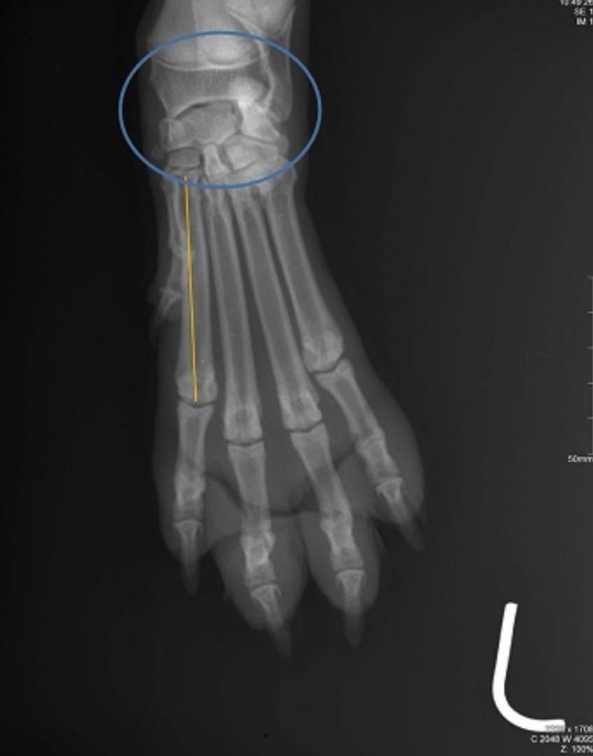
Radiographic anatomy--Latin name joint - blue, and bone with number yellow colour
articulatio carpi os metacarpi II
the radiograph shows a direct fracture of the tibia and fibula True False
False
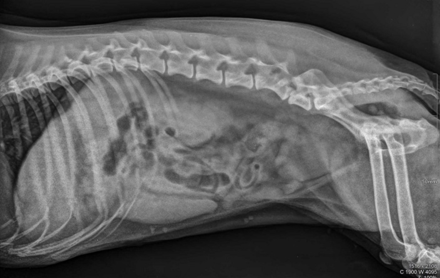
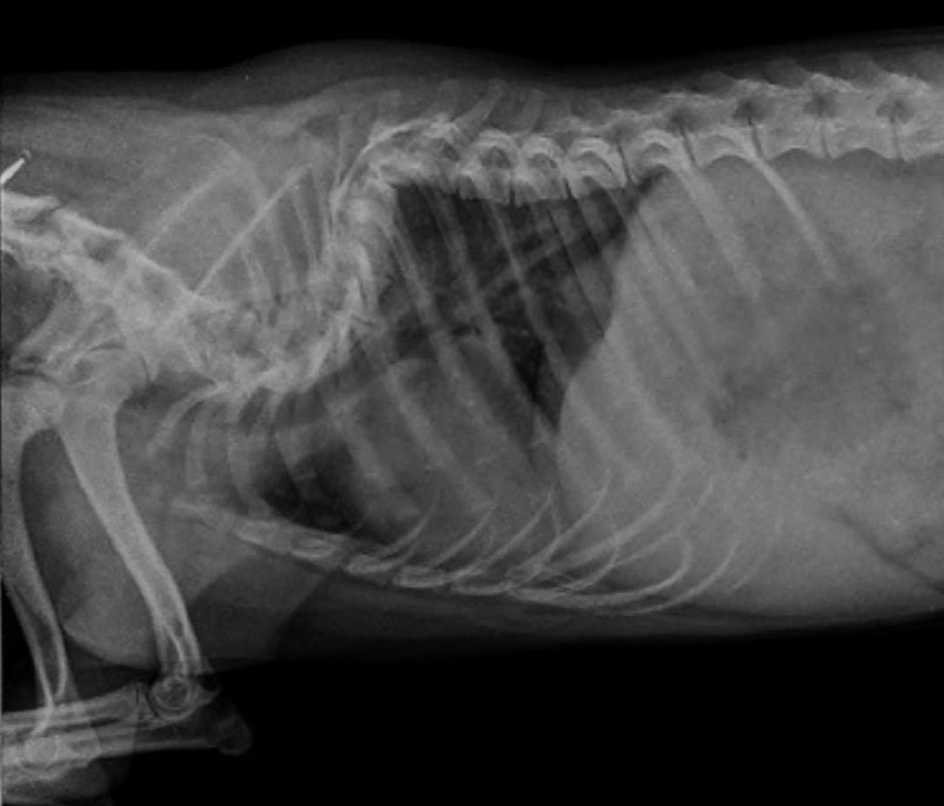
Radiographic report
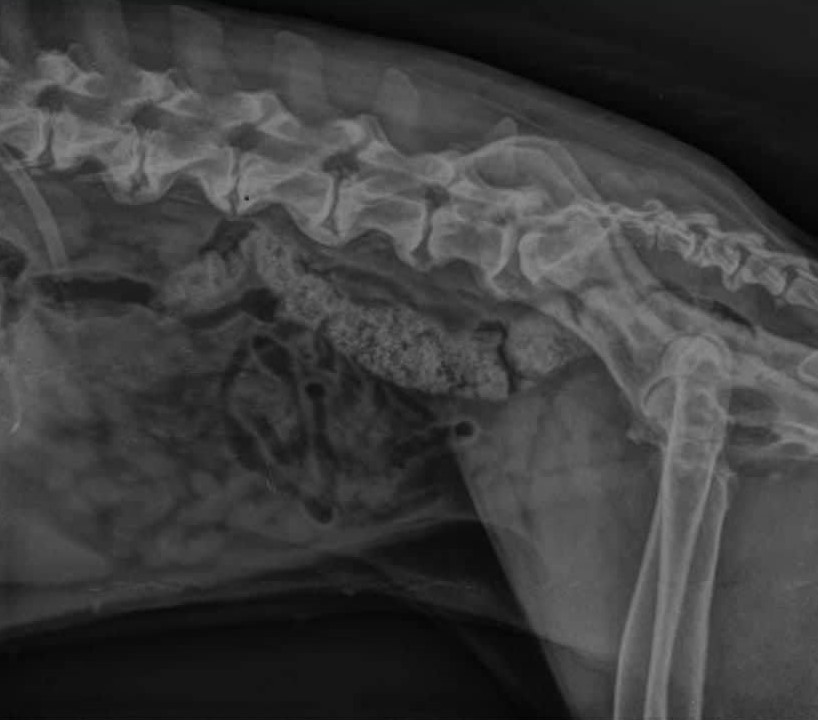
LL projection Adult dog Vertebrae cervicales, vertebrae thoracicae, vertebrae lumbales, articulatio humeri, articulatio Cubito, thorax, cranial part of the abdomen Pathology: hemivertebrae TH8
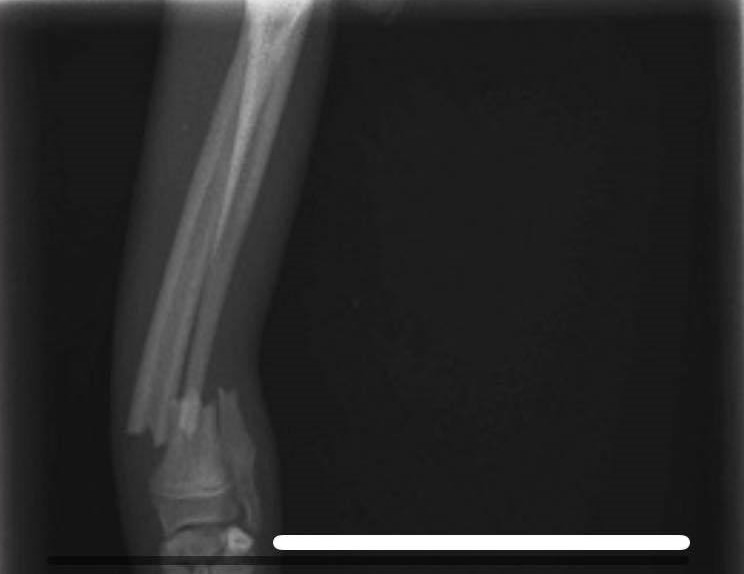
Radiographic report - pathology
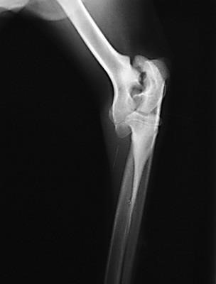
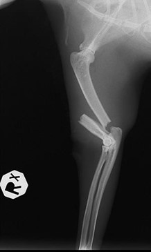
medio-lateral projection Adult dog os tibiae and os fibulae including articulatio genus and articulatio tarsi Complex, complete fractura of os tibiae and os fibulae
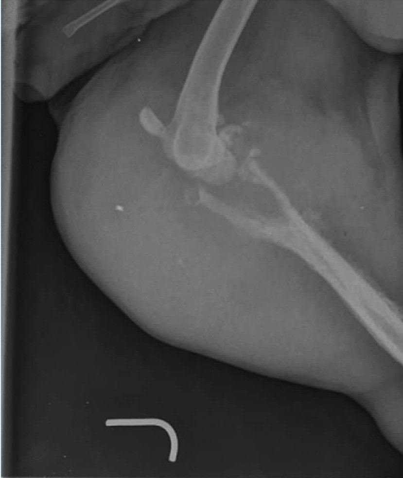
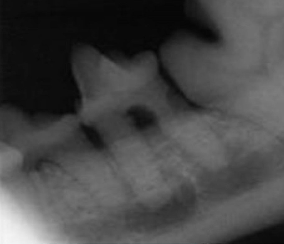
Radiographic report - pathology
dorso-ventral projection adult dog (deciduous teeth are not present, thin radiolucent pulp in most teeth) Mandibula left I3 (303) is missing canine (304) pulpitis and apical abscess
Bisecting Angle Technique is special technique for evaluation of Norberg Angle True False
False
Direct digitalization on x-ray department means a. I need FLAT panel, cassette with film for multiple use, dark room b. I need special cassette with film for one use , reader, and dark room c. I need FLAT panel , reader, computer, dark room d. I need FLAT panel, computer, dark room with developer and fixer e. I need FLAT panel, computer
I need FLAT panel, computer
Osteosarcoma is: a non-aggressive benign tumor of cartilage in the joints True False
False