Looks like no one added any tags here yet for you.
Describe the retinofungal projection.
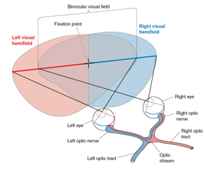
Describe the projection of an object in the left hemi-visual field in a of both eyes and through the retinofugal pathway.
Left hemifield projects to the right side of brain
Ganglion cell axons from nasal retina cross in optic chiasm (temporal axons do not)
Fixation point in center divides field
Binocular vision in center
Describe the pathway from the optic tract to the cortex.
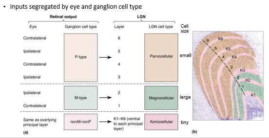
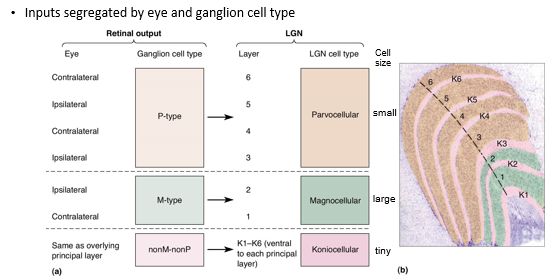
The optic tract is surrounded by LGN layers, labeled ventral (1) to dorsal (6)
Inputs to the LGN are segregated by eye and cell type
P-type RGCs → Parvocellular LGN cells (layers III to VI)
M-type RGCs → Magnocellular LGN cells (layers I and II)
nonM-nonP RGCs → Koniocellular LGN cells (in between LGN cell layer)
What visual deficit results when you cut the right optic nerve?
Loss of right peripheral vision
What visual deficit results when you cut the right optic tract?
Total loss of right vision
What visual deficit results when you cut the optic chiasm?
Total loss of peripheral vision
What are the non-thalamic projections of the optic tract and what are their functions?
Hypothalamus → role in biological rhythms, including sleep and wakefulness
Midbrain/pretectum → control the size of the pupil and certain types of eye movement
Superior colliculus → orientates the eyes in response to new stimuli
Explain the organization of the LGN and how it mirrors the retina.
Magnocellular LGN cells → large center surround receptive fields with transient response
Parvocellular LGN cells → small center-surround receptive fields with sustained response
What is the main synaptic input to the LGN?
RGCs
What is the proposed function of RGCs?
Name 4 names for the visual cortex.
Primary visual cortex
Brodmann’s area 17
Striate cortex
V1 (visual area 1)
What is retinotopy?
What is the role of radial connections?
What is the role of horizontal connections?
Describe Hubel and WIesel’s experiment using radioactivity.
Describe ocular dominance columns.
What are cytochrome oxidase blobs?
In the striate cortex, describe 3 types of monocular receptive fields.
Describe how the striate cortex contributes to binocular vision.
Describe orientation and direction selectivity.
What are complex cell receptive fields?
Describe blob receptive fields.
Describe the 3 parallel pathways to the extrastriate cortex and their functions.
Why is a cortical module necessary and sufficient to analyze a point in space?
What is the role of the dorsal and ventral streams?
Describe the location and role of Area MT.
Describe the location and role of Area MST.
Describe the location and role of Area V4.
Describe the location and role of Area IT.
Describe the location and role of fusiform face area.
What is the deficit in prosopagnosia?
Where is the lesion in prosopagnosia?
What is the deficit in achromatopia?
Where is the lesion in achromatopia?