human phys 14.2
1/46
There's no tags or description
Looks like no tags are added yet.
Name | Mastery | Learn | Test | Matching | Spaced | Call with Kai |
|---|
No analytics yet
Send a link to your students to track their progress
47 Terms
intercalated discs
specialized connections between myocardial cells containing gap junctions and desmosomes
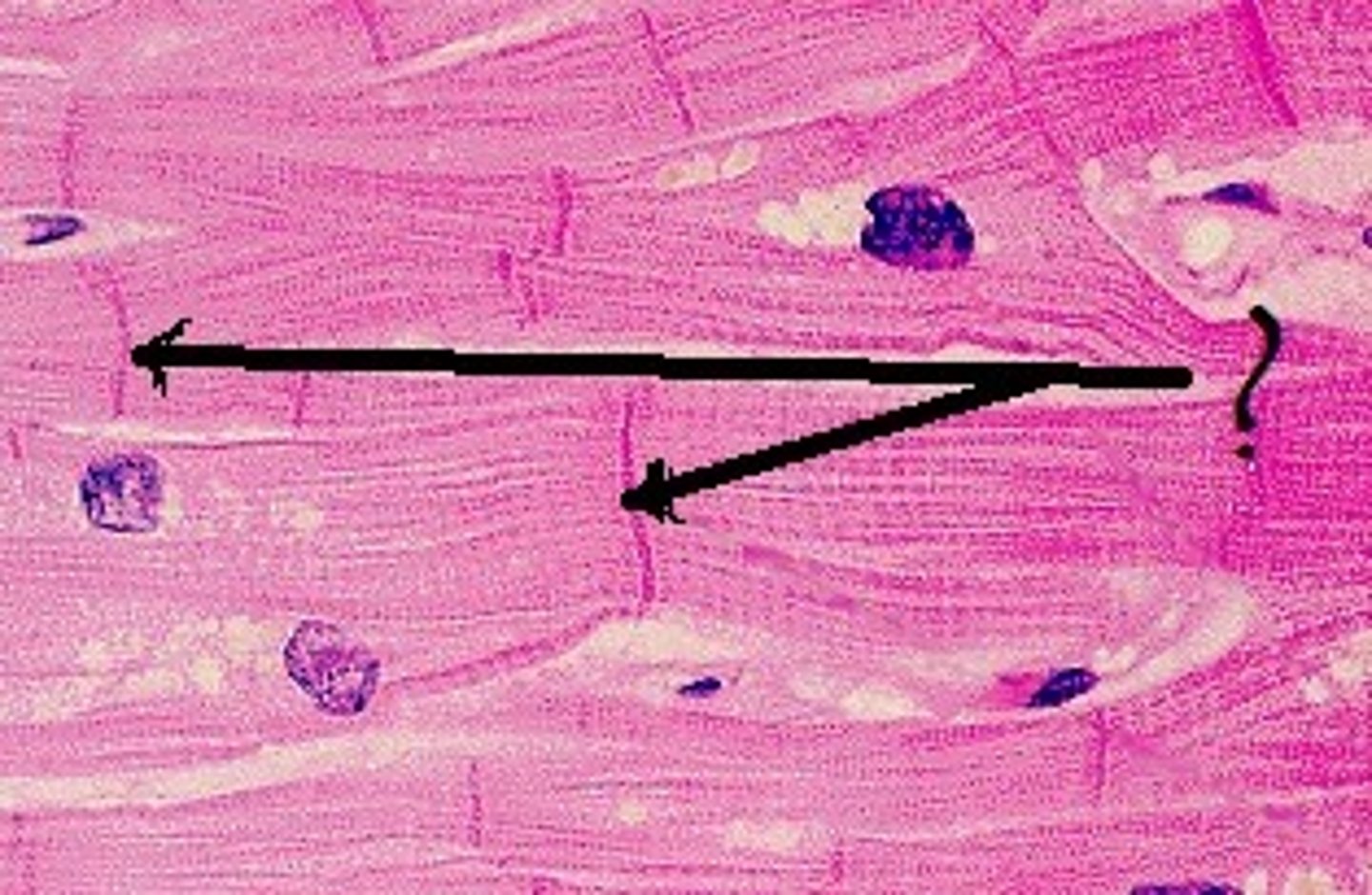
act as one, all cells depolarize and contract at same time, connected via intercalated discs
properties of cardiac muscles
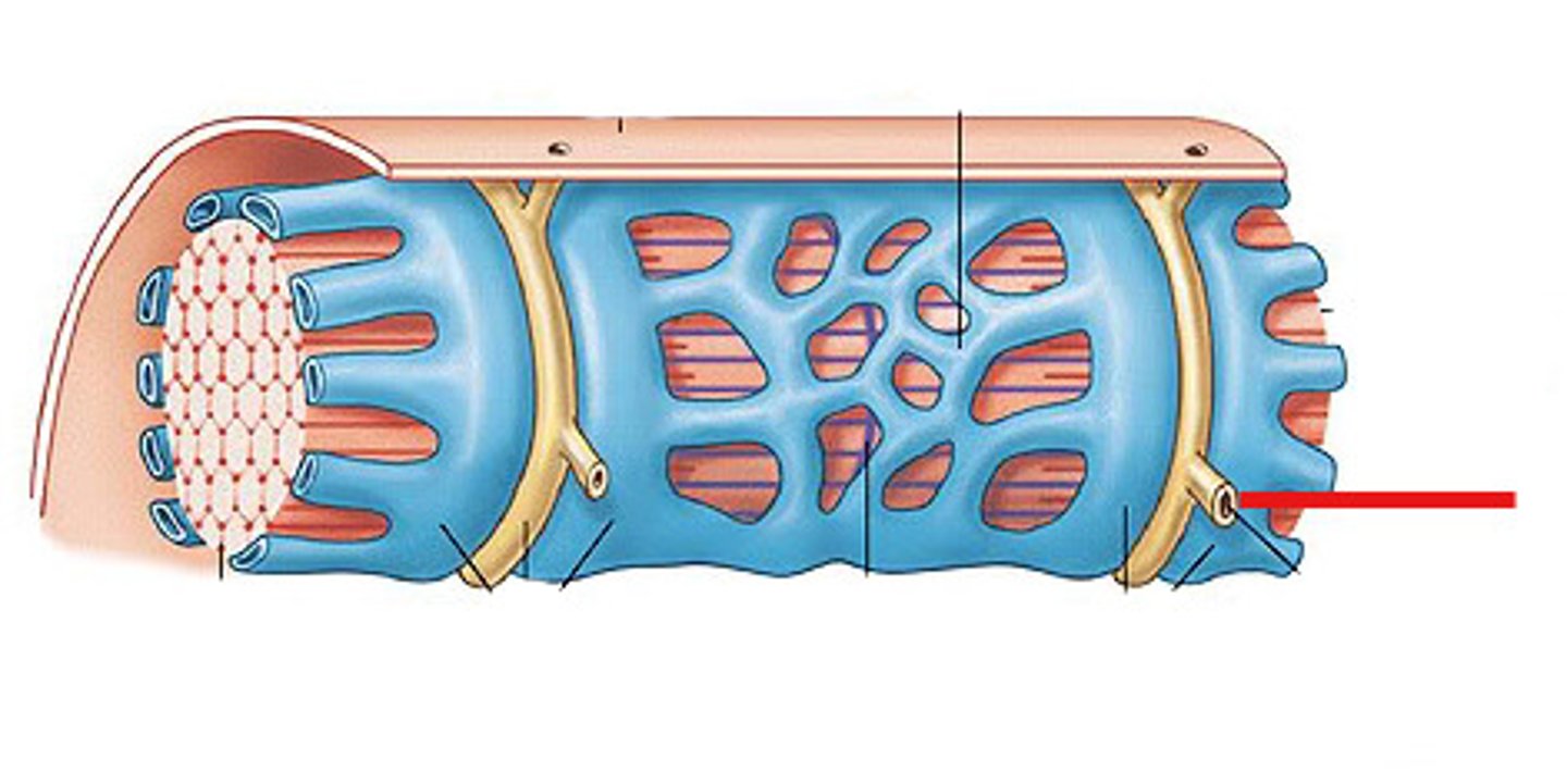
transverse tubules
System of tubules that provides channels for ion flow throughout the muscle fibers to facilitate the propagation of an action potential.
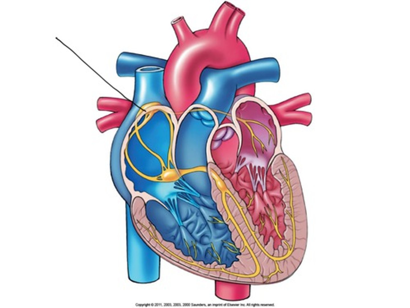
sinoatrial node
pacemaker of the heart
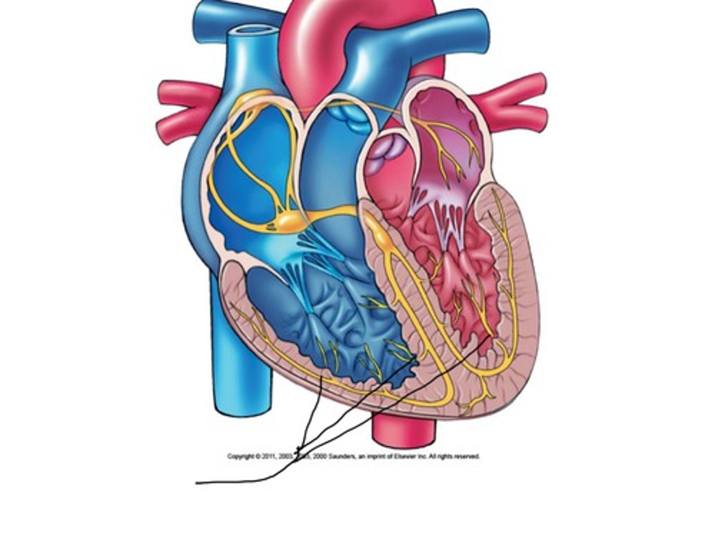
purkinje fibers
fibers in the ventricles that transmit impulses to the right and left ventricles, causing them to contract
SA node, AV node, AV bundle, right and left bundle branches, purkinje fibers
structures that contain autorhymic fibers
ensures chambers contract in a coordinated way
conduciton system
act as pacemaker, form conduction system
functions of autorhytmic fibers
SA node cells spontaneously depolarize and generates AP, moves to AV node, AV bundle, right and left bundles, purnkinje fibers
steps in AP conduction system
first half is K+ channels close and F type Na+ opens, second is T type Ca2+ opens
pacemaker potential: pacemaker phase (YELLOW)
L type ca2+ channels open
pacemaker : depolarizing phase (GREEN)
L type Ca2+ close, K+ channels open
pacemaker : repolarizing phase
produce force, don't depolarize on its own, AP produced only in response to signals from nodal cells
contractile cells
fast Na+ channels open
contractile AP: depolarizing phase
Na+ closes, K+ opens
contractile AP: intial repolarizing phase
L type Ca2+ opens, fast K+ close, slow K+ partially opens
contractile AP: plateau phase
L type Ca2+ closes, slow K+ fully open
contractile AP: final repolarizing phase
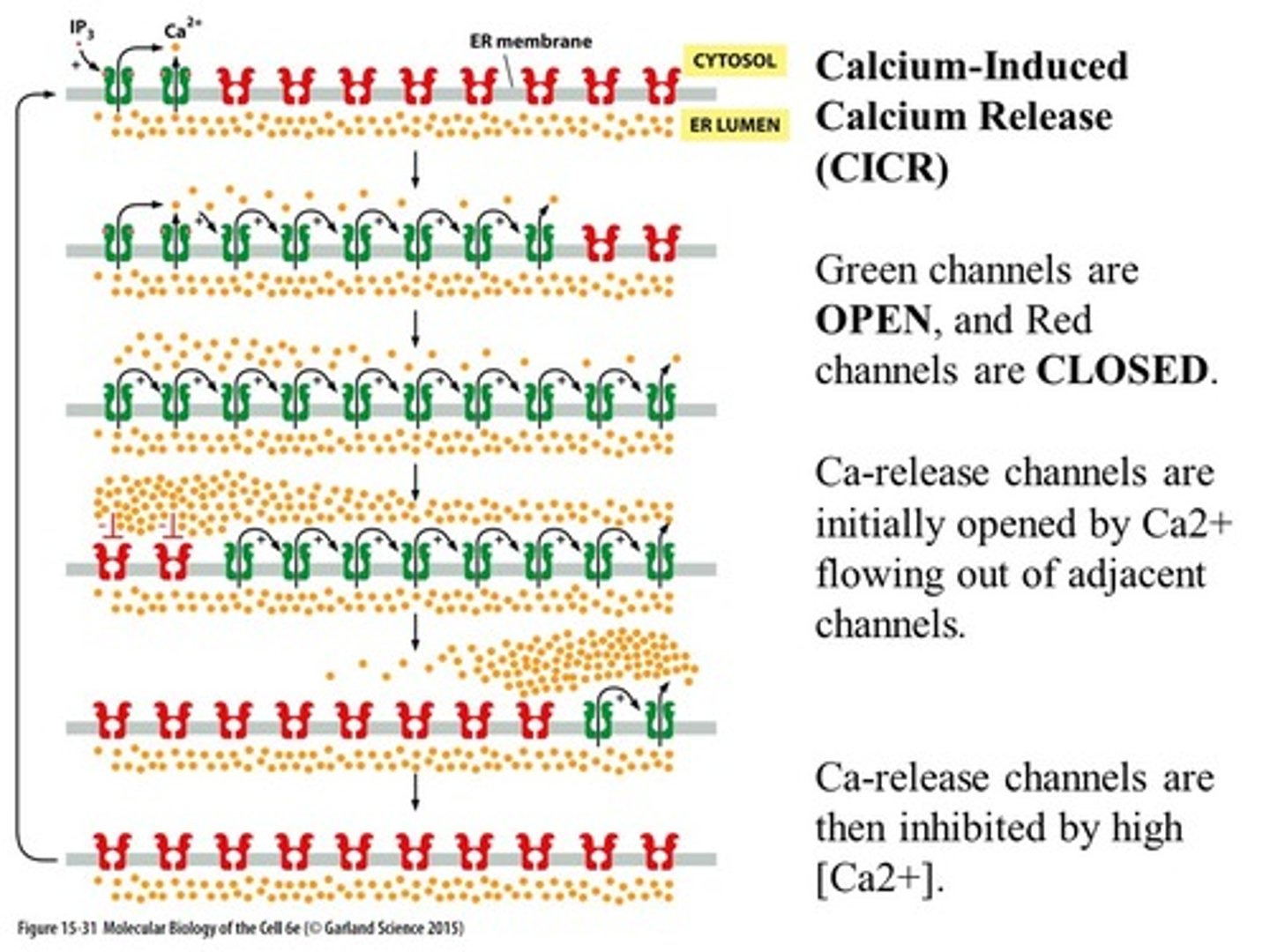
Ca2+ induced Ca2+ release
influx of Ca2+ causes release of more Ca2+ from SR through ryanodine receptors
long, almost as long as duration of contraction
refractory period of cardiac muscle
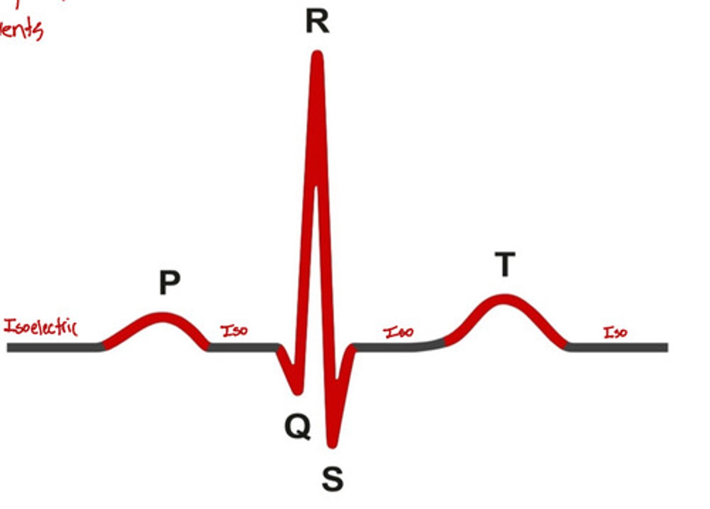
electrocardiogram
A recording of the electrical activity of the heart
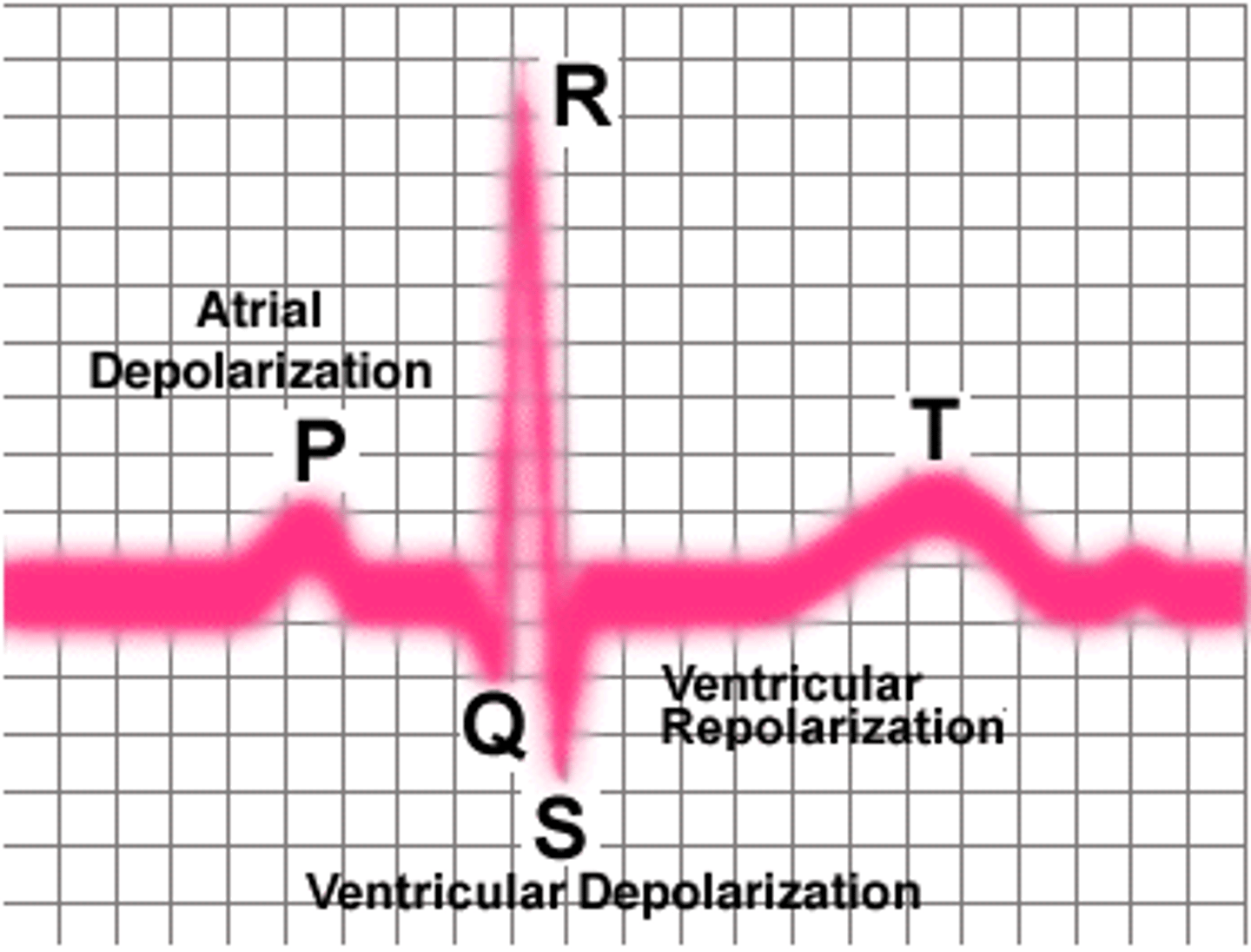
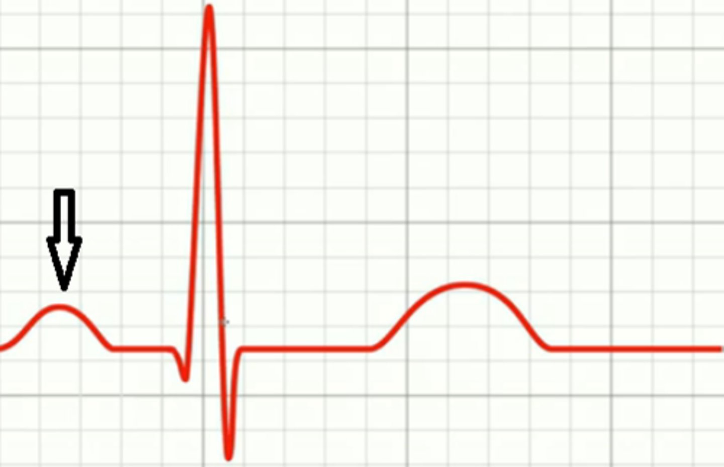
atrial depolarization, spreads from SA node to contractile fibers
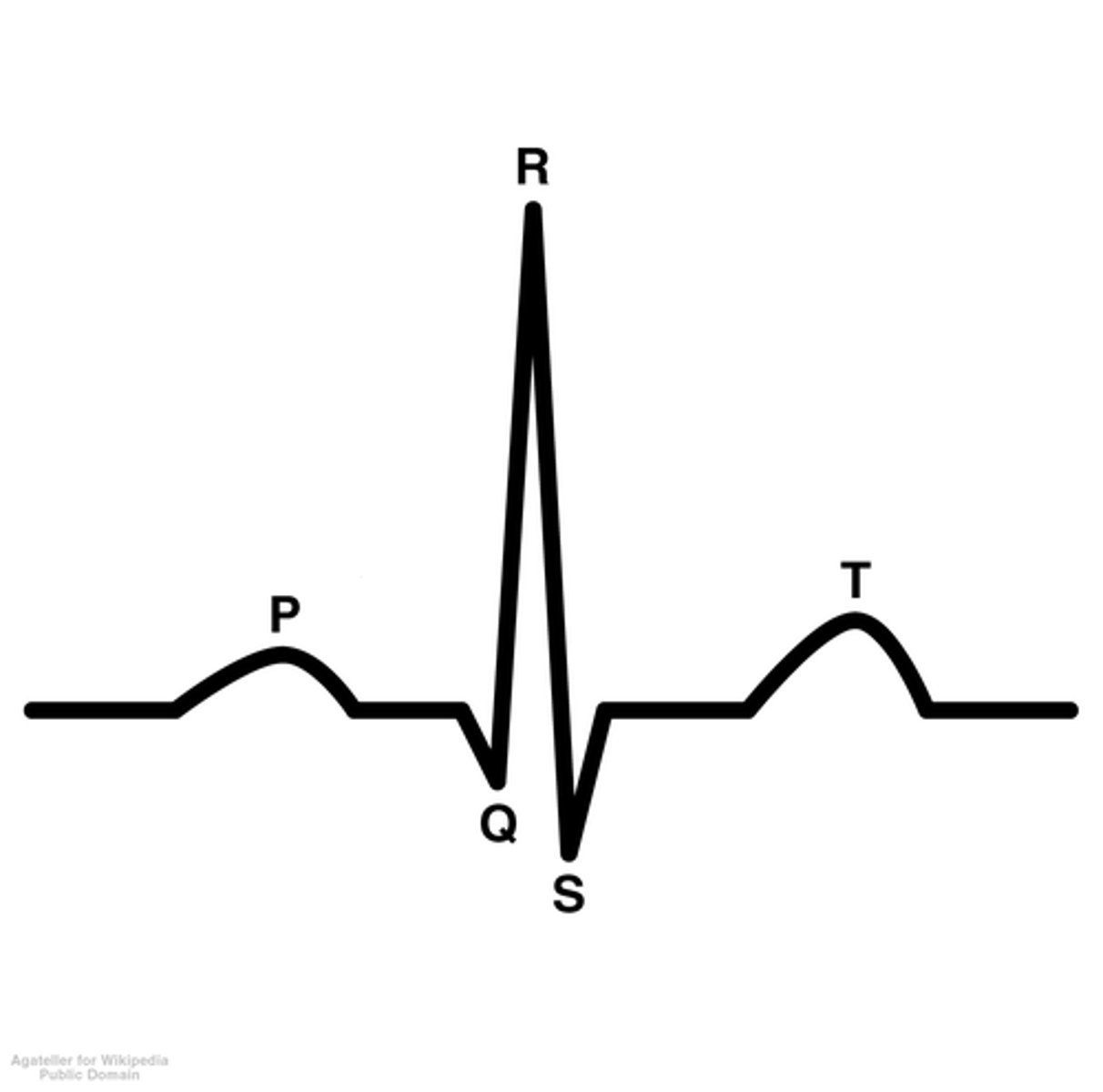
P wave
ventricular depolarization, AP spreads through ventricular contractile fibers
QRS complex
ventricular repolarization, smaller and wider than QRS complex
T wave
time from beginning of P wave to beginning of QRS complex
P-R interval

time when ventricular contractile fibers are depolarized during plateau phase of AP
S-T segment
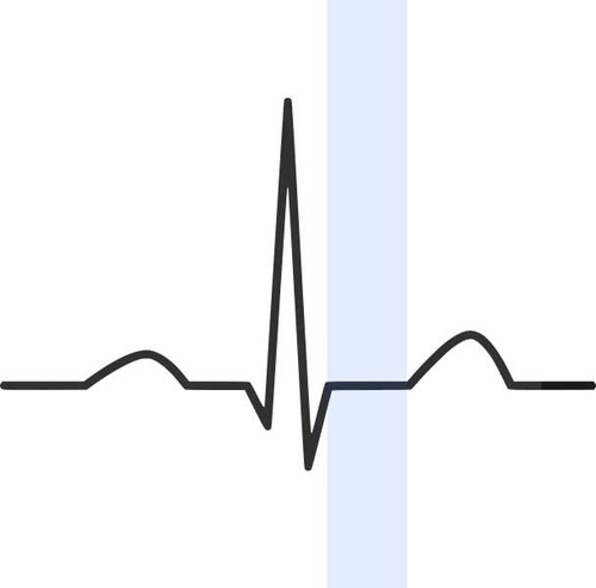
time from beginning of QRS to end of T wave
Q-T interval

P wave, P-R interval, QRS complex, S-T segment, T wave, Q-T interval
components of ECG/EKG

systole
Contraction of the heart
diastole
Relaxation of the heart
depolarization of atrial contractile fibers produces P wave, atrial systole contraction
step 1 of ECG waves
depolarization of ventricular contractile fibers produces QRS complex, ventricular systole contraction
step 2 of ECG waves
repolarization of ventricular contractile fibers produces T waves, ventricular diastole relaxation
step 3 of ECG waves
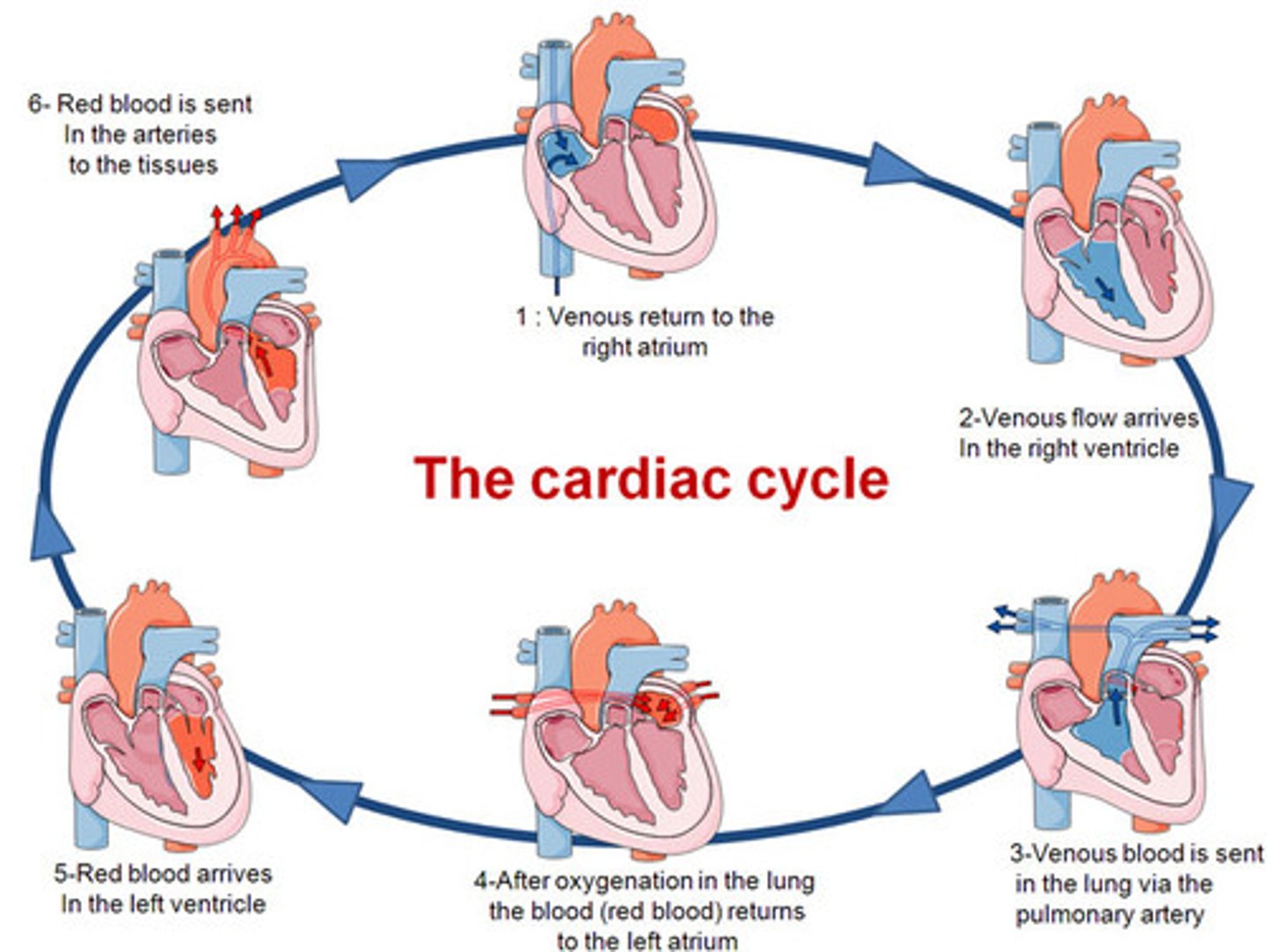
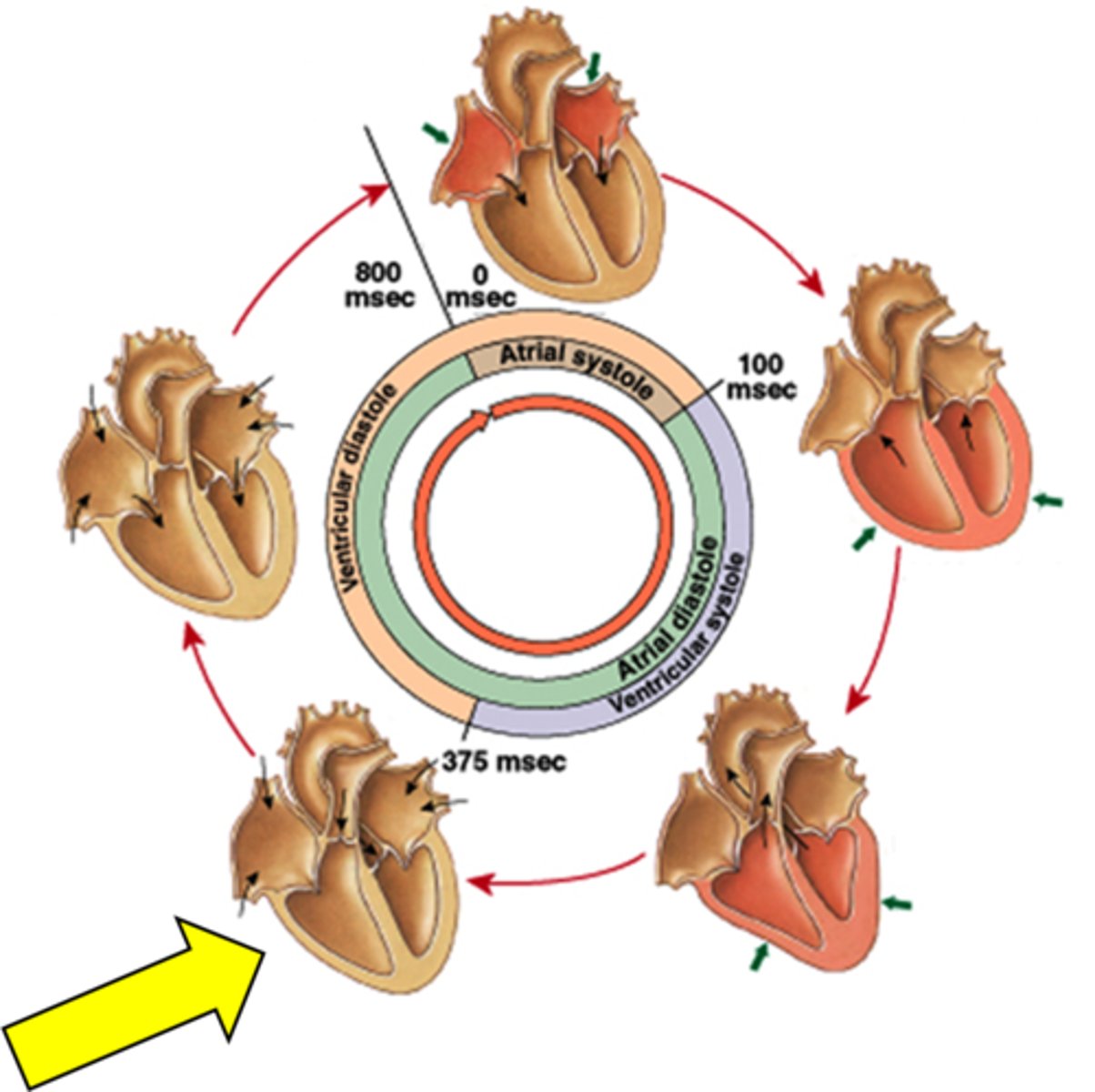
cardiac cycle
A complete heartbeat consisting of contraction and relaxation of both atria and both ventricles
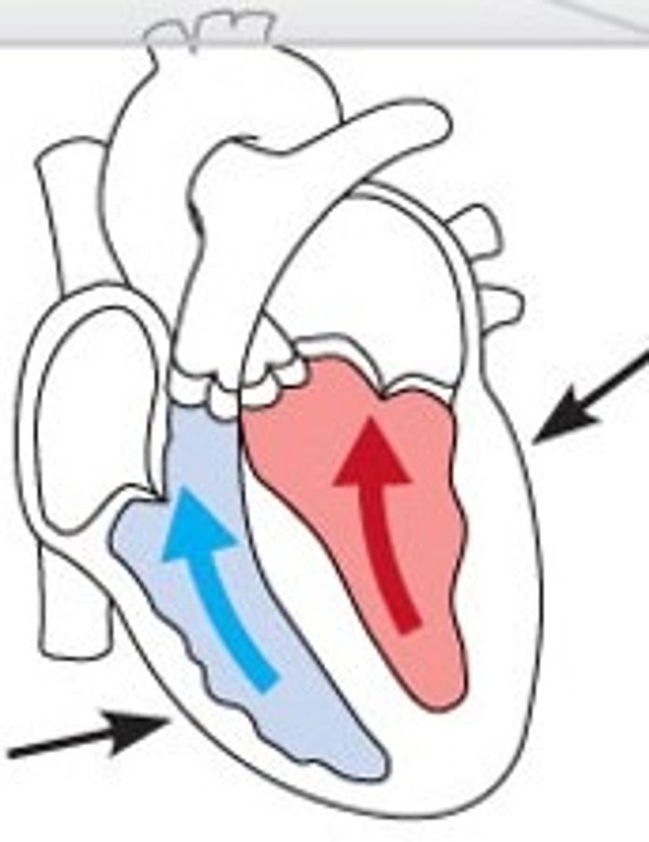
passive ventricular filling
Blood flows into ventricles without atrial contraction.
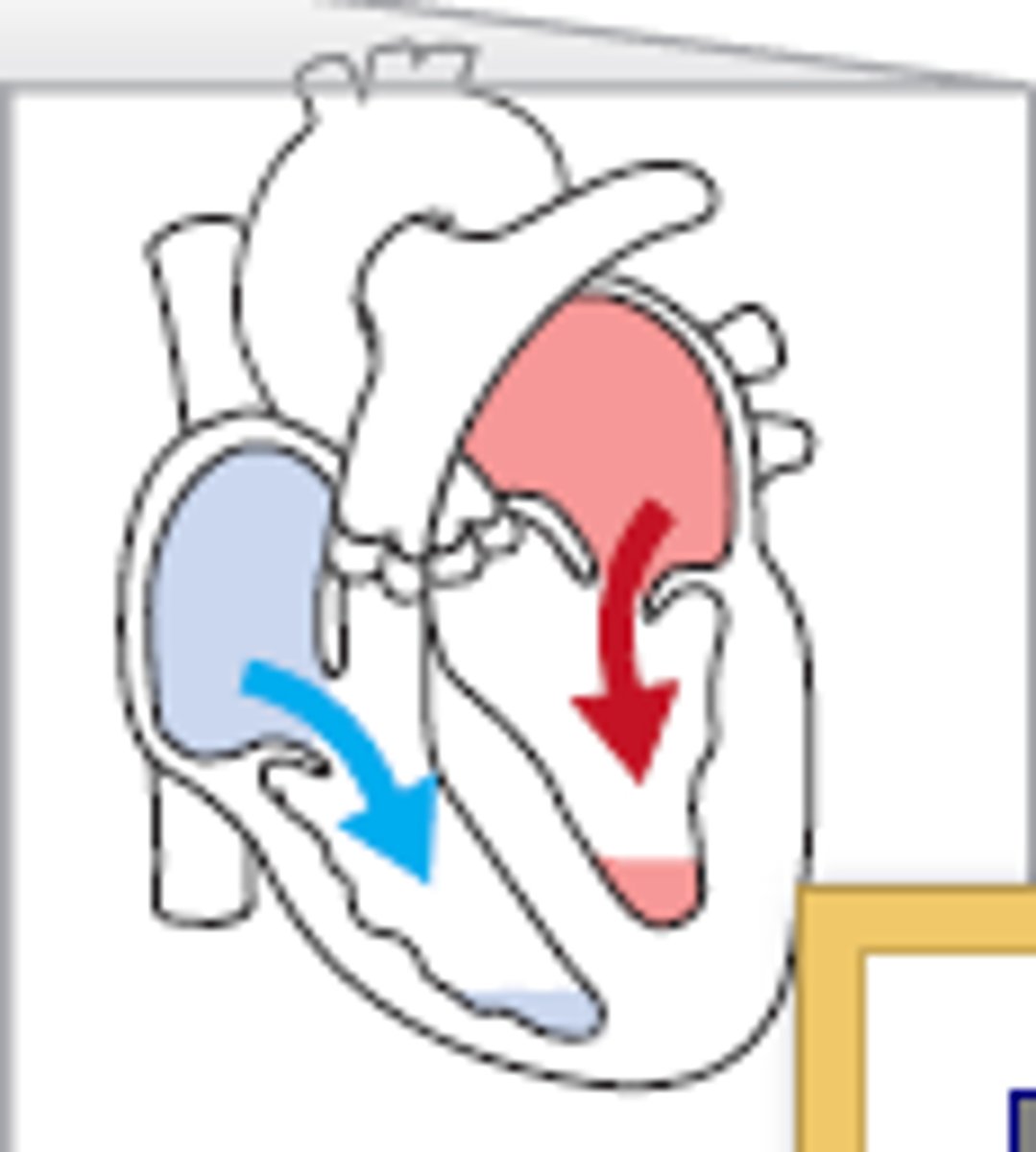
AV valves open, blood flows from atria into ventricles, no contractions
passive ventricular filling
atrial contraction
Atria contract to push blood into ventricles.
isovolumetric ventricular contraction
ventricles contract, all 4 valves are closed, pressure rises
ventricles contract, all 4 valves close, pressure rises, ventricular volume remains same
isovoluemtric ventricular contraction
ventricular ejection
as ventricular pressure rises and exceeds pressure in the arteries, the semilunar valves open and blood is ejected
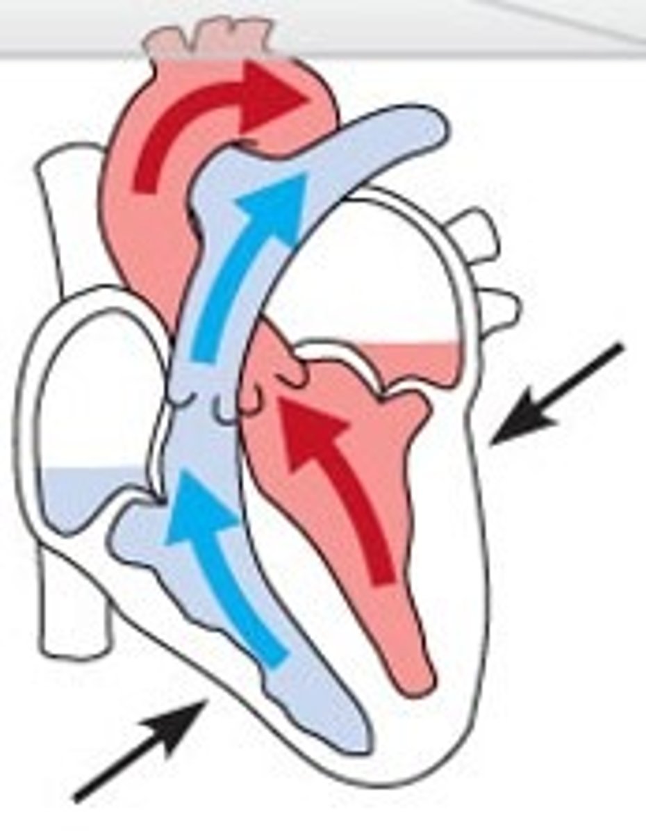
pressure rises, semilunar valves open, blood pumped out of heart
ventricular ejection
isovolumetric ventricular relaxation
all valves closed, no movement of blood in early diastole
all valves closed, pressure falls
isovolumetric ventricular relaxation
passive ventricular filling, atrial contration, isovolumetric ventricular contraction, ventricular ejection, isovolumetric ventricular relaxation
5 phases of cardiac cycle
lubb
closure of the tricuspid and mitral valves at the beginning of systole
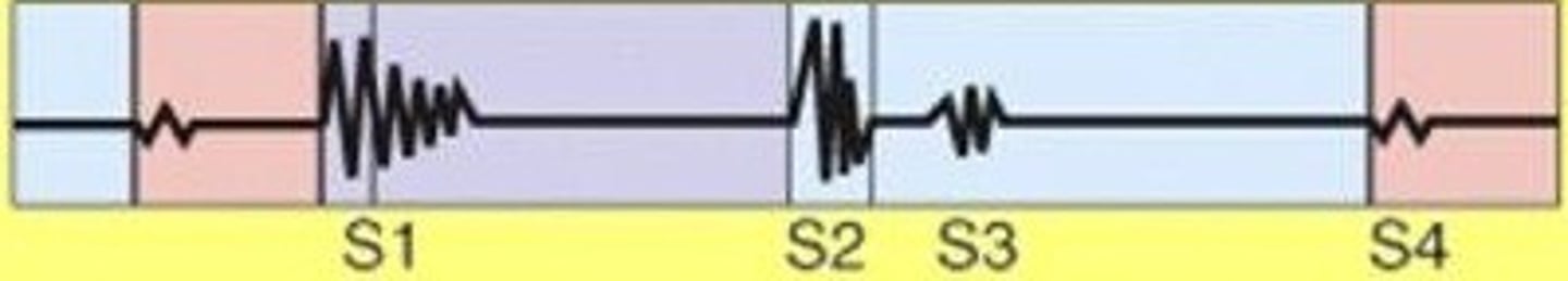
dubb
closure of the aortic and pulmonary valves at the end of systole
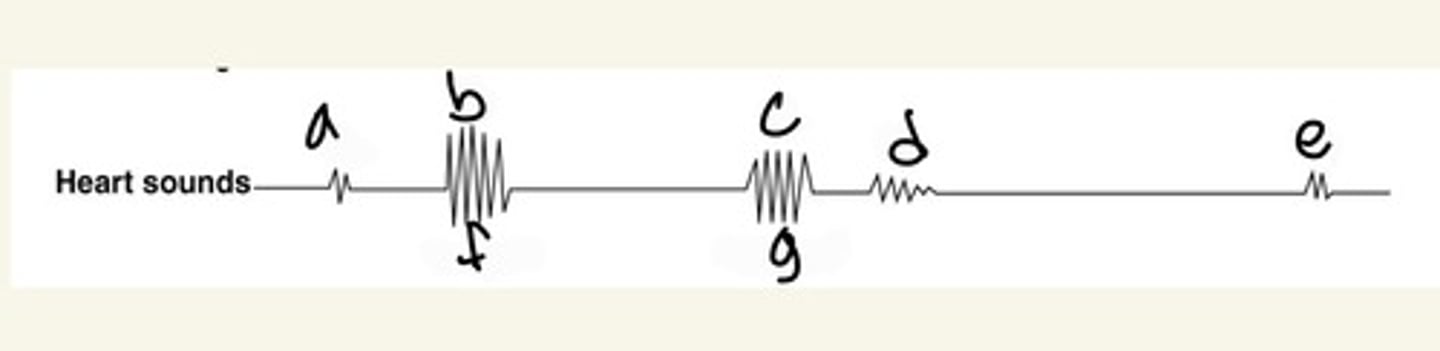
louder and a bit longer, caused by vibrations with closure of AV valves
first sound - Lubb
shorter and not as loud, caused by vibrations with closure of SL valves
second sound - Dubb