appendicular skeleton markings
1/109
Earn XP
Description and Tags
for lab quiz 3
Name | Mastery | Learn | Test | Matching | Spaced |
|---|
No study sessions yet.
110 Terms
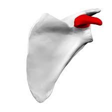
coracoid process of scapula
whats this? anterior view
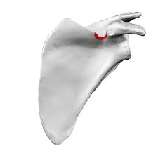
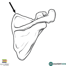
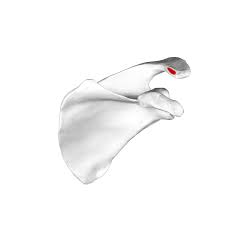
suprascapular notch
posterior view whats this?
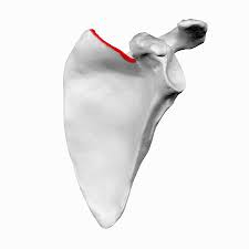
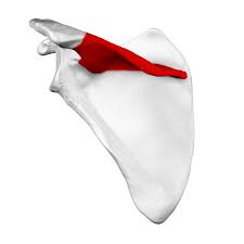
superior border of scapula
whats this border?
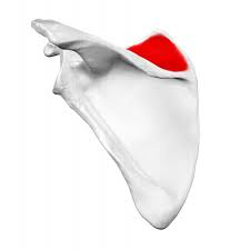
supraspinous fossa of scapula
whats this fossa? posterior view
superior angle of scapula
what angle is this? posterior view
spine of scapula
whats this?
medial border of scapula
what border is this ? posterior view
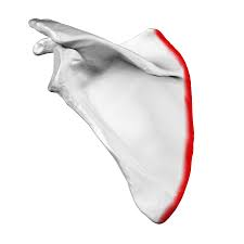
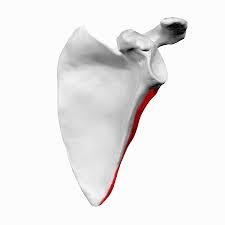
lateral border of scapula
what border is this? anterior view
infraspinous fossa of scapula
what fossa is this? posterior view
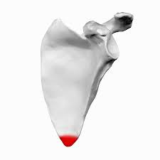
inferior angle of scapula
what angle is this? anterior
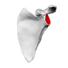
glenoid fossa of scapula
what fossa is this? anterior view
clavicular facet of scapula
seen at the top.
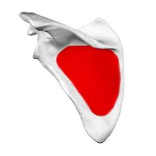
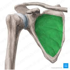
subscapular fossa of scapula
what fossa is this? anterior view
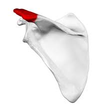
acromion process of scapula
what process is this? seen posteriorly
acromical end of clavicle
what end of clavicle is this? flatter end (lateral)
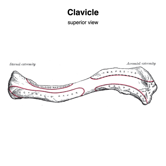
sternal end of clavice
what end of clavicle is this? round end (medial)

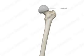
head of humerus
red of this

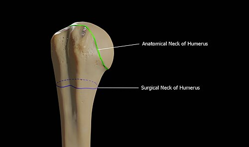
surgical neck of humerus
yellow of this
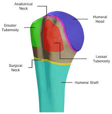
anatomical neck of humerus
green of this
greater tubercle of humerus
the blue of this

lesser tubercle of humerus
the green of this

intertubercular groove of humerus
between the green and blue (seen anteriorly)
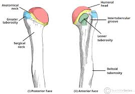
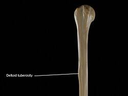
deltoid tuberosity of humerus
what tuberosity is this? seen anteriorly
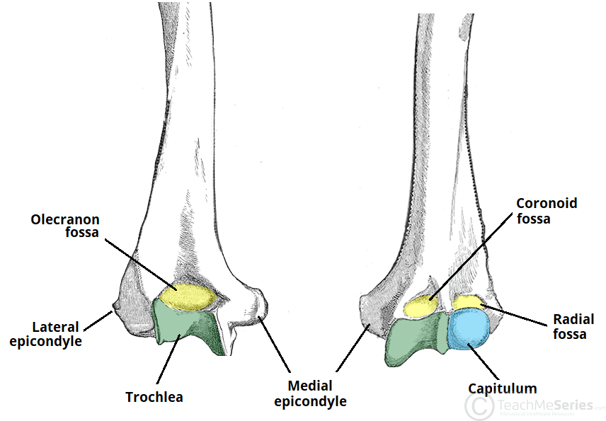
coronoid fossa of humerus
fossa on the ulna side of the humerus. seen anteriorly

radial fossa of humerus
fossa seen on the radius side of the humerus seen anteriorly

capitulum of humerus
under the radial fossa

trochlea of humerus
green, seen anteriorly and posteriorly. near bottom of humerus

lateral epicondyle of humerus
epicondyle of humerus seen posteriorly
medial epicondyle of humerus
epicondyle of humerus seen anteriorly and posteriorly

olecranon fossa of humerus
fossa seen at the near bottom of humerus. posteriorly seen
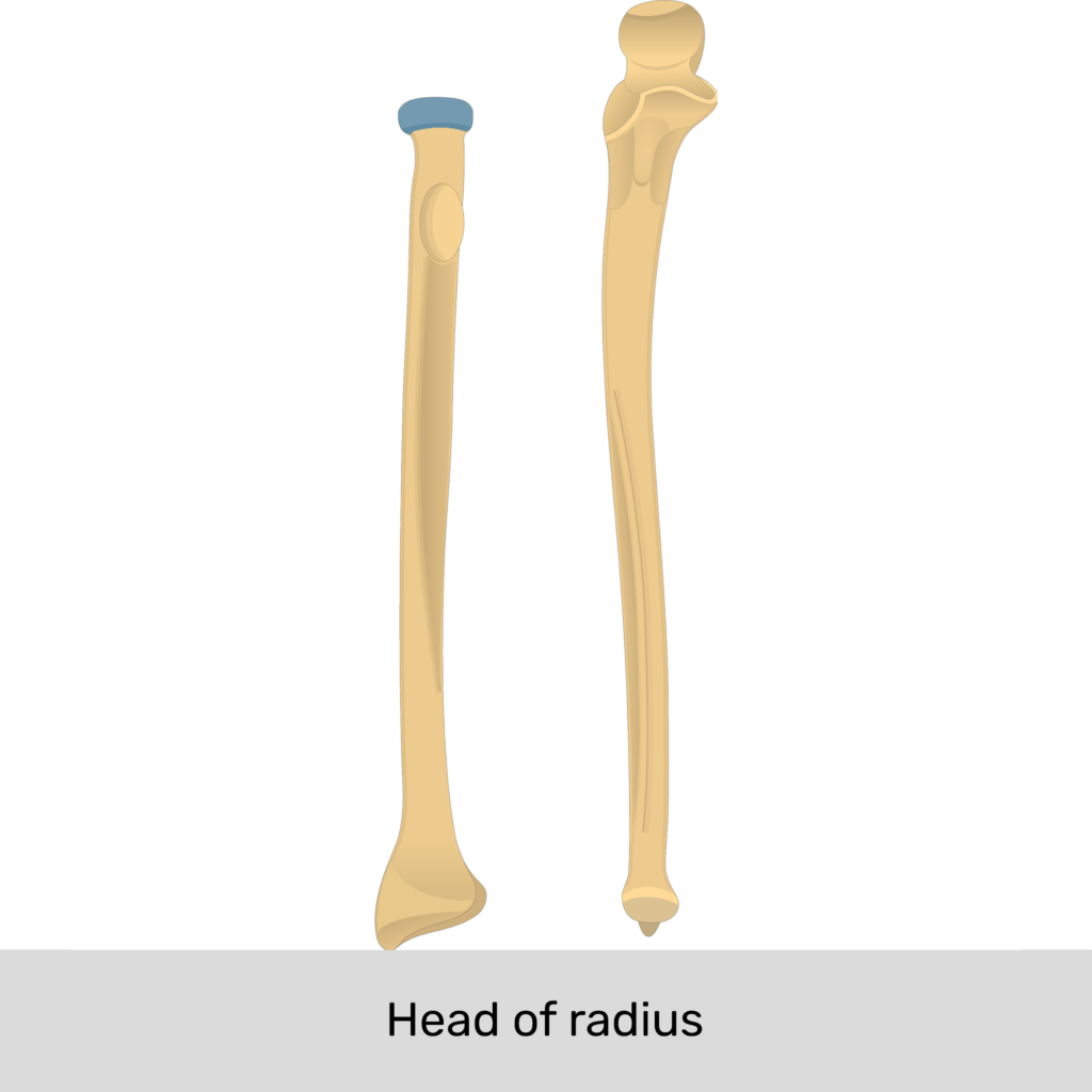
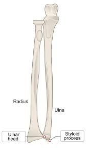
head of radius
the circle at the top of the radius
neck of radius
under the head of the radius
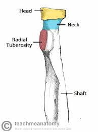
radial tuberosity
prominence on the radius on the medial line.

interosseous border of radius
middle border of the radius towards the medial line

styloid process of radius
sharp process of radius ( lateral side on the anatomical position)
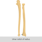
ulnar notch of radius
notch on the radius by the carpals (points towards the medial line in anatomical position)
surface for scaphoid of radius
surface for carpal bone (1st)
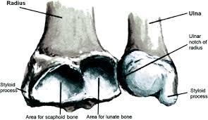
surface for lunate of radius
surface for carpal bone (2nd in nmeumonic)

olecranon of ulna
outside scoop of ulna

trochlear notch of ulna
inside scoop of ulna

coronoid process of ulna
process under the scoop of the ulna
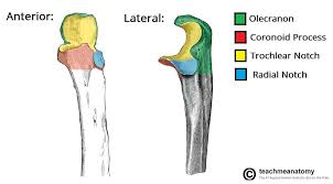
radial notch of ulna
notch on ulna faces the radius

interosseous border of ulna
middle border of ulna, towards the lateral side
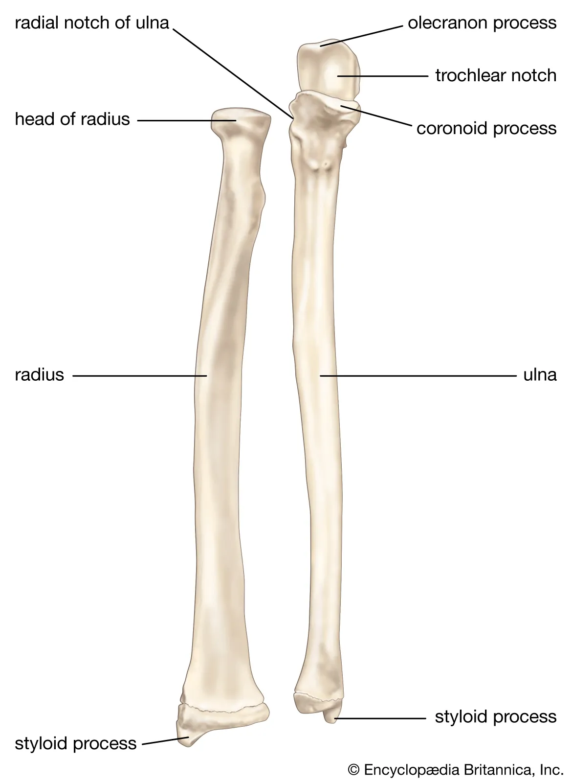
styloid process of ulna
process of ulna by the carpals
head of ulna
end of ulna by the carpals (not process)
scaphoid of carpals
purple (1st) (starts at thumb)

lunate of carpals
2nd carpal starts by thumb

triquetral of carpals
3 carpal starts by thumb, under fourth carpal

pisiform of carpals
4th carpal, on top of the 3rd carpal

trapezium of carpals
5th carpal, starts again by the thumb
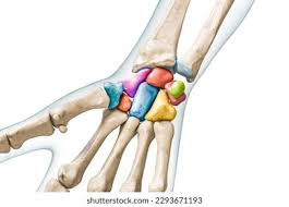
trapezoid of carpals
6th carpel, (2nd t of the nmeuonic)

capitate of carpals
7th carpal,

hamate of carpals
8th carpal, at the top by the pinky

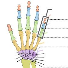
metacarpals of hand
the middle carpals (1 is thumb and 5 is pinky)

phalanges of hand
end of hand (1 is thumb 5 is pinky) (proximal-distal)
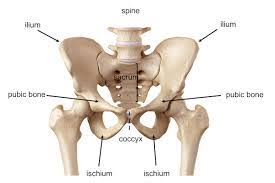
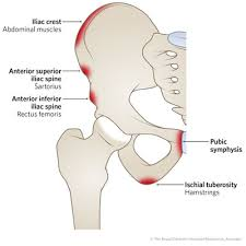
crest of ilium
top of the ilium, smooth top
ala of ilium
wing like side of ilium on the posterior
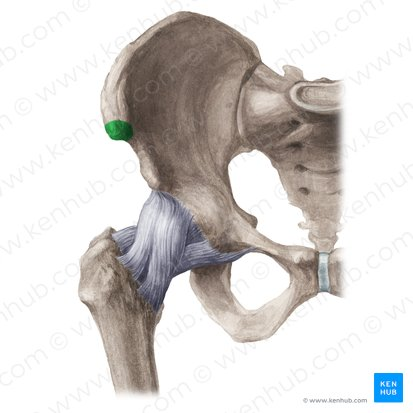
anterior superior iliac spine
what spine is this of ilium ?
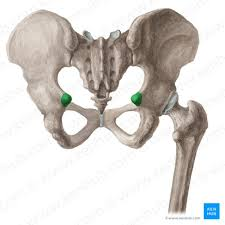
anterior inferior iliac spine
ilium spine on the inferior anterior side
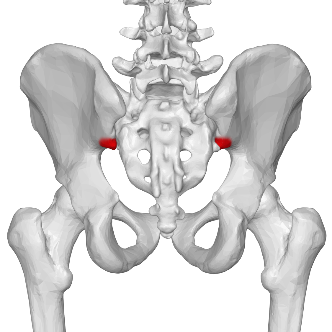
posterior superior iliac spine
spine found on the back of ilium
posterior inferior iliac spine
what spine of ilium is this?
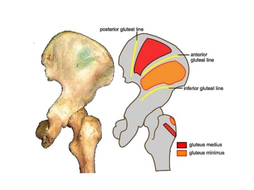
gluteal lines of ilium
ala side of ilium. lines for….
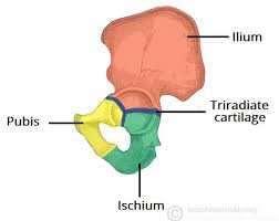
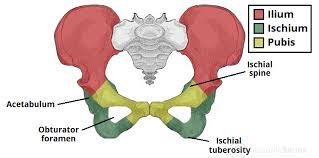
greater sciatic notch of ilium
yellow shown notch
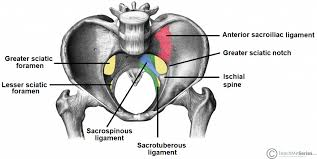
ischial spine
below ilium, spine seen posteriorly
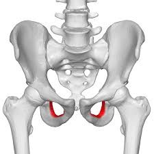
lesser sciatic notch
notch between ischium and pubis
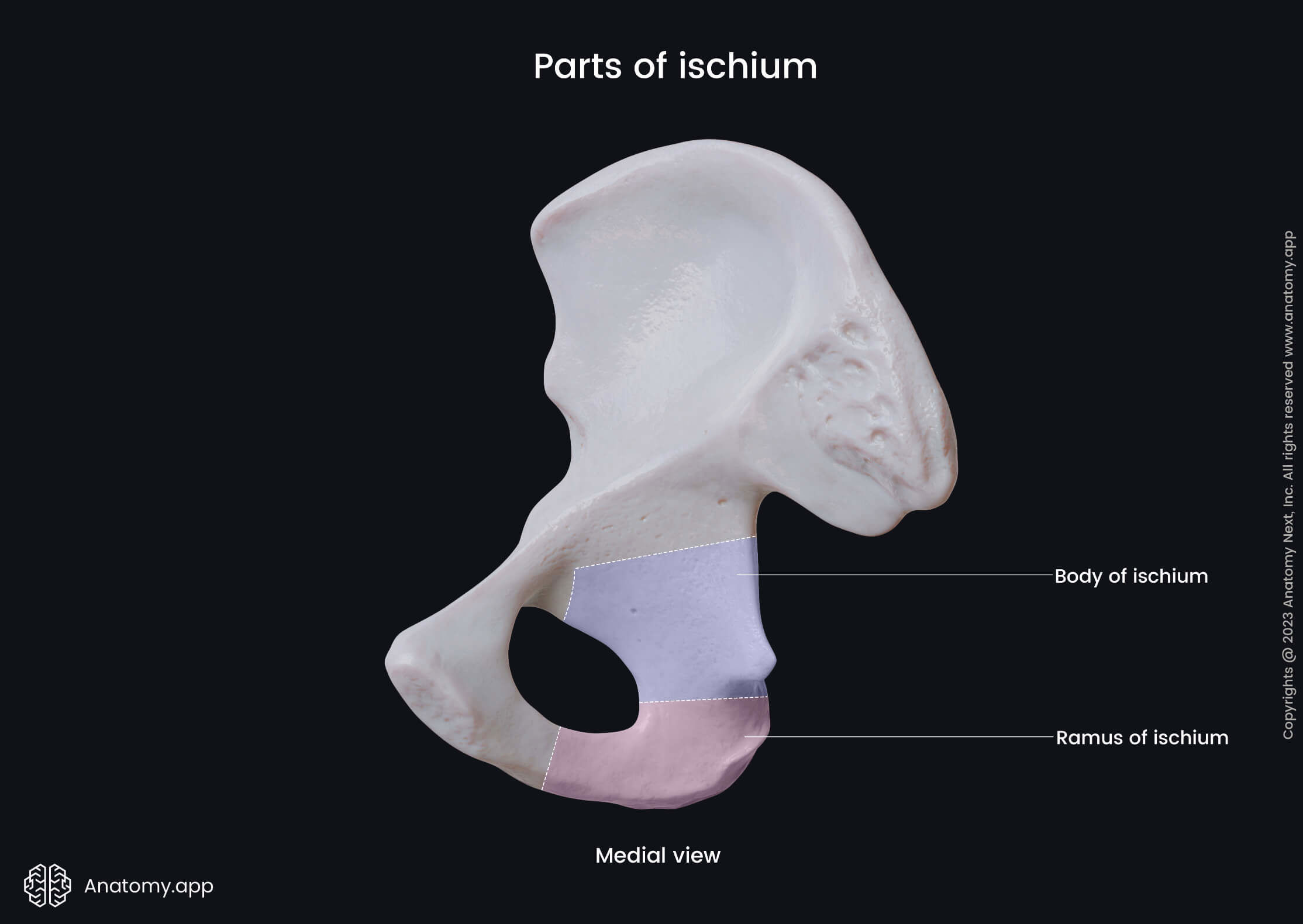
ischial tuberosity
whats this?
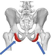
ischial ramus
pink past (flip words)
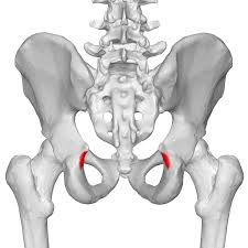
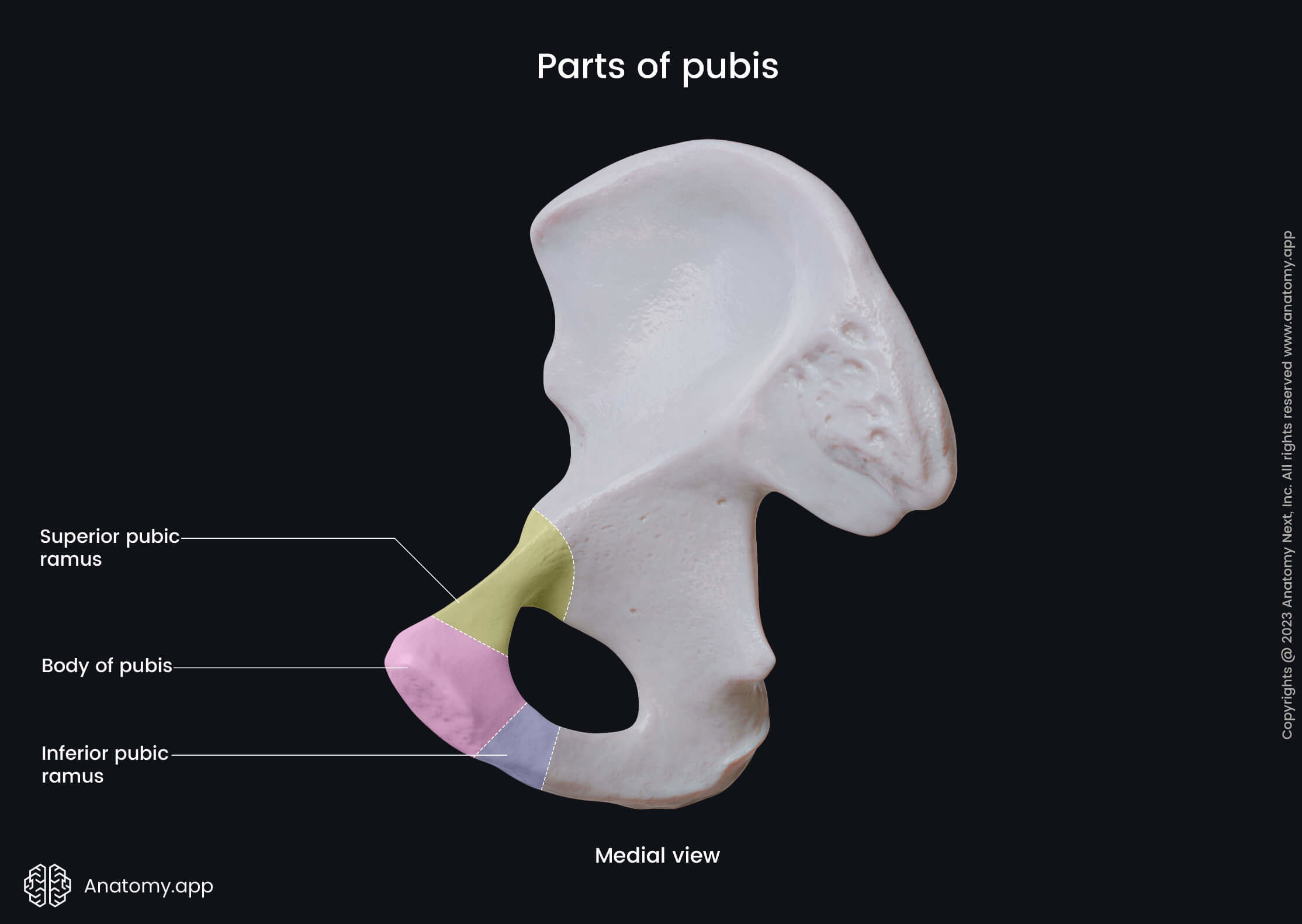
inferior pubic ramus
ramus between pubic bone and ischium ramus

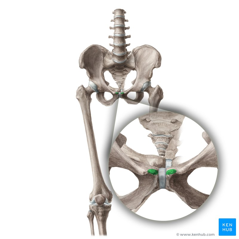
pubic symphysis
where the pubic meets
pubic crest
meets the symphysis
superior pubic ramus
top ramus of pubic (connects to ilium
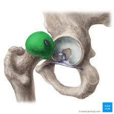
acetabulum
big hip socket where the femur connects
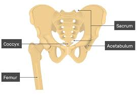
obturator foramen
what foramen is this?
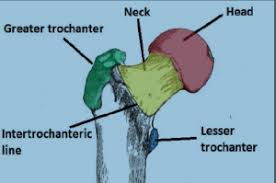
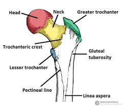
head of femur
what is this of the femur?
neck of femur
literally right under the head
greater trochanter of femur
green (only really seen posteriorly)

intertrochanteric fossa of femur
that fossa right under the green (not colored)

lesser trochanter of femur
blue (only really seen posteriorly)

intertrochanteric line
line on the anterior (opposite to the greater trochanter)
intertrochanteric crest
right by the greater trochanteric (has that fossa mentioned earlier)
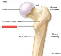
linea aspera of femur
line on the back of the femur ( where thigh muscles connect)
popliteal surface of femur
smooth surface of front of femur where its opposite of linea aspera
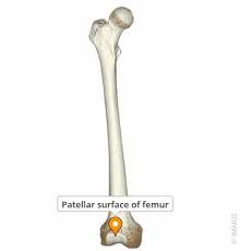
patellar surface of femur
where the patella goes
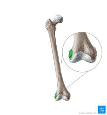
medial condyle of femur
medial bump next to patellar surface at the BOTTOM
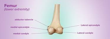
lateral condyle of femur
lateral condyle of femur next to patellar surface at the BOTTOM

medial epicondyle of femur
bump on the side next to the patellar surface on the midline
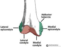
lateral epicondyle of femur
bump on side next to the patellar surface
adductor tubercle of femur
bump ABOVE the medial epicondyle
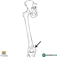
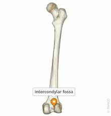
intercondylar fossa of femur
depression between the medial and lateral condyles of femur
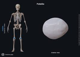
patella
knee cap
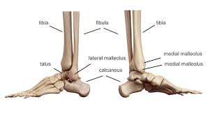
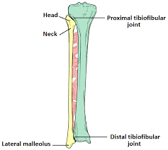
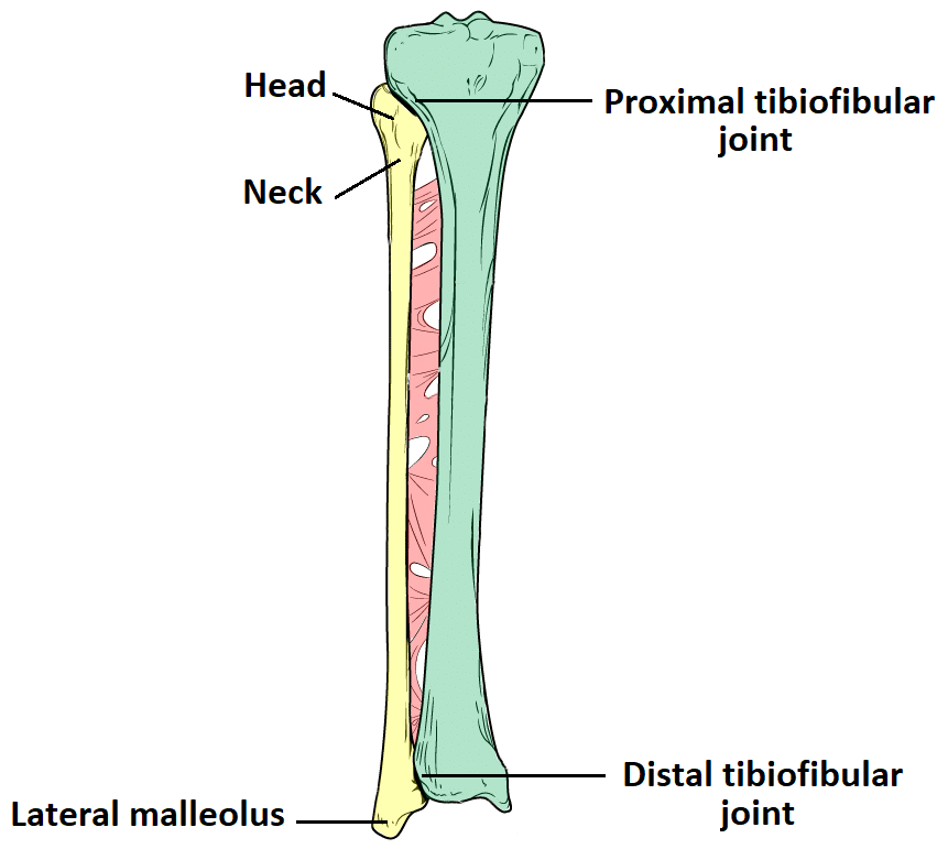
head of fibula
top of lateral bone of the lower leg
styloid process of fibula
process at the top of the fibula

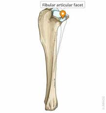
articular facet of fibula
top where fibula connects with tibia
lateral malleolus of fibula
end of fibula that connects with tarsals not as sharp as the top
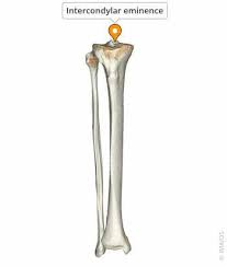
intercondylar eminence of tibia
pebble like raised area where the tibia meets patella
lateral condyle of tibia
bump on the side of the top of the tibia by the lateral side
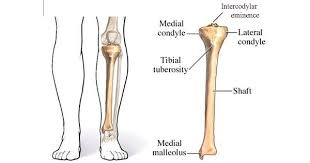
medial condyle of tibia
medial bump at the top of the tibia

tibial tuberosity
anterior protuberance of the tibia (knee protuberance of tibia)

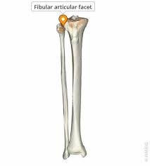
articular facet for fibula
medial side facet on tibia connecting to fibula
medial malleolus of tibia
above the calcenous still on the tibia