visual - blind spot and sensory cortex
1/25
Earn XP
Description and Tags
visual - blind spot and sensory cortex
Name | Mastery | Learn | Test | Matching | Spaced |
|---|
No study sessions yet.
26 Terms
Ganglion cells when stimulated
photoreceptors sends signals to one ganglion called neural convergence and creates receptive fields on the ganglion.
Ganglion cells can be excited or inhibited by the same stimulus received in the retina.
Stimulating can be exibited if it occurs in the center=fovea or inhibited when the periphery=edges receives signals (center-periphery antagonism).
antagonism
The center and periphery often respond to stimuli in opposite ways:
In bright light, the center (cones) is good at detailed, sharp vision.
In low light, the periphery (rods) takes over, helping you see things in dim light, but without the detail.
Light function in our brain
It is controlling our circadian rhythm through the suprachiasmatic nucleus in the hypothalamus.
Distribution of cones and rodes
• The cones and rods are sandwiched in the retina.
• The ratio of cones to rods depends on their location in the retina.
Blind spot
Angel degree of 20° indicates the place on the retina where there are no receptors (which is the place where ganglion cells leave the eye to form the optic nerve)
Distribution of cones and rodes in fovea and retina
There is a small area on the retina, the fovea, that only contains cones.
1) The peripheral retina (all the part other than the fovea) contains cones and rods.
2) There are many more rods than cones in the peripheral retina because most of the receptors in the retina are located there.
3) There are about 120 million rods and 6 million cones.
Why do we not notice the black spot?
Because it's located on one side of our visual field, where objects aren't in sharp focus.
How do Churchland & Ramachandran, 1996 explain the blind spot?
Some brain mechanisms "fill in" the part of the image that disappears into the blind spot.
How does increased convergence of an neuron work
When sending info to 1 ganglion cell, however they will fire on a different rate, we will only measure the signal from one neuron, which will be constant.
When all of them in a group fire together on one ganglion cell, we will measure the input depending on where the fire is, which might lead to an increase or convergence.
Neural processing LINEAR CIRCUIT
Linear circuit and neurons responses generated as we increase stimulated receptors.
What happens when we add convergence to the linear circuit?
Neurons (such as ganglion cells) receive signals from multiple bipolar cells, and as the size or intensity of the stimulus increases, the response of the neuron also increases.
Neural circuit that would produce a center‐periphery
Neural circuit that would produce a center‐periphery receptive field.
The signals from the receptors in the periphery reach the cell through the inhibitory synapses, while the signals from the receptors in the center do so through the excitatory synapses.
Thus, stimulation of the receptors in the center increases the firing rate recorded by our electrode, while stimulation of the receptors in the periphery decreases it.
In the retina, horizontal and amacrine cells carry these inhibitory signals.
NEURAL PROCESS OF LATERAL INHIBITION - CONTRAST EFFECT
Center (bright light): When light hits the center of the retina, the photoreceptors excite the ganglion cell, making it more active.
Surrounding (darker light): The photoreceptors around the center send inhibitory signals to the ganglion cell, making it less active.
Center-surround effect: The difference in light (center bright, surround dark) makes the intersection look darker, helping the brain notice edges and contrast between light and dark areas.
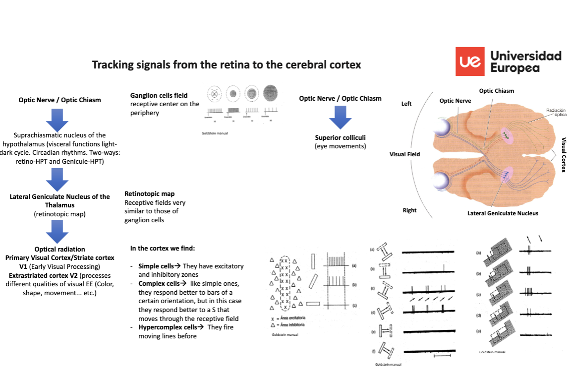
Tracking signals from the retina to the cerebral cortex
Primary pathway: (vision)
90% of the signals in the retina travel from the back of the eye to the optic nerve and from there to the lateral geniculate nucleus in the thalamus.
From the LGN the signals, the received signals are regulated into a flow of information and organization, before descended to the primary visual receptor area of the occipital lobe of the cortex = also called striated cortex.
Secondary pathway: (eye movement)
10% of the signals which involves control of the eye movement travel from the retina to the superior colliculi.
Why is occipital lobe called striated cortex? (Glickstein, 1988).
Because of the white stripes/striations created by nerve fibers (Glickstein, 1988).
The layers of lateral geniculate nucleus
It has 6 layers of neurons.
The 2 inner layers are called magnocellular (movement).
The 4 are called parvocellular (colors, texture and patterns, fines, and depth)
Tracking signals from the retina

Optic nerve/ chiasm → LGN of thalamus → optical radiation Primary visual cortex/striated cortex → extrastriated cortex V2
Visual information in the cerebral PRIMARY visual cortex (PATHWAYS)
From the primary visual cortex (V1), visual information fundamentally follows two paths:
the what pathway (ventral pathway, or parvocellular pathway)
the where pathway (dorsal pathway, or magnocellular pathway).
The what pathway in cerebral cortex
Processes intrinsic properties of objects (color, shape, etc.). Inferior temporal lobe.
The where pathway
Processes extrinsic properties of objects (distance, motion, etc.). Posterior parietal lobe
Kanwisher et al. (1997)
Analyzed the differences between the areas of the brain activated when processing images of faces and those that were activated when processing information from other objects
Processing information about places
The parahippocampal area of place or parahippocampal gyrus is activated by images of indoor or outdoor scenes.
Mainly focused on the processing of information on spatial distribution.
Face processing information
They detected the area specialized in processing faces, and found that the fusiform area of the face (FFA) is located below the temporal lobe.
Injury in brain areas related to visual funtion
Cortical blindness - loss of vision due to an injury in the brain area involved in visual function.
The cause for cortical blindness
Occipital lobe injuries (ischemia, trauma, etc.)
Which can present with anosognosia.
Different disturbances of vision
Visual agnosias
Agnosia for objects
Agnosia for drawings
Simultagnosia: inability to synthesize all the details of an stimulus as a whole, only perceiving its parts independently. It is due to bilateral parieto‐occipital lesions.
Agnosia for color
Color anomie (zone disconnection)
Perceptual achromatopsia: deficit in distinguishing colors due to cortical lesions
Prosopagnosia
Bilateral lesions of the parietal cortex may result in visuospatial agnosias and movement agnosias.