Skull Practice
1/262
There's no tags or description
Looks like no tags are added yet.
Name | Mastery | Learn | Test | Matching | Spaced | Call with Kai |
|---|
No analytics yet
Send a link to your students to track their progress
263 Terms
Fill in the total number of bones for the Cranium and Facial Bones
Cranium: 8 Facial Bones: 14
List the four cranial bones that form that calvaria (skull cap)
Frontal, Right Parietal, Left Parietal, Occipital
List the four cranial bones tha form the floor of the cranium
Right temporal Left temporal Sphenoid Ethmoid
The small horizontal plate of the ethmoid is called the
Cribriform plate
The vertical plate of the ethmoid bone forming the upper portion of the bony nasal septum is the
Perpendicular plate
A structure found in the middle of the sphenoid bone that surrounds the pituitary gland is the
Sella turcica
The posterior aspect of the sella turcica is called the ________________________
Dorsum sellae
Which structure of the sphenoid bone allows for the passage of the optic nerve and is the actual opening into the orbit? ________________________
Optic Foramen
Which structures of the sphenoid bone help to form part of the lateral walls of the nasal cavities? ________________________
Medial and Lateral Pterygoid Processes
Which radiographic projection best demonstrates the sella turcica and dorsum sellae?
Lateral Projection
Which aspect of the frontal bone forms the superior aspect of the orbit?
Oribital or horizontal portion
Cranial sutures are classified as being __________________ joints.
Fibrous or synarthrodial
Small, irregular bones that sometimes develop in adult skull sutures are called __________________ or __________________ bones and are most frequently found in the __________________ suture.
Sutural or wormian; lambdoidal
Which term describes the superior rim of the orbit? (Include the abbreviation also.)
Supraorbital margin (SOM)
What is the name of the notch that separates the orbital plates from each other?
Ethmoidal Notch
Which cranial bones form the upper lateral walls of the calvarium?
Right and left parietals
Which cranial bone contains the foramen magnum?
Occipital
A small prominence located on the squamous portion of the occipital bone is called the __________________.
External occipital protuberance, or inion
What is the name of the oval processes found on the occipital bone that help form the atlanto-occipital joint?
Occipital condyles, or lateral condylar portions
List the three aspects of the temporal bones.
A. __________________
B. __________________
C. __________________
Squamous, Mastoid, Petrous
True/False: The mastoid portion of the temporal bone is the densest of the three aspects of the temporal bone.
False (Petrous Portion)
Which external landmark corresponds with the level of the petrous ridge?
Top of the ear attachment (TEA)
Which opening in the temporal bone serves as a passageway for nerves of hearing and equilibrium?
Internal acoustic meatus
1B. List the three aspects of the temporal bone:
Squamous, Mastoid, Petrous
2b. Which aspect of the temporal bone is considered the densest?
Petrous portion
3b. Which structure makes up the cartilaginous external ear?
Auricle or pinna
4b. How long is the average external acoustic meatus (EAM)?
1 inch (or 2.5cm)
5b. Which small membrane marks the beginning of the middle ear?
Tympanic membrane (eardrum)
6b. What is the collective term for the small bones of the middle ear?
Auditory ossicles
7b. Which structure allows for communication between the nasopharynx and middle ear?
Eustachian or auditory tube
8b. What is the major function of the structure described in Question 7?
To equalize the atmospheric pressure within the middle ear
9b. Which structure serves as an opening between the mastoid portion of the temporal bone and the middle ear?
Aditus
10b. What is the name of the thin plate of bone that separates the mastoid air cells from the brain?
Tegmen tympani
11b. Which of the auditory ossicles picks up sound vibrations from the tympanic membrane?
Malleus (Hammer)
12b. Which of the auditory ossicles is considered the smallest?
Stapes (Stirrup)
13b. Which of the auditory ossicles resembles a premolar tooth?
Incus (Anvil)
14b. What is the name of the small membrane that connects the middle to the inner ear?
Oval or vestibular window
15b. Which two sensory functions occur within the inner ear?
Hearing and Equilibrium
16b. What is the name of the small membrane found at the base of the cochlea (two terms possible)?
Round or cochlear window
17b. True/False: The semicircular canals include a closed system specific to the sense of hearing.
False
19b. Match each of the following clinical indications for the temporal bone to the correct definition or description: Neoplasia
New and abnormal growth
19b. Match each of the following clinical indications for the temporal bone to the correct definition or description: Otosclerosis
Hereditary disease involving excessive bone formation of middle ear
19b. Match each of the following clinical indications for the temporal bone to the correct definition or description: Mastoiditis
Bacterial infection of the mastoid process
19b. Match each of the following clinical indications for the temporal bone to the correct definition or description: Acoustic Neuroma
Benign tumor of the auditory nerve sheath
19b. Match each of the following clinical indications for the temporal bone to the correct definition or description: Polyp
Growth arising from a mucous membrane
19b. Match each of the following clinical indications for the temporal bone to the correct definition or description: Cholesteatoma
Benign, cystic mass or tumor of the middle ear
20b. Which of the following radiographic appearances pertains to an acoustic neuroma?
Expansion of the internal acoustic canal
21b. Is CT the best imaging modalities that demonstrates otosclerosis?
Yes
Is the middle nasal conchae a facial bone?
No
2c. What is the largest immovable bone of the face?
Maxilla
3c. List the four processes of the maxilla.
Frontal process, Zygomatic process, Alveolar process, Palatine process
4c. Which of the processes mentioned in Question 3 is considered most superior?
Frontal process
5c. Which soft tissue landmark is found at the base of the anterior nasal spine?
Acanthion
6c. Which facial bones form the posterior aspect of the hard palate?
Horizontal portion of the palatine bones
7c. Which two cranial bones articulate with the maxilla?
Frontal and ethmoid
8c. Which facial bones are sometimes called the "cheek bones"?
Zygomatic or malar bones
9c.Does the mandible articulate with the zygomatic bone?
No, it articulates with the TMJ
10c. Which facial bone is associated with the tear ducts?
Lacrimal bones
11c. The purpose of the __________, or ___________, is to divide the nasal cavity into compartments and to circulate air coming into the nasal cavities.
Conchae; turbinates
12c. True/False: The right and left nasal bones form the largest part of the nose.
False (Most of the nose is composed of cartiliage)
13c. A deviated nasal septum is most likely to occur at the junction between _______ and _______
Septal cartilage, vomer (pushed laterally to one side)
14c. Match each of the following mandibular terms to the correct definition or description: A. Gonion
Mandibular Angle
14c. Match each of the following mandibular terms to the correct definition or description: Mandibular Notch
U-shaped notch
14c. Match each of the following mandibular terms to the correct definition or description: Body
Horizontal portion of mandible
14c. Match each of the following mandibular terms to the correct definition or description: Condyloid Process
Posterior process of the upper ramus
14c. Match each of the following mandibular terms to the correct definition or description: Coronoid Process
Bony process located anterior to mandibular notch
14c. Match each of the following mandibular terms to the correct definition or description: Ramus
Vertical portion of mandible
14c. Match each of the following mandibular terms to the correct definition or description: Mentum
Chin
14c. Match each of the following mandibular terms to the correct definition or description: Symphysis Menti
Point of union between both halves of the mandible
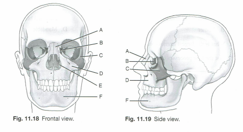
15c. The single facial bone and the one pair of facial bones not visible from the exterior and not demonstraed in Figs 11.18 and 11.19 are the __________ and the ___________ respectively.
Vomer; paleatine bones inferior surface view of the maxillae
20c. From anterior to posterior, the cone-shaped orbits project upward at an angle of _______ and toward the midsagittal plane at an angle of ________.
30 degrees, 37 degrees
21c. Which facial bone opening has the maxillary branch of the fifth cranial nerve passing through it?
Inferior orbital fissure
22c. Which of the facial bone openings is formed by a cleft between the greater and lesser wings of the sphenoid bone?
A. Superior orbital fissure
23c. What is another term for the second cranial nerve?
Optic nerve
1d. What is the older term for the maxillary sinuses?
Antrum, antrum of Highmore
2d. An infection of the teeth may travel upward and involve the _________ sinus.
Maxillary
3d. Specifically, where are the frontal sinuses located
Between the inner and outer tables of the skull, posterior to the glabella
4d. The frontal sinuses rarely become aerated before the age of _________
6 years
5d. Which specific aspect of the ethmoid bone contains the ethmoid sinuses?
Lateral masses or labyrinths
6d. The drainage pathway for the paranasal sinuses is called the:
Ostiomeatal complex
7d. Which sinus is projected through the open mouth with a PA axial transoral projection?
Sphenoid sinus
10d. What is the name of the passageway betweent the maxillary sinuses and the middle nasal meatus?
Infundibulum
11d. True/False: Most CT studies of the paranasal sinuses do not require the use of contrast media.
True
12d. Which position is most often used when performing a CT study of the sinuses?
Prone
1e. What are the three classifications of the skull?
Mesocephalic, Brachycephalic, Dolichocephalic
1e. What classification fits the width <75% of length?
Dolichocephalic
1e. What classification fits the width >= 80% of length?
Brachycephalic
1e. What classification fits the width between 75% and 80% of length?
Mesocephalic
2e. Central ray angles and degree of rotation stated for basic skull positions are based on the ___________ skull, which has an approximate angle of __________ between the midsagittal plane and the long axis of the petrous bone.
Mesocephalic, 47 degrees
3e. The long, narrow-shaped skull has an angle of approximately _______ between the midsagittal plane and the long axis of the petrous bone.
±40 (<47 degrees)
4e. True/False: Skull morphology has no impact on positioning considerations.
False (CR angles and head rotations may be different)
5e. There is a _______ difference between the orbitomeatal and infraorbitomeatal lines, and _______ between the orbitomeatal and glabellomeatal lines.
7–8 degrees; 7–8 degrees (same degrees of difference
6e. Match each of the following cranial landmarks and positioning lines with the correct definition: Lateral junction of the eyelid
Outer Canthus
6e. Match each of the following cranial landmarks and positioning lines with the correct definition: Posterior angle of the jaw
Glabelloalveolar Line (GAL)
6e. Match each of the following cranial landmarks and positioning lines with the correct definition: A line between the infraorbital margin and the EAM
Infraorbitometal Line (OML)
6e. Match each of the following cranial landmarks and positioning lines with the correct definition: Corresponds to the highest “nuchal” line of the occipital bone
Inion
6e. Match each of the following cranial landmarks and positioning lines with the correct definition: A line between the glabella and alveolar process of the maxilla
Glabelloalveolar Line (GAL)
6e. Match each of the following cranial landmarks and positioning lines with the correct definition: A line between the mental point and EAM
Mentomeatal Line
6e. Match each of the following cranial landmarks and positioning lines with the correct definition: Located at the junction of the two nasal bones and the frontal bone
Nasion
6e. Match each of the following cranial landmarks and positioning lines with the correct definition: The small cartilaginous flap covering the ear opening
Tragus