Clinical Thoracic Radiology
1/12
There's no tags or description
Looks like no tags are added yet.
Name | Mastery | Learn | Test | Matching | Spaced |
|---|
No study sessions yet.
13 Terms
What is the difference in color of Chest X-ray
Chest X-ray or CT Scans
→ anything that has air will appear black, while anything that absorbs the X-ray will be white
→ so things like bone and metal are the whitest while fat and soft tissue are greyish
What is the difference between a PA and AP view
Posterior-Anterior View
→ X-ray source is behind the person and the X-ray is viewed as if from the front
Anterior-Posterior View
→ X-ray source is in front of the person and the X-ray is viewed as if from behind
What is Rotation, Inspiration, and Penetration?
Three markers that determine quality of the radiograph
1) Rotation - X ray should be straight on, clavicles should be equally spaced from the spinous process
2) Inspiration - chest-x-rays should be taken with patient fully breathed in
→ 5th to 7th anterior ribs should dissect the mid clavicular line
→ 9-10 posterior ribs should be visible
3) Penetration - how well the X-rays have passed through the body
→ well penetrated film should have spinous processes visible behind the heart and left diaphragm should be visible to the spine
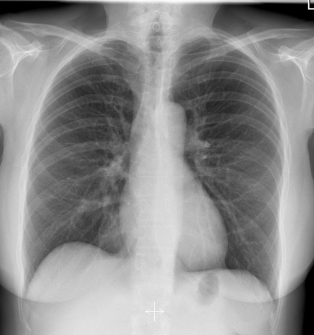
What are the airway structures seen on this film?
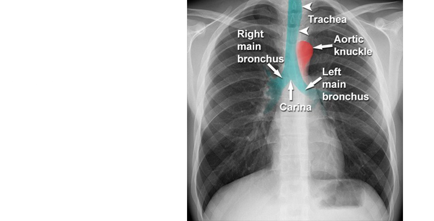
→ right main bronchus and left main bronchus
→ trachea
→ carina
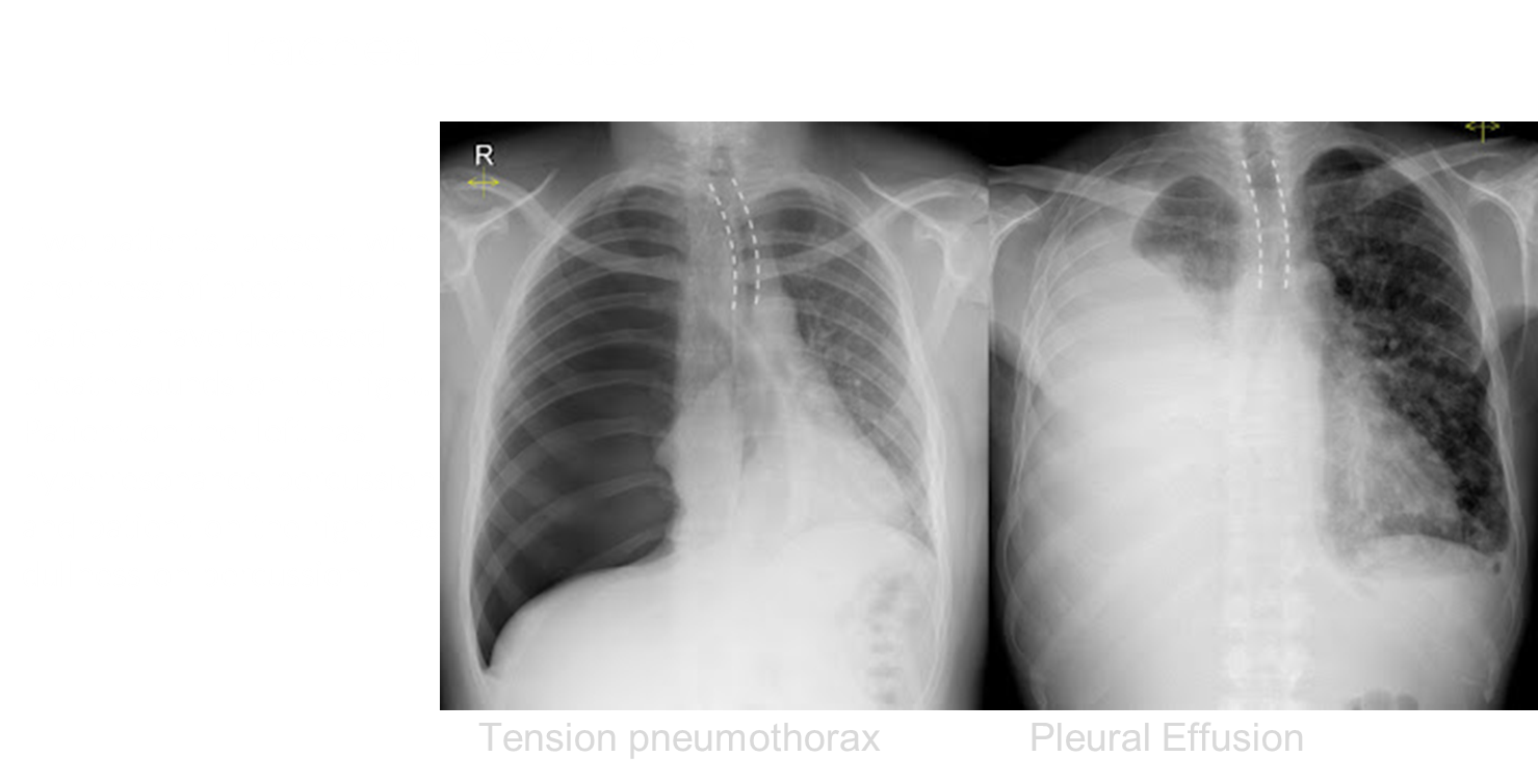
What is Tracheal Deviation?
Deviation of the trachea from its normal midline position
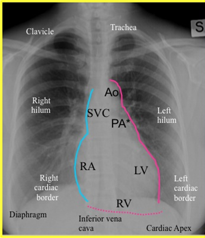
What are the cardiac structures on this chest x-ray

Cardiac shadow should be around less than 50 percent of the diameter of the chest
→ aortic arch
→ SVC
→ Right Atrium
→ Pulmonary Artery
→ Left Ventricle
→ Right Ventricle
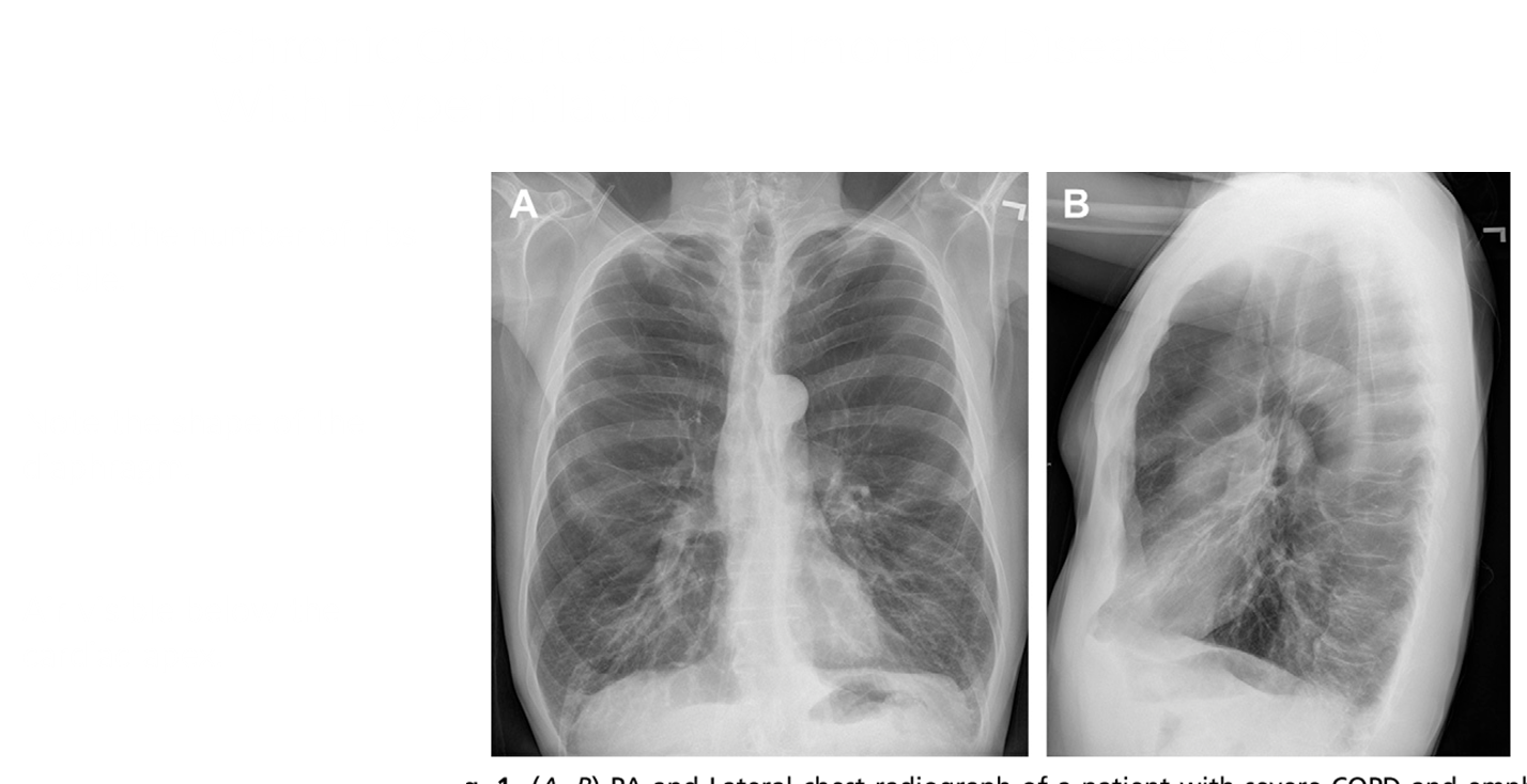
How can you identify COPD with hyperinflation on a chest X-ray
1) higher number of visible ribs (past ten)
2) the diaphragm are flattened in shape
3) air visible below the heart
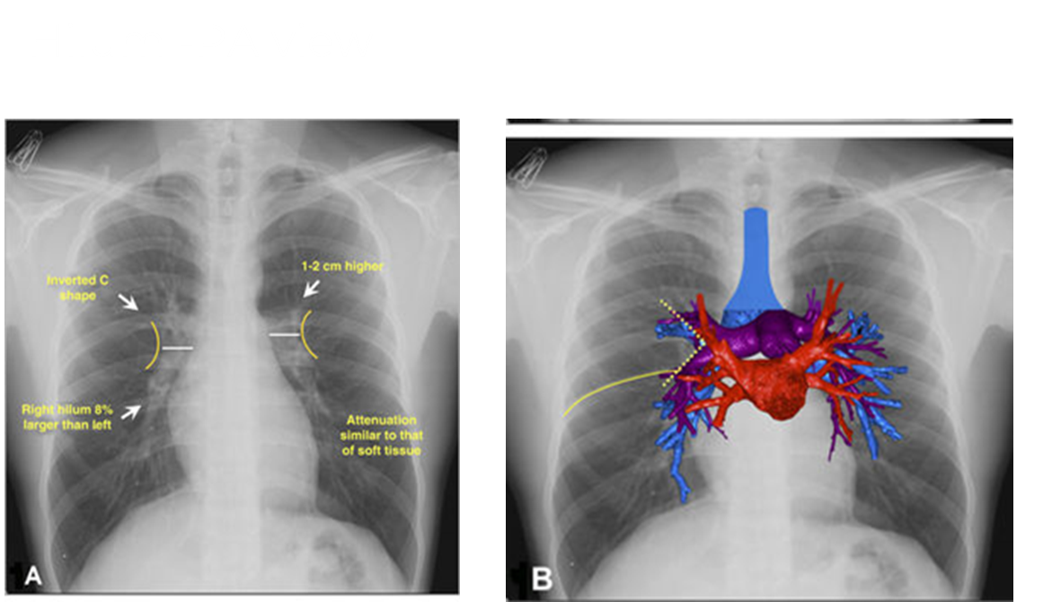
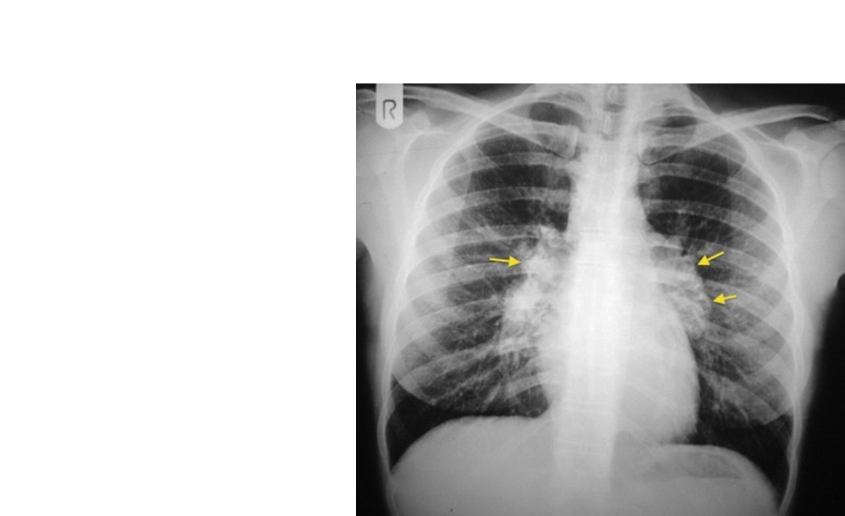
What does the hilum look like on a PA chest X-ray
Hilum should typically have a C shaped structure
→ if there is an issue it will make a convex shape outward
Which mediastinal compartment is the mass located?
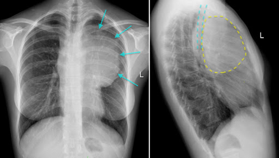
Located in the anterior compartment as the heart is not clearly visible
What are the structures on the CT scan (7)
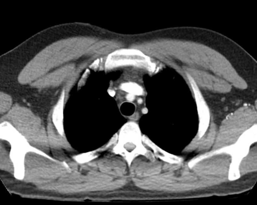
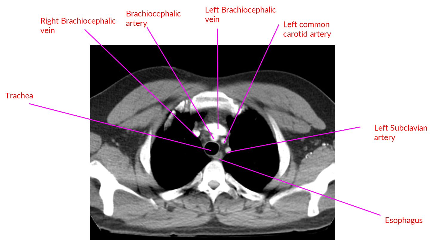
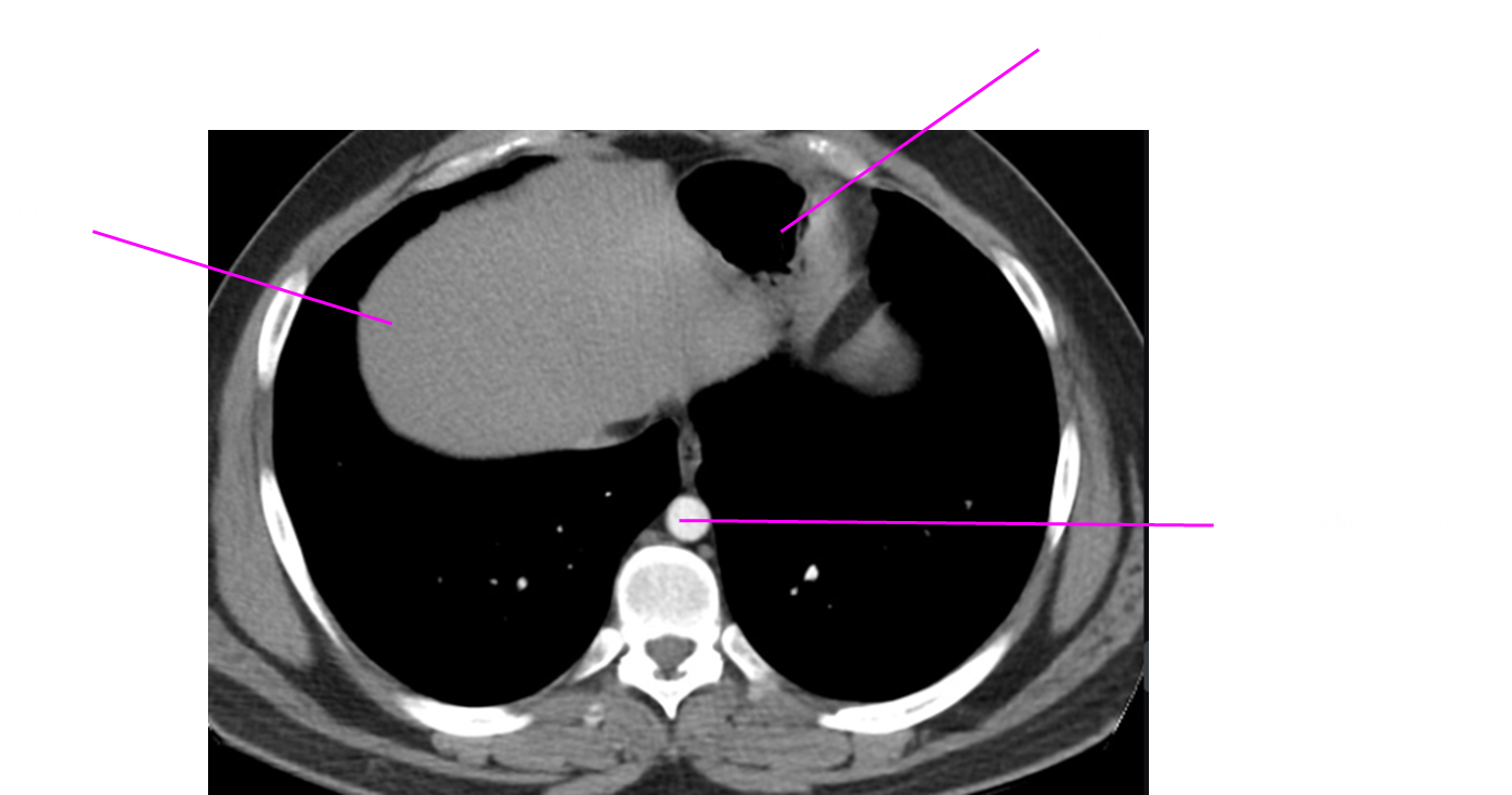
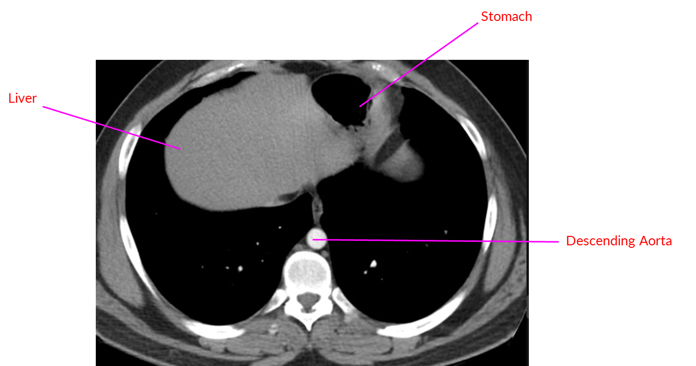
What structures are on this CT scan (8)
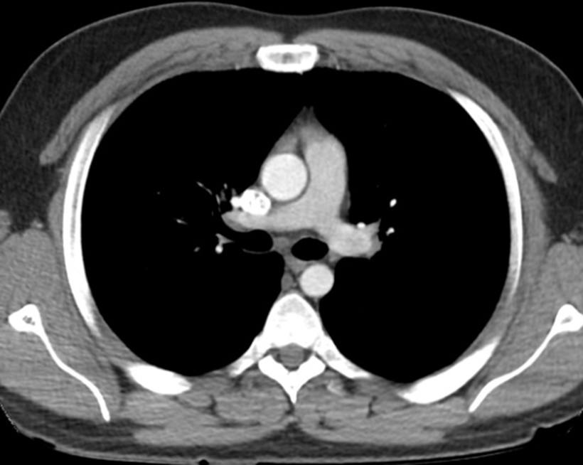
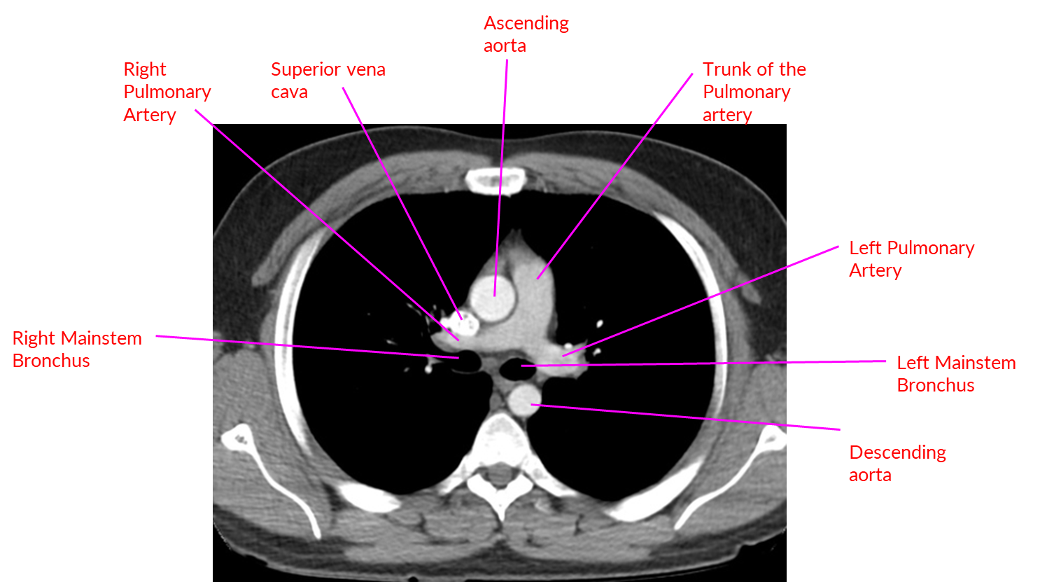
What structures (7)
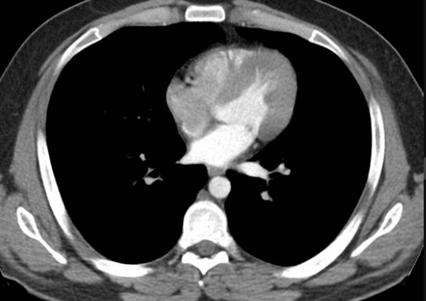
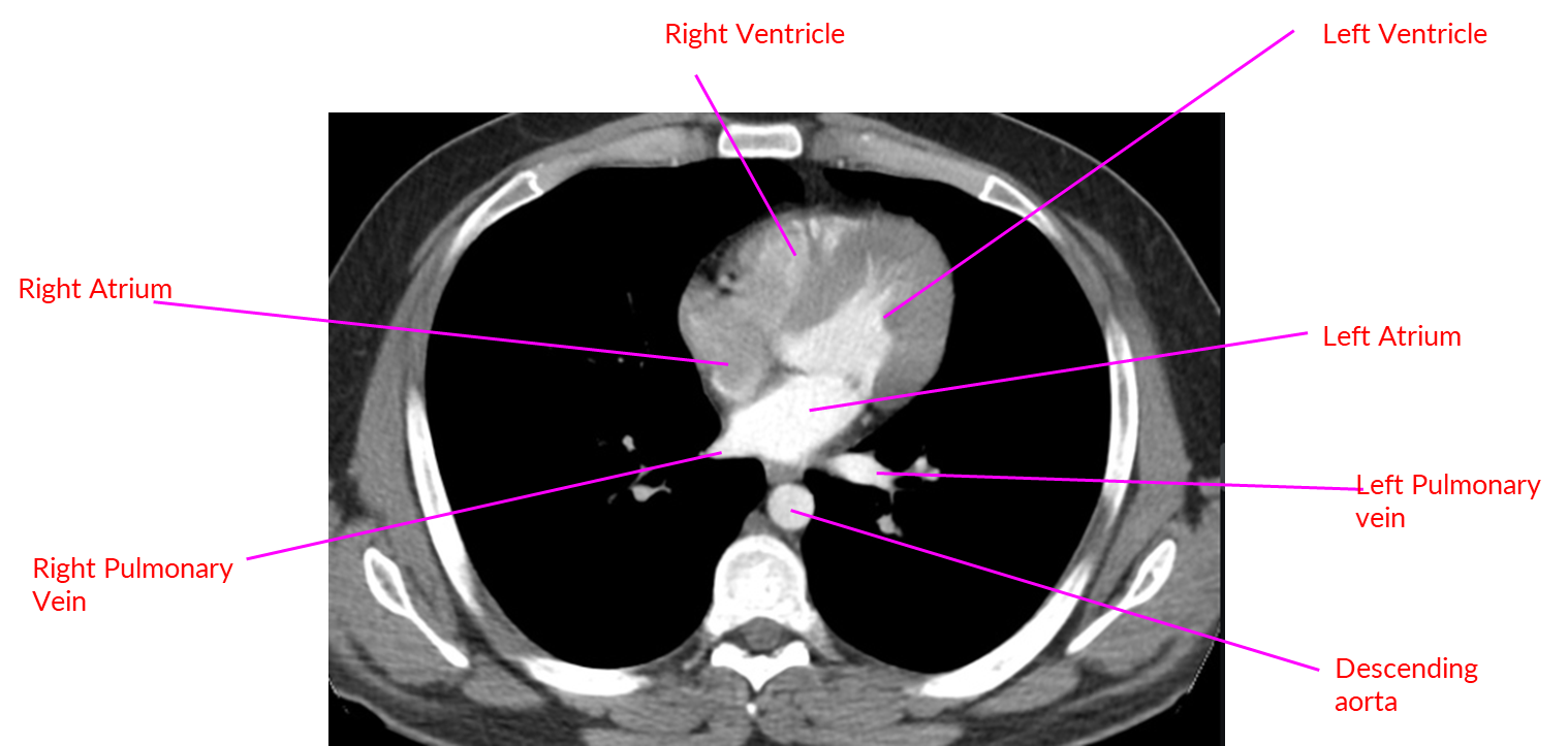
Structures (3)
