VCP - 2023
1/72
There's no tags or description
Looks like no tags are added yet.
Name | Mastery | Learn | Test | Matching | Spaced |
|---|
No study sessions yet.
73 Terms
Siderotic PCs in Lab w PIMA
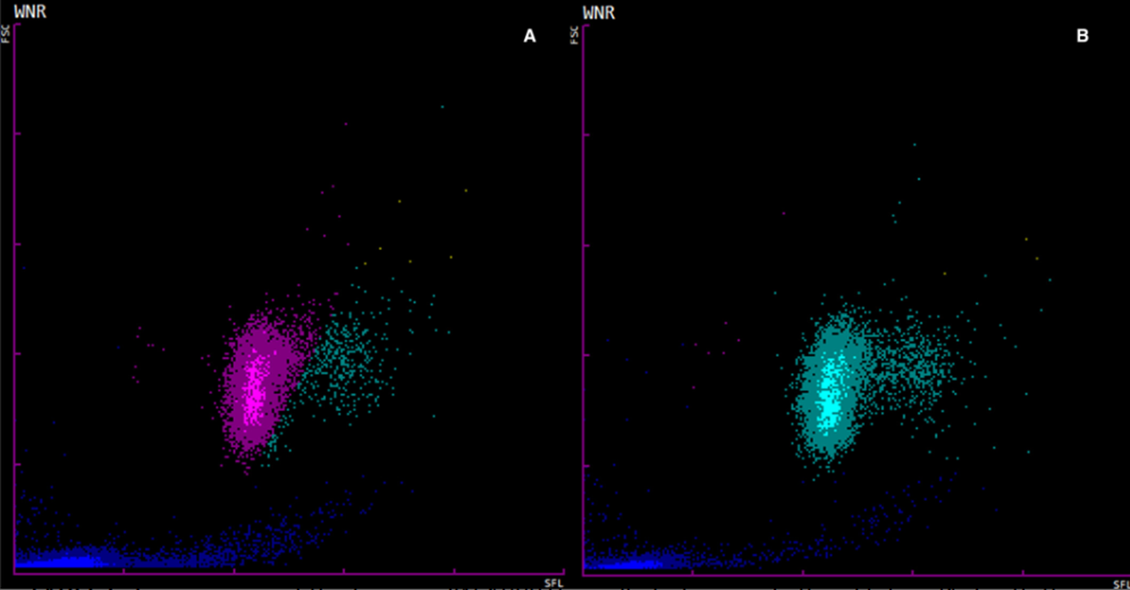
WNR channel graph from Dog 4, demonstrating an error in the automated gating of NRBCs. (A) Automated gating demonstrates a significant proportion of leukocytes counted as NRBCs (purple), while (B) demonstrates correct gating as leukocytes (turquoise) on the second run following dilution.
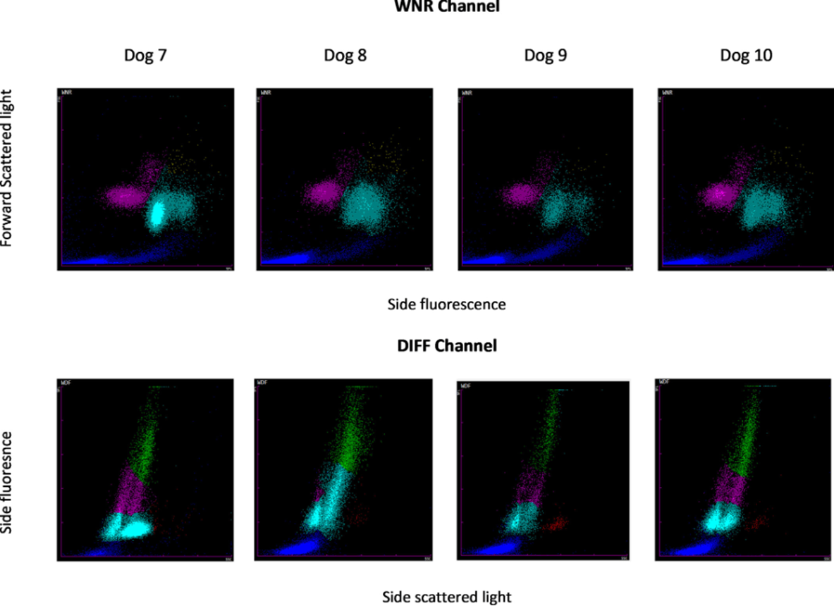


Unusual poorly defined cloud above & to the right of the nRBC cloud, extending into the basophil area. Also an abrupt delineation between the nRBC and leukocyte clouds, indicating errors in gating.
-Cases 10, 12: nRBC and leuko clouds abruptly delineated (gating error)
-Cases 8, 10, 12: signif merging of platelet/cell fragment cloud w the leuko cloud
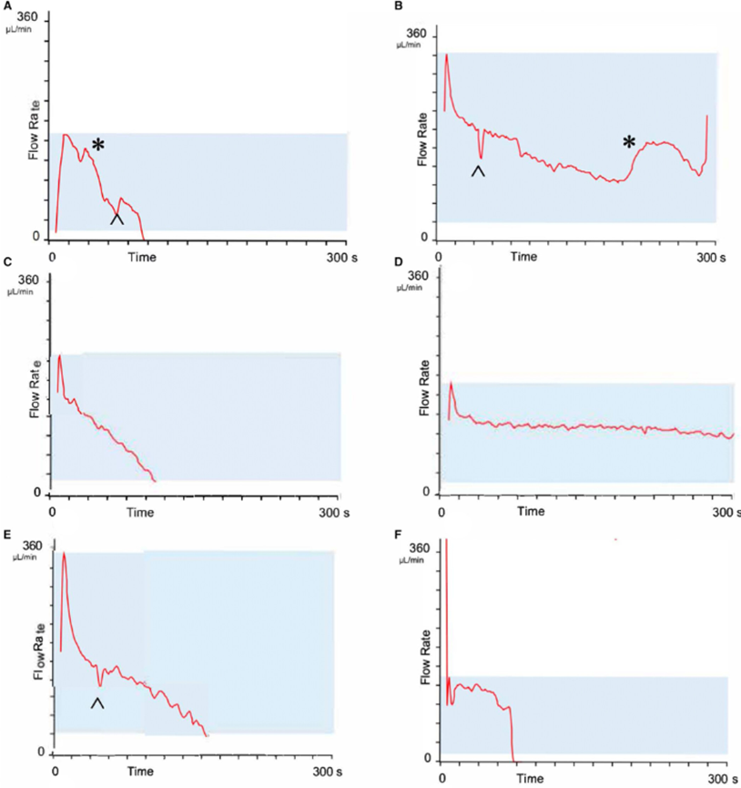
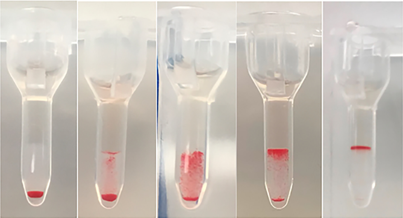
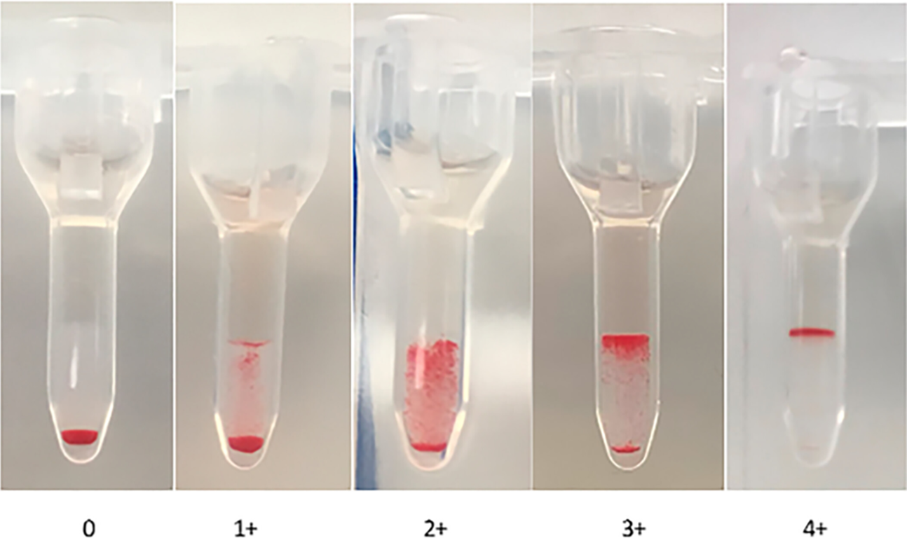
PFA-200
Several patterns: (closure, non-closure, & flow obstr)
•Peaks – elevations above the plane of curve, rapid return to baseline (A, B, *)
•Valleys – dips below the plane of curve, rapid return to baseline (B, E, ^)
•Rapid initial increase – initial peak formed at >45° angle
•Slow initial increase – initial peak formed at <45° angle
•Steady decline – essentially linear, no change in slope, reaching baseline (B, C)
•Flatline – approximately horizontal after initial peak (D)
•Multiphasic – one or more signif changes to slope
•Initial vertical peak – rapid incr apparently to infinity in first seconds of flow (F)
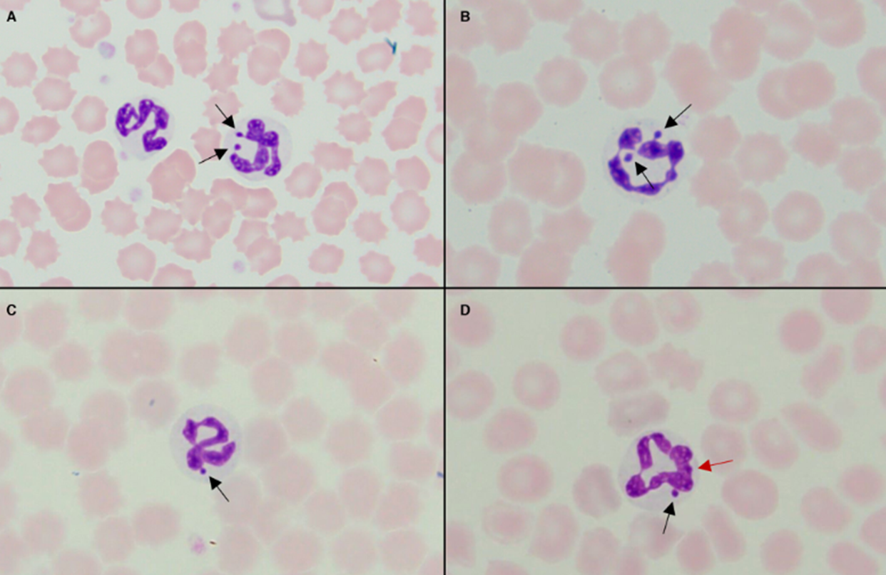
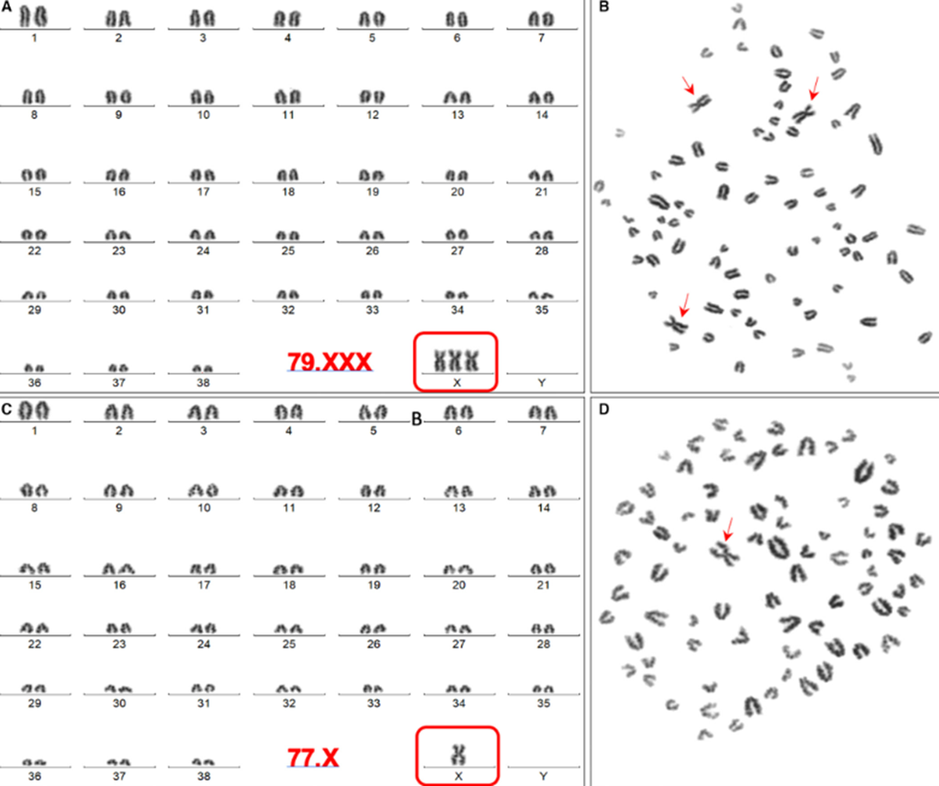
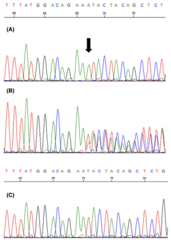
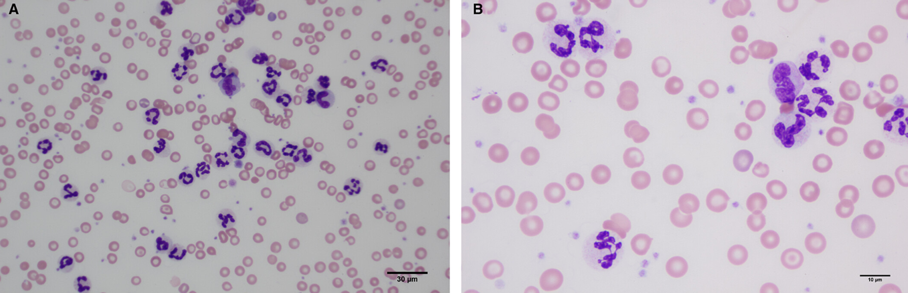
Double Barr bodies and X-monosomy/X-trisomy mosaicism in a dog with presumed idiopathic epilepsy (some had 1, many had 0)
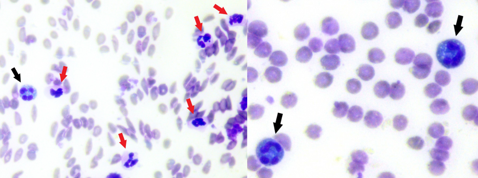
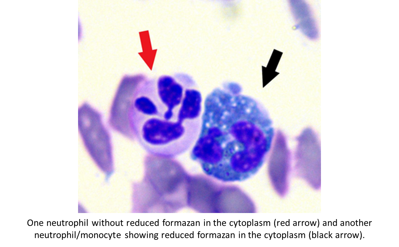
Nitro blue tetrazolium reduction test
Blue indicates activation (ROS in oxidative burst)
Border collie puppies with hematomas and RA, F8 gene:
Hemophilia A due to deletion, B is carrier, C is affected
All had prolonged aPPT, FVIII 5-6% activity
Lung mass ALP stain
Histiocytic sarcoma. Arrows = atypical round cells (ALP neg), arrowheads = respiratory epithelium (ALP pos)
Lung imprint, ALP stain

Mesothelial cells, ALP neg
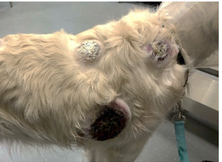
3yo Golden w multiple cutaneous masses rapidly growing

CD3- Pax5- MUM1+
MUM1/IRF4 labeling in Langerhans cell histiocytosis, initial misdx as PCT
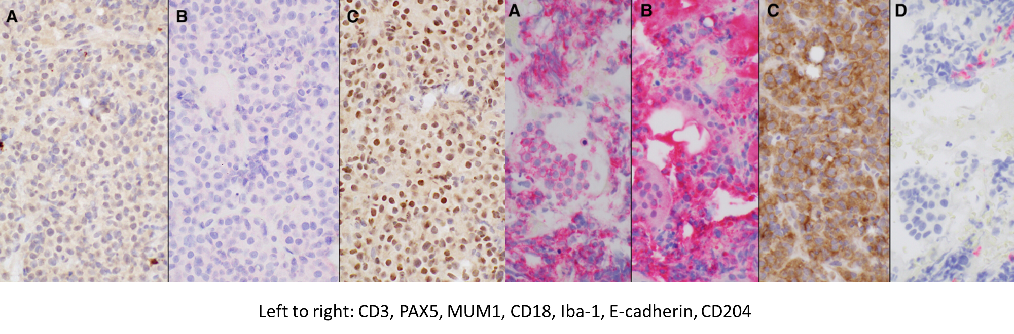
Cutaneous mass on flank of a dog
Keloidal fibroma

Unclear why aSMA+ in this case
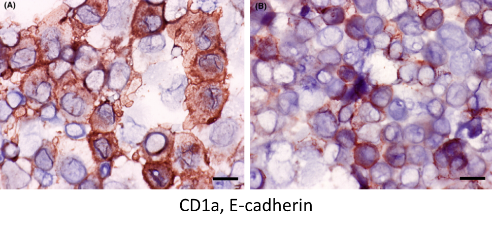
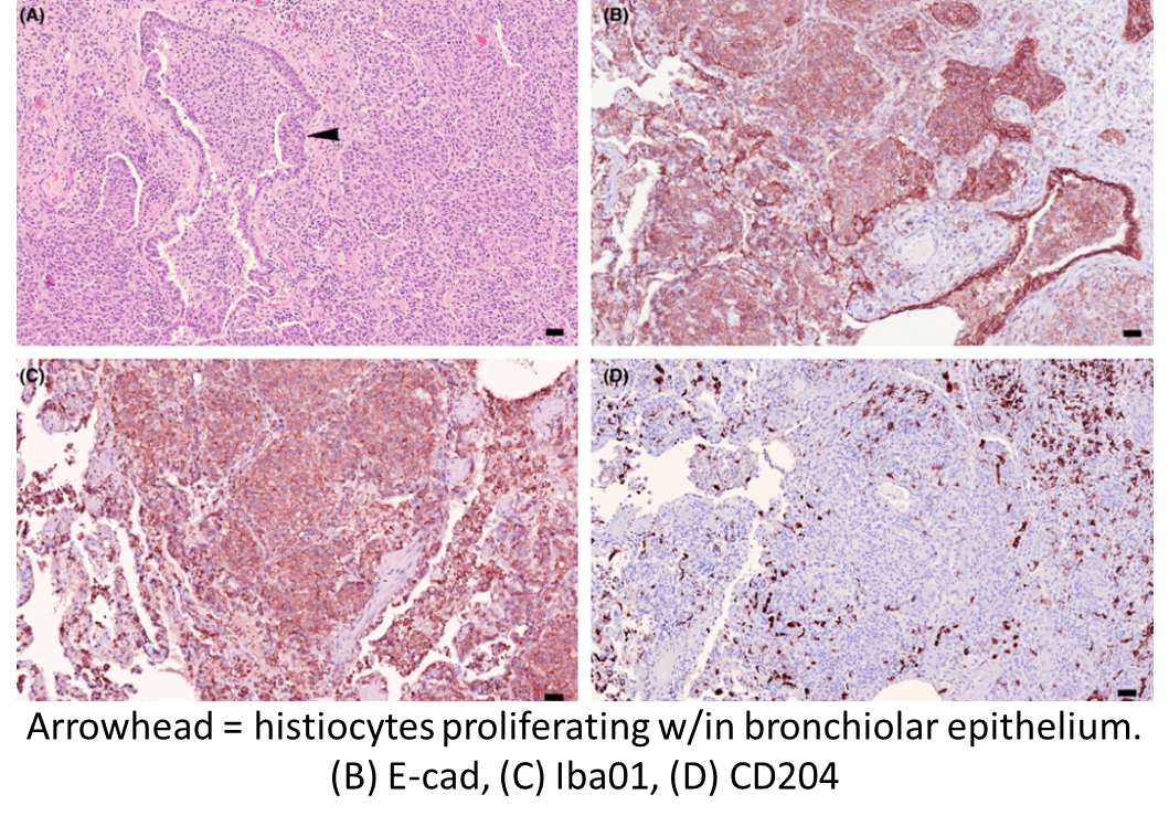
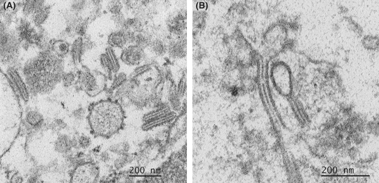
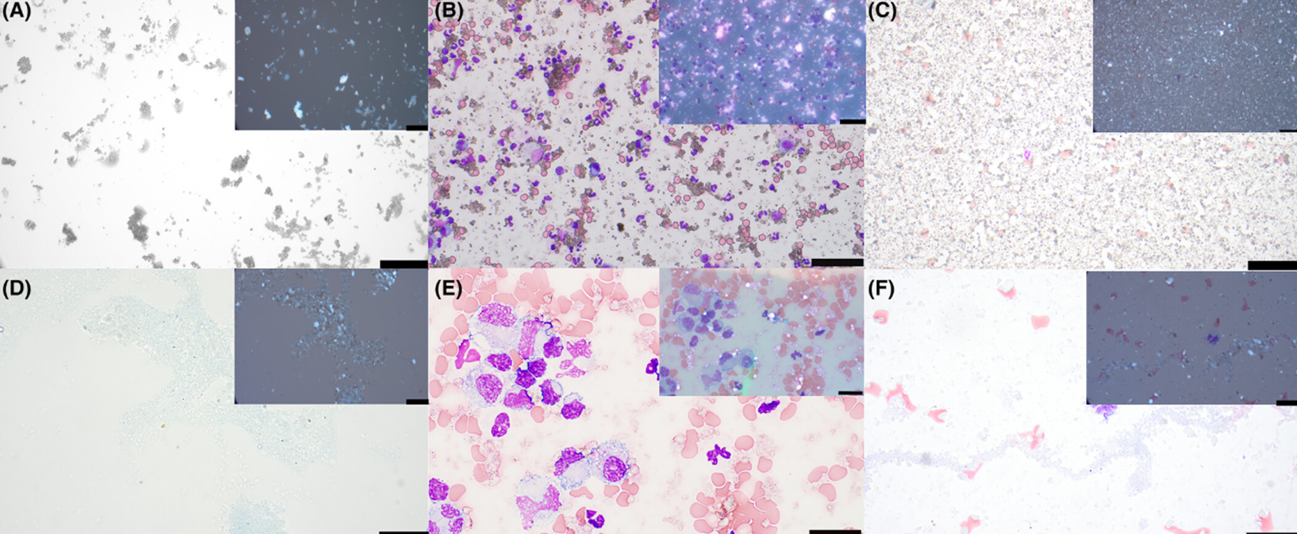
BAL from a 9yo British shorthair, 3mo resp difficulty refractory to steroids & antimicrobials
Feline pulmonary Langerhans cell histiocytosis
Final: E-cad+/Iba-1+/CD1a+/CD5+/CD18+/MHCII+/CD204−/CD4−
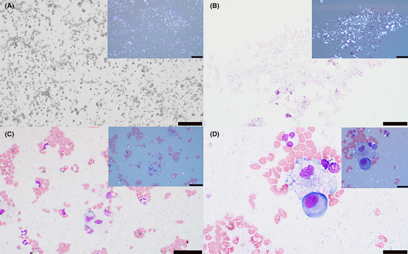
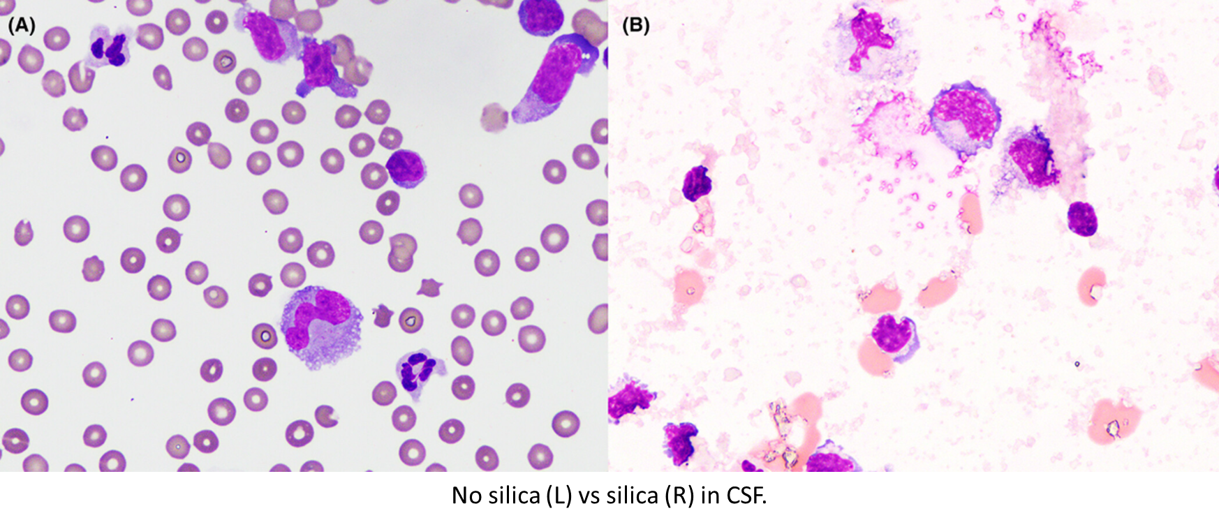
Left to right: DI water, peritoneal effusion, CSF
Top low power, bottom oil immersion
Silica particles
Silica particles in urine, low-power and oil immersion.
Top row DI water, bottom row pleural effusion (low and high mag)
Barium particles
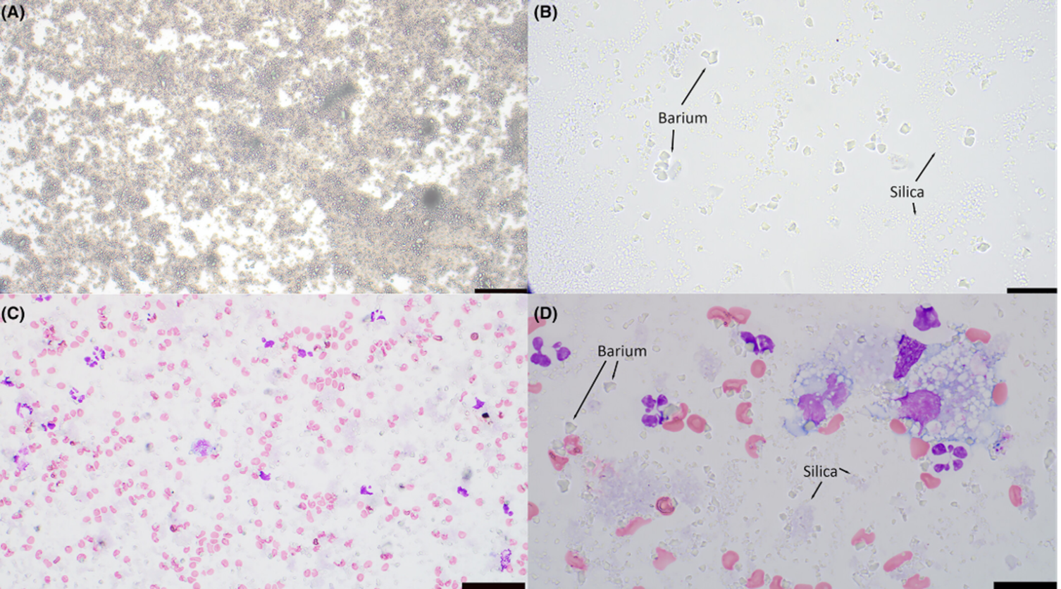
Both silica & barium. DI H2O top, pleural effusion bottom; low mag left, oil immersion right.
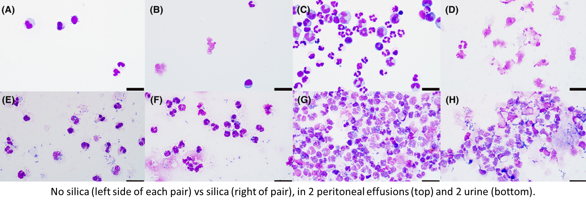
It’s labeled so not much of a flashcard, but good pic
Effects on preservation:
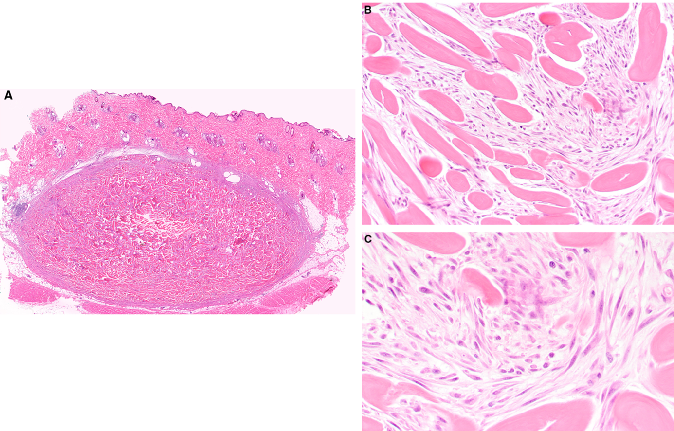
SQ mass on thigh of 14yo Border Collie
Extracardiac adult rhabdoyoma
Desmin top, NSE bottom
SQ mass flank of 13yo mixed dog
Extracardiac adult rhabdomyoma
Desmin top, NSE bottom
8yo MN yorkie with respiratory effort, lethargy, mild pleural exudate, mediastinal mass
Spindle-cell thymoma colliding with a bronchogenic cyst
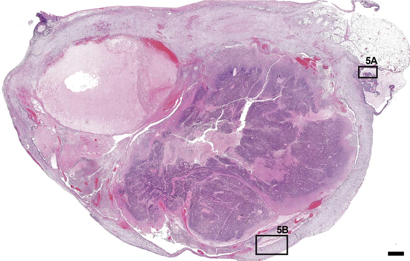
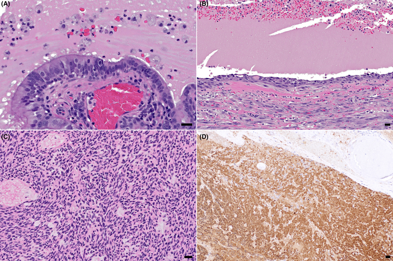
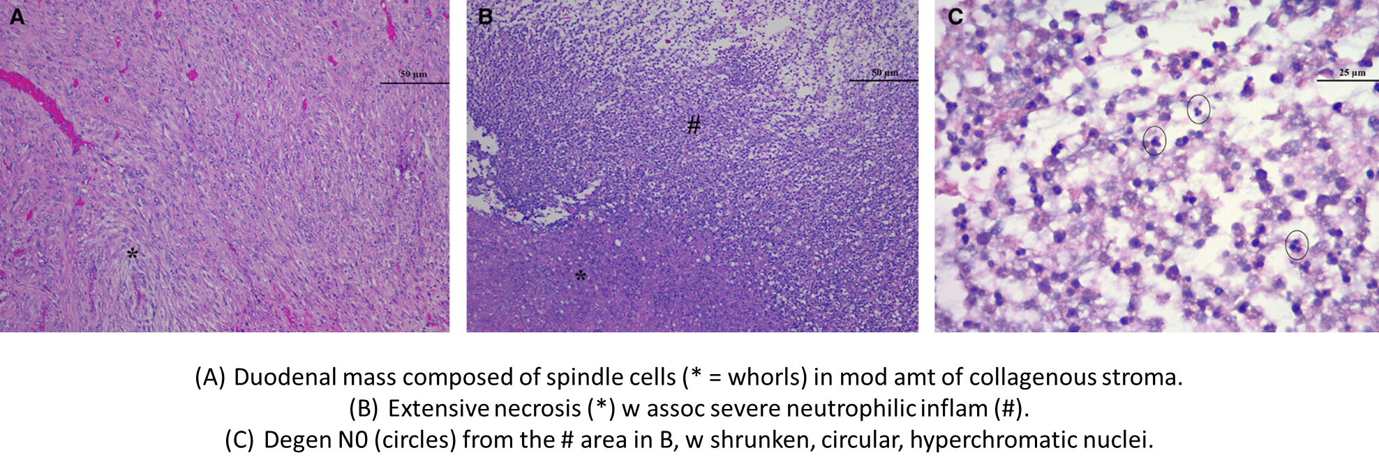
A) Well-diff’d pseudostrat ciliated resp epith, cyst lumen contains proteinaceous fluid, N0, histiocytes, RBCs.
B) Elsewhere, lining is attenuated and lined by squamous to stratified squamous epith.
C) In solid areas, neoplastic spindle to polygonal cells in streams/sheets w elongated to oval nuclei.
D) CK+
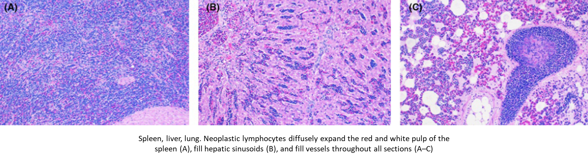
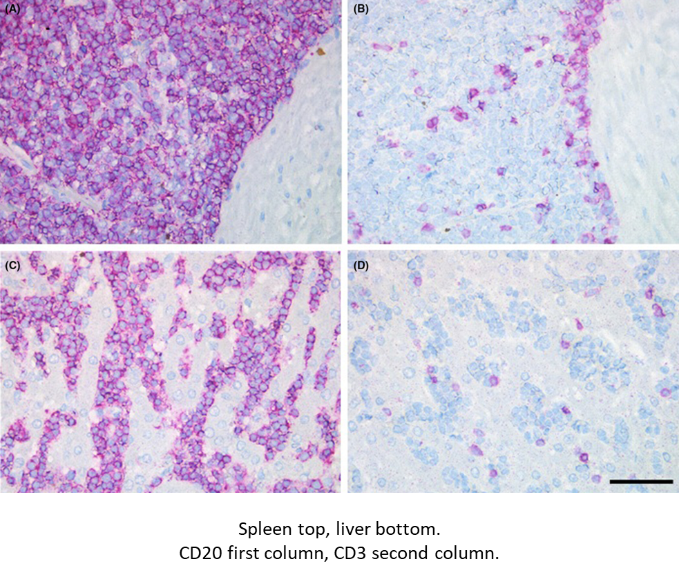
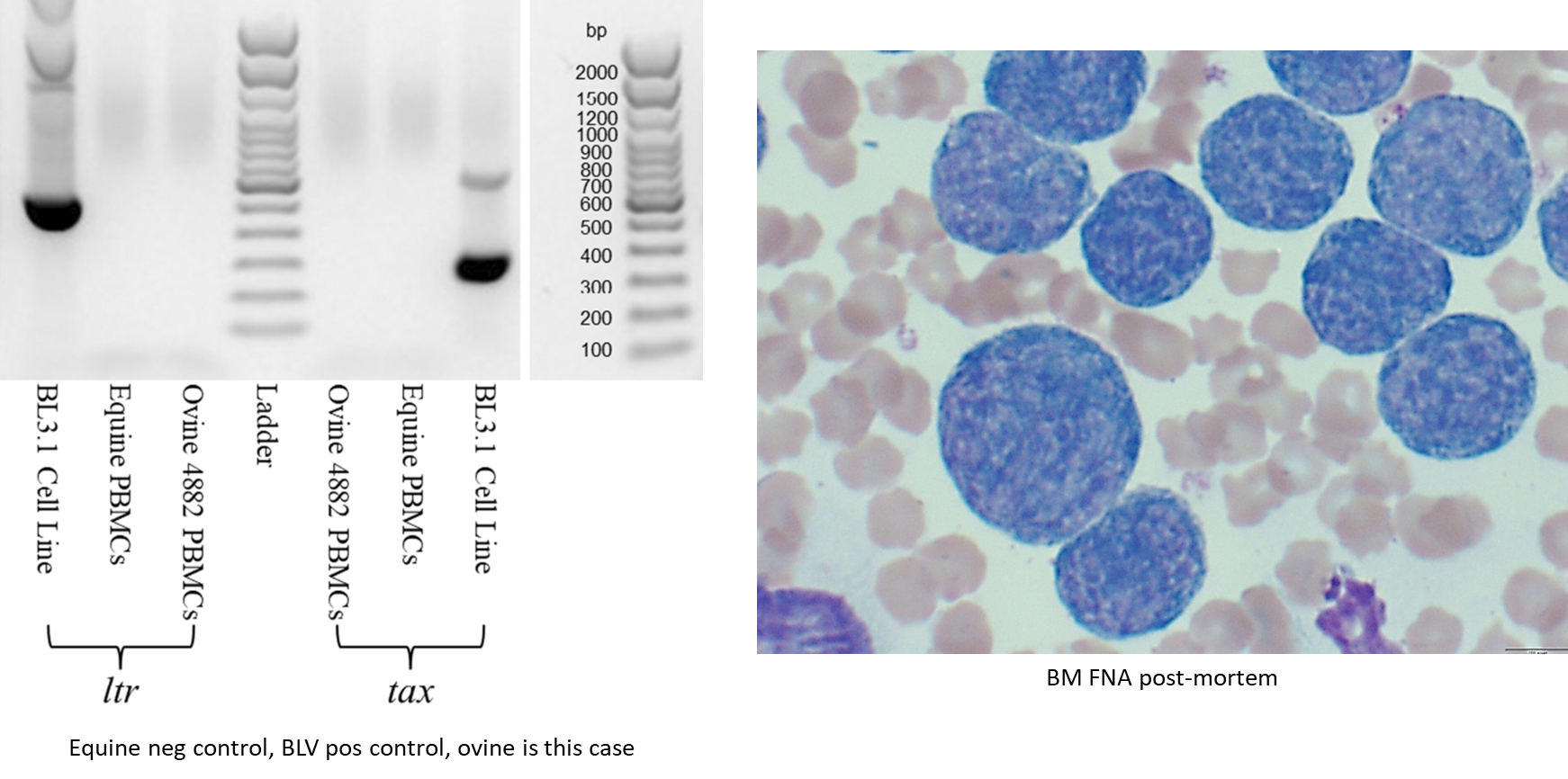
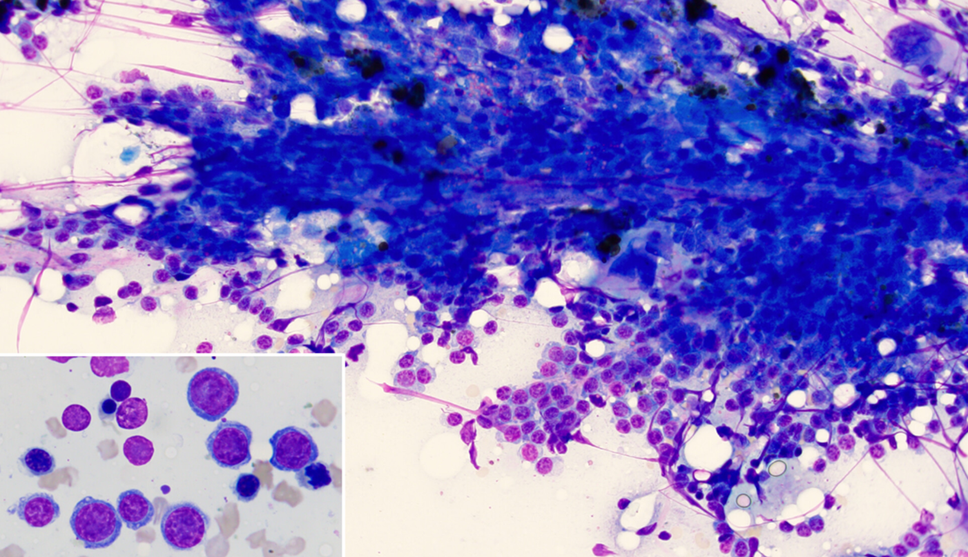
5yo Whiteface wether, WBC 708
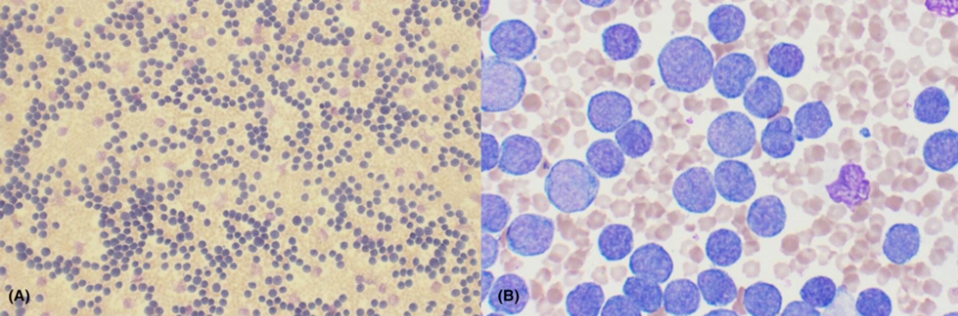
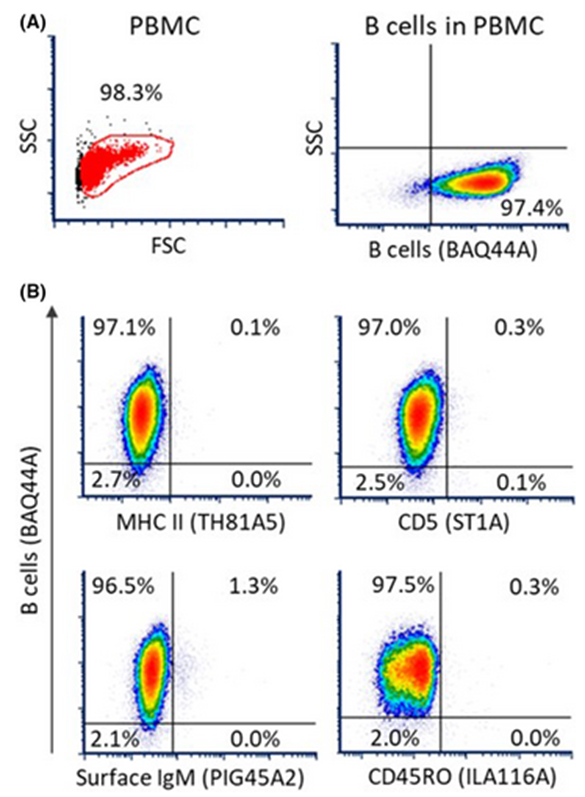
B-cell leukemia
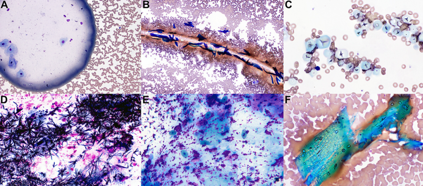
(A)Human spittle; (B) Flaky human hair; (C) Starch/glove powder; (D) Gram stain precipitate; (E) Medical lubricant; (F) Pleomorphic plant material
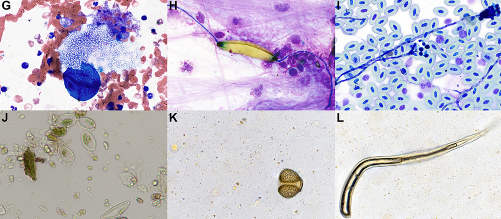
(G) Plant spore-filled spherule
(H) Fungal structure in a BAL from non-sterilized endoscope
(I) Fungal hyphae and conidial heads on a blood film left unstained for several months
(J) Diatoms from skin contamination in a urine wet-mount in a manatee
(K) Grain of pine pollen
(L) Plant hair/trichome
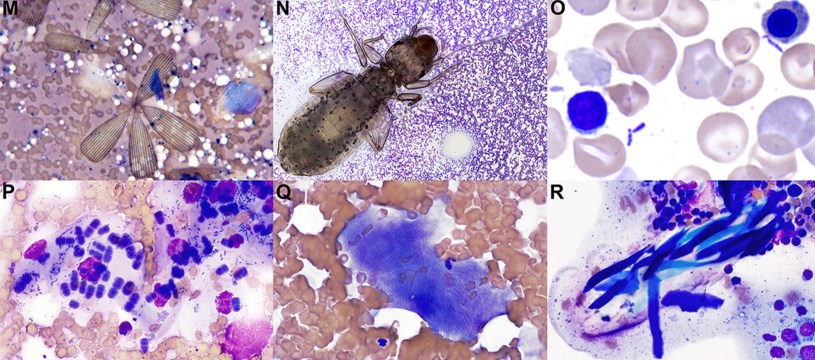
(M) Fragments of mosquito parts
(N) Booklouse trapped in immersion oil
(O) Superficial mixed bacterial on a blood film from a nail prick on a marmoset
(P) Oropharyngeal contaminants
(Q) Fragment of striated skeletal muscle
(R) Keratin flakes/bars
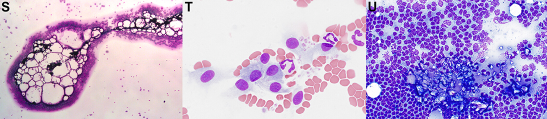
(S) Lipid in a blood film from a crested toad
(T) Endothelial cells in a blood film from a dog
(U) Glass shards on the edge of an FNA
Blood from a Zovawk pig
A: M0 and RBC
B: N0
C: E0
D : B0
Blood from a Zovawk pig
A: L0 and plts
B: M0
C: Plts (pleomorphic)
D : Plt
Blood from wild anurans
A: Intraerythrocytic haemogregarine
B: Lankesterella and a neutrophil
C: Intracytoplasmic structure and a thrombocyte
D: Hepatozoon sp. and a microfilaria
Blood from wild anurans
E: Trypanosoma sp.
F: Lankesterella sp.
G: Trypanosoma and Hemolivia sp.
H: Trypanosoma sp.
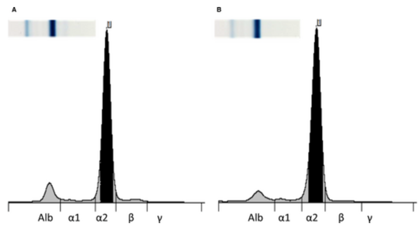
Bald eagle protein electrophoresis
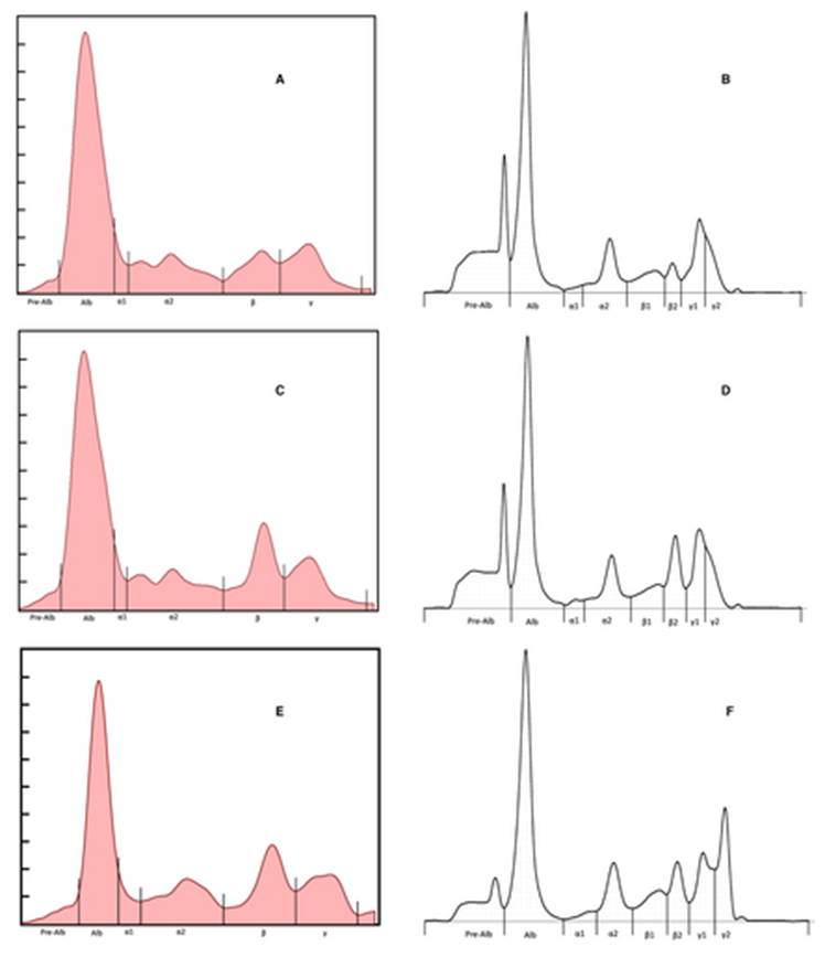
Left AGE, right CZE
Paired serum + plasma
First 2 rows clinically normal
Last row clinically abnormal
- Note 2 defined gamma globulin fractions
AGE 1-6: prealb, alb, a1, a2, b, g
CZE 1-8: prealb, alb, a1, a2, b1, b2, g1, g2
Greyhound BM sample stored for 3d
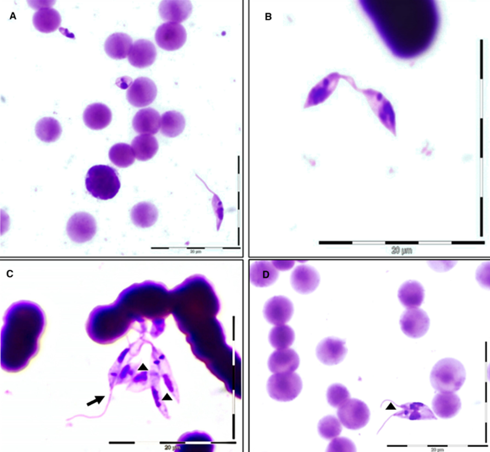
Leishmania promastigotes
- (A) Procyclic promastigotes and amastigotes
- (B) Procyclic promastigotes
- (C, D) Elongated nectomonads (arrowheads) and leptomonad (arrow)
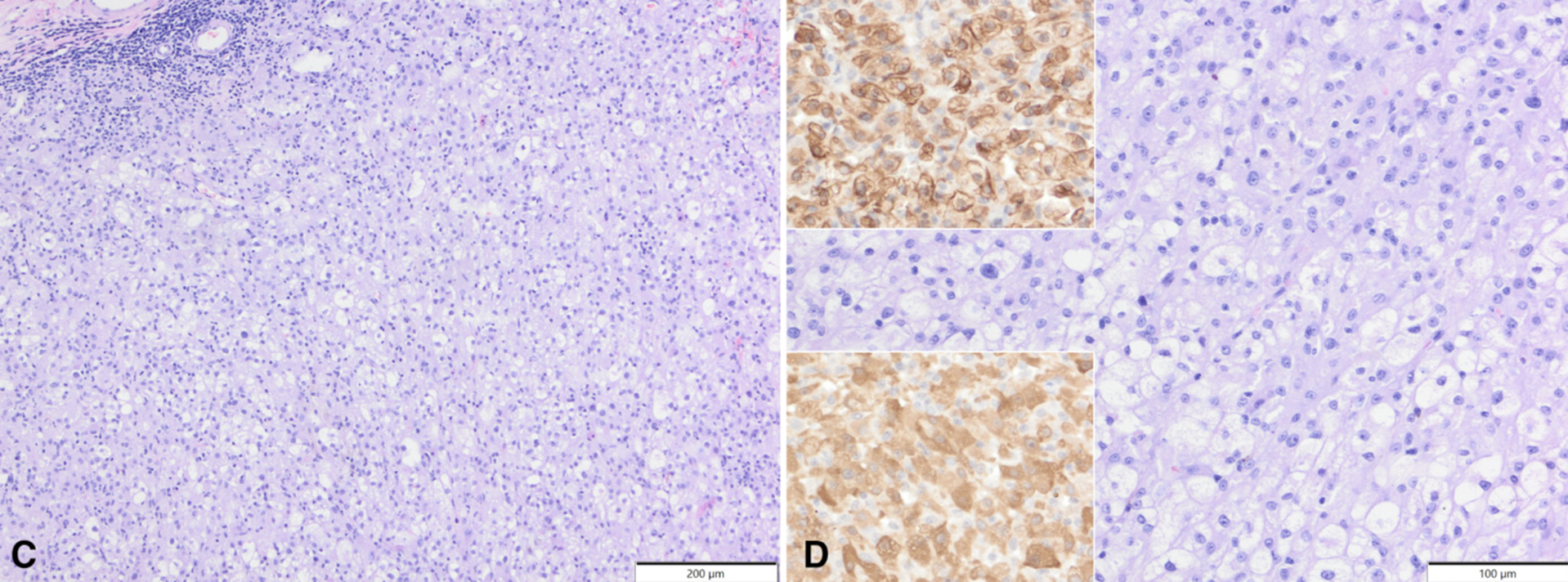
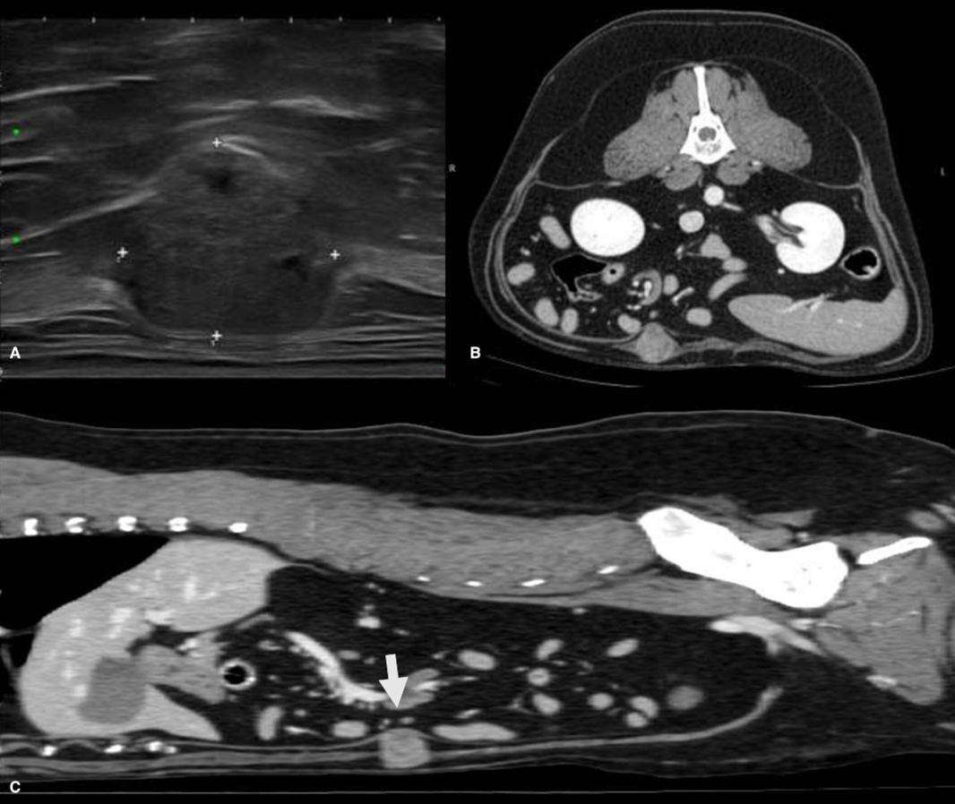
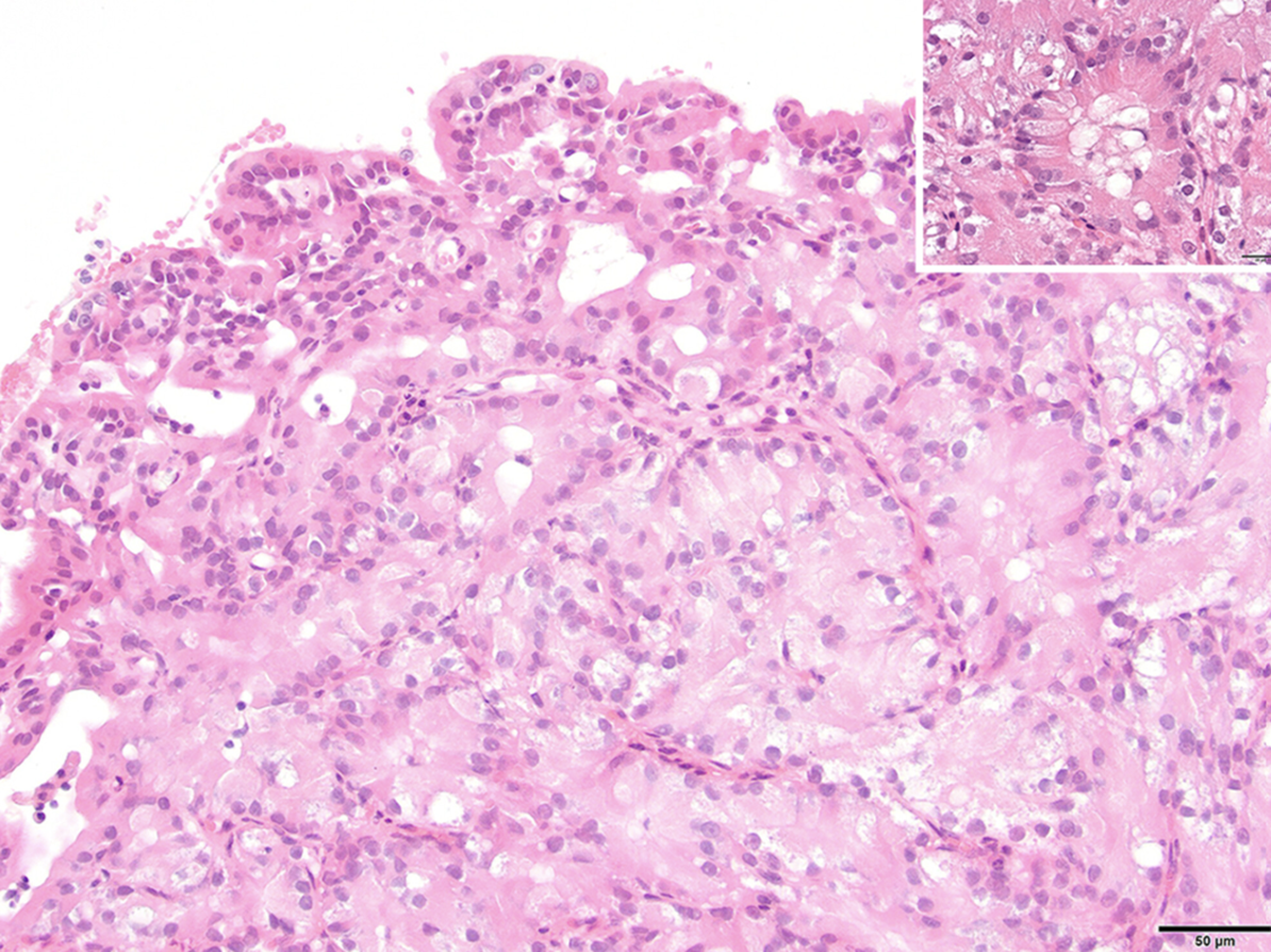
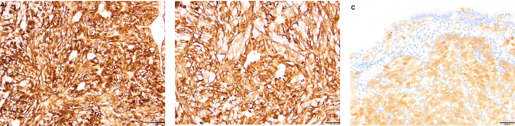
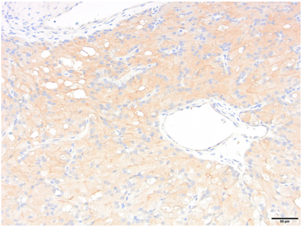
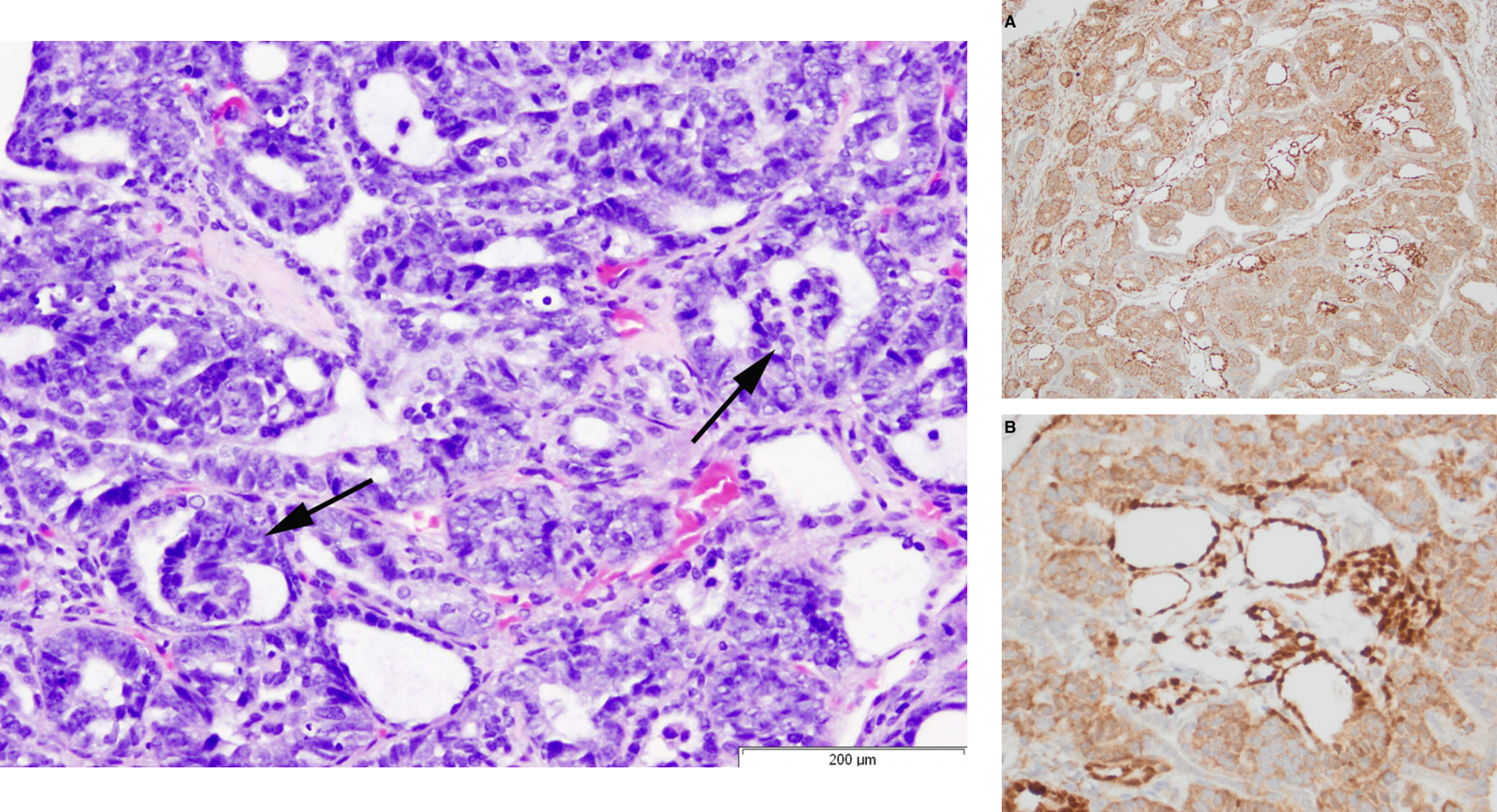
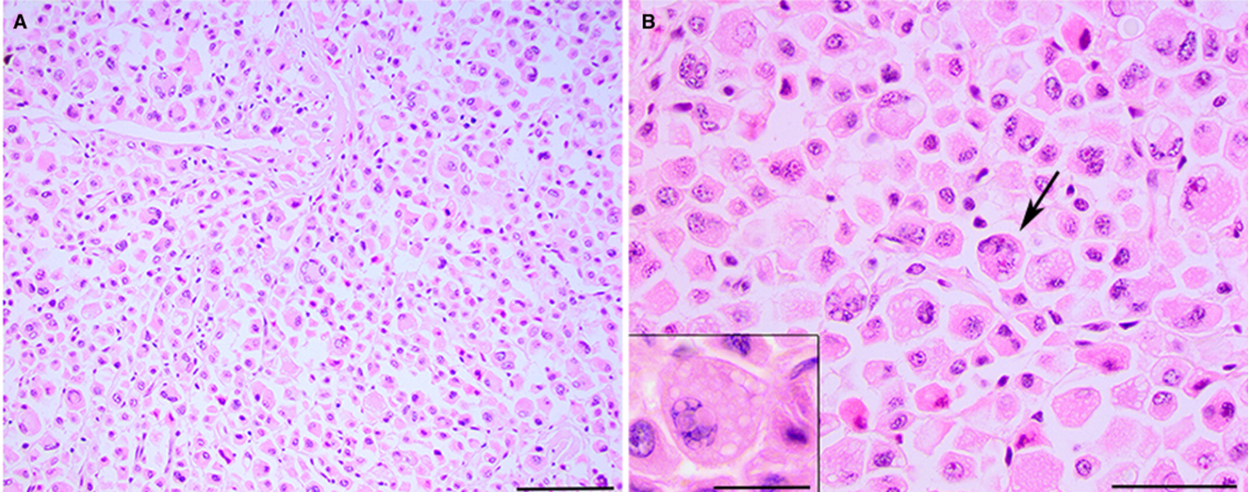
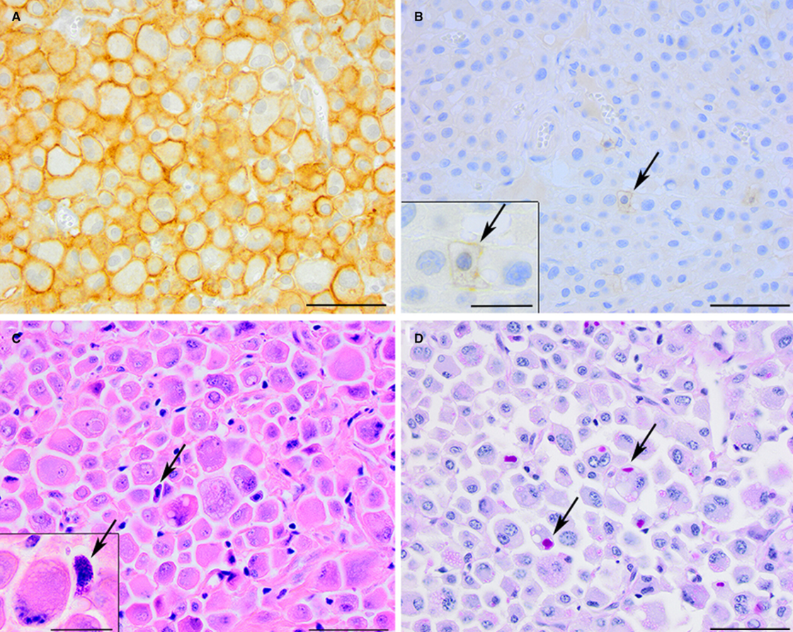
11yo FS Golden with SQ nodule, elevated liver enzymes
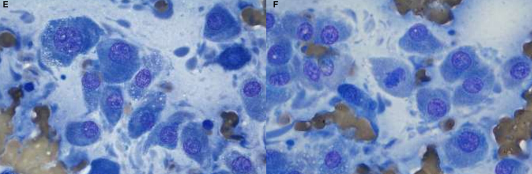
Needle tract seeding and malignant transformation of hepatocellular adenoma into well-differentiated hepatocellular carcinoma, SQ mass is at the core bx site
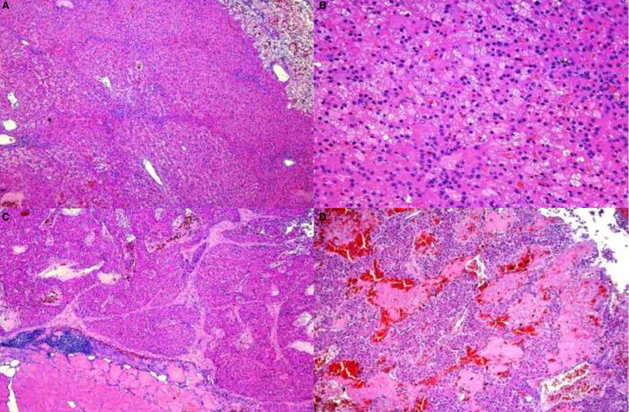
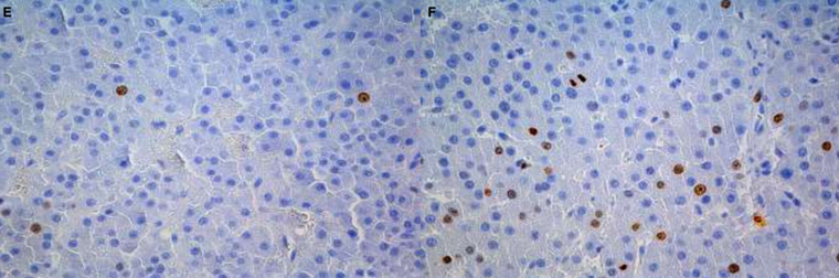
(A, B) HCA. Well-demarcated with adjacent normal liver, forms pseudolobular structures composed of regular thick trabeculae
(C) HCC. Effacing muscle & adipose, loss of lobar architecture, irregular thickness of trabeculae (>5 hepatocytes thick), moderate desmoplasia.
(D) Randomly distributed lytic necrosis & hemorrhage.
(E, F) Ki-67. Hepatic mass mild positivity (2.9 pos nuclei/HPF) vs intramuscular mass highly increased positivity (21.1/hpf)
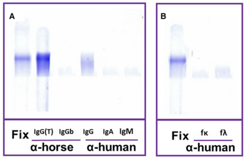
BM from a horse with blepharitis and incidental hyperglobulinemia
Multiple myeloma with IgG(T) M-protein (IgG3 and 5)
Serum and plasma
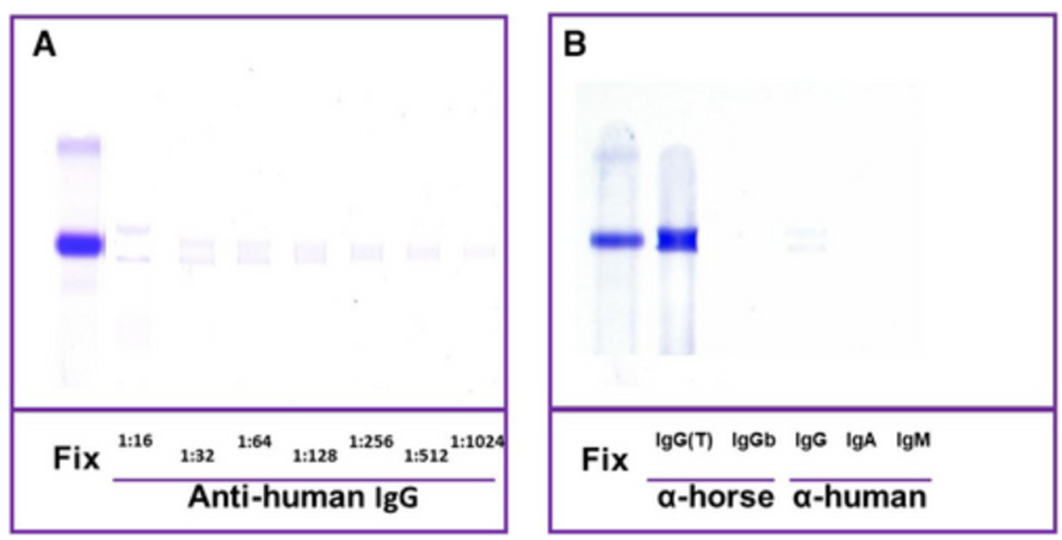
Urine
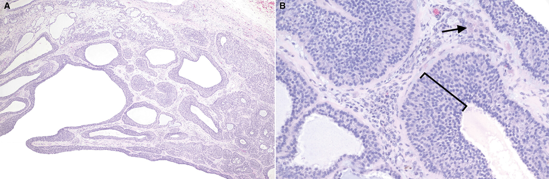
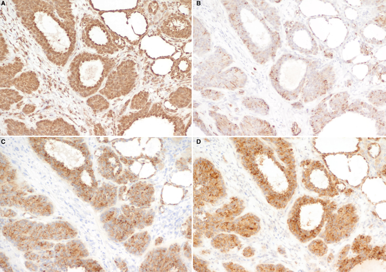
Nasal mass in a 7yo Burmese
Olfactory neuroblastoma
(A) CK AE1/AE3, diffuse, strong membranous and cytoplasmic
(B) Vimentin, diffuse, strong membranous and cytoplasmic
(C) III beta-tubulin, diffuse, strong cytoplasmic
COX-2, diffuse, strong, 95% of cells, overlying resp epith & supportive stroma are negative
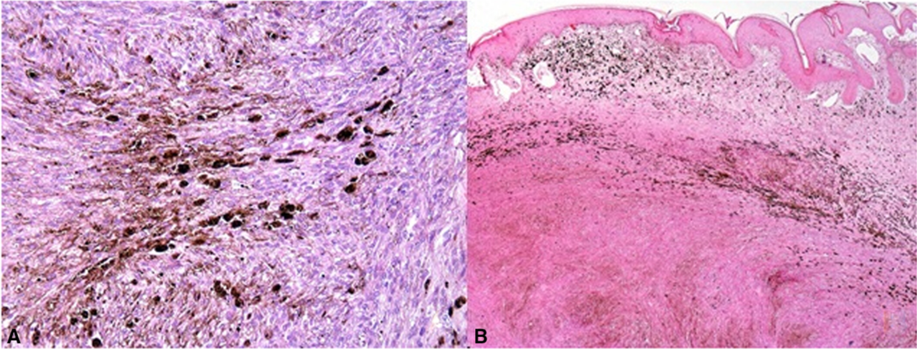
Lichtenstein’s green racer snake
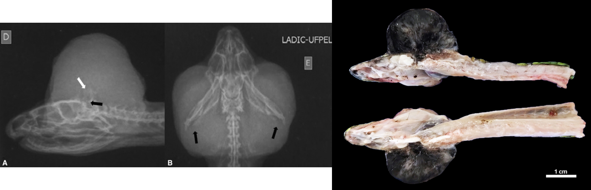
Melanophoroma
4yo Aus cattle dog with lethargy and generalized pain, CSF
TNCC 348/uL, RBC 310/uL, TP 423 mg/dL
Mycobacterium haemophilum (leprae clade)
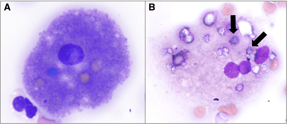
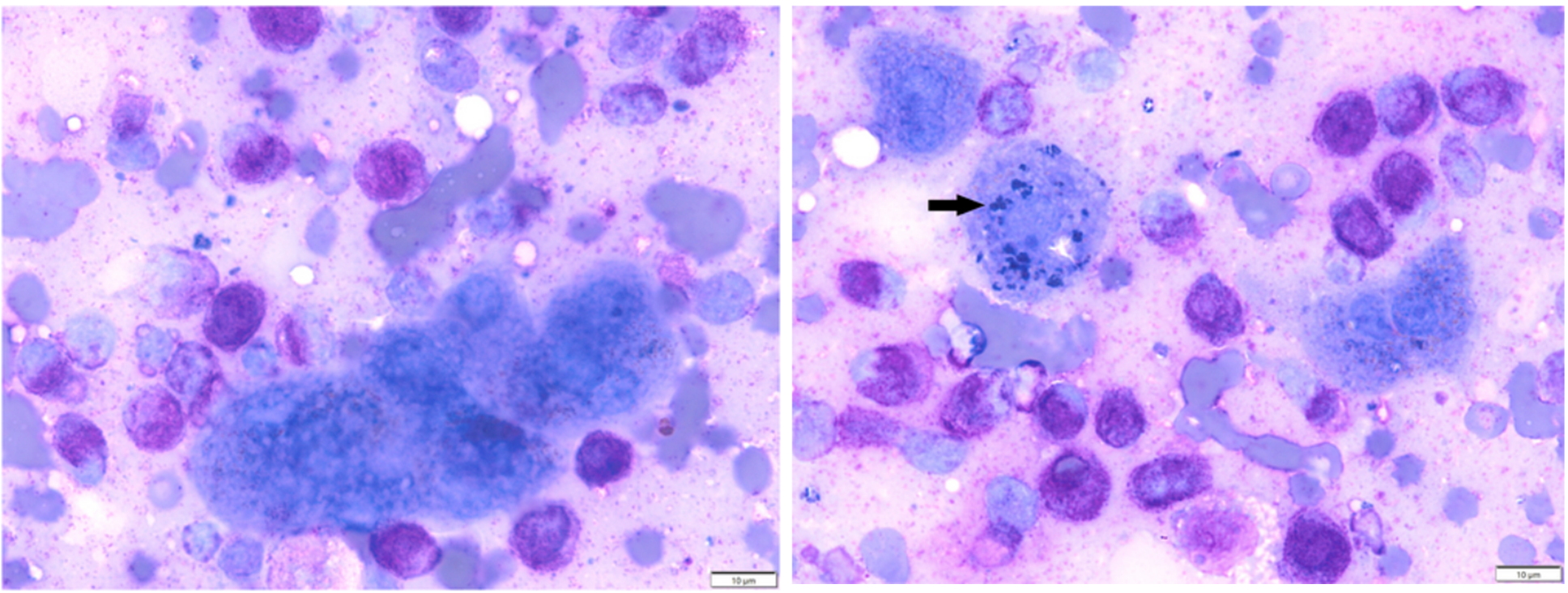
10yp DSH acute depression and anorexia, mod RA (16.3), severe leukopenia (1,200, N0 672), mild thrombocytopenia, FIV+, splenomegaly
Hemophagocytic syndrome in a cat with immune-mediated hemolytic anemia
6yo DSH 1wk lethargy, acute anorexia, marked NRA (9.7%), inappropriate rubricytosis (17), mod thrombocytopenia (59)
Spleen
BM
Mycoplasma haemofelis identified on PCR, secondary hemophagocytic syndrome
BM from a 9yo DSH with FIV+, panleukopenia (N0 0.27, L shift, toxic), HCT and plt WNL, splenomegaly
Presumptive hemophagocytic syndrome associated with co-infections with FIV, Toxoplasma gondii, and Candidatus mycoplasma haemominutum in an adult cat
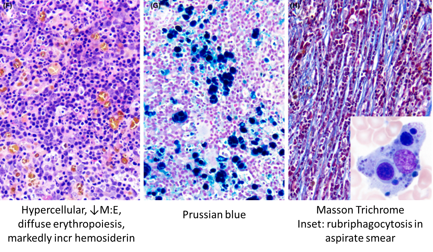
Bone Marrow
A: highly cellular, granulo hyper, L shift, incr immatures
B: erythrophagocytosis
C: rubriphagocytosis
D-F: neutrophagocytosis
Spleen at splenectomy:
A-B: erythrophagocytosis
C: rubriphagocytosis
D-F: neutro and erythrophagocytosis
Post-mortem liver
Top: nodules, neutro and erythrophago
Bottom: hepatocytes w high N:C, indistinct borders, and degen N0 w cocci
Post-mortem BM:
(Later stage of dz)
A: Erythrophago
B: Eryth and neutrophago
C-D: Rubriphago
E-F: neutrophago
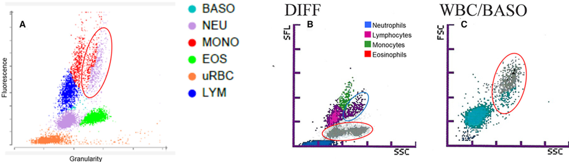
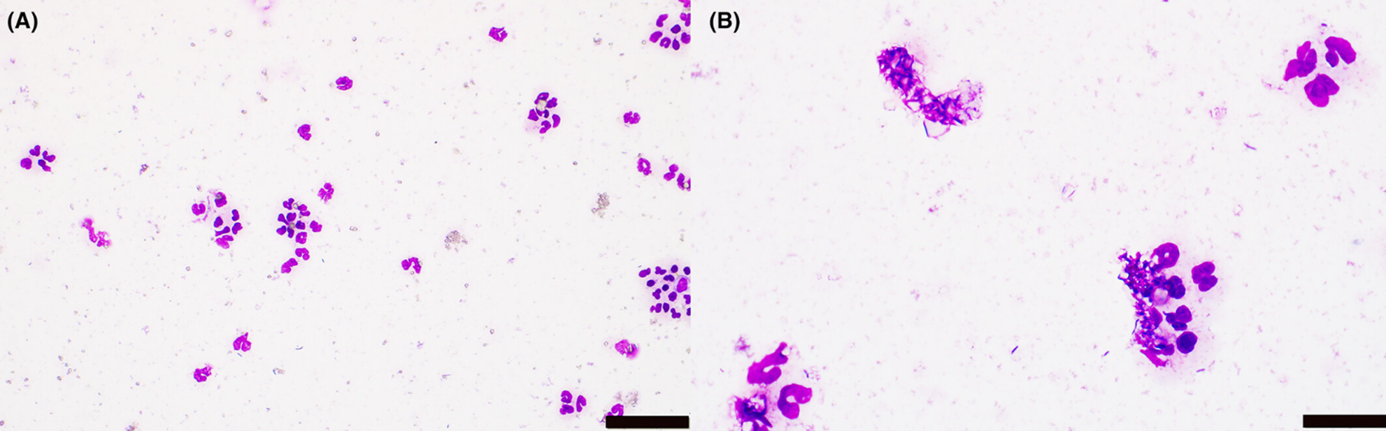
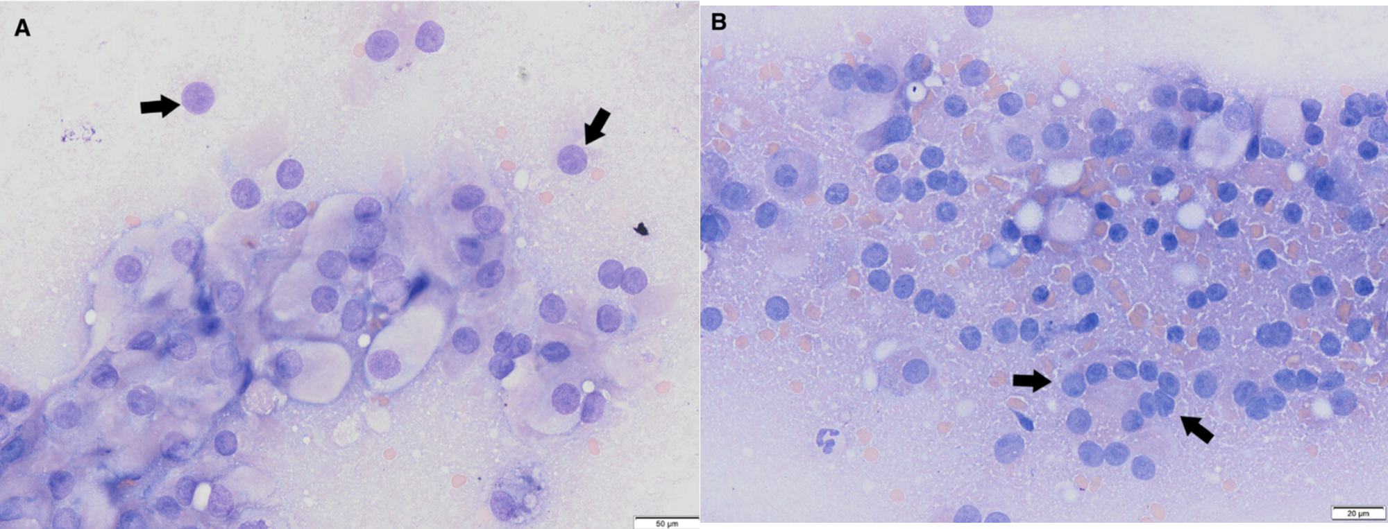
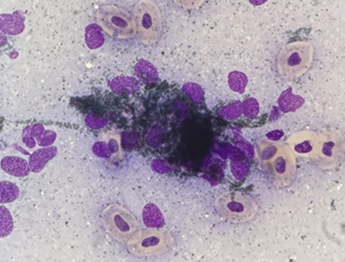
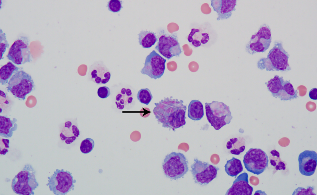
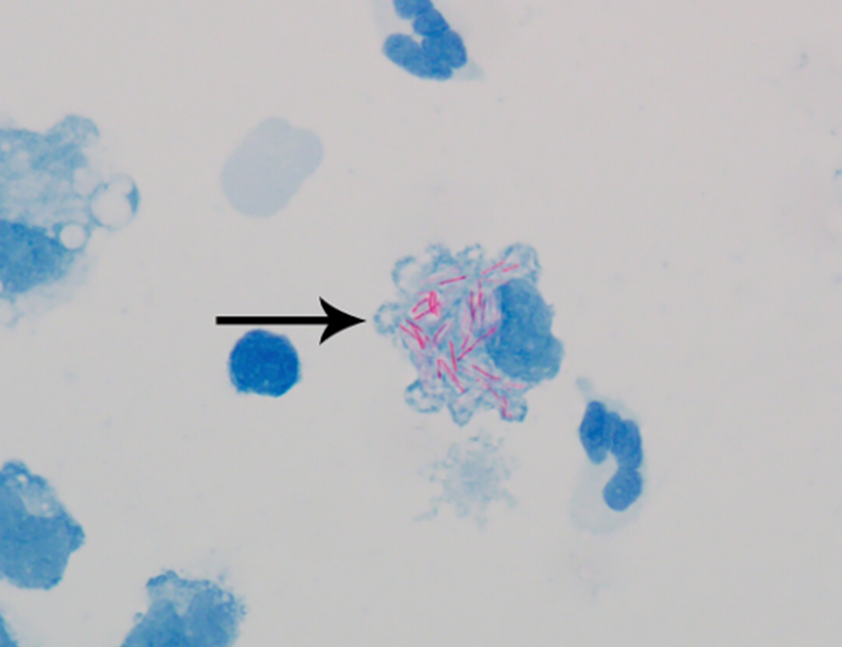
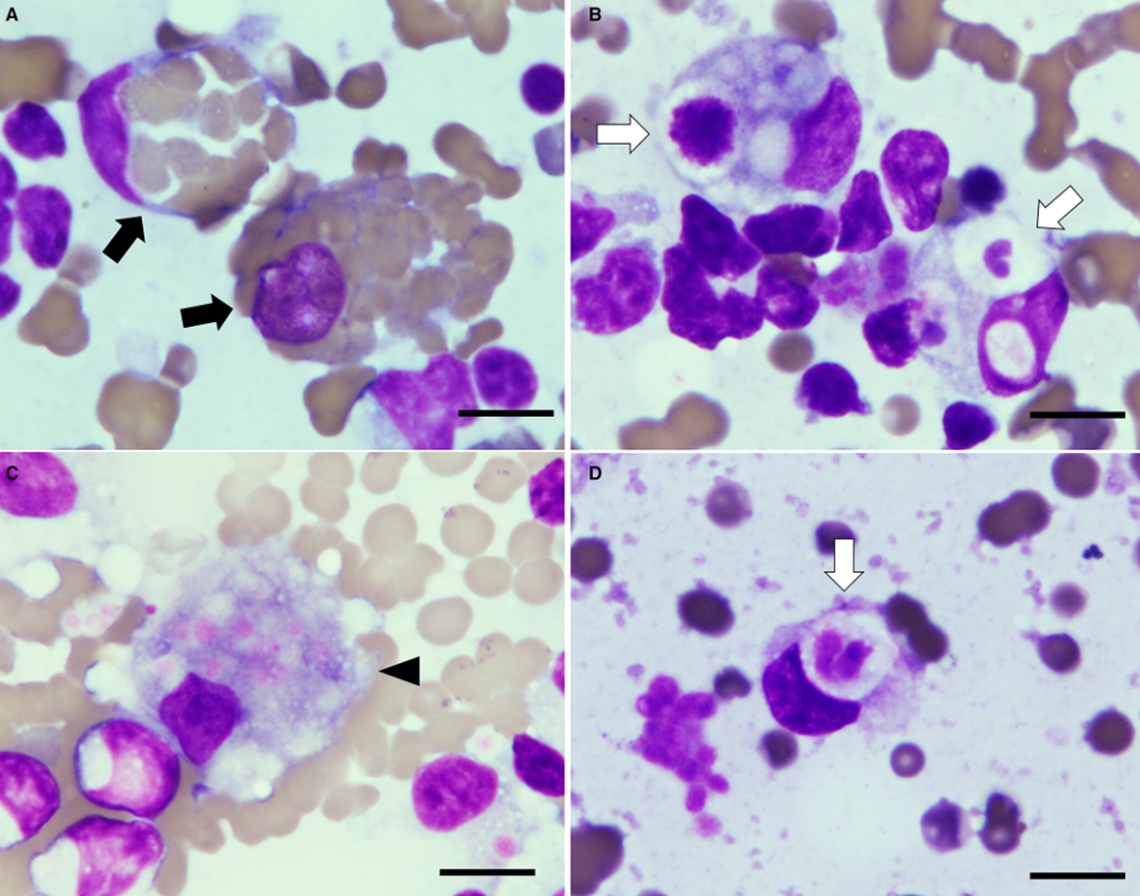
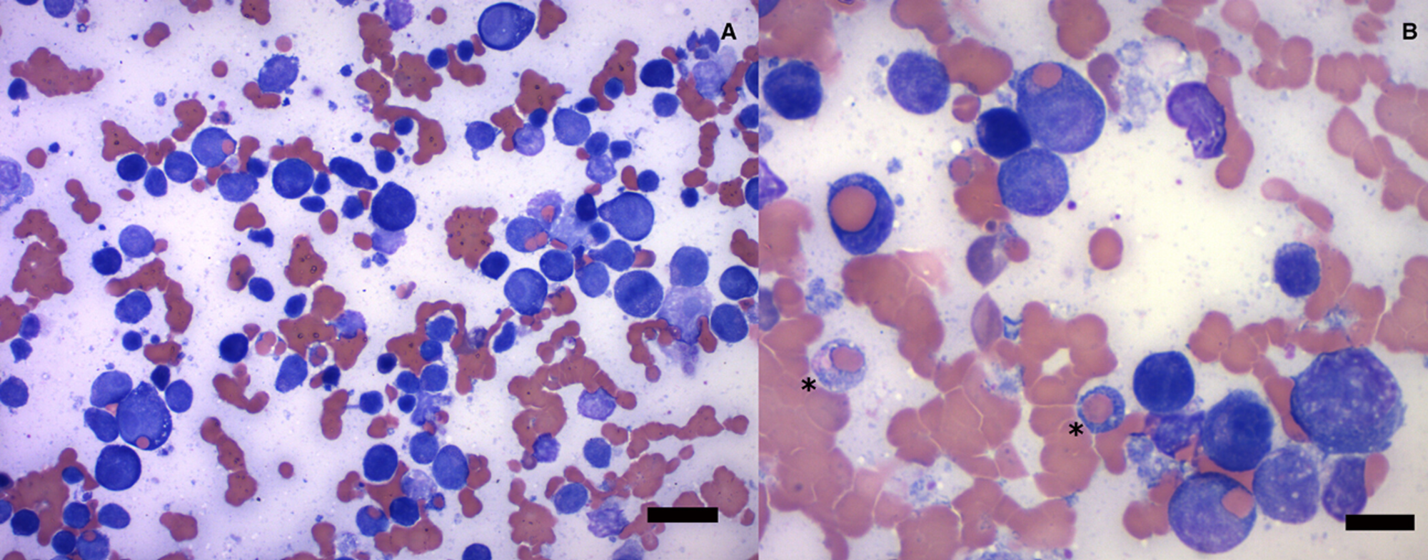
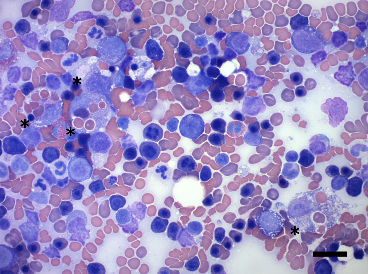
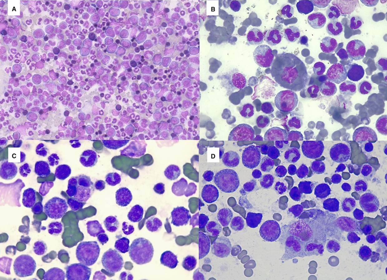
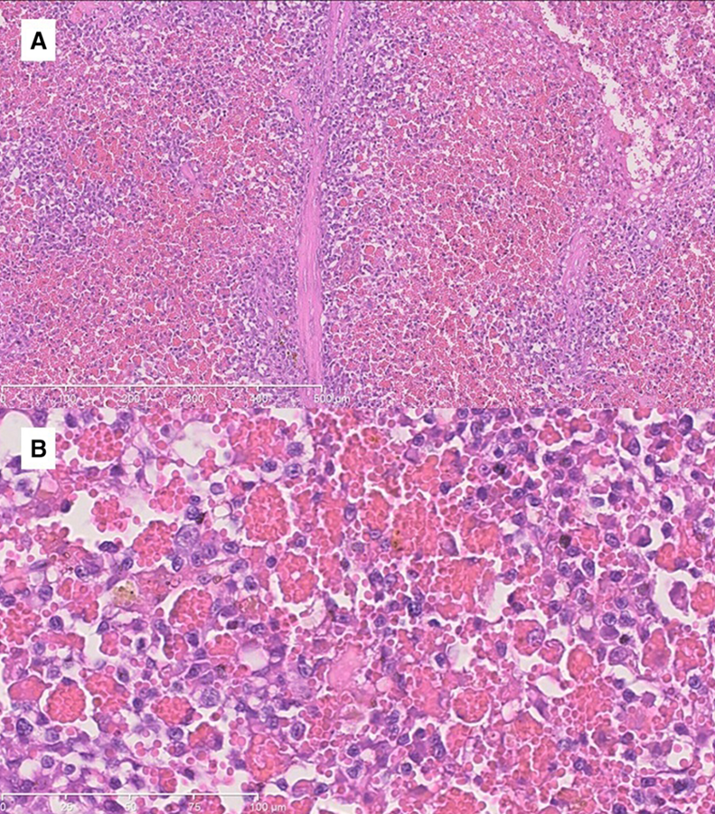
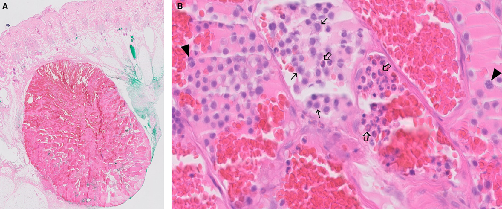
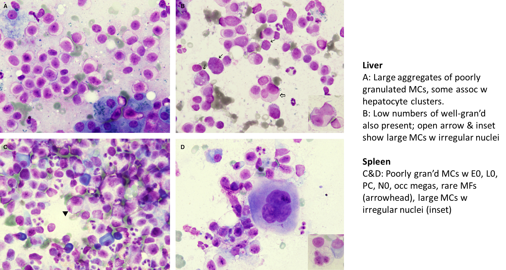
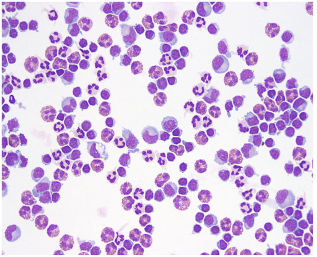
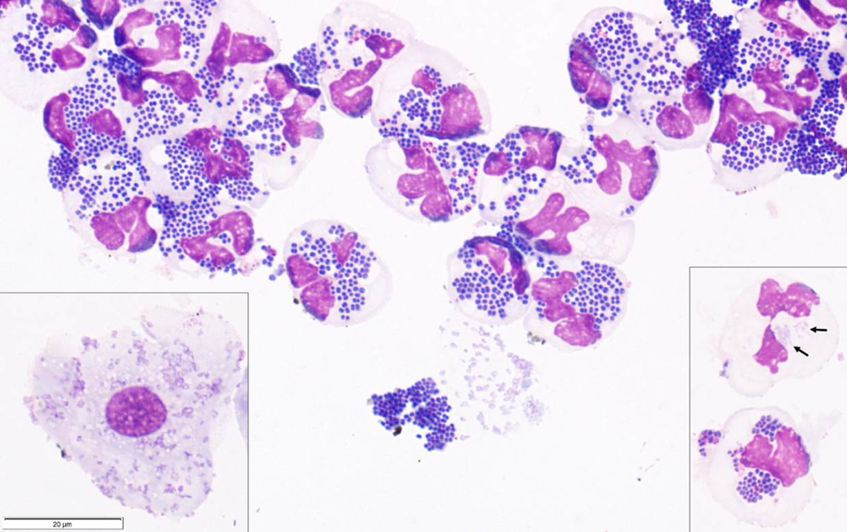
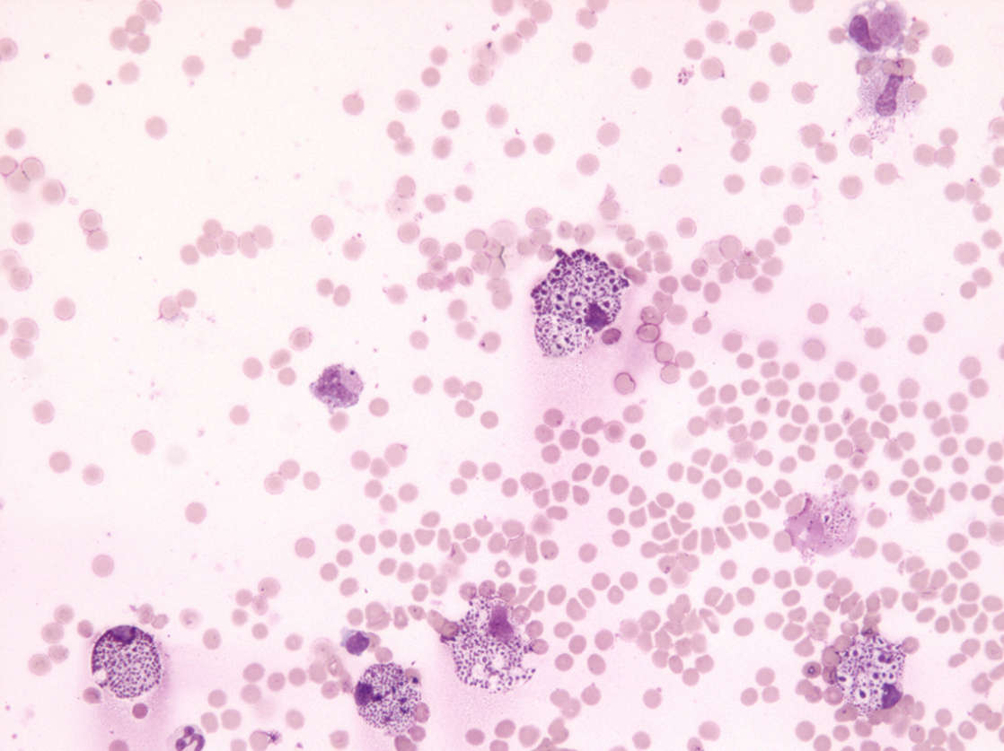
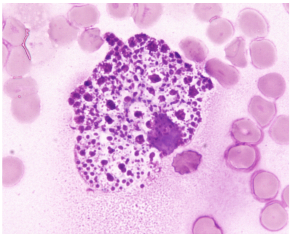
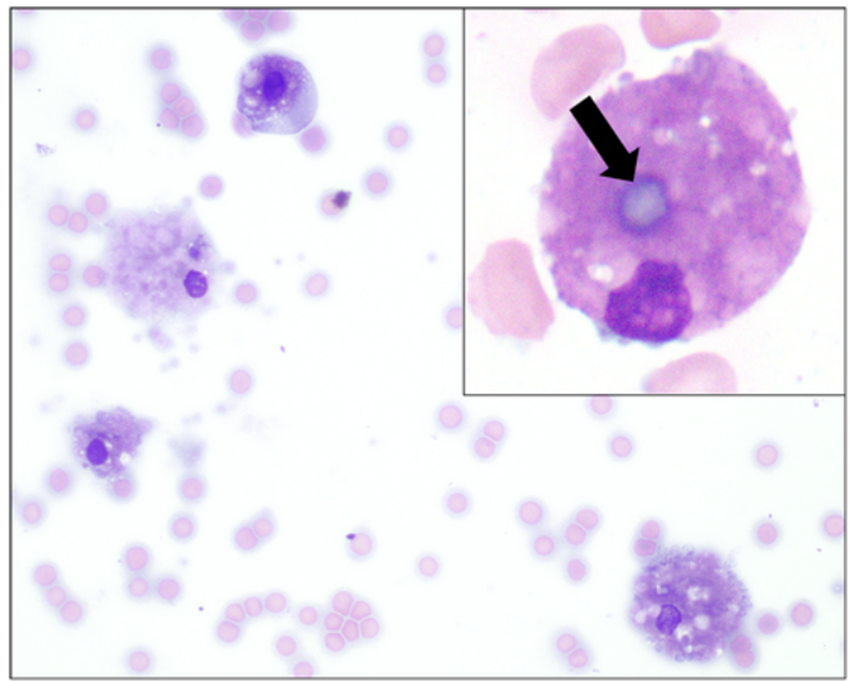
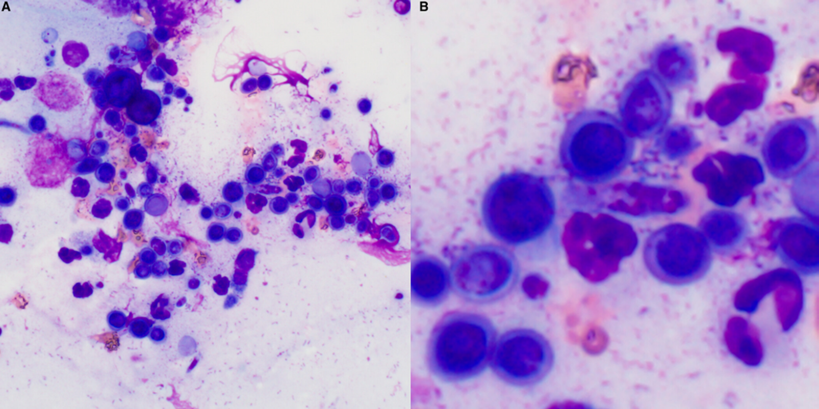
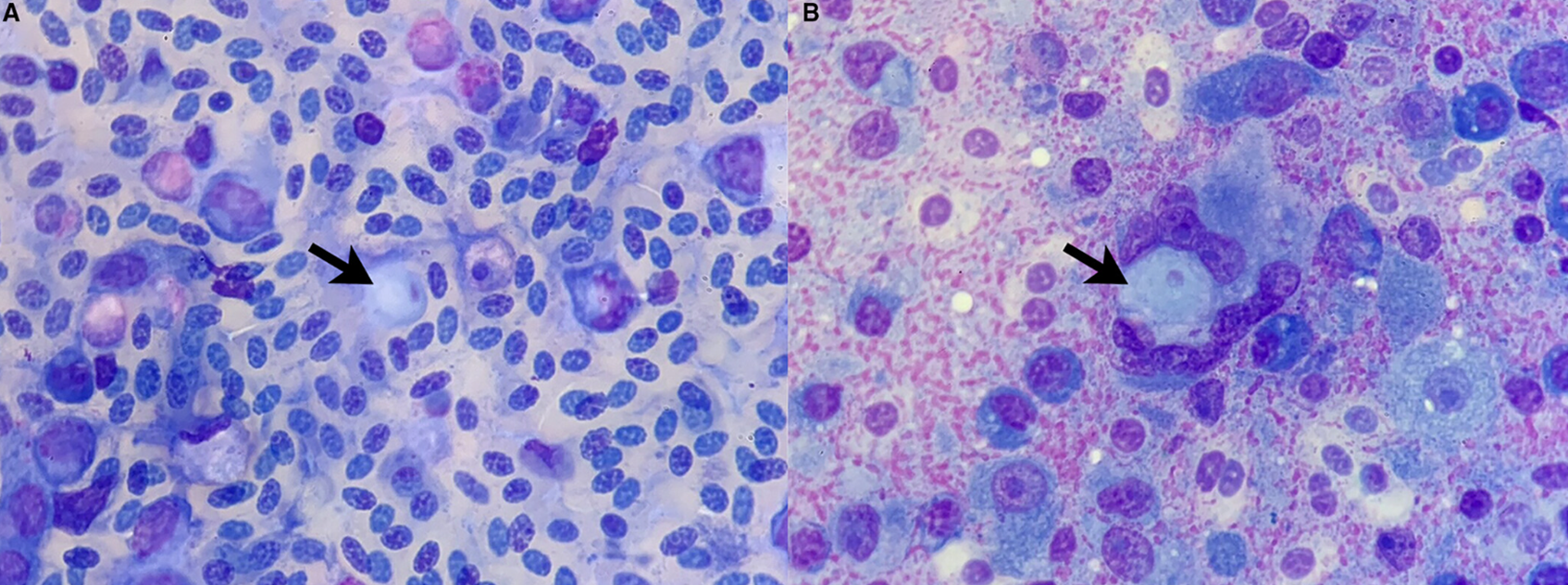
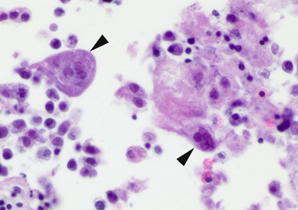
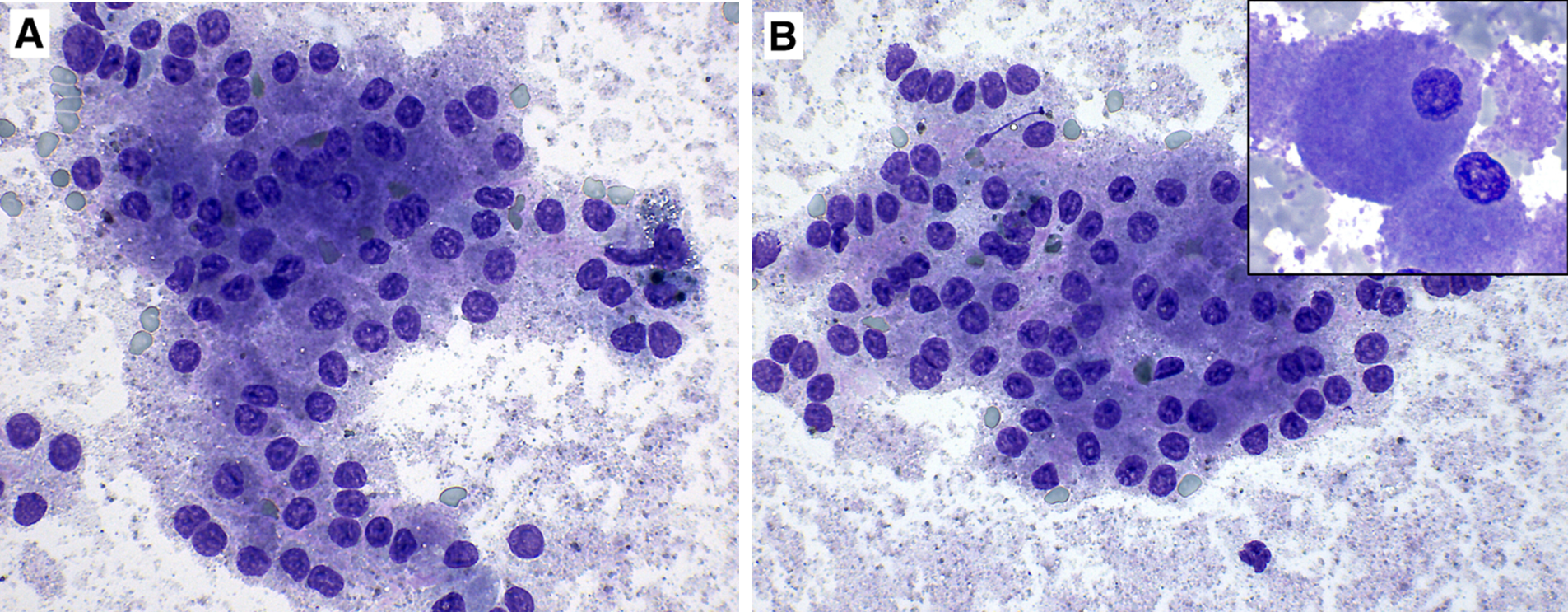
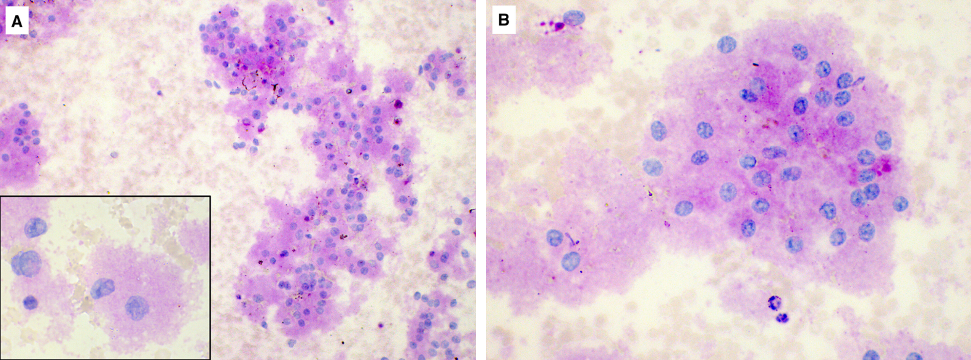
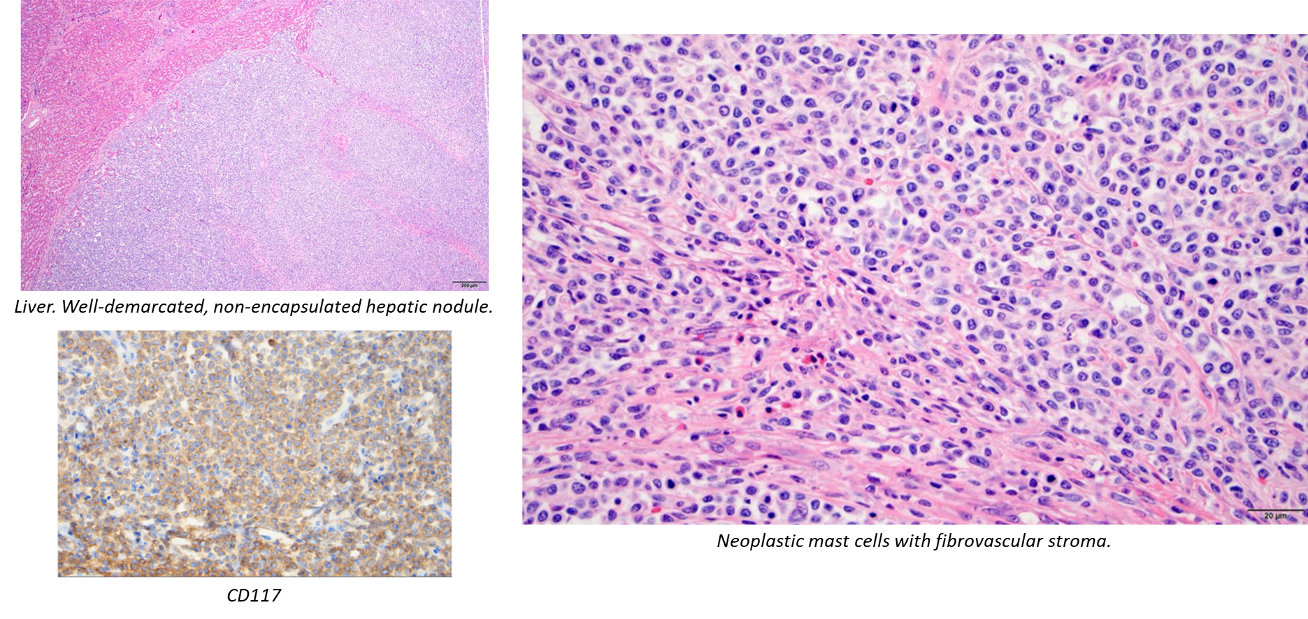
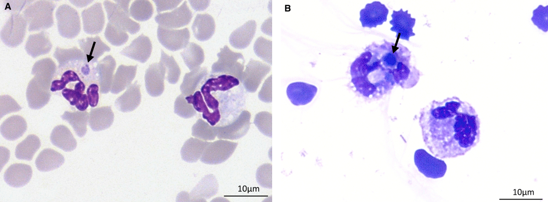
Mastocytemia and systemic mastocytosis in a dog
ProCyte Dx (A), Sysmex XT-2000iV DIFF (B), and WBC/BASO (C) scattergrams.
(A) A cloud was observed in the unusual location parallel and to the right of the monocyte dot plot location (red circle). Cells in that cloud were classified as either monocytes or neutrophils with no clear separation.
(B) Neutrophil and eosinophil dot plots were present at the respective locations (red circle) and appeared separated, but the instrument did not report numerical results for neutrophils and eosinophils. In addition to the normal lymphocyte dot plot location, the second cloud of cells classified as lymphocytes was displayed to the right of the monocyte dot plot area (blue circle).
(C) The second population of cells (above and to the right of the blue leukocyte cluster) is present (red circle), which was likely comprised of mast cells resistant to lysis in the basophil reagent. FSC indicates forward scatter; SSC indicates side scatter
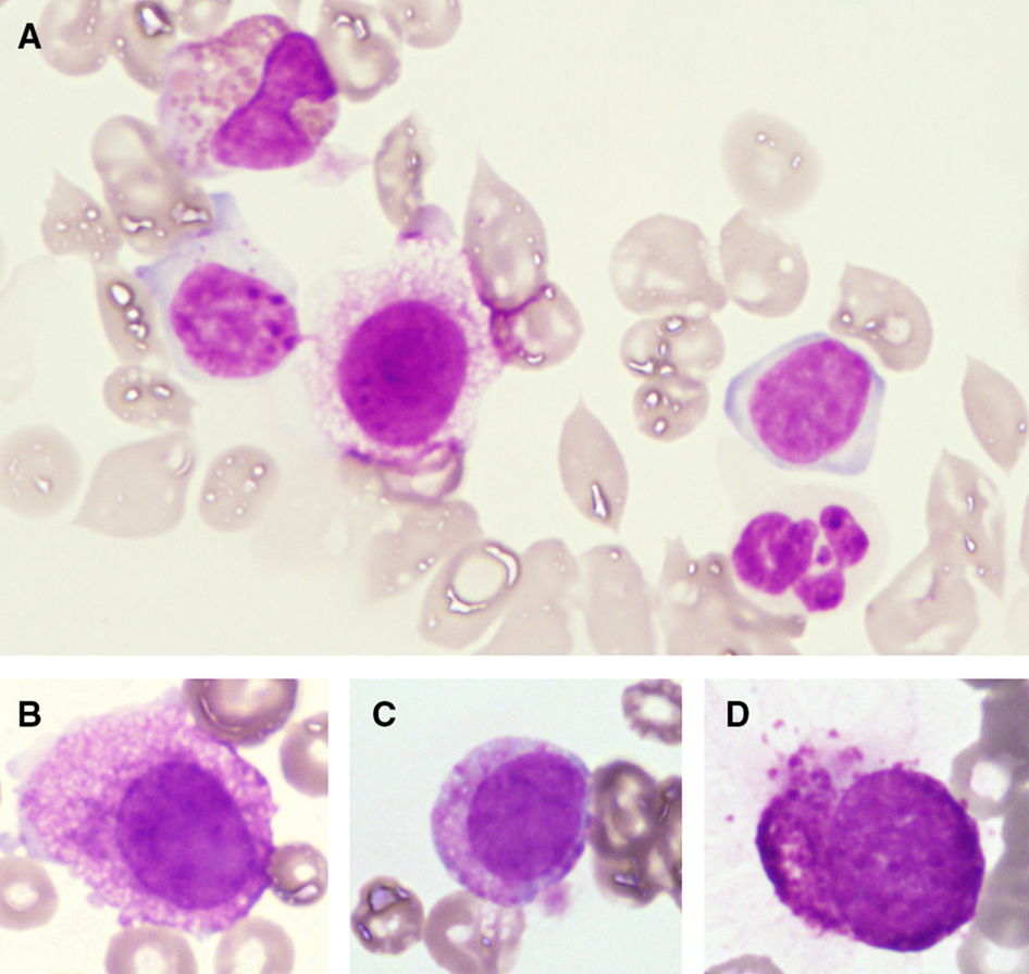
Cutaneous hemangioma w MC in the blood channels:
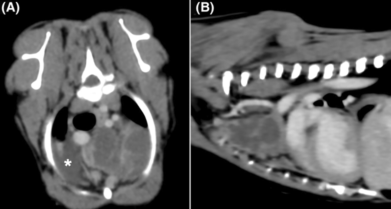
10yo mixed dog with multifocal bone lesions after prior removal of ventral neck mass
Metastatic medullary thyroid carcinoma

CSF from a 6yo Jack Russel with episodic seizures, ataxia, and obtundation
•TNCC 635/uL (RI <5), TP 1.38 (RI <0.35), minimal RBC (10/uL, RI: 0)
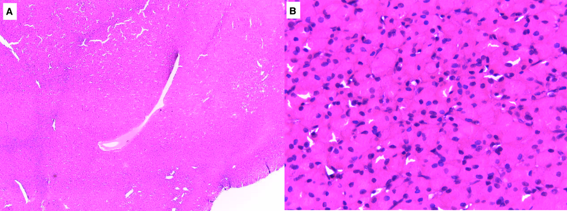
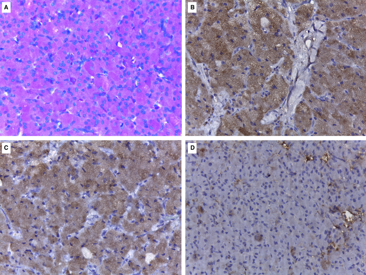
Eosinophilic pleocytosis due to primary CNS histiocytic sarcoma
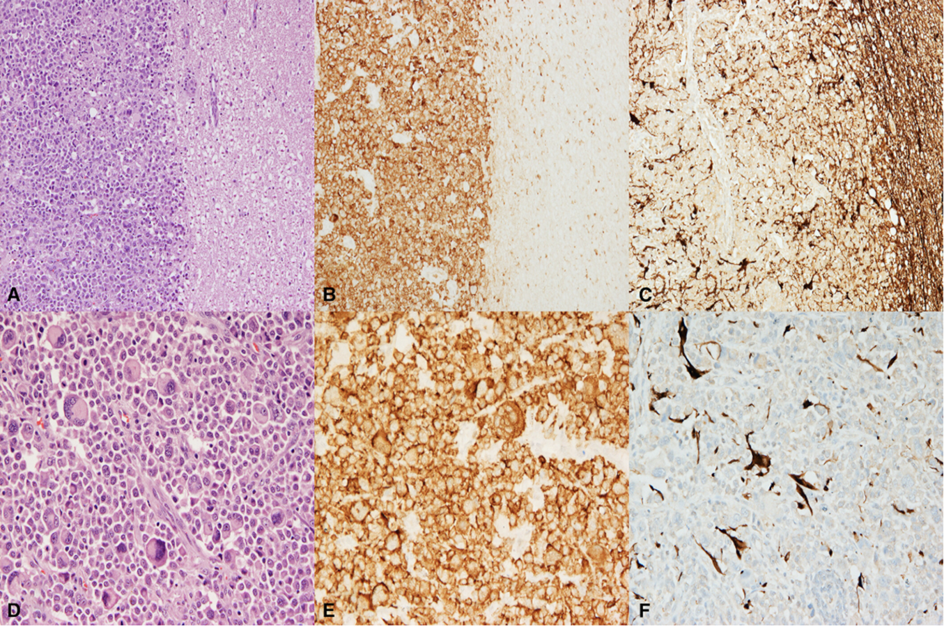
A: Well-demarc, unencapsulated, effacing WM
B: Iba-1
C: GFAP (normal tissue on R side is pos)
D: Numerous MNGC
E: Iba-1, membranous
F: GFAP+ astrocytes surrounded by neg neoplastic cells
Peritoneal effusion from an 8yo dachshund with HCT 5%, thrombocytopenia
Mycoplasma haemocanis
Initially had a duodenal ACA resected, received 2 blood transfusions, 2nd donor was known negative but nothing known about 1st
Blood smear:
2yo Golden with progressive paraparesis, intradural extramedullary mass L2
Nephroblastoma
WT1: multifocal nuc & cyto immunolabeling
Urine sediment from a 5yo Borzoi with erythema multiforme managed by pred
USG 1.011, pH 7.5, protein 3+, WBC 20-50 with clumps
Ddx Mcyoplasma or ureasplasma
Culture: Staph pseudointermedius, Mycoplasma spp, E. coli
DNA analysis: Mycoplasma canis
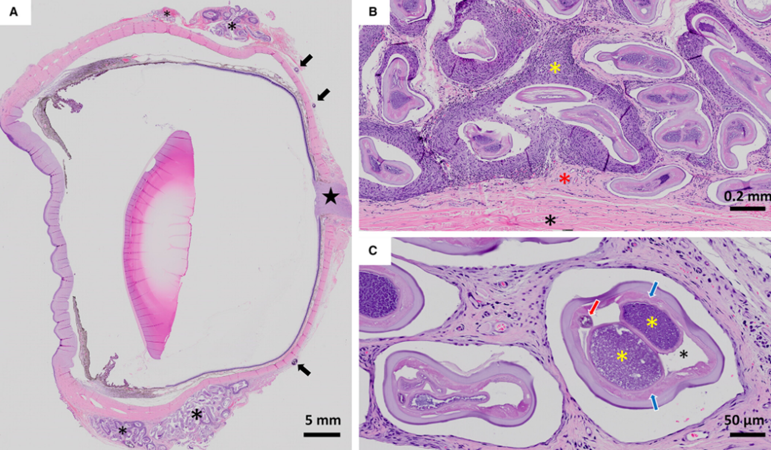
Conjunctival mass from 8yo mixed dog
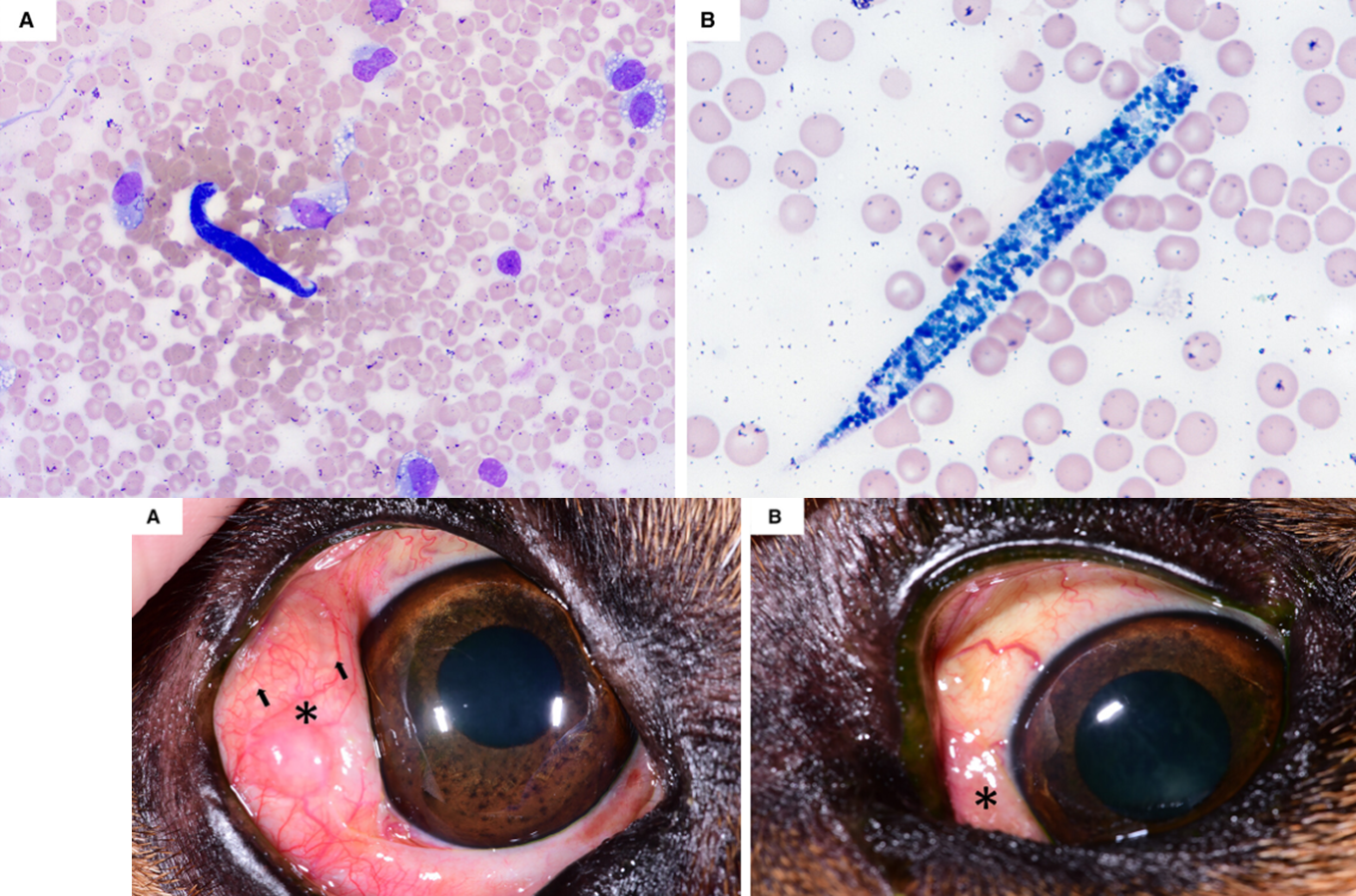
Onchocerca lupi
2yo MN Terrier mix with ill-defined, soft, fluctuant mass R dorsal perineal area x 1wk. ~10cm, fluid-filled, non-painful, non-reducible
Initial concern carcinoma, but turned out it was, in fact reducible
Perineal hernia with entrapped fluid and reactive mesos
CSF from 2.5yo Boston Terrier with progressive tetraparesis, ataxia
MPS I (Hurler’s disease)
Deficiency of α-L-iduronidase
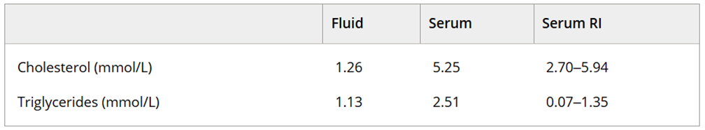
Recurrent inguinal mass in a 7yo FS Pug wtih fever, lethargy, inappetance
Ddx lymphatic neoplasm, lymphangiectasia, lymphocele, mild lymphocytic inflammation
NOT supportive of chyle or pseudochyle
Histo: Cutaneous lymphangioma, PROX-1 nuclear labeling
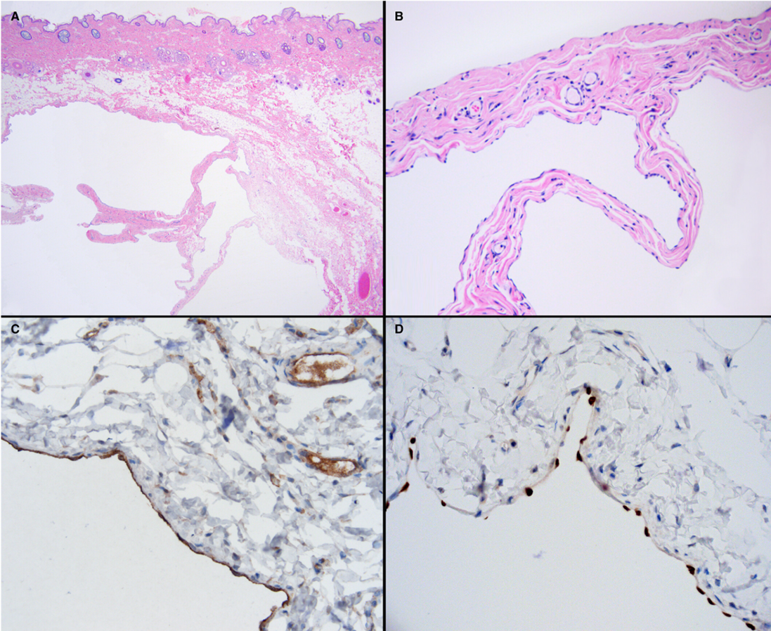
(A) SQ expanded by arborizing, empty vessels.
(B) Lined by single layer of uniform, flattened endothelial cells.
(C) Intracytoplasmic labeling for FVIIIra
(D) Intranuclear labeling for PROX-1
Later dev’d similar lesion near tarsus - poss lymphangiomatosis of the pelvic limb
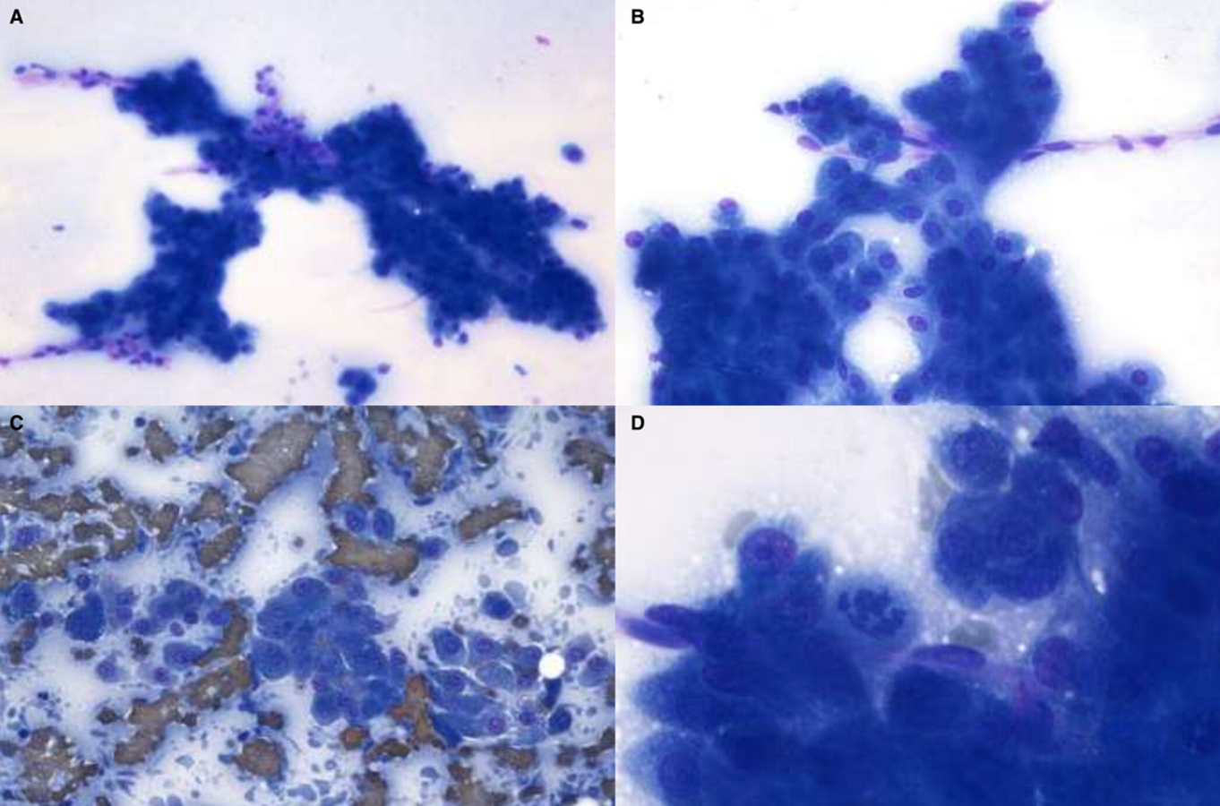
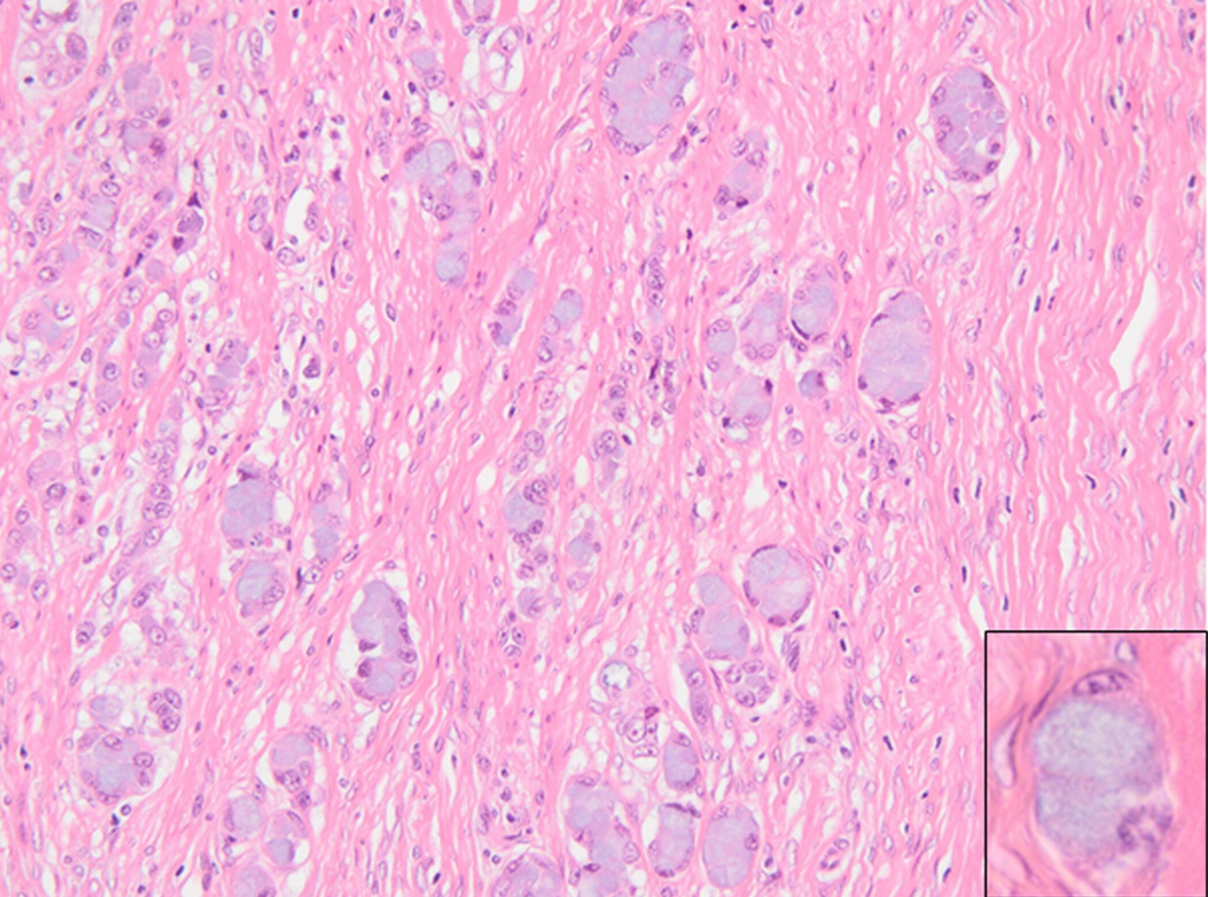
8yo mixed dog, diffuse generalized thickening of the gastric wall
Signet ring gastric adenocarcinoma
17 wk English Bulldog with urinary incontinence, stranguria, hematuria
Grossly red-brown, turbid, USG 1.016, pH 7.5, mod proteinuria, 50+ RBC, 0-2 WBC, 5-9 epith
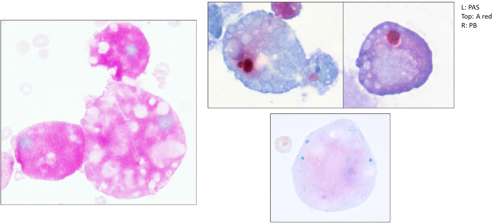
Malakoplakia, inclusions consistent with Michaelis-Gutmann bodies
PAS+ confirms macros
Alizarin red+ (calcium) with faint multifocal PB+ (iron)
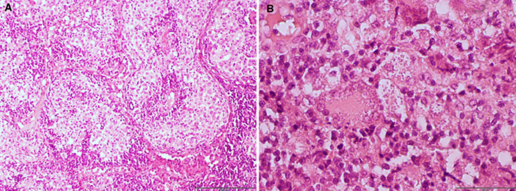
Intracranial mass in 13yo Chihuahua w cluster seizures, CSF protein-cytologic dissociation (4 cells/uL, TP 47.6 mg/dL), mass in piriform lobe
Meningioma, meningothelial histomorphology
9yo Gordon Setter non-wt-bearing and swelling of 4th RF digit
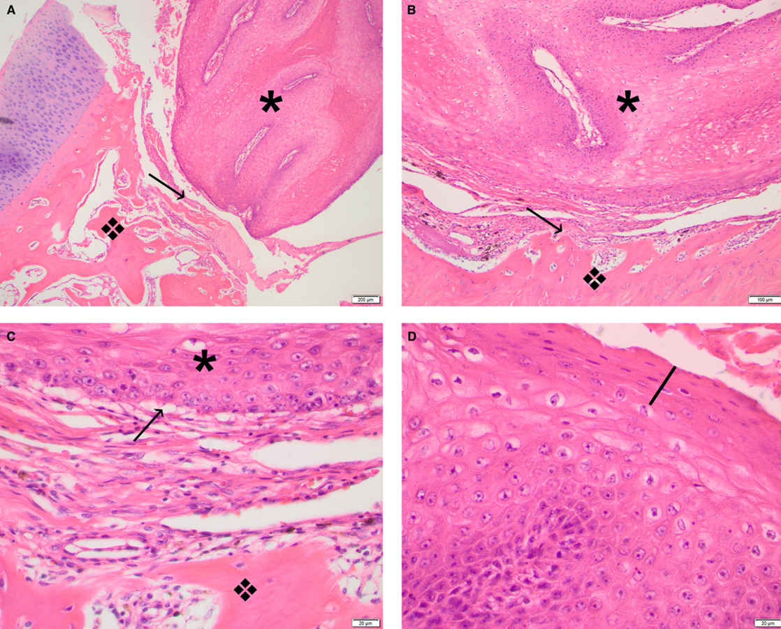
Inflamed subungual keratoacanthoma
4yo DSH with swollen paws, mod NRA (0.20), hyperphosphatemia, mild azotemia
Metastatic mineralization due to CRF
18yo Hanoverian mare ovarian mass
Granulosa cell tumour
A) Strong, diffuse vimentin
B) Variable pancytokeratin
C) Moderate to strong, cytoplasmic inhibin-α
D) Moderate to strong, cytoplasmic, anti-Müllerian hormone
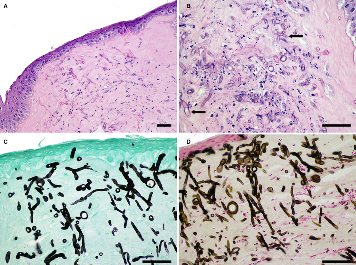
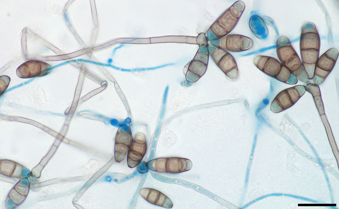
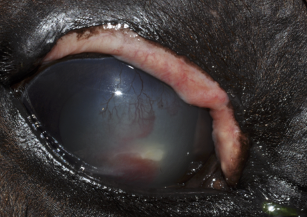
22yo QH gelding scraping from a corneal ulcer
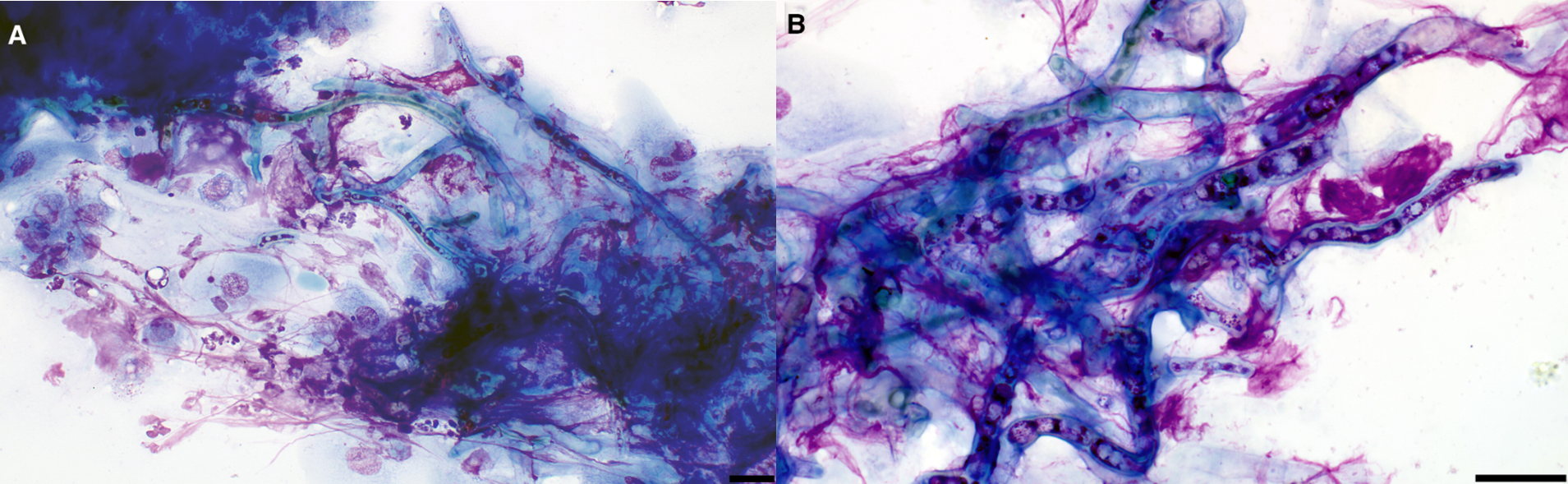
Phaeohyphomycosis due to Cochliobolus sp. (anamorph Curvularia sp.)
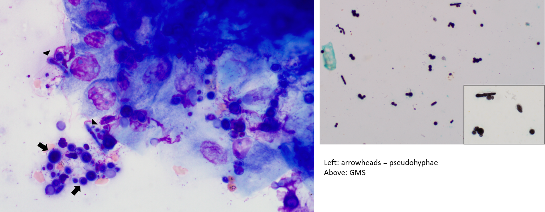
9yo QH mare scraping from a corneal ulcer
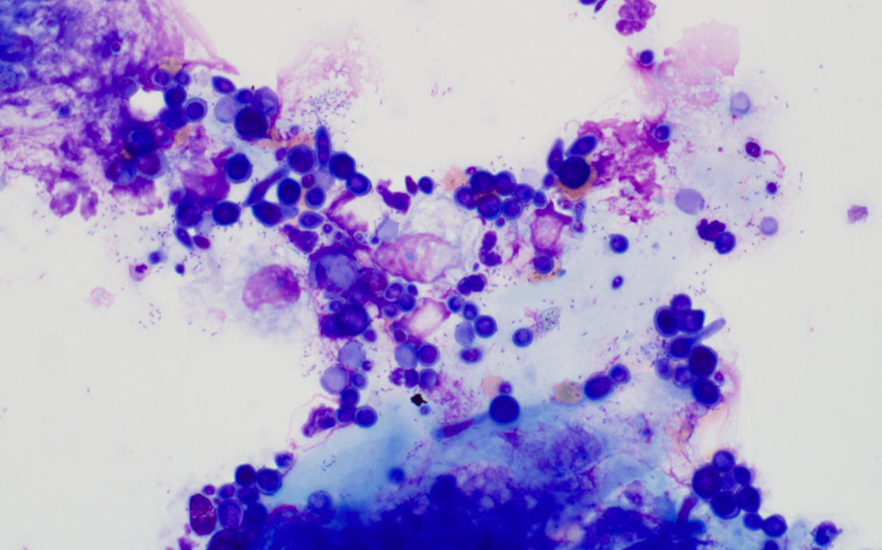
Candida tropicalis
2mo F Bourbon red turkey poult from a backyard flock, submitted for necropsy. Several birds died in prev weeks, had yellow-coloured droppings, hunched w ruffled feathers, stopped eating. Impression from multifocal liver nodules.
Histomonas meleagridis
3mo F chicken found dead along w several others over prev weeks, no signs of illness. Postmortem tracheal swab.
Infectious laryngotracheitis caused by Gallid Herpesvirus-1
Syncytial cells w eos intranuc inclusion bodies in trachea, eyelid, & lung are diagnostic
Wild juvenile mourning dove with masses on the cere
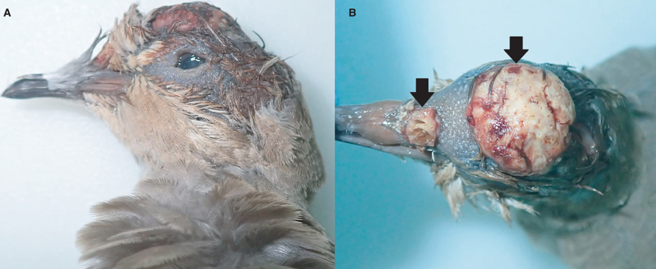
Avipoxvirus, Bollinger bodies containing Borrel bodies
6yo M pet rabbit testicular mass
Granular cell tumour, ddx interstitial cell tumour
PAS:
A: PAS
B: Vimentin
C: Melan-A
D: CK AE1/AE3
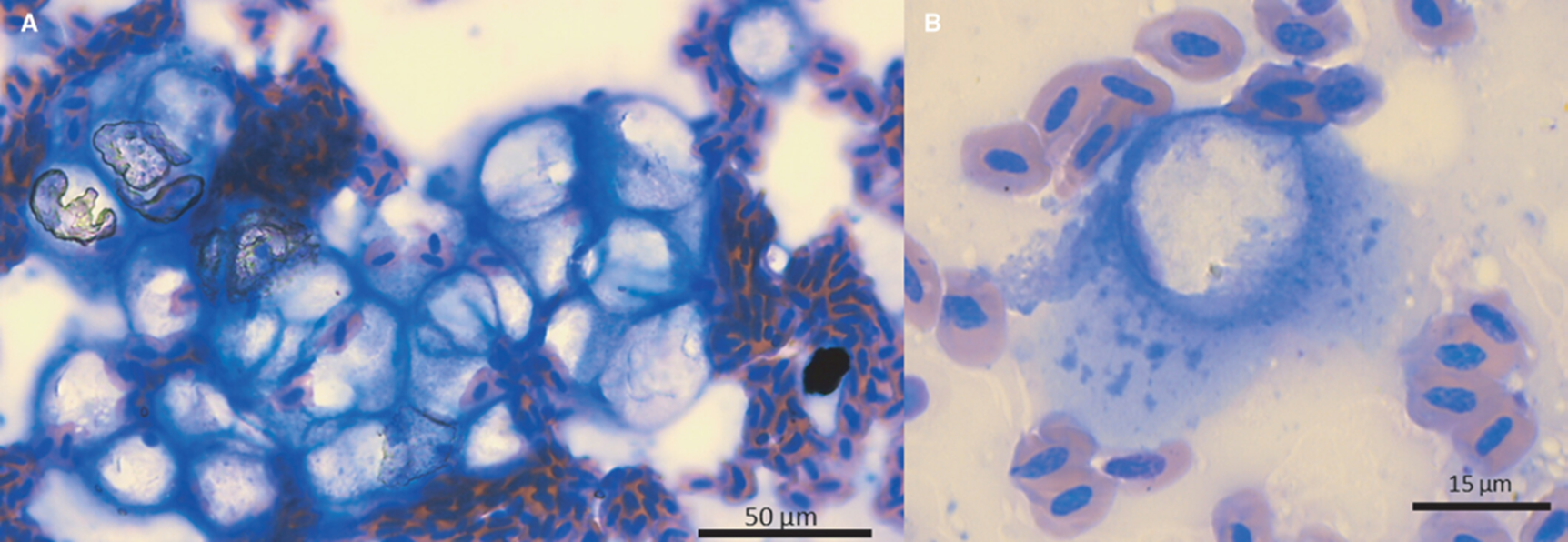
Coelomic fluid in an Eastern River Cooter (Pseudemys concinna concinna)
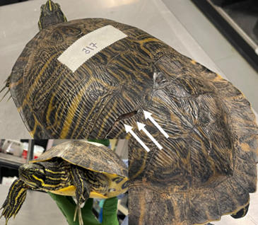
Transudate with spermatozoa
Rectal scraping from a 14yo mixed dog with intermittent, recurrent large bowel diarrhea
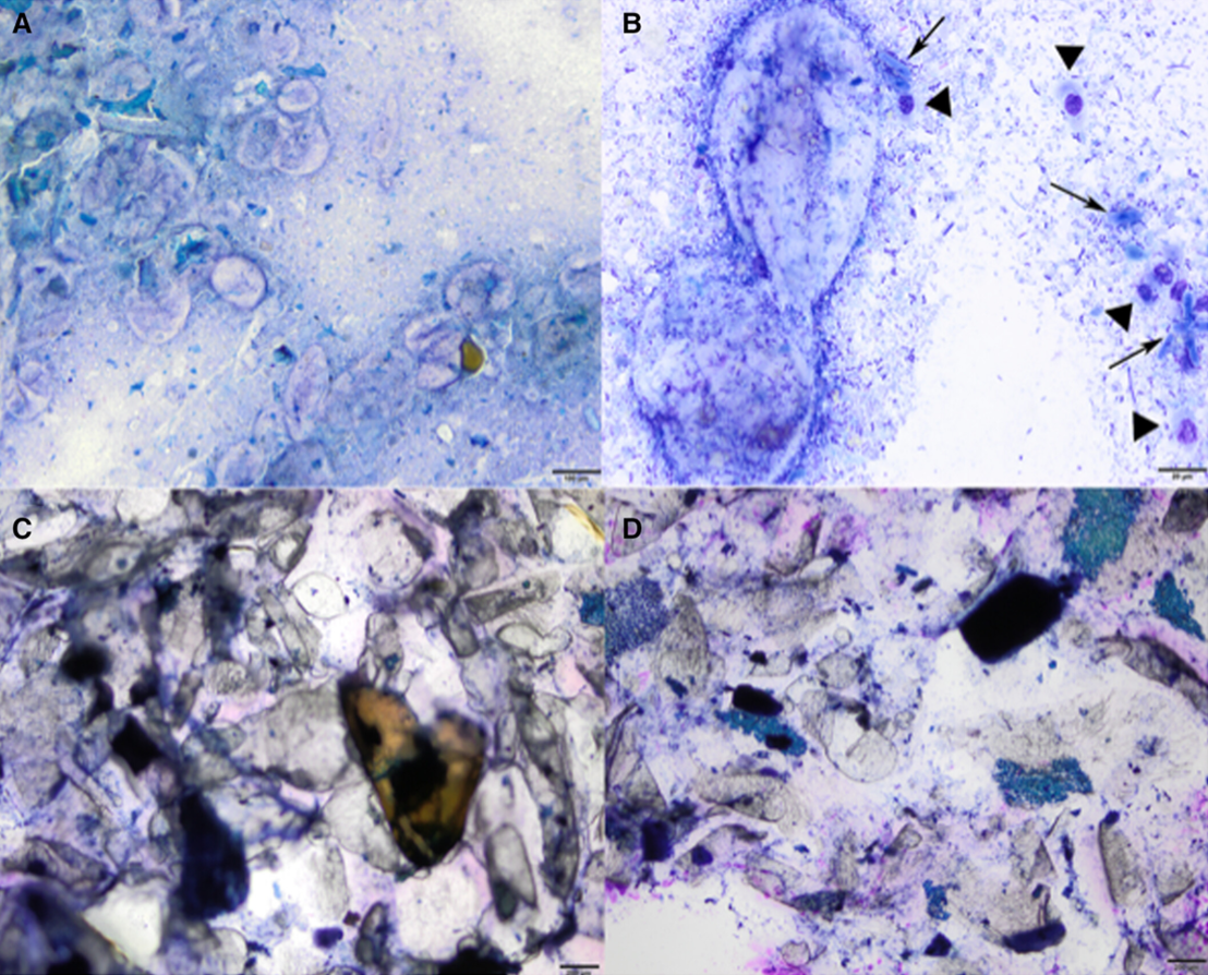
Psyllium husk (tx’d w metronidazole, psyllium, fenbendazole)
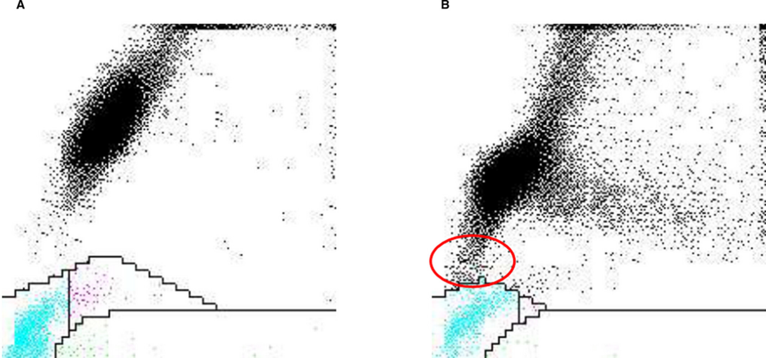
ADVIA 2120 platelet absorption cytograms
A: Normocytic, normcohormic, B: microcytic, hypochromic RBCs causing high interference, see how gate is smaller than it should be for reticulated platelets (purple)
Gel column crossmatches
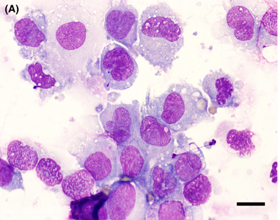
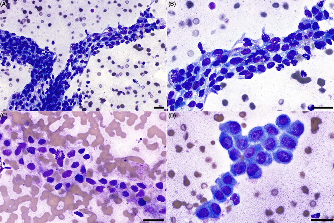
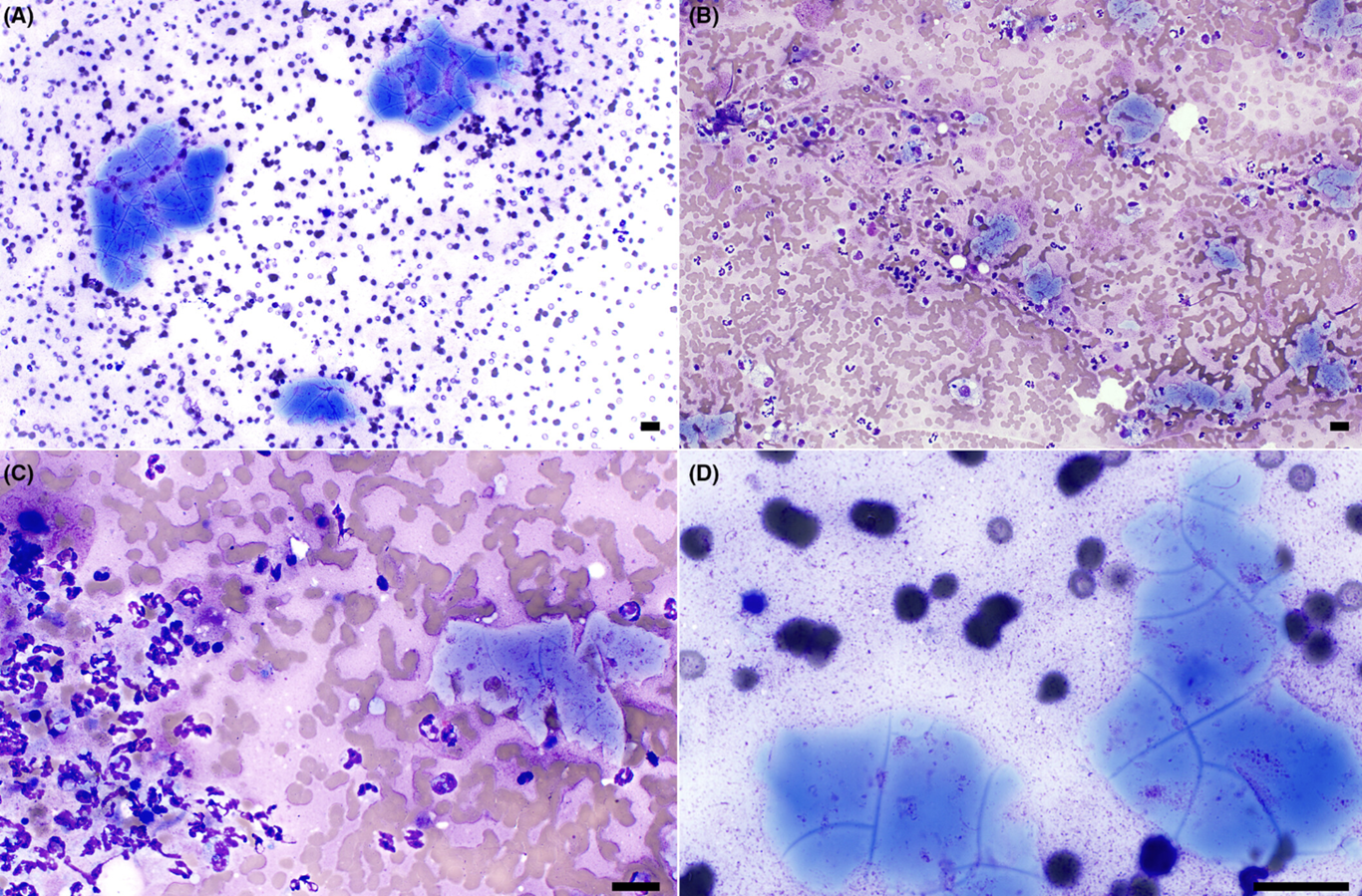
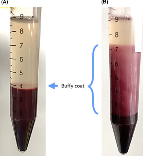
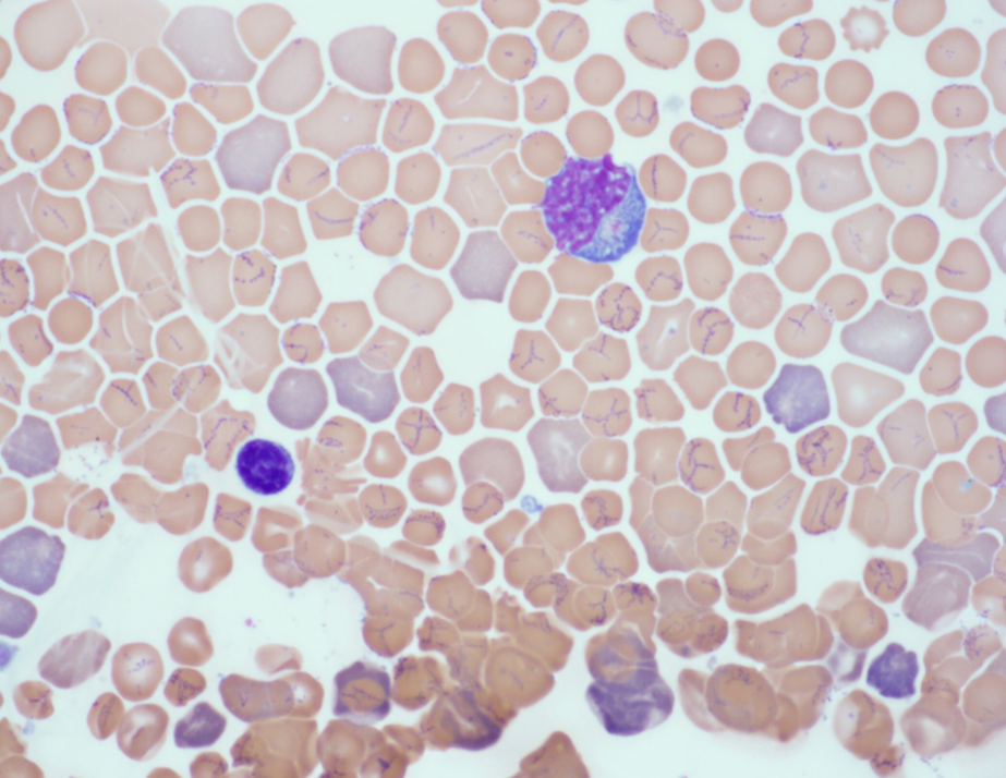
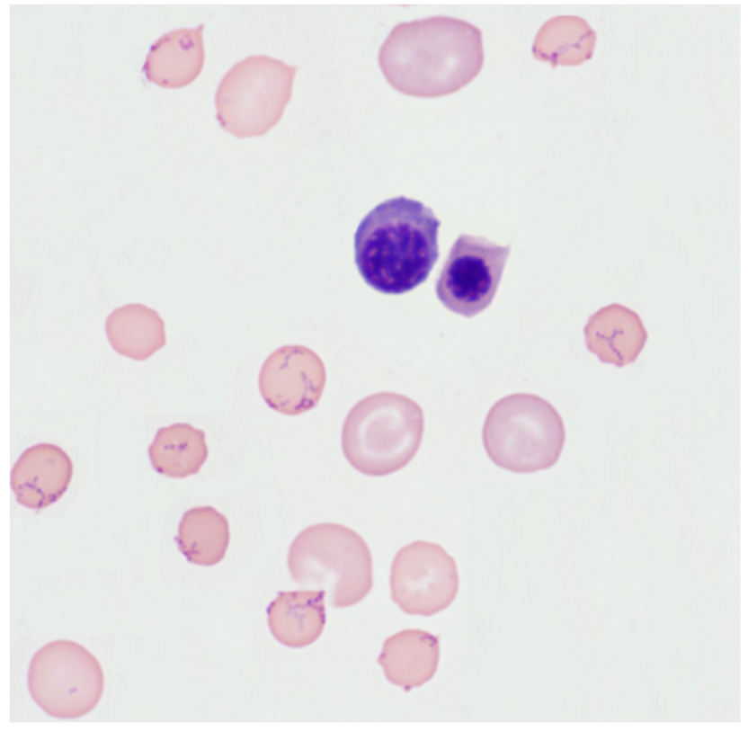
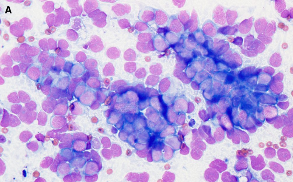
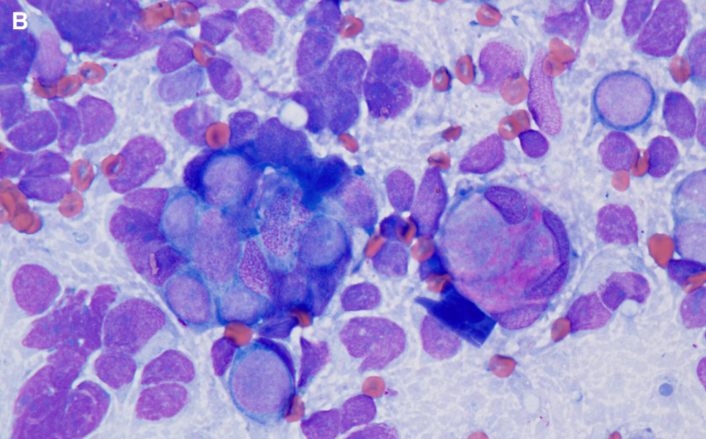
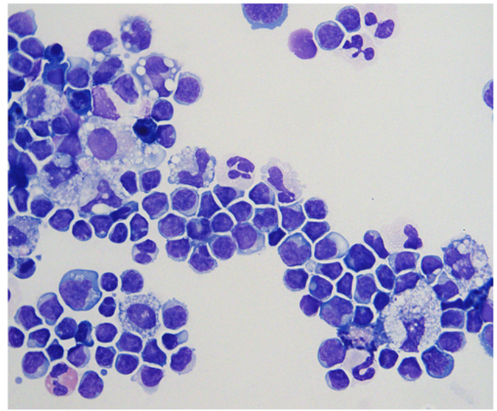
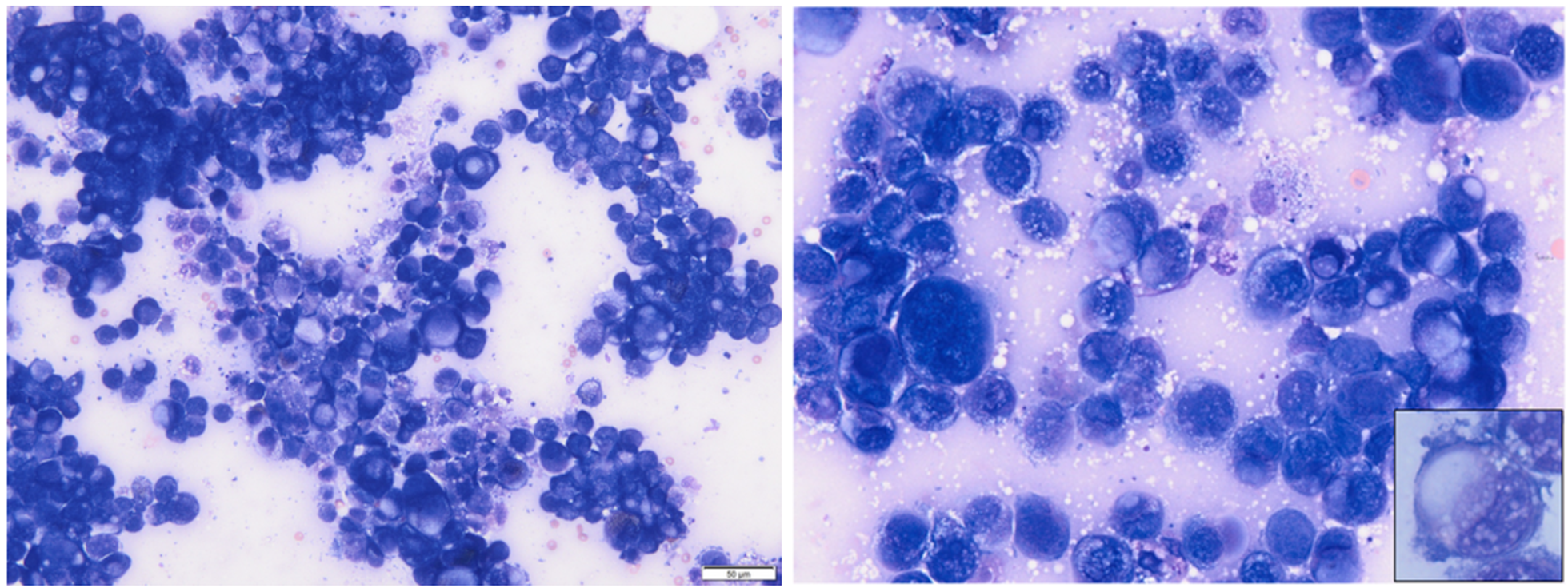
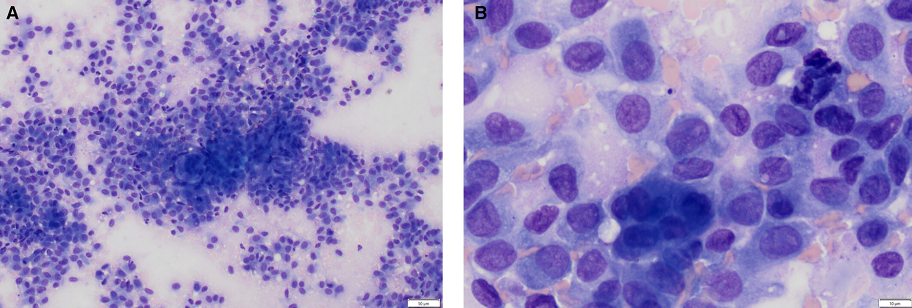
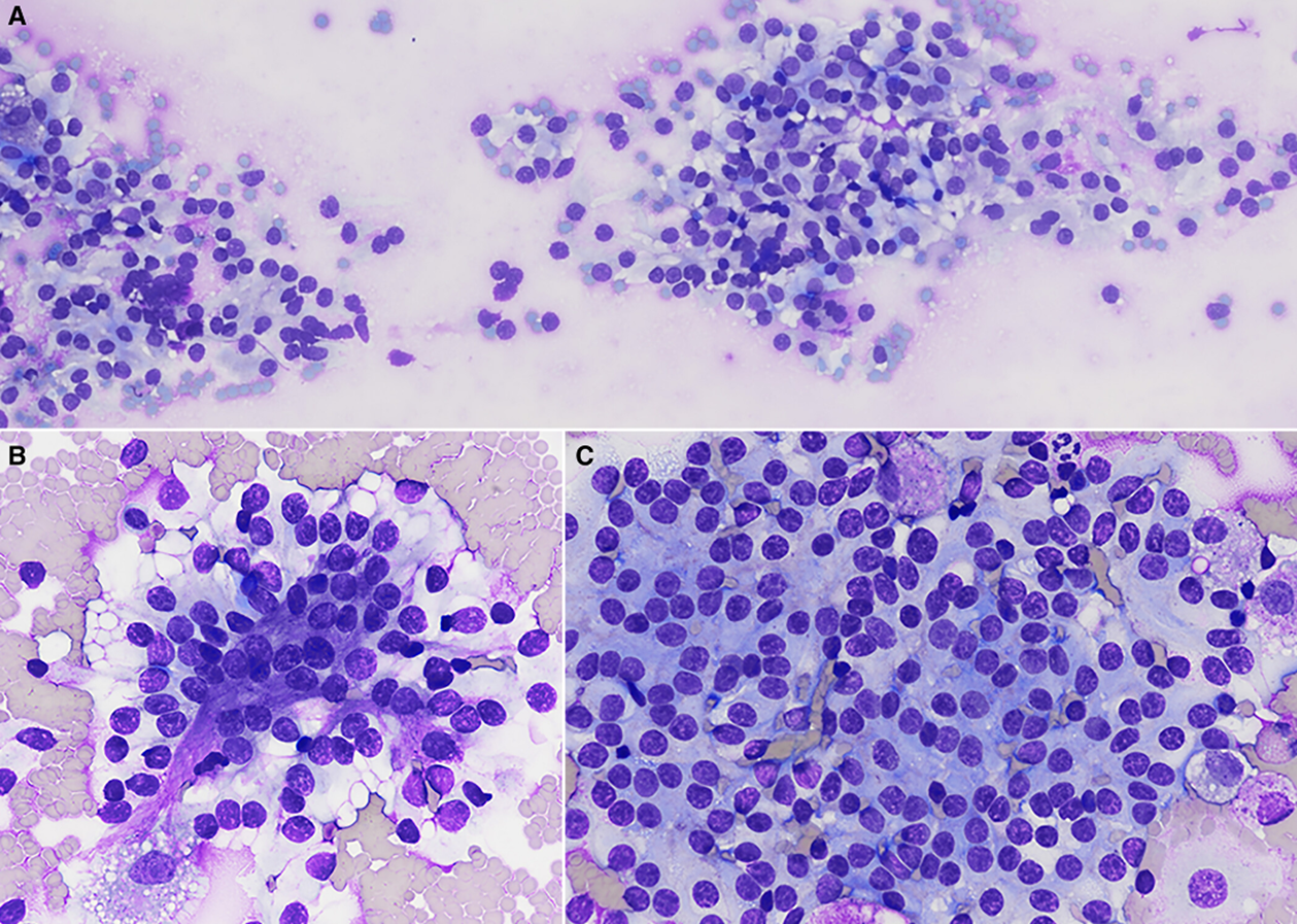
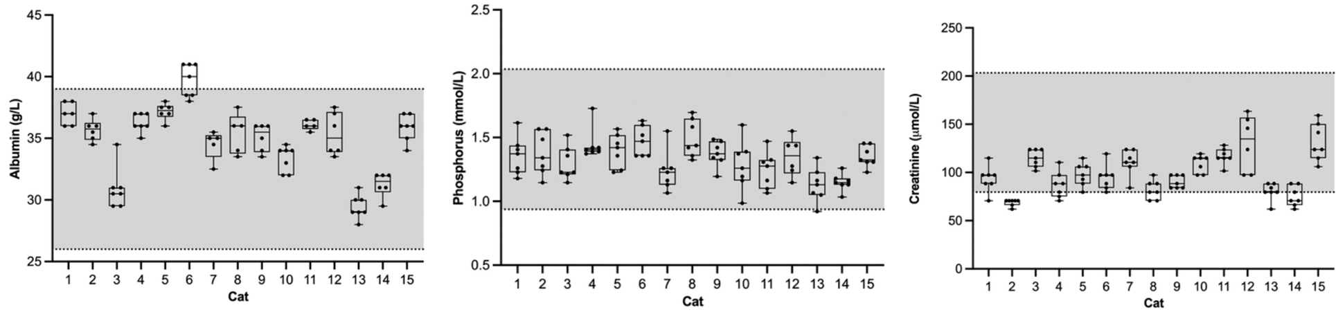
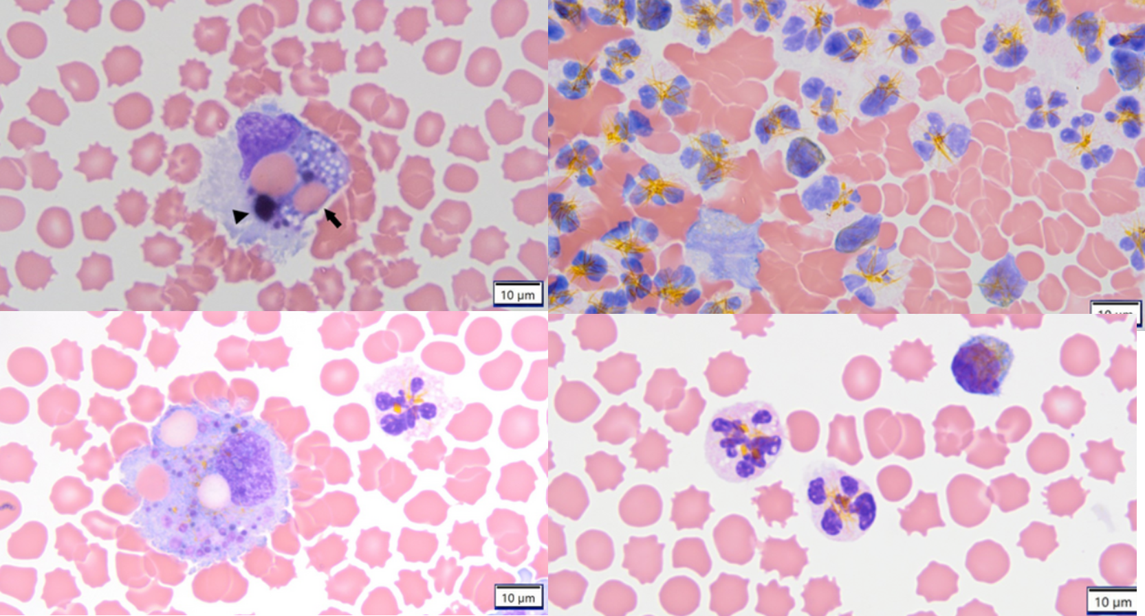
9yo Boston with severe leukocytosis (81,700)
Blood smear
Bone marrow
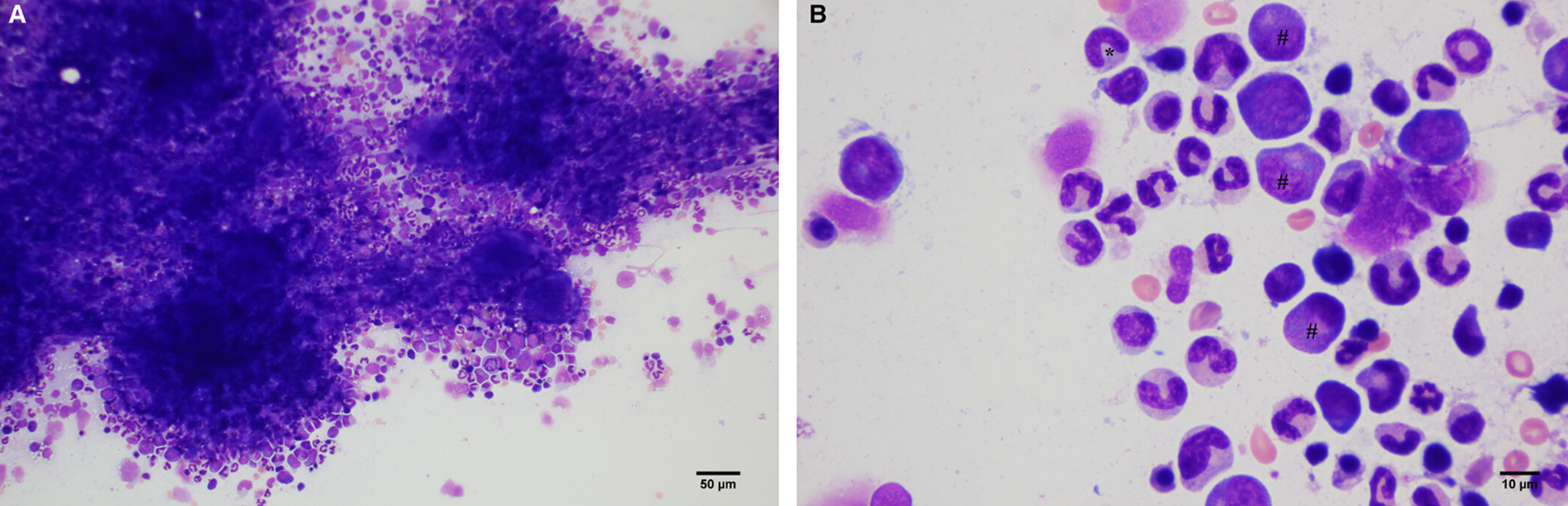
Paraneoplastic leukocytosis found secondary to GIST in duodenum
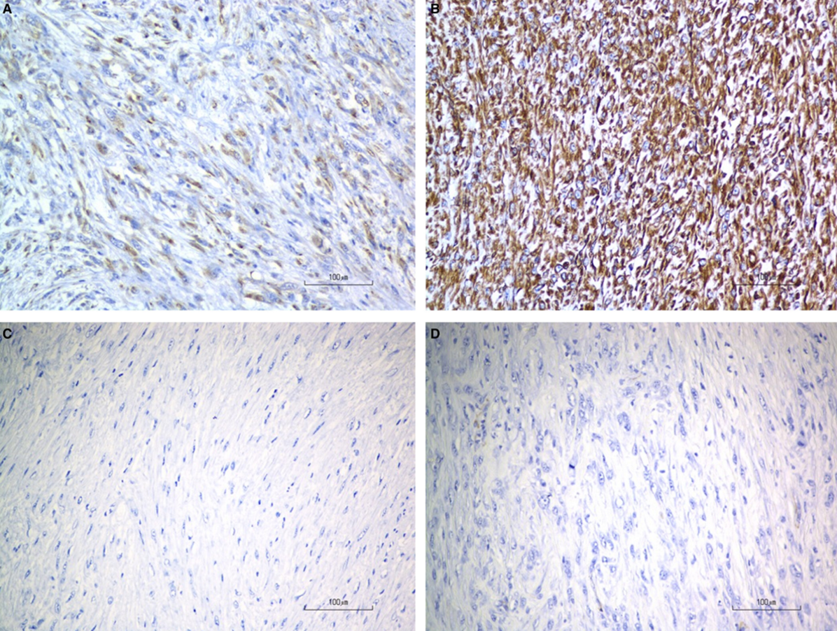
A: CD117 (KIT)+
B: Vimentin+
C: Desmin-
D: SMA-
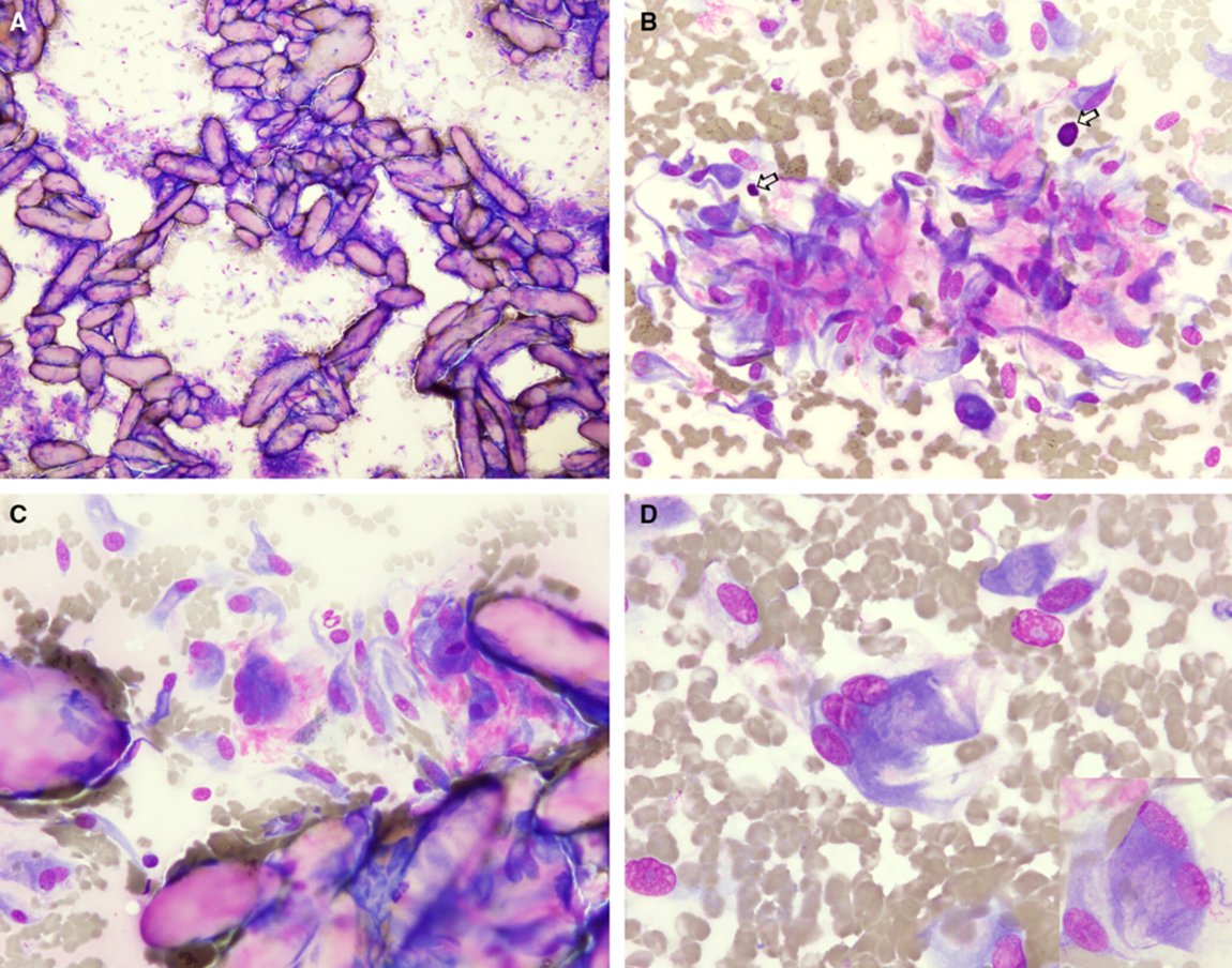
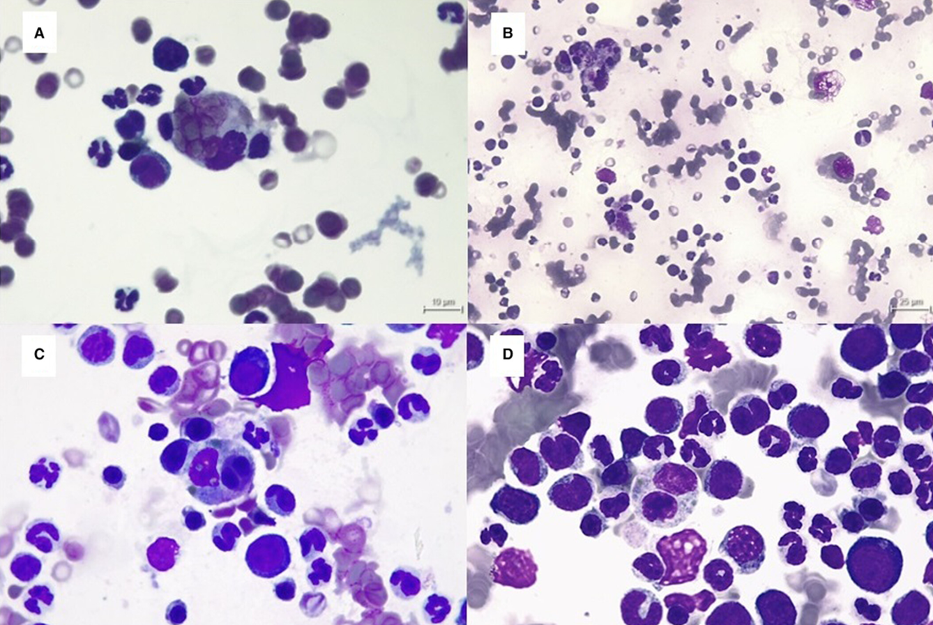
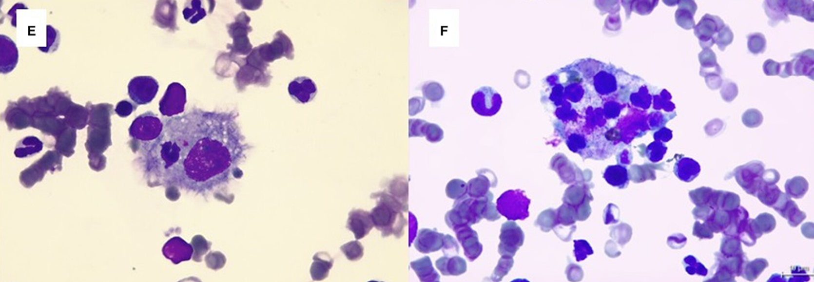
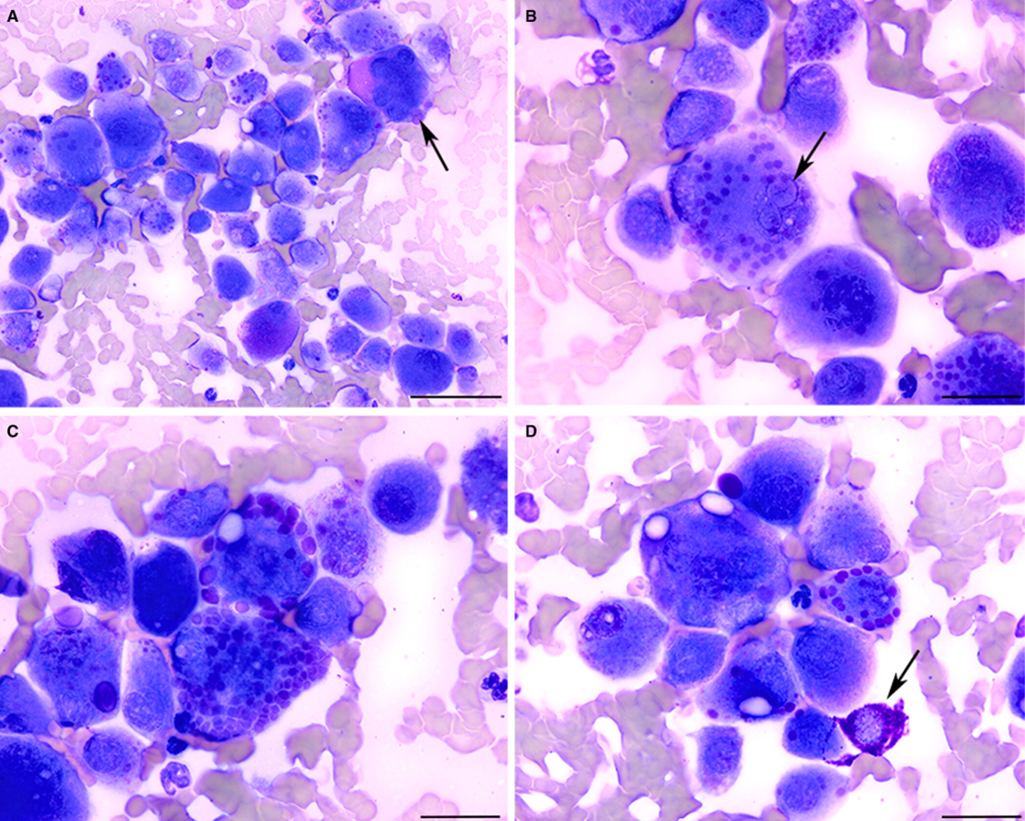
Individuality examples
Left to right: High, low, intermediate
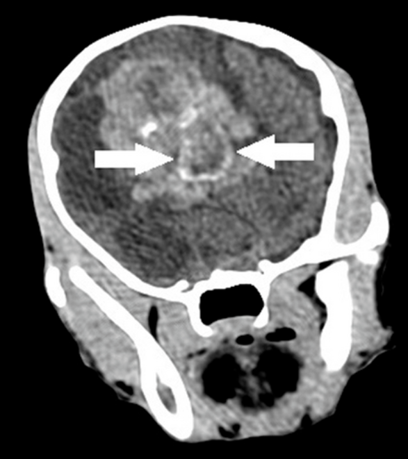
9wk F Irish Setter with progressive refractory seizures
CSF TNCC 1100/uL, TP 1939 mg/dL
Cerebral vascular hamartoma
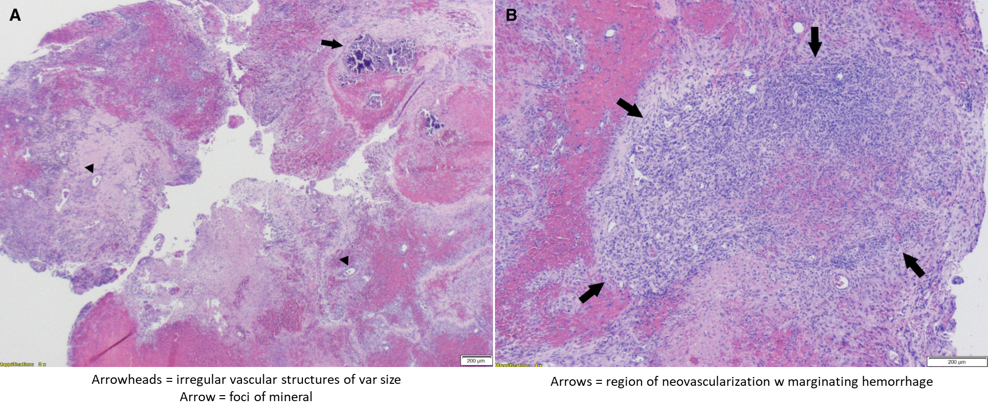

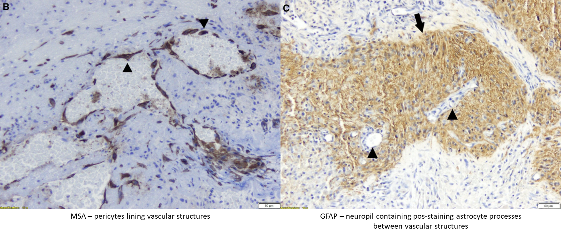
MSA = muscle specific actin
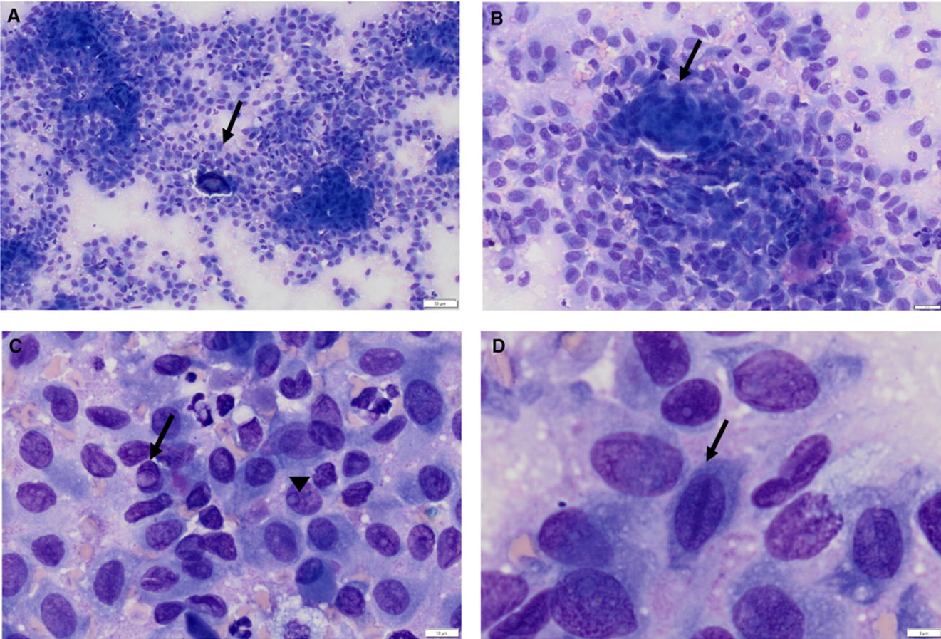
•16yo FS DSH 0.5cm soft pink mass P3 of D4 RHL
Cyto ddx: neural or neurosecretory tumour (e.g. GCT), synovial sarcoma, HS, PCT, anaplastic carcinoma, pleomorphic MCT
Histo: presumptive pleomorphic MCT
IHC: Granular variant of a histiocytic tumour

A: Iba1
B: CD117 (staining MC)
C: Giemsa (in MC)
D: PAS (5% of T cells had stained grans, esp larger ones)
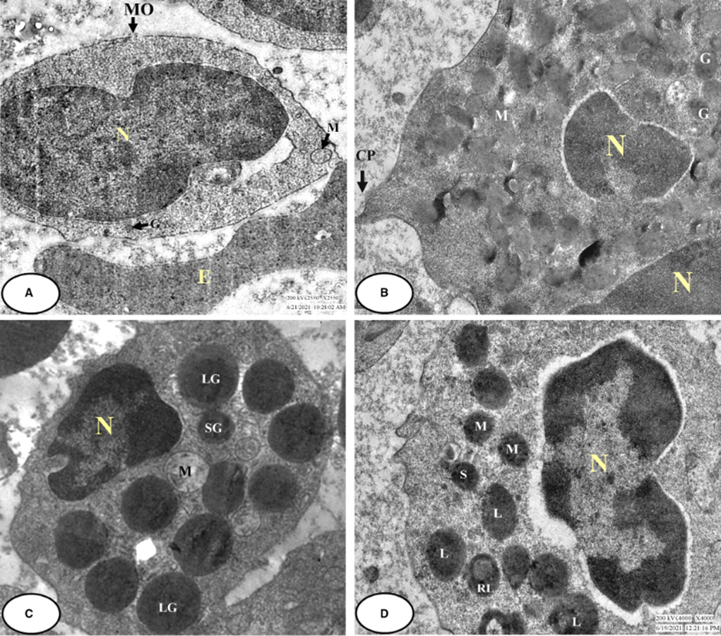
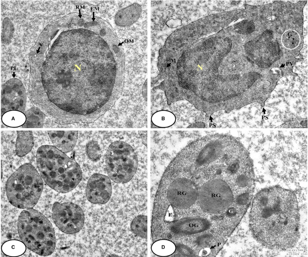
•TEM: Prominent filopodia or interdigitating plasma membranes, abundant ER, mod numbers of low-density membrane-bound granules w granular to fibrillar substance (few more electron-dense or w dense spherules, arrays, fragments)

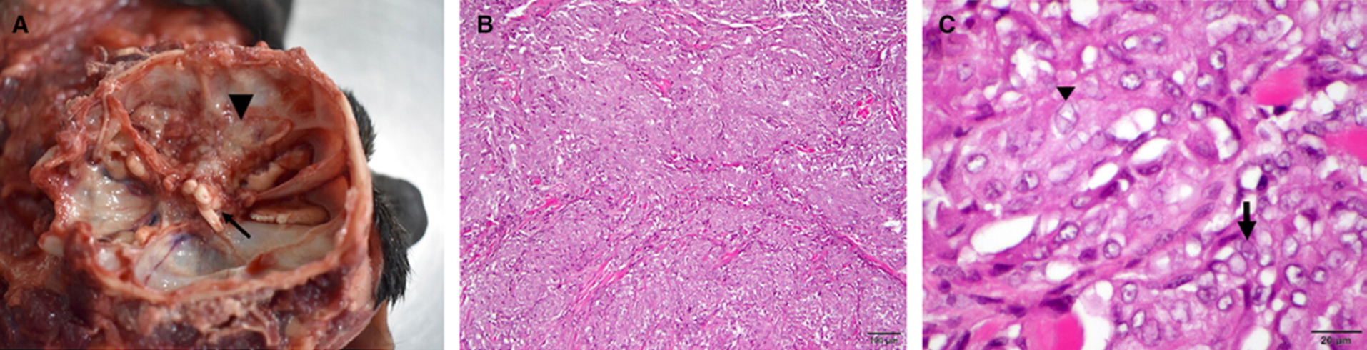
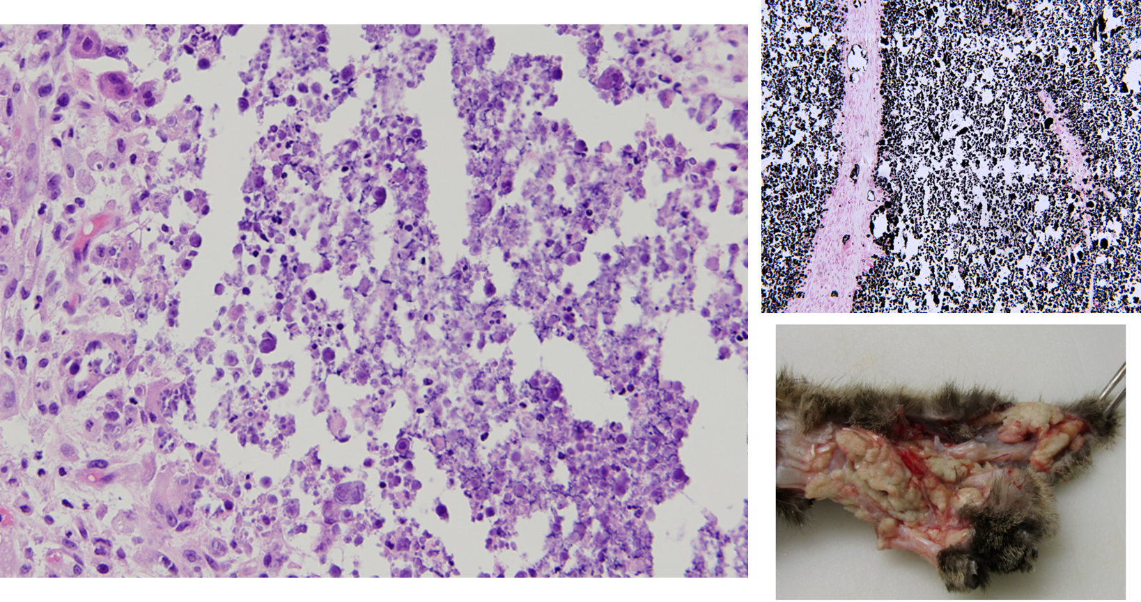
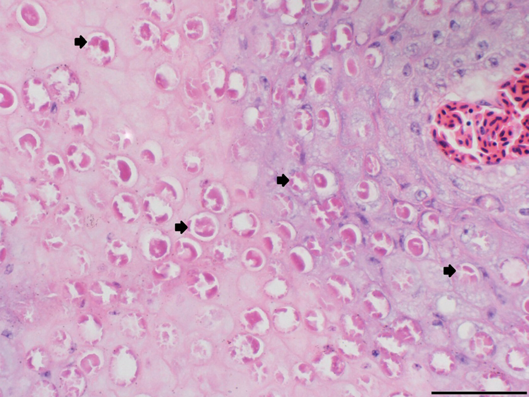
12yo F Vietnamese Pot-Bellied Pig anorexia/lethargy x2d, open-mouth breathing, pale mm, liver nodules:
Blood smear:
Visceral MCT and mastocytemia, CD117+, CD3- CD79a-
Arrow is lipofuscin in hepatos
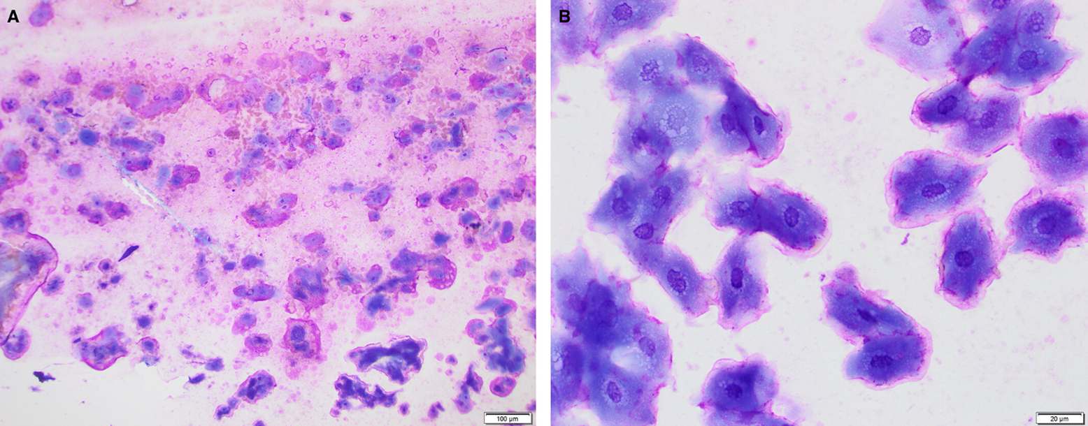
Urine sediment from an 11 mo dachshund w PU/PD, anorexia/lethargy, pyrexia
1.005, 2+ prot, 4+ blood, 0-5 RBCs, 4-8 WBCs
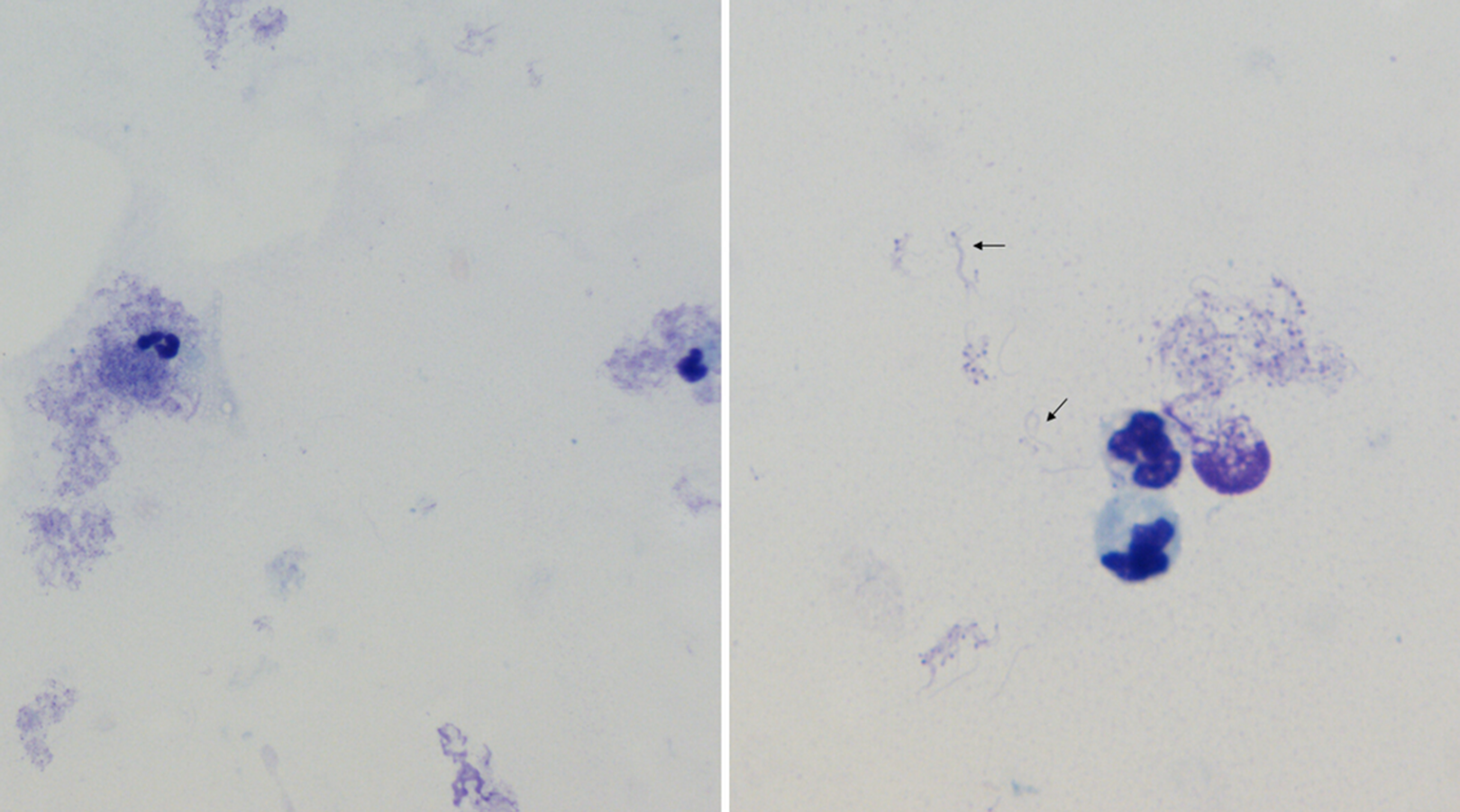
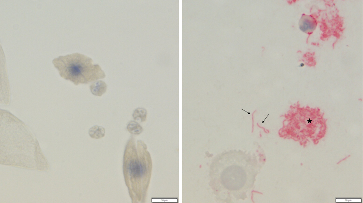
Leptospiral organisms
IHC for leptospira:
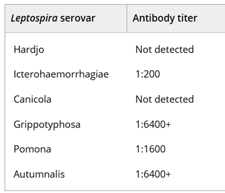
MAT:
2yo MN Creole Shepherd w chronic HL paraparesis and progressive neuro deficits
Suppurative spinal meningomyelitis in a dog with intra-neutrophilic cerebrospinal fluid cells Ehrlichia canis morulae
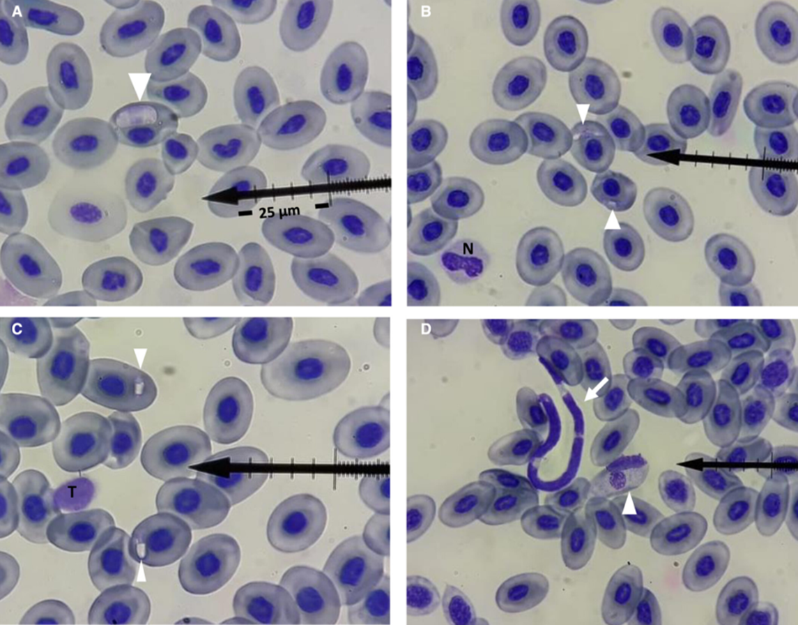
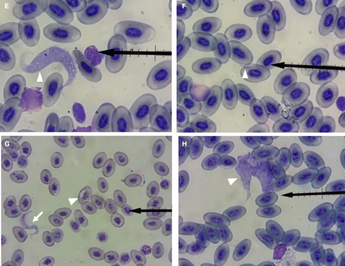
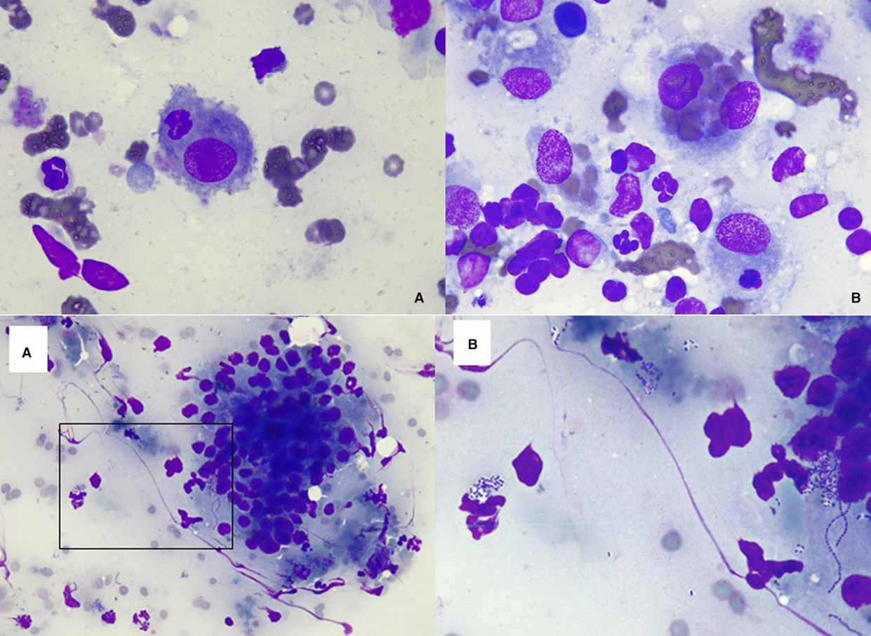
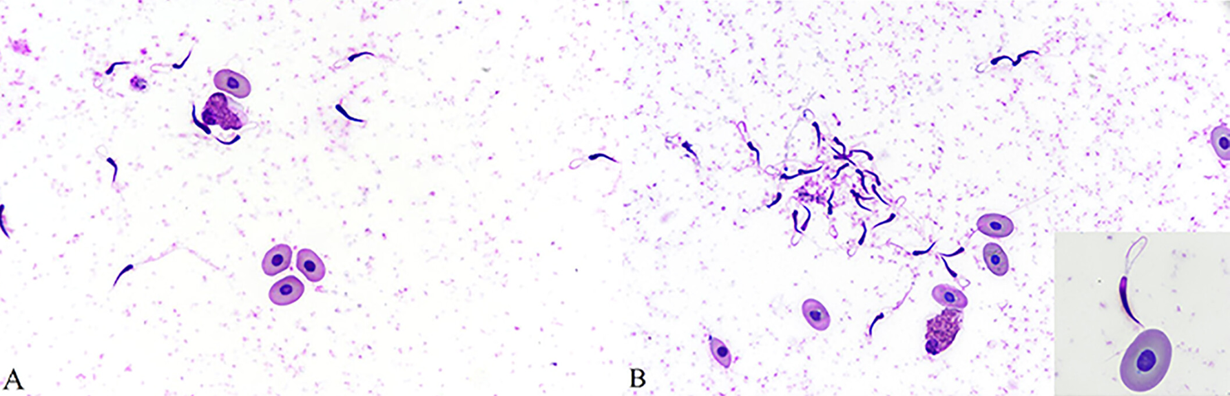
Blood cells in big-bellied searhorses