Computer Simulated Clinical Examination-Dental Hygiene CDCA
1/172
There's no tags or description
Looks like no tags are added yet.
Name | Mastery | Learn | Test | Matching | Spaced | Call with Kai |
|---|
No analytics yet
Send a link to your students to track their progress
173 Terms
Neurofibroma
A benign tumor most commonly on the tongue; can be on the lateral border of tongue. This tumor is derived from nerve tissue.
MRSA
Methicillin-resistant Staphylococcus aureus-is a strain of the staph bacteria that lives on 20-30% of the population but does not cause infection in all who are colonized with it. MRSA resides on the skin and in the nasal passage, and once you contract a MRSA staph infection it can be very difficult to eradicate the bacteria.
MRSA antibiotics
Many antibiotics can be used to treat this condition. Look for any of these: vancomycin, linezolid, bactrim, septra, tetracycline, minocycline, doxycycline, and clindamycin.
Prednisone
Delayed wound healing, poor healing, with increased risk of infection. This medication affects the immune response.
Ultrasonic insert with plastic tips
Can be used with implants for removal of calculus. Also, titanium and plastic hand instruments can be used on implants.
Oraqix duration
Approximately 20 minutes.
Hutchinson's Incisors
Congenital syphillus- notched incisal edges on incisors, also mulberry molars- tiny globules of enamel instead of cusps on first molars.
Class I Occlusion
(Mesognathic)-normal; mesiobuccal cusp of the maxillary first molar is positioned in the buccal groove of the mandibular first molar. Maxillary canine occluded with the distal half of the mandibular canine and the mesial half of the mandibular first premolar.
Class II Occlusion
(Retrognathic) buccal groove of the mandibular first permanent molar is distal go the mesiobuccal cusp of the maxillary first permanent molar by at least the width of a premolar. Distal portion of the maxillary canine is mesial to the mesial portion of the mandibular canine by at lease the width of a premolar.
Division I-retruded mandible with one or more maxillary anterior teeth protruded facially.
Division II- retruded mandible with one or more maxillary anterior teeth inclined lingually.
Class III Occlusion
(Prognathic) Buccal groove of the mandibular first permanent molar is mesial to the mesiobuccal cusp of the maxillary first permanent molar by at lease the width of a premolar. Mesial portion of the maxillary canine is distal to the distal surface of the mandibular canine by the width of a premolar.
Arch development
Each dental arch goes through phases of development as the permanent teeth erupt and the primary teeth are shed.
During this time, the ramus and body of the jaw develops and undergoes lengthening and horizontal growth to achieve its mature form and accommodate the larger permanent teeth.
Leeway space
This difference in size, mesiodistally between the two types of teeth. The contour of the bone covering the narrower roots of the premolars, in addition to the state of flux of the bone formation in this area, furnishes adjustment for dental arch measurements, making the middle segment of the arches important architecturally. Leeway space allows the future forward movement of the permanent molars.
The canine relationship between the arches in primary teeth is the same as:
As that of the permanent dentition.
Terminal plane
The ideal molar relationship in the primary dentition, when in centric occlusion.
Flush terminal plane
In which the primary maxillary and mandibular second molars are in an end‑to‑end relationship.
Mesial step
In which the primary mandibular second molar is mesial to the maxillary second molar.
Distal step
In which the primary mandibular second molar is distal to the maxillary second molar, is not an ideal molar relationship in the primary dentition and thus is not a type of terminal plane relationship.
Primate spaces
Within a primary dentition, primate spaces may occur between the primary teeth; a space is noted between the maxillary lateral incisor and the canine, and between the mandibular first molar and canine.
The most common occlusion related OMD is:
Tongue thrusting, which is the functional deviations occurring with habitual incorrect placement and use of the tongue, lips, and mandible during physiological rest, chewing, swallowing, and/or functional speech patterning.
Myofunctional considerations-mandible
The mandible may be more retruded during development in these situations or when a parafunctional habit, such as digit sucking, is present due to the excessive pressures of the thumb or hand resting against the mandible or the increased muscle activation of the mentalis muscle to help support an incompetent or everted lower lip pattern.
Myofunctional considerations-lingual frenum
Another issue is the length of the lingual frenulum. If the lingual frenulum is restricted, as with ankyloglossia, it limits the possibility of creating appropriate pressure against the maxillary arch for normal expansion.
Scalloped tongue
Indentations in the lateral border; harmless.
Macroglossia
Large tongue.
Hairy tongue
Elongation of filiform papillae (dorsum) clean tongue.
Hairy leukoplakia
Lateral border, white, corrugated lesions, associated with HIV, Epstein Barr virus.
Geographic tongue
Benign, inflammatory condition, irregular patches look like map.
Anemia
Impaired O2 delivery, 1st pale tongue, flattening of filiform papillae, finally painful and red.
Fissured tongue
Associated with xerostomia, clean tongue.
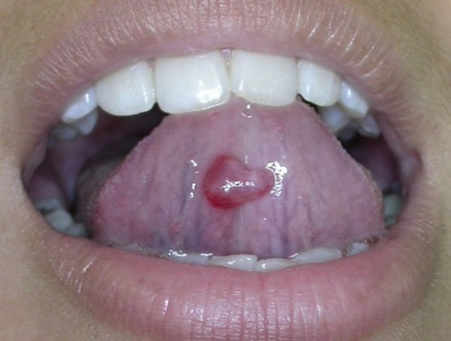
Cyst of Blandin-Nuhn
Accessory salivary glands on ventral surface, cyst develops from trauma.
Median Rhomboid Glossitis
Raised, red, glossy, slanted rectangular shape on dorsum due to Candida albicans infection.
Granular Cell Tumor
Benign tumor, granular tissue, circumscribed.
Lingual thyroid
Posterior 1/3 of tongue, thyroid gland fails to migrate toward trachea as developing, difficulty swallowing.
Piercing
Tongue common site, watch for infection and tooth fractures, remove for radiographs, keep clean.
Mucocele
Swelling due to mucus from accessory salivary glands, common, most mandibular labial mucosa.
Accessory salivary gland tumor
Nodular, benign, rare.
Nasolabial cyst
In canine-lateral region of maxillary lip.
Implantation cyst
Foreign-body reaction, after trauma.
Mesenchymal nodules and tumors
Fibromas, lipofibromas, neuromas, and neurofibromas.
Angioedema
Localized swelling of the fluid beneath the skin, most are allergic reaction.
Cheilitis glandularis
Chronic inflammatory disorder of labial salivary glands.
Orofacial granulomatosis
Nonpainful swelling forming granulomas.
Cellulitis
Inflammation of the cellular tissue, bacterial infection.
Actinic cheilosis
Premalignant lesion on vermilion border of the lip from the sun.
Candidal cheilitis
Candida albicans, inflammatory response.
Angular cheilitis
Red fissures at commissures, C. albicans and Staphylococcus aureus, nutritional deficiency, denture irritation.
Exfoliative cheilitis
With desquamation.
Odontogenic infection
Orofacial infection where microbes invade tissue, replicate, and overwhelm the immune system
4 classic features of infection
Heat
Pain
Redness
Swelling
4 places they spread
Buccal space
Masseteric space
Infraorbital space
Ludwig's angina
Buccal space
Near buccinator muscle, maxillary and mandibular molars.
Masseteric space
Between masseter muscle and side of ramus, MN molars
Infraorbital space
Just below eye to nose area, MX premolars, anteriors.
Ludwig's angina
Submandibular, submental, and sublingual, bilateral, MN molars or fractured mandible.
Angioedema
Accumulation of fluid, lips.
Emphysema
Abnormal presence of air in tissue
postoperative bleeding.
Purpura
Purplish discoloration.
Petechiae
Pinpoint, red spots.
Ecchymosis
Small bruise.
Hematoma
Larger localized bruise, black eyes, mistakenly injecting posterior superior alveolar vein.
Bell's Palsy
Weakness and paralysis of 7th cranial nerve, one side of face.
Traumatic ulcer
Chemicals (aspirin burn), heat (food), mechanical force, ill-fitting denture, broken tooth, dental injection, kids chewing numb lip after anesthesia, thumb sucking.
Recurrent aphthous stomatitis
3 types.
Minor aphthae
Canker sores, movable mucosa, red border.
Major aphthous
Severe form, > 1 cm, multiple lesions.
Herpetiform
Least common, resemble primary herpes, smaller (pinpoint) at 1st, any mucosa.
Pseudoaphthae
Nutritional deficiency, Crohn's disease often.
Behçet's syndrome
Affects triad of oral, eye, and genital surfaces primarily.
Aphthae
Red border.
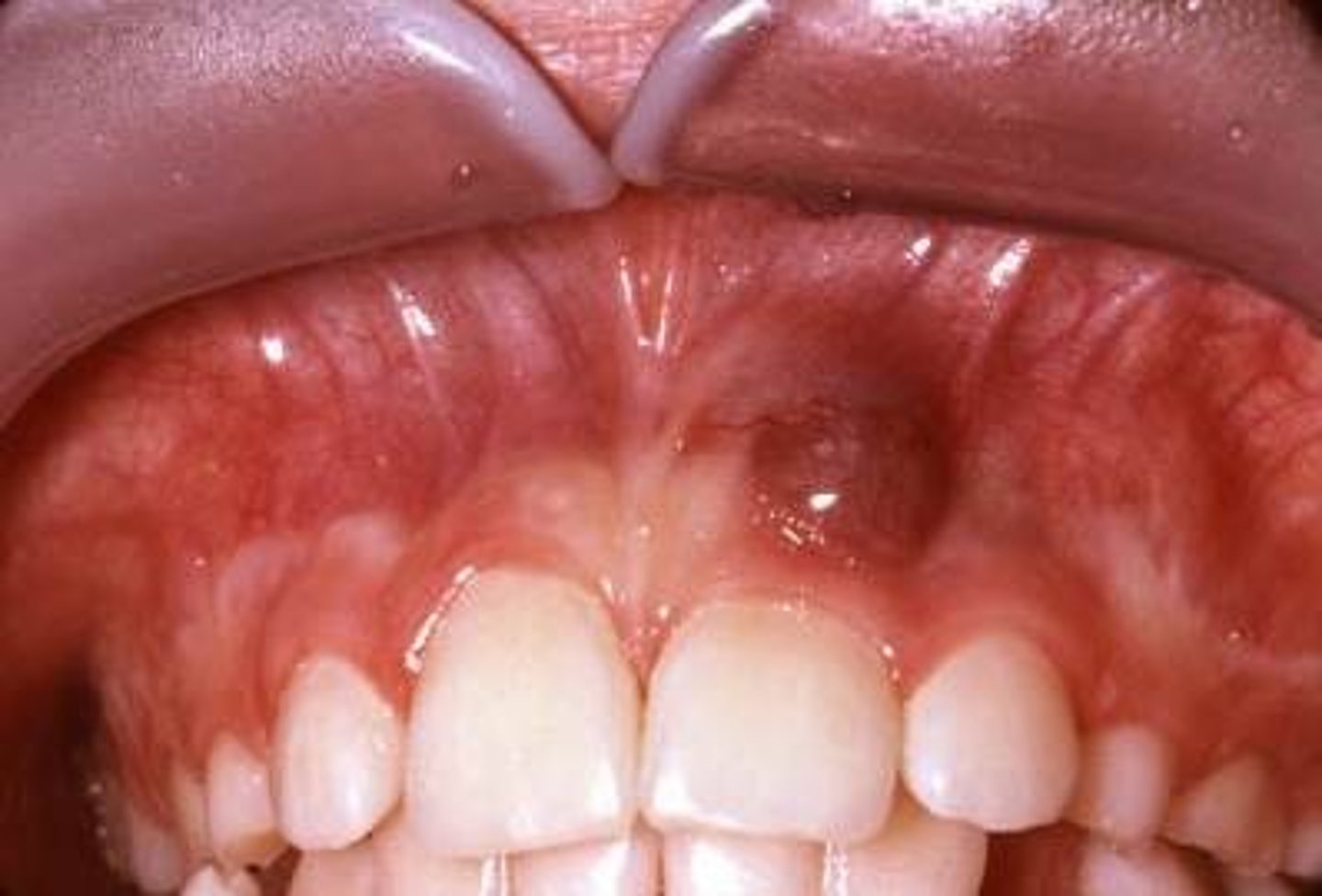
Eruption cyst
Soft tissue cyst surrounding the crown of an unerupted tooth.
Small, dome-shaped, translucent swelling filled with blood or serum and thus is red, brown, or blue-gray.
Herpetic gingivostomatitis
Painful vesicles, become ulcers, contagious, can become herpetic whitlow (finger) or conjunctivitis (eye), postpone treatment.
Recurrent herpes simplex infection
Dormant in nerves, recurs, clusters, contagious, postpone treatment.
Recurrent herpes labialis
Lip, vesicle to scab.
Recurrent herpes stomatitis:
Intraoral.
Varicella Zoster
Human herpes virus 3. Primary infection (chickenpox): rash, face and trunk, vaccine prevents.
Recurrent infection-herpes zoster or shingles
Mostly in elderly, unilateral, immunosuppressed vulnerable.
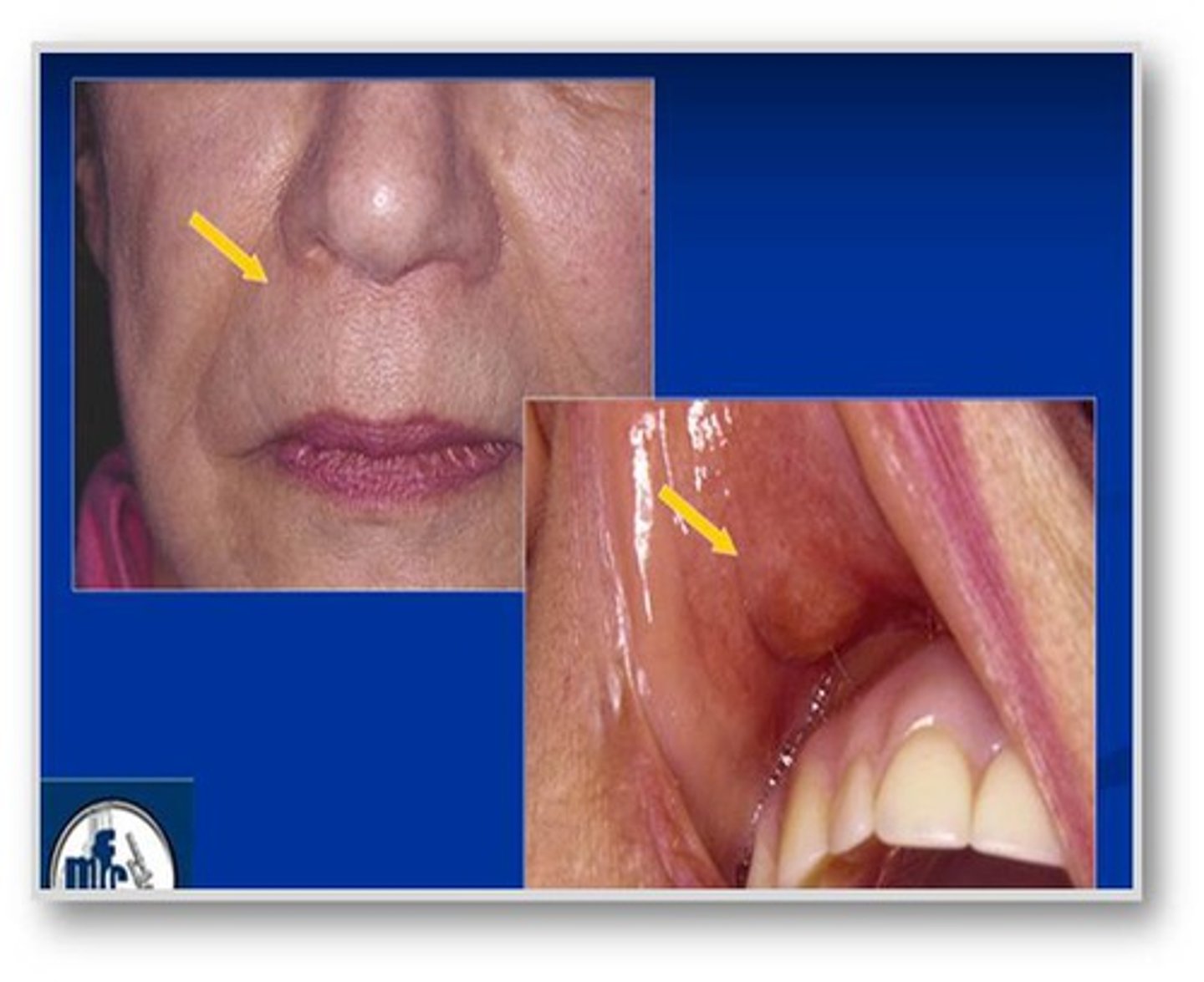
If you see a photograph of a geriatric patient with one side of the face appearing to have some bumps and redness by the eye, what would this most likely be?
Herpes Zoster
Herpes Zoster
Also called Shingles. It is a painful, blistering, skin rash due to the Varicella Zoster Virus-the virus that causes chickenpox. Shingles may develop in any age group but you are more likely to develop the condition if: you are older than 60, you had chickenpox before age 1, your immune system is weakened by medications or disease.
Measles
Also known as morbilli, an infection of the respiratory system caused by a virus specifically a paramyxovirus of the genus Morbillivirus. This is the condition where you get Kopliks spots inside the mouth. It appears primarily in children. You can get a rash that starts on the head and spreads to other areas, moving down the body that appears as flat, discolored areas (macules) and solid, red, raised areas (papules) that later join together.
Herpangina
A viral illness ordinarily in children that involves ulcers or sores (lesions) inside the mouth, a sore throat, and fever.
Herpes Simplex
A virus that produces most cold sores and genital herpes. It usually affects the lips, and in some primary attacks, the mucous membranes in the mouth. A herpes infection may occur on the cheeks or in the nose, but facial herpes is very common.
Which medications cause bleeding?
Blood thinners and chemotherapy agents. The main ones are antihistamine, anticholinergic, anorexiants, antidepressants, antihypertensives, anti-parkinsons agents, diuretics and sedatives.
Medications:
Anticoagulants such as warfarin (coumadin)
Antiplatelet drugs such as aspirin and clopidogrel (plavix)
Certain epilepsy, convulsion and seizure medications
Chemotherapy drugs
Antihistamine drugs
Immunosuppressant drugs
Drug classes that commonly cause xerostomia
Antiemetics, antianxiety agents, decongestants, analgesics, antidiarrheals, bronchodilators and skeletal muscle relaxants.
If a patient with infective endocarditis has no contraindications of premedication, when would they need to be premidicated?
Periodontal probing is an invasive procedure requiring medication if indicated. The remaining options are not considered invasive and therefore do not require it.
If you see a picture of fordyce granules and it asks which treatment was neccessary
No treatment is necessary. It is just a sebaceous gland.
If you see a question about fluoridated water; a child drinking fluoride water and taking supplements is at greater risk for?
Hypocalcification. Dental fluorosis is a form of enamel hypocalcification which results from the ingestion of excessive fluoride during the period of enamel formation. To cause mottling, fluoride must be present in concentrations several times that found in controlled fluoridated water supplies.
After being called to the reception area and seeing an unresponsive patient what do you do next?
Activate EMS procedures.
Which hepatitis shot is given to the dental hygienist employee?
All dental staff should be protected from hepatitis B virus.
Which method of fluoride application immediately gets absorbed into the enamel?
Varnish has the highest PPM and whichever product has the highest PPM gets absorbed into the enamel quicker.
If you have an elderly patient with BP 153/120mmHG what do you do?
1. Re-check BP in 5 minutes.
2. Consult MD immediately.
3. Do NOT perform dental treatment until the elevated pressure decreases.
Free gingiva
The area from the free gingival groove to the top of the top of the gingiva (free gingival margin).
Neurofibroma
A benign nerve sheath tumor derived from the myelin sheath of peripheral nerves in the peripheral nervous system. It's an autosomal dominant genetically-inherited disease, which can result in a range of symptoms from physical disfiguration and pain to cognitive disability.
Neuroma
A growth or tumor of nerve tissue. Neuromas are tumors of any part of a nerve (including the surrounding myelin); sometimes the term is used more broadly to refer to any tumor of neural tissue.
What % of alcohol in hand disinfection solution to use in a dental office?
60%
Median rhomboid glossitis
Currently thought to represent a chronic fungal (candidiasis) infection in this area of the tongue. In general, no treatment is necessary for median rhomboid glossitis. For those with symptoms (pain or burning sensation), an antifungal medication may be prescribed to kill the yeast and thereby reduce the symptoms.
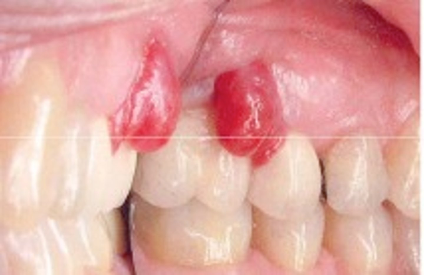
How do you determine is it Pyogenic granuloma or a Peripheral giant cell granuloma?
This can be hard to distinguish. The appearance of peripheral giant cell granuloma is similar to pyogenic granuloma. Peripheral giant cell granulomas color ranges from red to bluish -purple, but is usually more blue in comparison to pyogenic granulomas. It can be sessile or pedunculated with the size usually being less than 2cm.
Pyogenic granuloma
Small, reddish bumps on the skin that bleed easily due to an abnormally high number of blood vessels. It is a primarily oral disease which appears as an overgrowth of tissue due to irritation, physical trauma or hormonal factors. It is not an infectious but a reactive lesion due to local irritating factors.
Peripheral giant-cell granuloma
An oral pathologic condition that appears in the mouth as an overgrowth of tissue due to irritation or trauma. It presents as a lobular, purplish-blue exophytic nodule exclusively on the gingiva, both edentulous and dentate, and usually anterior to the molars. It originates from the periodontal ligament or the periosteum. It occurs across a wide age range, commonly in children, young adults, and in females. It presents as sessile or pedunculated and smooth surfaced or lobular, and though usually painless it can occasionally be ulcerated, painful and accompanied by bleeding. Like pyogenic granuloma, it is usually present either on the buccal or lingual gingiva or between teeth, but it can occasionally surround the teeth and act aggressively by displacing teeth much like a sarcoma. It appears only on the gingiva or on an edentulous (without teeth) alveolar ridge. It is more often found in the mandible rather than the maxilla but can be found in either anterior or posterior areas. The underlying alveolar bone can be destroyed, leaving a unique appearance referred to as "cupping resorption" or "saucerization."
Peripheral giant-cell granuloma target population
There is a gender difference with 60% of the disease occurring in females. The prevalence of peripheral giant cell granulomas is highest around 50-60 years of age.
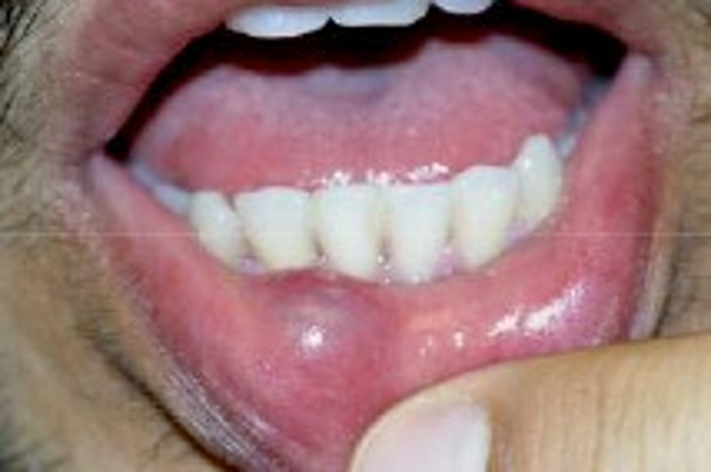
Mucocele
A mucous cyst and is painless, thin sac on the inner surface of the lips. It contains clear fluid. Symptoms: a thin fluid-filled sac appears on the inside of the lip. The sac is bluish and clear.
Ranula
A type of mucocele found on the floor of mouth. Ranulas present as a swelling of connective tissue consisting of collected mucin from a ruptured salivary gland duct, which is usually caused by local trauma. Only appears UNILATERALLY! Not bilateral.