chapter 19 : ultrasound evaluation and mapping of the superficial venous system
1/43
There's no tags or description
Looks like no tags are added yet.
Name | Mastery | Learn | Test | Matching | Spaced | Call with Kai |
|---|
No analytics yet
Send a link to your students to track their progress
44 Terms
competency
duplex ultrasound is used to assess the — of superficial veins for use as bypass conduit
types of bypass procedures superficial veins can be used for
coronary bypass grafting
lower extremity bypass grafting
hemodialysis access fistulas
information gathered about the superficial veins
vein patency
position
depth
size
vein mapping
allows for selection of optimal vein, can alter planned surgery and surgical approach, and minimizes the amount of surgical dissection
5 common figurations of the great saphenous vein
single trunk medially in thigh with several large tributaries
single trunk that courses anterolaterally in the thigh
various degrees of duplication and communication
closed loop system in thigh
partial double system
saphenous compartment
main trunk of the saphenous vein lies in —
bounded by saphenous fascia superficially and muscular fascia deeply
dominant vessel
when duplication of the GSV is present, one system may be larger
— it’s important to note for proper selection
superficial
duplicate systems of the GSV often course — of the saphenous compartment and are likely to be an accessory saphenous vein
large tributaries of the GSV
anterior accessory GSV
posterior accessory GSV
variations in calf are less common and include
anterior dominant GSV
posterior dominant GSV
double system
cutaneous tributaries
GSV system has multiple —
perforating veins
perforate the muscle fascia and connect the superficial system to the deep system
have valves to ensure one-way flow (superficial to deep)
arteriovenous fistula
can be created during in situ bypass surgery if valves of perforating valves are not ligated
small saphenous vein
typically a single trunk, which courses through the middle posterior aspect of calf
terminates in the popliteal vein
variations of the small saphenous vein
in about 20% of patients, it continues above the popliteal fossa (cranial extension of the - )
can terminate directly into the femoral vein or inferior gluteal vein, or can communicate with GSV
cephalic vein
begins at wrist, courses along radius and into upper arm, and terminates into the subclavian vein
basilic vein
begins at wrist, courses along ulnar and into upper arm, and terminates into the brachial vein to form the axillary vein
cephalic & basilic veins
can be amped for bypass; however, more common to evaluate as part of preoperative assessment for creation of dialysis fistula
groin
mapping the GSV begins at the —
new mark should be placed every 2-3 cm
transverse
— orientation is bets used to identify tributaries when mapping out the GSV
vein size
is also measured in TRV orientation when mapping the GSV
should be measured at the prox, mid, and distal thigh and calf
inner vein diameter
— should be measured (inner wall/blood interface to inner wall/blood interface) when taking images of the GSV
popliteal vein
when mapping the small saphenous vein, the same techniques are used
identify at confluence with — first
brachial vein
basilic vein can be followed from its termination into the — . then followed to ulnar aspect of wrist
biceps muscle
cephalic vein is easiest to follow in the upper arm over the —
then followed to its termination into the subclavian vein
vein diameters are measured proximally and distal in forearm and upper arm
higher frequency transducer (10MHz+)
you want to use a — for superficial imaging
wall status
planar arrangement (how it sits in one of the compartments)
diameter
vein mapping provides information not only about the presence or absence of vein but also about the suitability of vein or use as a conduit
normal, healthy vein should :
have smooth, thin walls
be compliant and easily compressible
have freely moving valve leaflets
2 mm
most surgeon will not use a vein with a diameter less than —
2.5-3 mm
most prefer vein diameter of —
spasm
smaller veins may be prone to — and are difficult to suture
segmental thrombus
may be encountered in superficial veins
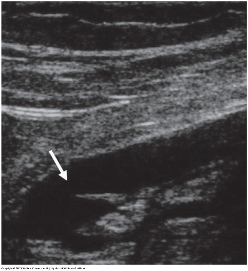
valve leaflets
thrombus is usually visualized adjacent to —
varicosities
dilated, tortuous portions of the saphenous system
subdermal branches
varicosities may be in — with main saphenous trunk spared
recanalization
presents as irregular intima surface or wall thickening
usually not considered adequate for se as conduit
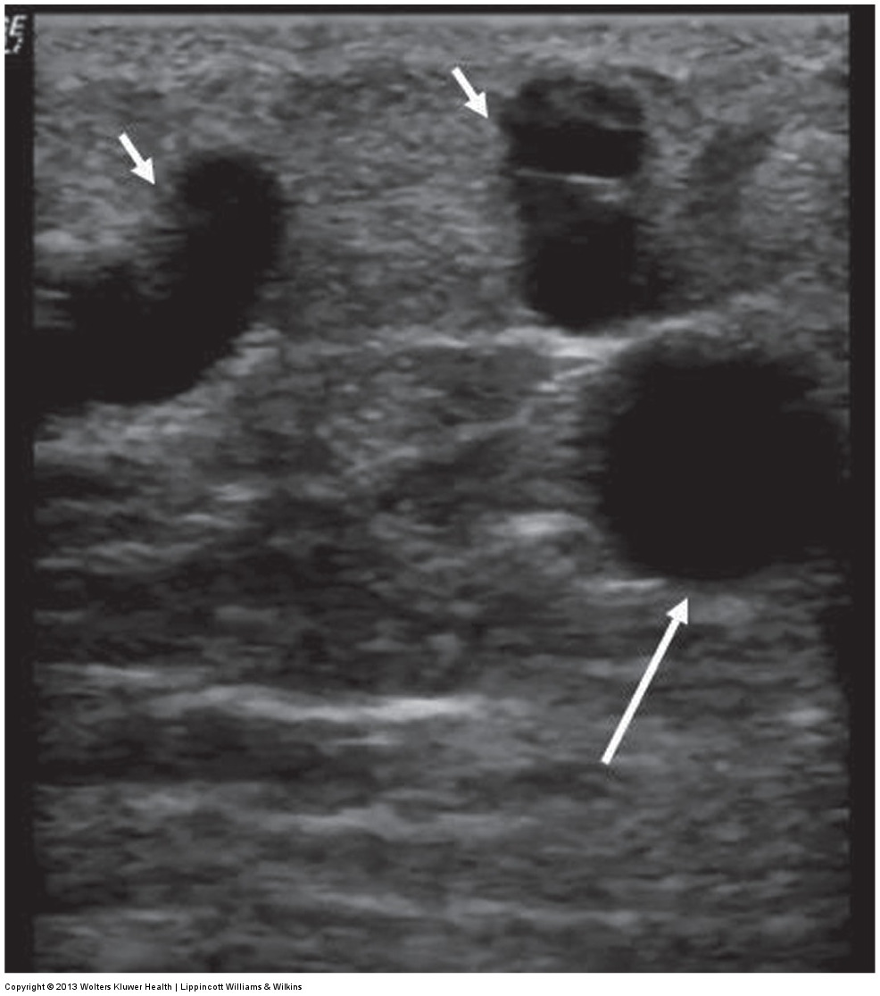
calcification
not as common as in arterial walls; often occurs in diabetic patients
presents as bright echoes within the vein wall producing acoustic shadowing
vein still may be used if it is isolated
vein inadequate
diffuse, intermittent calcification renders —
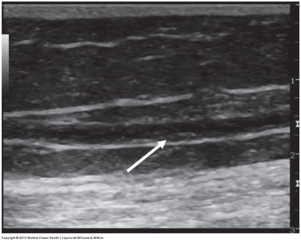
valve abnormalities
stenosis or frozen valve
often encountered in vein that as previously thrombosed
if isolated, healthy vein segment can be used
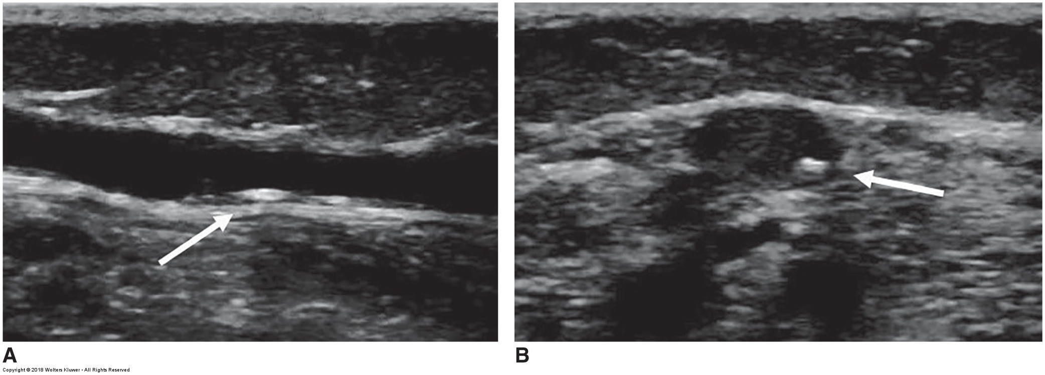
ultrasound image of thrombus next to valve leaflets
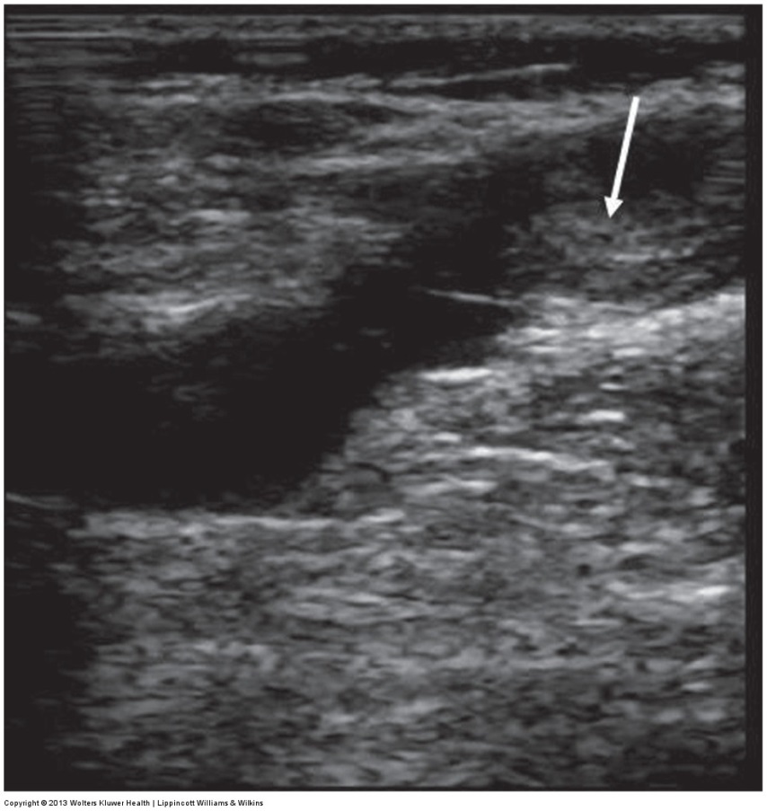
ultrasound of superficial varices with the main system beneath it
ultrasound image of a thickened recanalized vein
ultrasound of the SSV with wall calcification
ultrasound image of a frozen valve leaflet