GRISTO Module 3 Anatomy: Infratemporal Fossa
1/55
There's no tags or description
Looks like no tags are added yet.
Name | Mastery | Learn | Test | Matching | Spaced | Call with Kai |
|---|
No analytics yet
Send a link to your students to track their progress
56 Terms
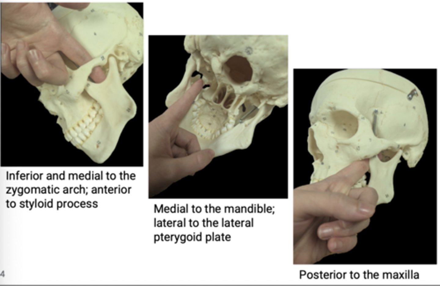
Infratemporal fossa
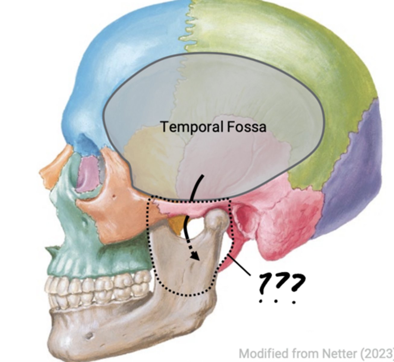
Anterior: Maxilla
Posterior: Styloid process
Medial: Lateral pterygoid plate
Lateral: Mandibular ramus
Borders of the infratemporal fossa
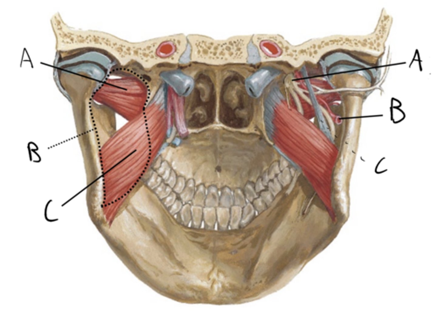
A. Temporalis
B. Lateral pterygoid muscle
C. Medial pterygoid muscle
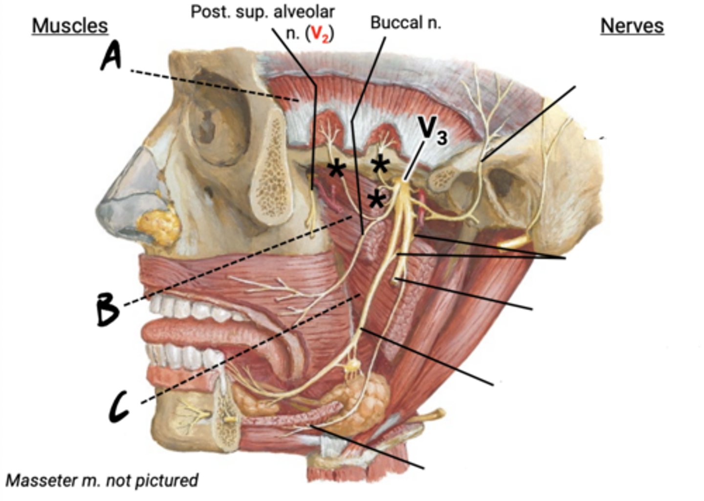
Buccal
A. Auriculotemporal nerve
B. Chorda tympani nerve (CN VII)
C. Inferior alveolar nerve
D. Lingual
E. Nerve to mylohyoid
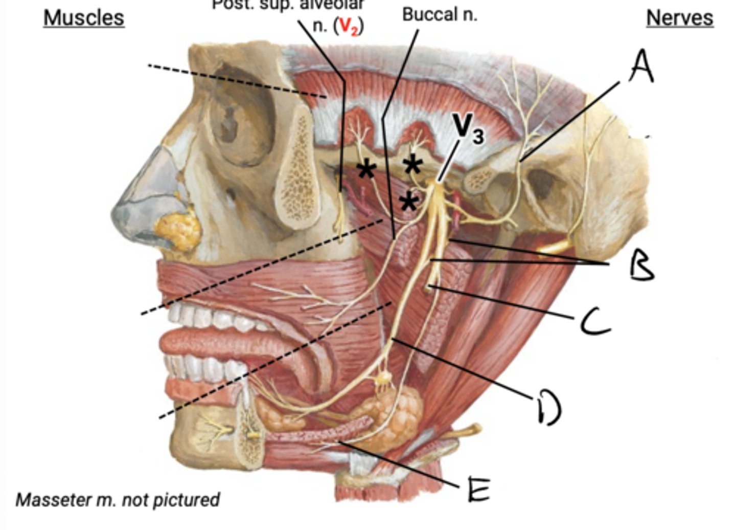
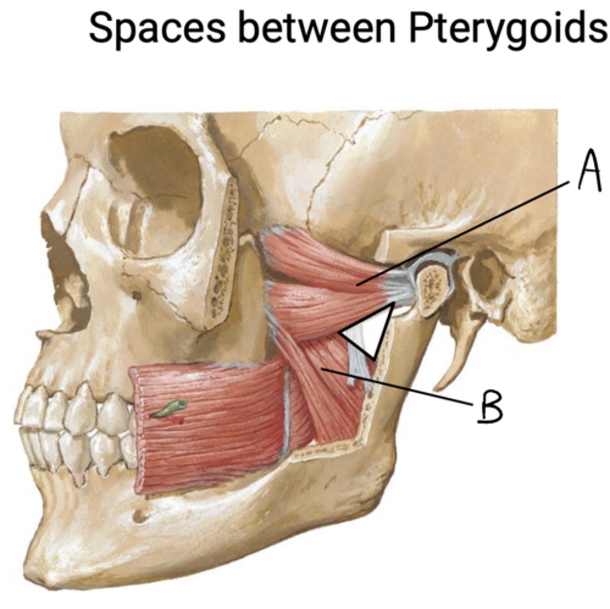
the medial and lateral pterygoid muscles
The lingual and inferior alveolar nn are found between
A. Lateral pterygoid muscle
B. Medial pterygoid m
CN V3 and the maxillary artery
The space between the pterygoid muscles is a hotspot for
LEFT:
A. Lateral pterygoid muscle
B. Infratemporal fossa
C. Medial pterygoid muscle
Right:
A. V3 (and branches)
B. Maxillary artery
C. Sphenomandibular ligament
VASCULATURE OF THE HEAD AND DEEP FACE
VASCULATURE OF THE HEAD AND DEEP FACE
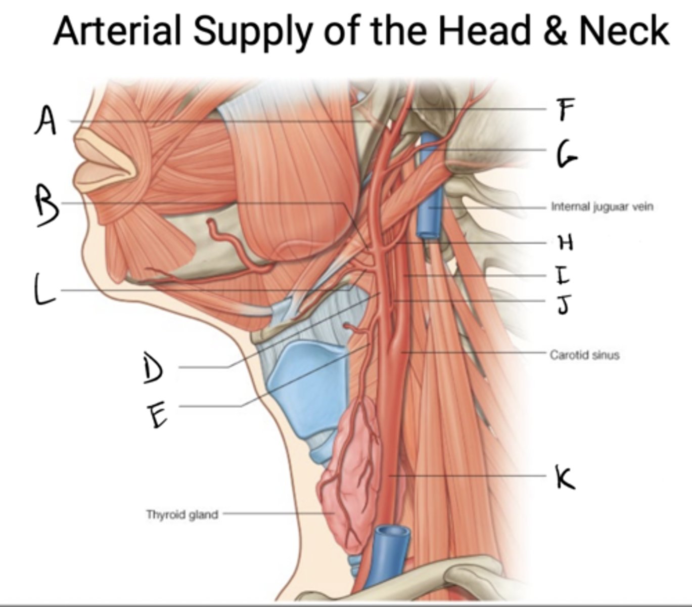
A. Maxillary artery
B. Facial artery
C. Lingual artery
D. External carotid a.
E. Superior thyroid a
F. Superficial temporal a
G. Posterior auricular a
H. Occipital a
I. Internal carotid a
J. Ascending pharyngeal a
K. Common carotid a
angle of the mandible
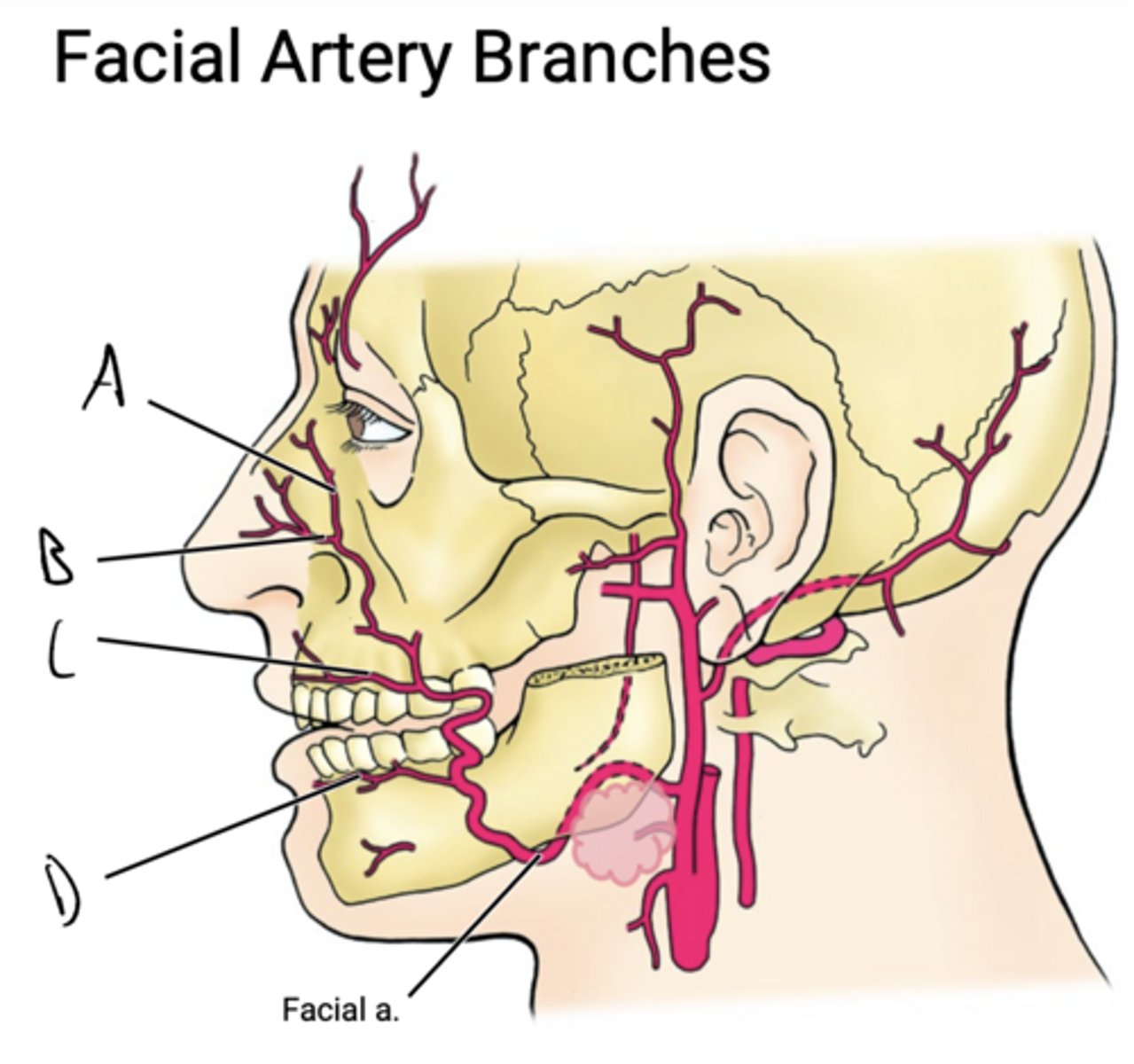
The facial artery branches near the
mandible (and posterior to the submandibular gland in the neck)
The facial artery runs posterior to the
inferior mandible to enter the face
The facial artery loops around the _____ to enter ____
the bridge of the nose, giving off several branches along the way
The facial artery then runs diagonally from the mandible up to
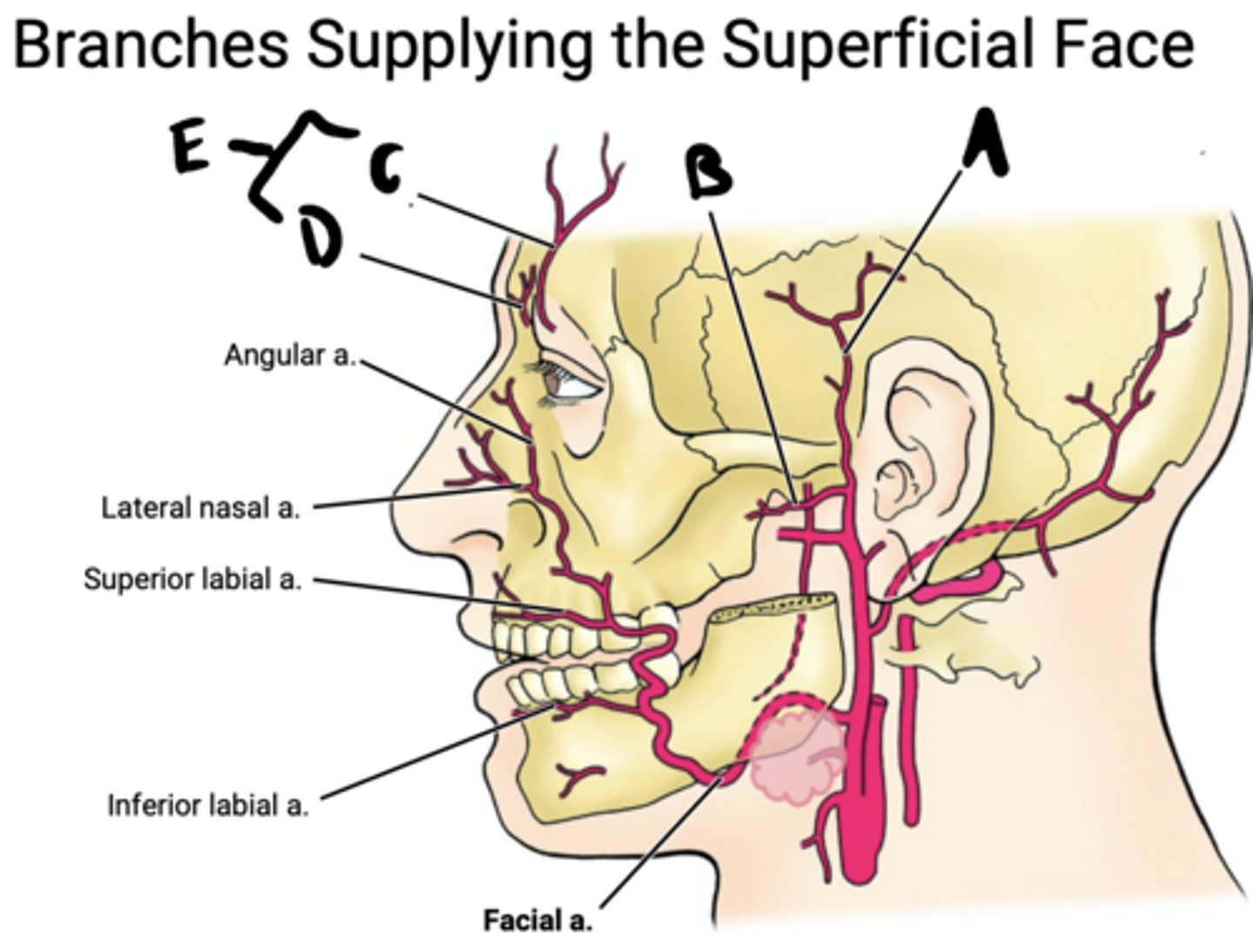
Facial artery
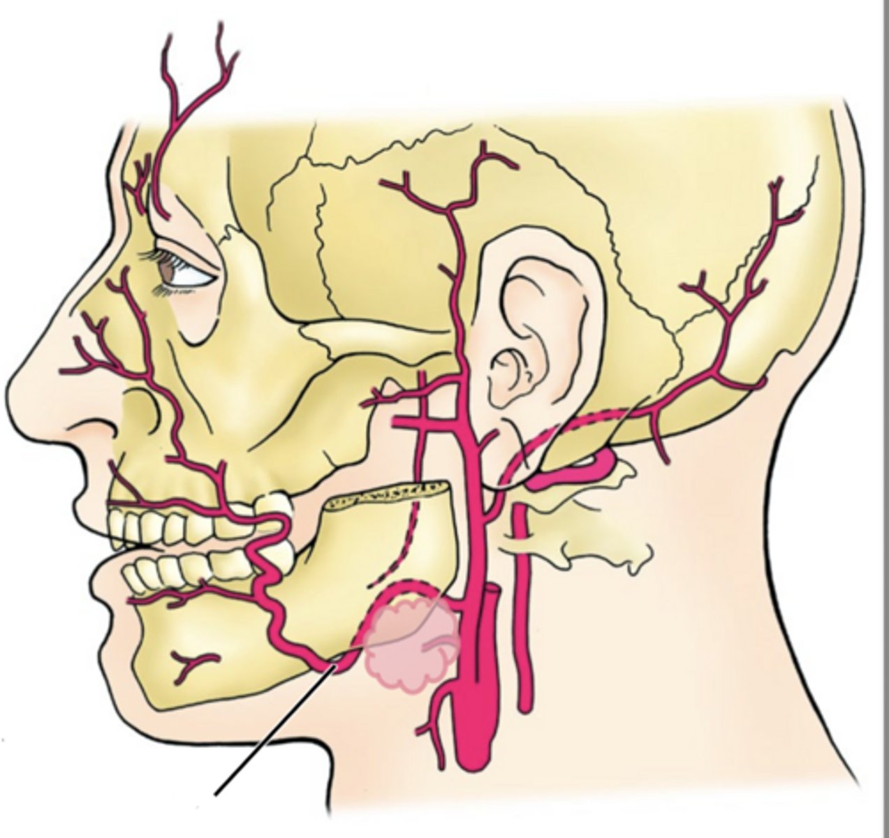
A. angular a.
B. lateral nasal a.
C. Superior labial a.
D. inferior labial a.
A. Superficial temporal artery
B. Transverse facial artery
C. Supraorbital artery
D. Supratrochlear artery
E. Internal carotid a. branches
A. Maxillary artery
B. Lateral pterygoid m
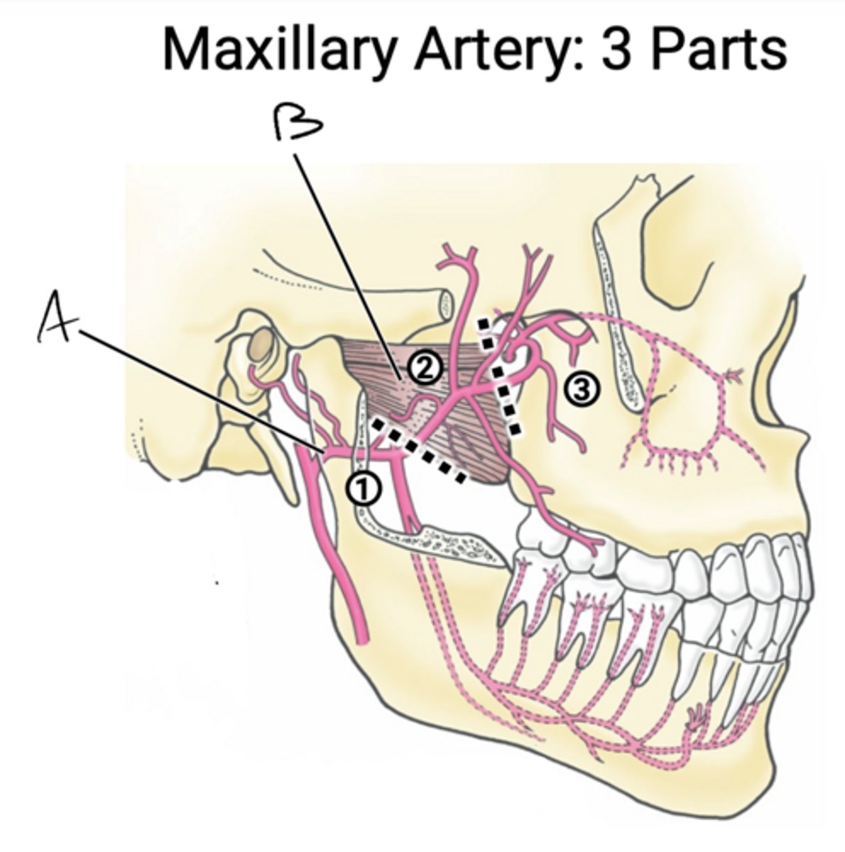
Portion before lateral pterygoid
- branches run with named V3 branches
Maxillary artery: Part 1
Portion over/under lateral pterygoid
- branches run with V3 muscular nn
- branches go directly to structure (no foramina)
Maxillary artery: Part 2
Portion after lateral pterygoid
- Branches run with V2 branches
Maxillary artery: Part 3
A. Inferior alveolar artery
B. Deep auricular artery
C. Anterior tympanic artery
D. Middle meningeal artery
Follows inferior alveolar nerve (V3) into mandibular foramen
Supplies mandibular teeth, chin
Inferior alveolar artery
Follows auriculotemporal nerve (V3)
Supplies external acoustic meatus
Deep auricular artery
Follows chorda tympani nerve (VII)
Supplies tympanic membrane
Anterior tympanic artery
Enters foramen spinosum
Supplies dura mater
Middle meningeal artery
A. Deep temporal aa
B. Masseteric aa
C. Pterygoid aa
D. Buccal aa
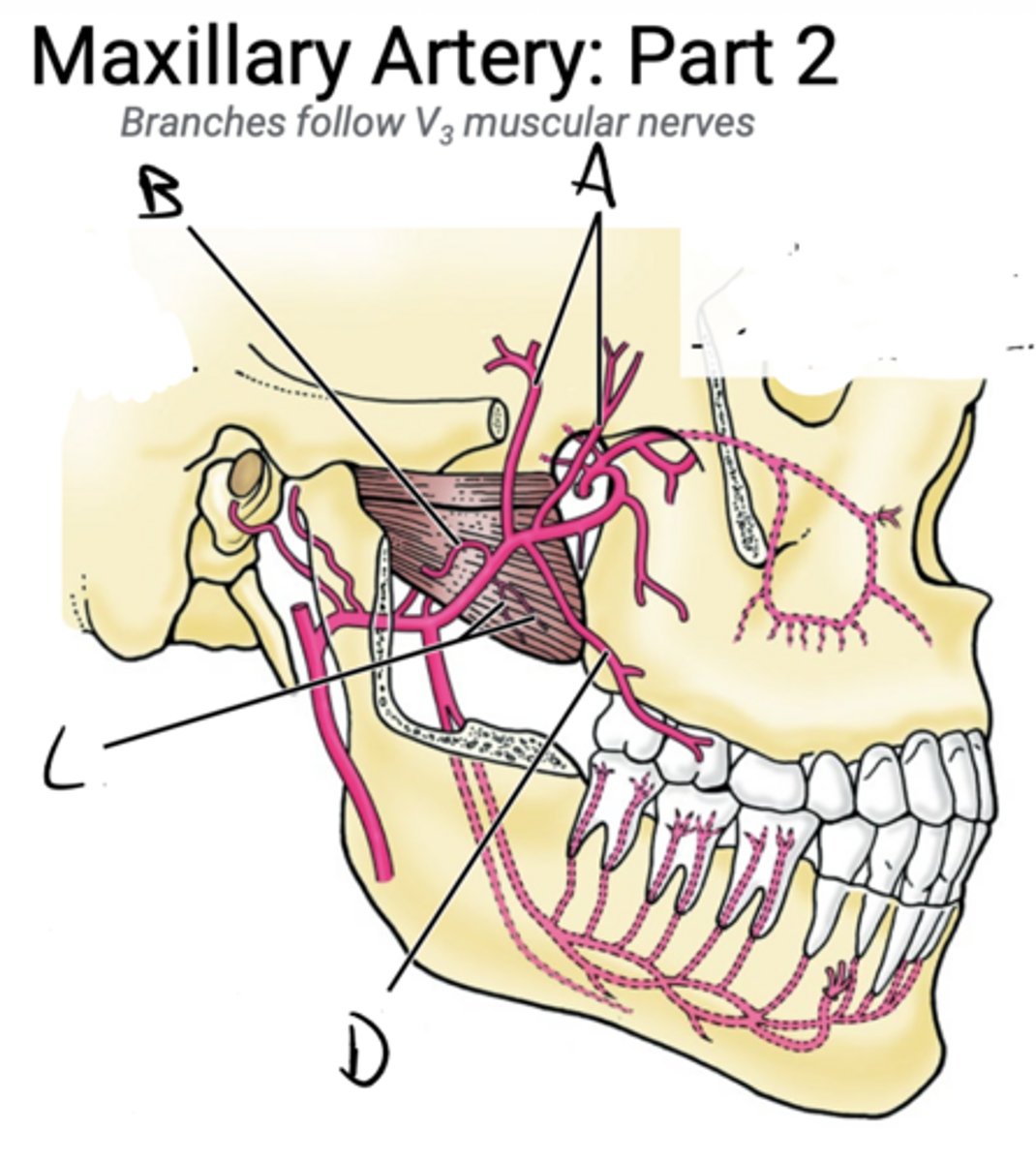
Follow nerve to temporalis m (V3)
Supply temporalis muscle
Deep temporal aa
Follow nerve to masseter m (V3)
Supply masseter muscle
Masseteric aa
Follow nerve to pterygoid mm (V3)
Supply pterygoid muscle
Pterygoid aa
Follow buccal n (V3)
Supply buccinator muscle
Buccal aa
A. Artery of pterygoid canal
B. Sphenopalatine artery
C. Infraorbital artery
D. Posterior superior alveolar artery
F. Descending palatine artery
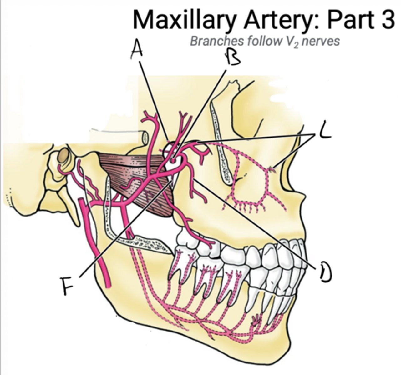
Follows greater petrosal n (VII)
Supplies pharynx, auditory tube, middle ear
Artery of pterygoid canal
Follows nasopalatine n (V2)
Supplies nasal cavity, anterior palate
Sphenopalatine artery
Follows infraorbital nerve (V2)
Supplies maxillary teeth, maxillary sinus, anterior face
Infraorbital artery
Follows posterior superior alveolar nerve (V2)
Supplies maxillary teeth, maxillary sinus
Posterior superior alveolar artery
Follows greater/lesser nn (V2)
Supplies palate
Descending palatine artery
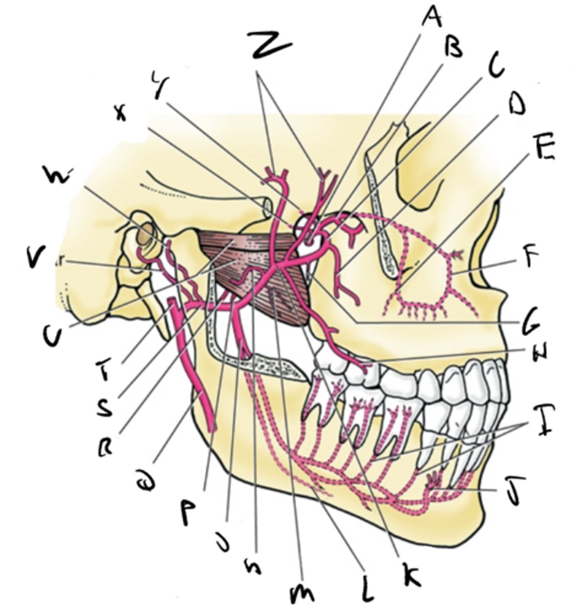
A. Sphenopalatine
B. Infraorbital
C. Maxillary (3rd part)
D. Posterior superior alveolar
E. Middle superior alveolar
F. Anterior superior alveolar
G. Descending palatine
H. Buccal branch
I. Dental branches
J. Mental branch
K. Maxillary (2nd part)
L. Branch to mylohyoid
M. Pterygoid branch
N. Masseteric branch
O. Inferior alveolar
P. Accessory mingeal
Q. External carotid
R. Middle meningeal
S. Maxillary (1st part)
T. Superior temporal artery
U. Lateral pterygoid muscle
V. Deep auricular
W. Anterior tympanic
X. Artery of pterygoid canal
Y. pharyngeal artery
Z. Deep temporal branches
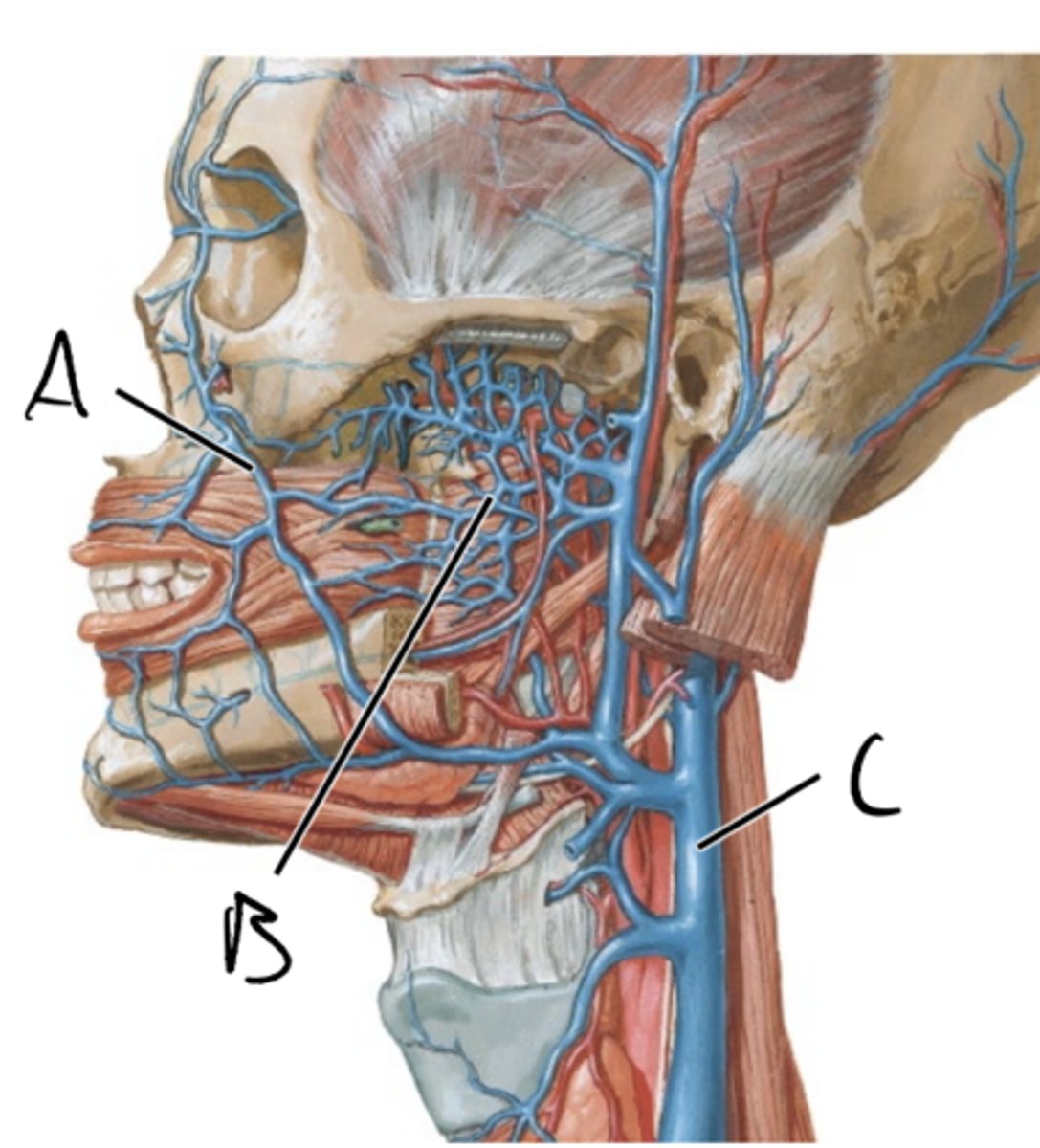
arterial system
In general venous return follows
pterygoid venous plexus
The __________ is a network of veins in infratemporal fossa
- Down to the internal jugular veins
- Back to Retromandibular veins
- Up to the ophthalmic veins
- Up to the Cavernous sinus (Meningeal blood space)
Pterygoid plexus and facial veins have multiple draining pathways
A. Facial vein
B. Pterygoid venous plexus
C. internal jugular vein
- blood/nutrient supply to the teeth
- Spread of infection
- Vascular injury during anesthetic injection
- Inadvertent intravascular injection of anesthetic
Clinical significance of infratemporal vasculature
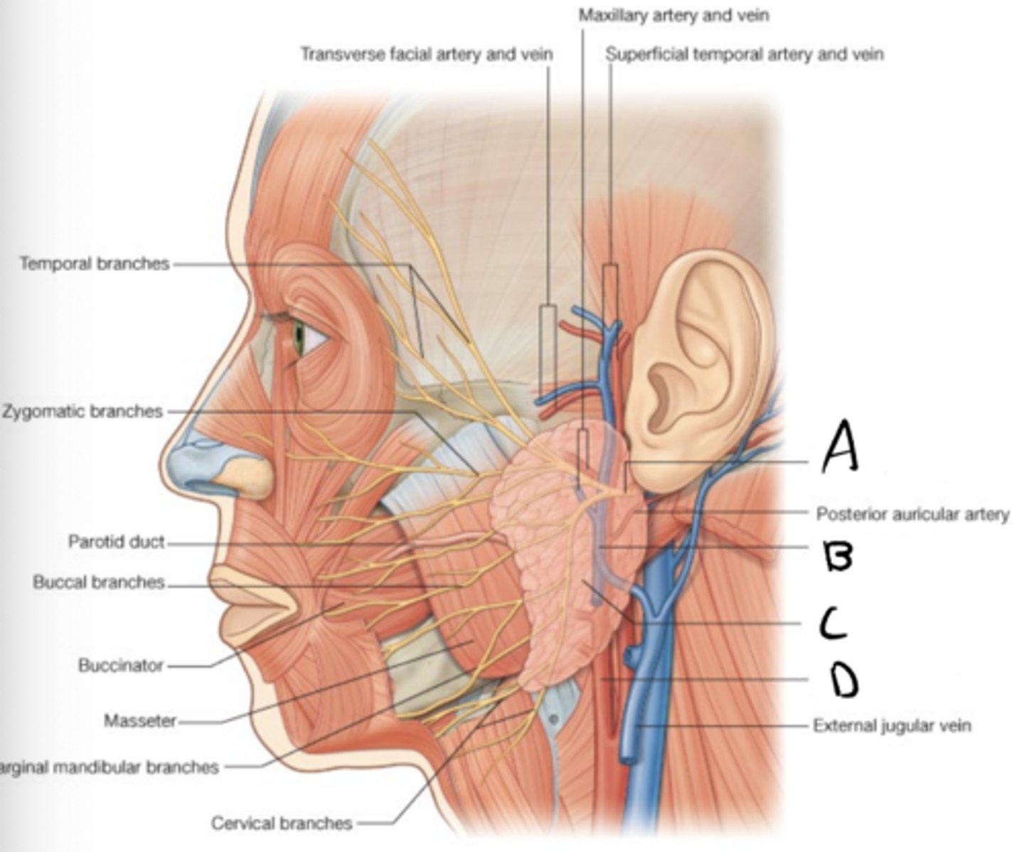
Parotid region
Superficial to the infratemporal fossa is the
Parotid gland
Facial nerve
External carotid artery
Retromandibular vein
Structures in the parotid region
A. Facial nerve
B. Retromandibular vein
C. Parotid gland
D. External carotid artery
salivary gland
The parotid gland is a
produces serous secretions
Parotid gland primarily
CN IX
The parotid glands is stimulated by parasympathetics from
Lesser petrosal n (CN IX), Auriculotemporal n (V3)
Preganglionics are carried in the _______ and postganglionics are carried via the
saliva to the oral cavity
The parotid duct conveys
Masseter muscle, buccinator muscle
The parotid duct passes out of the parotid gland superfifcal to ______. Then it dives down through the fibers of the ________ to enter the oral cavity
2nd maxillary molar
The parotid duct enters the oral cavity adjacent to the
infection (E.g mumps)
Clinical note: poor oral hygiene can lead to what spreading through the parotid duct to the parotid gland
infratemporal fossa.
This has important implications when considering anesthetic injections
The Parotid gland often wraps around the posterior mandible, meaning small portions of the gland are located in the