PSB3340 EXAM 3: CH 8 General Principles of Sensory Processing
1/41
There's no tags or description
Looks like no tags are added yet.
Name | Mastery | Learn | Test | Matching | Spaced | Call with Kai |
|---|
No analytics yet
Send a link to your students to track their progress
42 Terms
How is the notion of Five Senses challenged from the neurobiological perspective?
Each sensory processes multiple features of one stimulus
Each sensory system has multiple types of sensory receptors
Example: pressure, temperature, balance, pain, proprioception
What are the submodalities of somatosensation?
Touch
pressure, vibration, and texture
Proprioception
sense of body position and movement
Temperature
detects hot and cold
Pain (Nociception)
What types of sensation are processed from the inner ear
Auditory
sound waves
cochlea
hair cells in the organ of corti convert vibrations to electrical signals
signals travel auditory nerve to brain
Vestibular (Balance)
head movements and position
both info is sent to the vestibulochochlear nerve (cranial nerve VIII!
Distinguish sensory receptors and neurotransmitter receptors.
Sensory receptors detect specific physical stimuli
Don’t have axons, have cell bodies that synapse on dendrites or cell bodies of other neurons
convert stimuli into eletrical signals thru transduction
Neurotransmitter receptors detect chemical signals released by neurons
mediate communication between neurons by initiating post synaptic potentials
What is meant by sensory salience?
How noticeable or attention grabbing a particular stimulus is to the sensory systems
ex: brighter lights, unexpected stimuli,etc
What is the Doctrine of Specific Nerve Energies?
Johannes Muller hypothesized that Each sensory nerve, no matter how it is stimulated, produces its own specific type of sensation.
concluded that the brain is funcitonally divided and specific nerves convey specific types of info
all sensory info is eventually encoded as action potentials
For example:
Stimulating the optic nerve (even by pressure or electricity) will cause you to see light (a visual sensation), even if no light is present.
Stimulating the auditory nerve will produce a sound sensation.
Do sensory receptors fire action potentials? Explain
✅ Some do fire action potentials directly:
These receptors generate action potentials themselves, especially in unipolar neurons (e.g., mechanoreceptors in skin or olfactory receptors).
They convert a stimulus into an electrical signal called a receptor potential, and if it's strong enough, it triggers an action potential that travels along the sensory neuron's axon to the CNS.
🛑 Others do not fire action potentials directly:
These receptors (e.g., photoreceptors in the retina, hair cells in the ear) don’t fire action potentials.
Instead, they produce graded potentials (like depolarization or hyperpolarization) and release neurotransmitters to activate adjacent neurons, which then fire action potentials.
In general terms, how does sensory information reach the brain?
reception: absorption of physical energy by sensory receptor
hair cells in your ear bend from sound waves
transduction: conversion of physical energy to electrochemical pattern in neurons
touch receptors convert pressure into depolarization > action potentials
coding: turning the signal into a pattern of neural activity that tells your brain what the stimulus is
brighter light causes more frequent firing in certain neurons
Define umwelt.
the way a species perceives the world
ex: dogs can hear higher frequencies than us
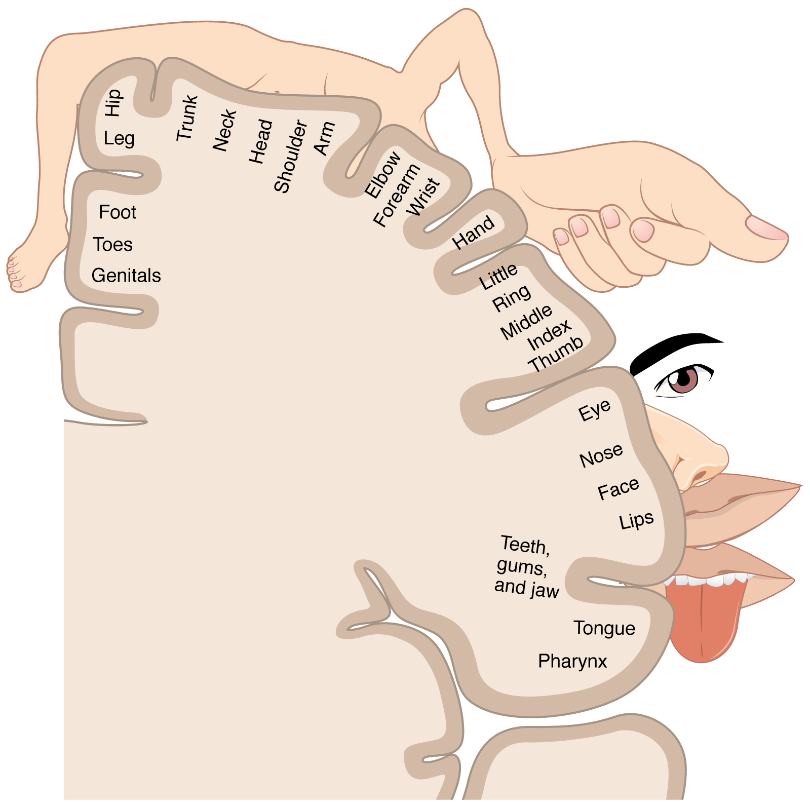
Do all sensory modalities occupy brain regions equally in size? Explain
Sensory modalities do not take up equal brain space. The brain allocates more space to senses and body parts that are more important or require more precise processing.
How do neurons encode stimulus frequency?
Neurons encode frequency (like the pitch of a sound or the rate of a flickering light) by:
Phase locking: Neurons fire in sync with each wave cycle (used in hearing).
Population coding: Different neurons are tuned to specific frequencies — kind of like channels on a radio.
✅ Example: In the cochlea, different parts respond to different sound frequencies — high pitch = base of cochlea, low pitch = apex.
How do neurons encode stimulus intensity?
range fractionation: different receptor cells are “specialists” in particular segments on an intensity scale
more intense stimuli leads to more action potentials firing per second
How do neurons encode stimulus location?
position of excited receptors on sensory surface
if a bug is on ur back, a receptor there will get excited
receptor pathways convey a specific position (labeled lines)
cells at all levels of the nervous system are arranged in an orderly, maplike manner
relative time of arrival of the stimulus at the two receptors, or the relative intesity is directly related to location of stimulus
comparing inputs of multiple receptive fields inputs
What is a receptive field?
stimulus region and features that affect the activity of a cell in a sensory system
Function of receptive field
localize stimuli: where is it coming from
discriminate detail: close apart
filter sensory input: analyze compelx stimuli, some receptive fields respond best to specific shapes, angles, or directions of motion
What is lateral inhibition?
Interconnected neurons inhibit their neighbors, enhancing contrast at the edges of regions
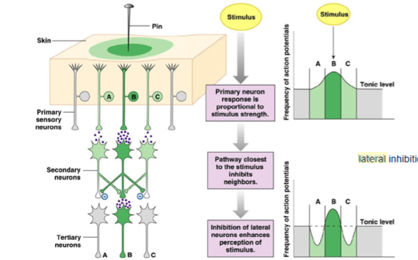
Whats the function of lateral inhibition
Sharpens the acuity of sensory recognition
Describe the mechanisms of adaptation to a stimulus
receptors become less sensitive and stop firing action potentials
neuron slows it firing rate cause ion channels change, affecting firing
thalamus or cortex may inhibit the sensory input when its no longer meaningful
Hierarchical processing
sensory info is processed in stages w each stage handling increasingly complex features of the stimulus
spinal cord > brainstem > thalamus > primary sensory cortical areas to nonprimary sensory cortical areas or nonprimary sensory cortical areas
Which receptors show slow or no adaptation to a stimulus?
Tonic
they keep firing as long as the stimulus is present
detecting duration
ex: pain, pressure, proproception
Which receptors undergo rapid adaptation
Phasic
respond quickly at the beginning or end of a stimulus
then stop firing even if stimulus continues
touch, smell, temp
why do organisms adapt to sensory stimulus exposure
so we dont get overstimulated
describe plasticity in cortical maps
brains ability to reorganize and adapt the structure and function of its cortical areas in response to experience, learning, injury, or environmental changes
ex: monkey experiment where their somatosensory input got bigger
Describe the pathways that process sensory information
Sensory information enters the CNS through the brainstem or spinal cord and then reaches the thalamus. The thalamus transmits the information to the cerebral cortex; the cortex directs the thalamus to suppress some sensations. Primary sensory cortex swaps information with nonprimary sensory cortex. This organization is present in all sensory systems except smell, which bypasses the thalamus, going directly to cortex
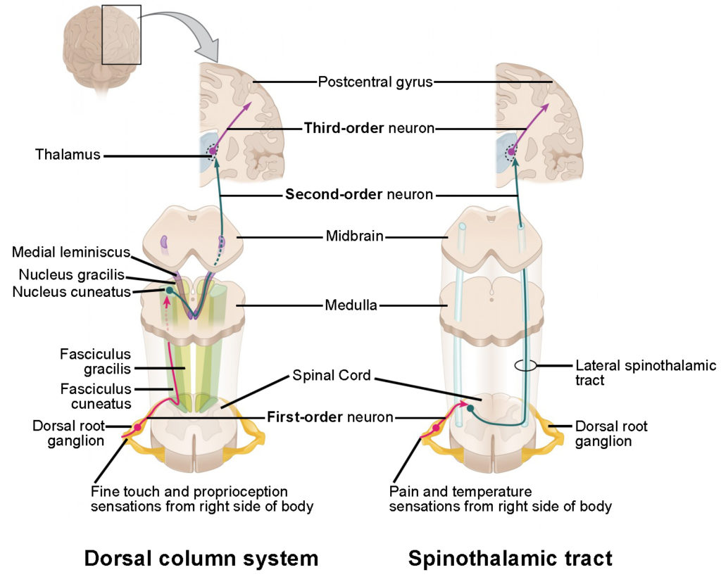
Describe the organization of somatosensory inputs through the dorsal column system
1. Feel it → Receptors
Fine touch, pressure, proprioception
Uses mechanoreceptors/proprioceptors
2. Climb it → Dorsal Columns (1st-order neurons)
No synapse yet
Climb up spinal cord on the same side (ipsilateral)
Two tracks:
Gracilis = Ground (legs/lower body)
Cuneatus = Ceiling (arms/upper body)
3. Flip it → Medulla (2nd-order neurons)
Synapse at nucleus gracilis/cuneatus
FLIP sides here! (Decussation = crossing over)
Now it's contralateral
4. Ship it → Medial Lemniscus → Thalamus
Axons ride the medial lemniscus through the brainstem
Synapse at the VPL of the thalamus
5. Sense it → Somatosensory Cortex
Final stop: Postcentral gyrus
Body map = Sensory homunculus
Medial = legs
Lateral = face/hands

Describe the organization of somatosensory inputs through the ventral spinothalamic tract
1. Receptors (Feel it)
Mechanoreceptors in the skin detect crude touch/pressure.
Signals travel via Aδ and C fibers (slower than dorsal column fibers).
2. First-Order Neurons (Enter spinal cord)
Enter via the dorsal root of the spinal cord.
Synapse immediately in the dorsal horn (unlike dorsal column where they ascend first).
3. Second-Order Neurons (Cross it)
After synapsing, these neurons decussate (cross over) in the spinal cord (usually within 1–2 segments).
They then ascend contralaterally in the ventral (anterior) funiculus as the ventral spinothalamic tract.
4. Thalamus (Relay station)
The tract ascends to the ventral posterolateral nucleus (VPL) of the thalamus.
Still somatotopically organized: lower body more lateral, upper body more medial (opposite of cortex layout).
5. Third-Order Neurons (To the brain!)
From the thalamus, third-order neurons project to the primary somatosensory cortex in the postcentral gyrus.
Epicritic Touch
-fine and discriminative touch
-proproception
What’s included in epicritic touch
Pacinian corpuscle: sudden, deep pressure
- Meissner corpuscle: sudden, light touch
- Ruffini corpuscle: gradual, stretch
- Merkel’s Disk: gradual, light touch
- proprioceptors (muscle spindles, GTOs,
joint proprioceptors)
Protopathic touch
non discriminating, pain, and temperature
whats included in protopathic touch
temp and pain, free nerve endings that respond to tissue damage, release intracellular K+ from nerve itself, bradykinins, histamine due to damage
ascending pain fibers release substance P (and others)
- analgesia mediated by descending projections from PAG, raphe etc.
- descending analgesic action mimicked by opiate drugs
- pain may also sensitize (inflammation, neuralgia)
meissener’s corpuscles
Location: Dermal papillae (fingertips, palms, lips)
Stimulus: Light touch & low-frequency vibration
Adaptation: Rapid
Function: Detect texture & fine touch, especially changes over time
merkel’s discs
Location: Epidermis (especially fingertips)
Stimulus: Sustained pressure & texture
Adaptation: Slow
Function: Detect shape, edges, and steady touch (e.g., reading Braille)
pacinian corspucles
Location: Deep dermis & subcutaneous tissue
Stimulus: Deep pressure & high-frequency vibration
Adaptation: Very rapid
Function: Detect sudden changes or vibration (e.g., power tools, phone vibrations)
ruffini endings
Location: Deep dermis, ligaments, tendons
Stimulus: Skin stretch
Adaptation: Slow
Function: Detect continuous pressure and stretching (important for grip adjustment)
hair follicle receptors
Location: Wrapped around hair follicles
Stimulus: Hair displacement
Adaptation: Rapid
Function: Detect light touch via hair movement (e.g., a breeze or bug on skin)
What is the current knowledge about the stimuli to which pain receptors respond?
responsive to tissue damage, release of intracellular K+ from nerve itslef, bradykinins, histamine
Describe descending pathways for analgesia (pain relief)
1 activate periaqueductal gray to process both emotional and sensory info related to pain
2 pag neurons signal rostral ventromedial medulla, integrates pain info and coordinates the modulation of spinal pain transmission thru two groups of neurons
-on cells: facilitate pain transmission by increasing nociceptive signaling
-off cells: inhibit pain transmission by suppressing nociceptive signaling
3 rvm sends descending projections to the spinal cord that release serotonin and norepinephrine
4 release of serotonin and norepinephrine interacts w/ specific receptors on primary nociceptive neurons
transient receptor potential vanilloid type 1 (TRPV1):
a receptor that binds to capsaicin (spicy chemical) to transmit the burning sensation from chili peppers and normally detects sudden increase in temp
TRPM8: (cool-menthol receptor)
a sensory receptor found in some free nerve endings, that opens an ion channel in response to a mild temperature drop or exposure to menthol.
responds to cool temps rather slowly
located on small C fibers
Describe the range of treatments that are currently used to control pain
social rejection and pain
social rejection activates the anterior cingulate cortex and that the extent to which a person is upset by rejection correlates with activation of this region
more distress or feelings of rejection the participant felt, the greater the activity in anterior cingulate
strategies to alleviate pain
cannabis
when endogenous cannabinoid receptors and free nerve endings of nociceptors are stimulated by Cannabis sativa, pain is reduced significantly
opiates
a class of compounds that exert an effect like that of opium, including reduced pain sensitivity
brain will release substances that work like opiates