NHA CET Exam
1/302
There's no tags or description
Looks like no tags are added yet.
Name | Mastery | Learn | Test | Matching | Spaced |
|---|
No study sessions yet.
303 Terms
HIPAA
Data privacy & security provisions laws 2 protect med info
PHI
Any health status & healthcare service info that's personally IDable & can link a person
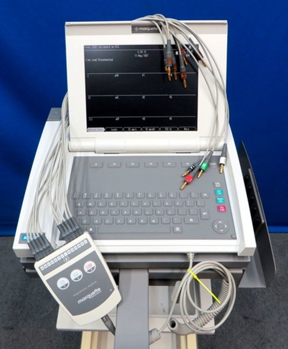
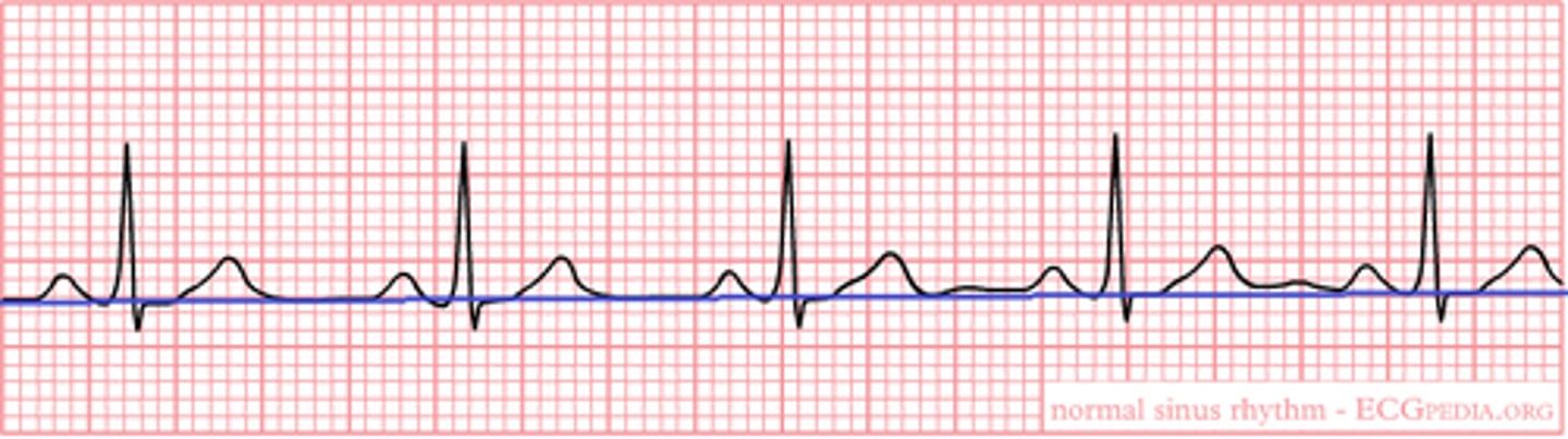
EKG
Written tracing made by an EKG machine
Implied consent
The PT's actions implies they're giving procedure consent
Holter monitor
Portable battery-operated device that continuously monitors ♡ activity ≥24hr; AKA ambulatory monitor

Event recorder
Outpatient cardiac test involving PT triggering the machine 2 record when having symptoms
Med asepsis
Practice designed 2 ↓ pathogen transfer & break the infection chain
Standard precautions
Basic level infection-control practices healthcare workers must do b4, during, & after every PT encounter 2 prevent infection spread
HAI
Infection from the healthcare workers/setting
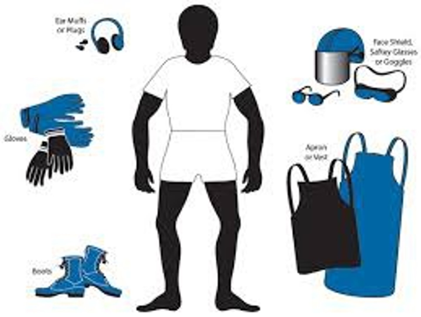
PPE
Protective clothes & equipment (gloves, mask, gown, goggles) that protect a worker from chem/infectious material exposure
Leads
Wires that conduct electrical impulses from electrodes 2 an EKG machine

EKG machine
Instrument that gives the ♡'s electrical activity a graphic rep
SDS
Manufacturer's documentation of potentially harmful substances listing the chem parts, associated hazards, & exposure treatment
Therapeutic communication
Using strategies incorporated thru verbal & nonverbal techniques 2 ensure a message is received, interpreted & understood properly
Baseline
An electrically neutral area on the EKG, AKA the isoelectric line or TP segment; makes a comparison pattern across time
Sphygmomanometer
BP measurement equipment that typ appears as a dial w/ #s, bulb & bp cuff
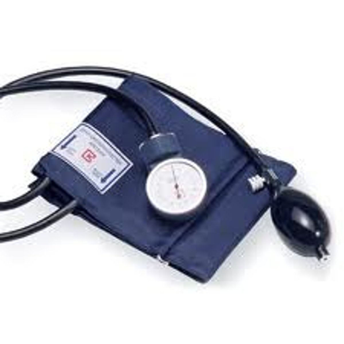
Auscultation
Listening 2 bod sounds (♡, lungs, GI tract) using a stethoscope
SBP
When ♡ is contracted, recorded as the top BP #; the 1st korotkoff sound heard during auscultated BP
DBP
When ♡ is relaxed, recorded as the bottom BP #; the last korotkoff sound heard during auscultated BP
<120mmHg
Expected ref range SBP
<80mmHg
Expected ref range & elevated DBP
120-129mmHg
Elevated SBP
130-139mmHg
↑ BP (stage 1) SBP
80-89mmHg
↑ BP (stage 1) DBP
≥140mmHg
↑ BP (stage 2) SBP
≥90mmHg
↑ BP (stage 2) DBP
≥180mmHg
Hypertensive crisis SBP
≥120mmHg
Hypertensive crisis DBP
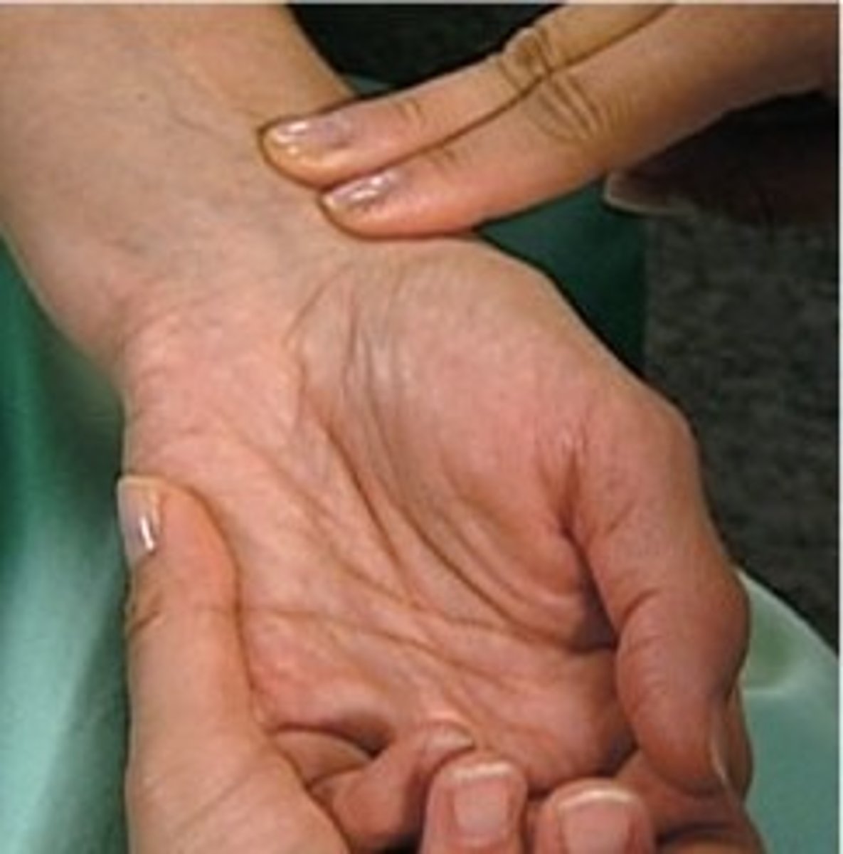
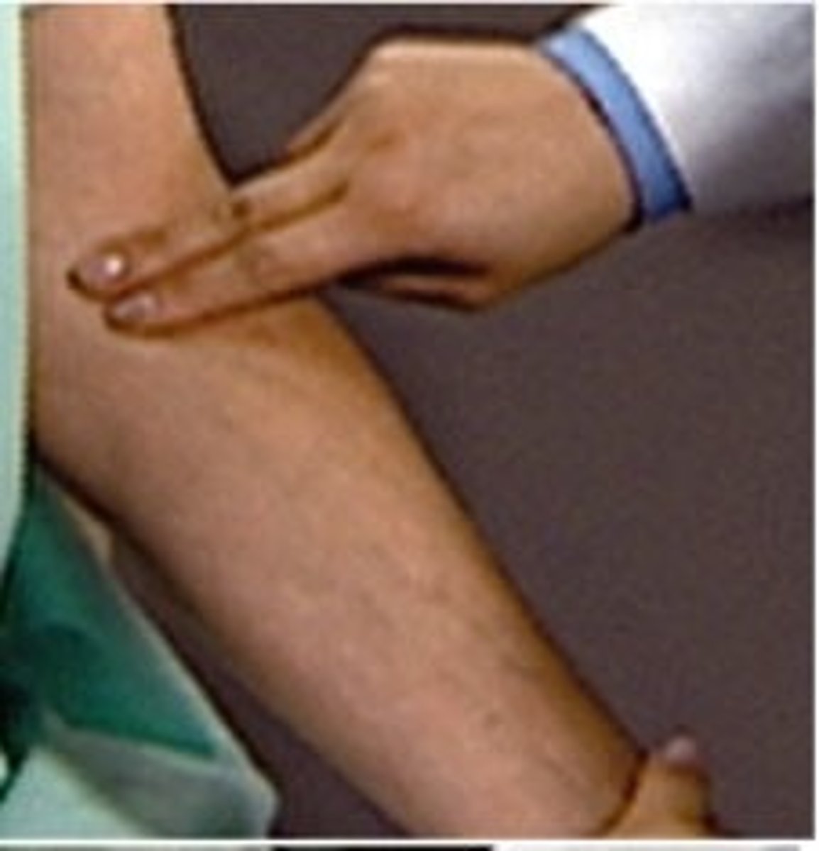
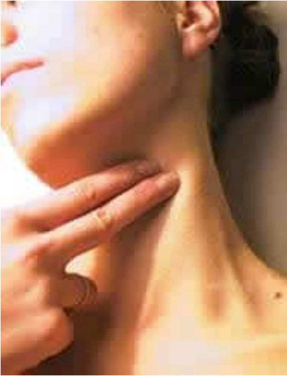
Palpation
Using hands 2 exam the bod
Radial pulse site
Thumb side of the wrist, most typ adult site
Brachial pulse site
↑er arm, most typ kid site
Carotid pulse site
On the neck below the jaw bone, most typ site 4 emergencies
Stress test
Ischemia/related EKG ∆s provoking test 2 help diagnose cardiac abnormalities; AKA exercise stress test
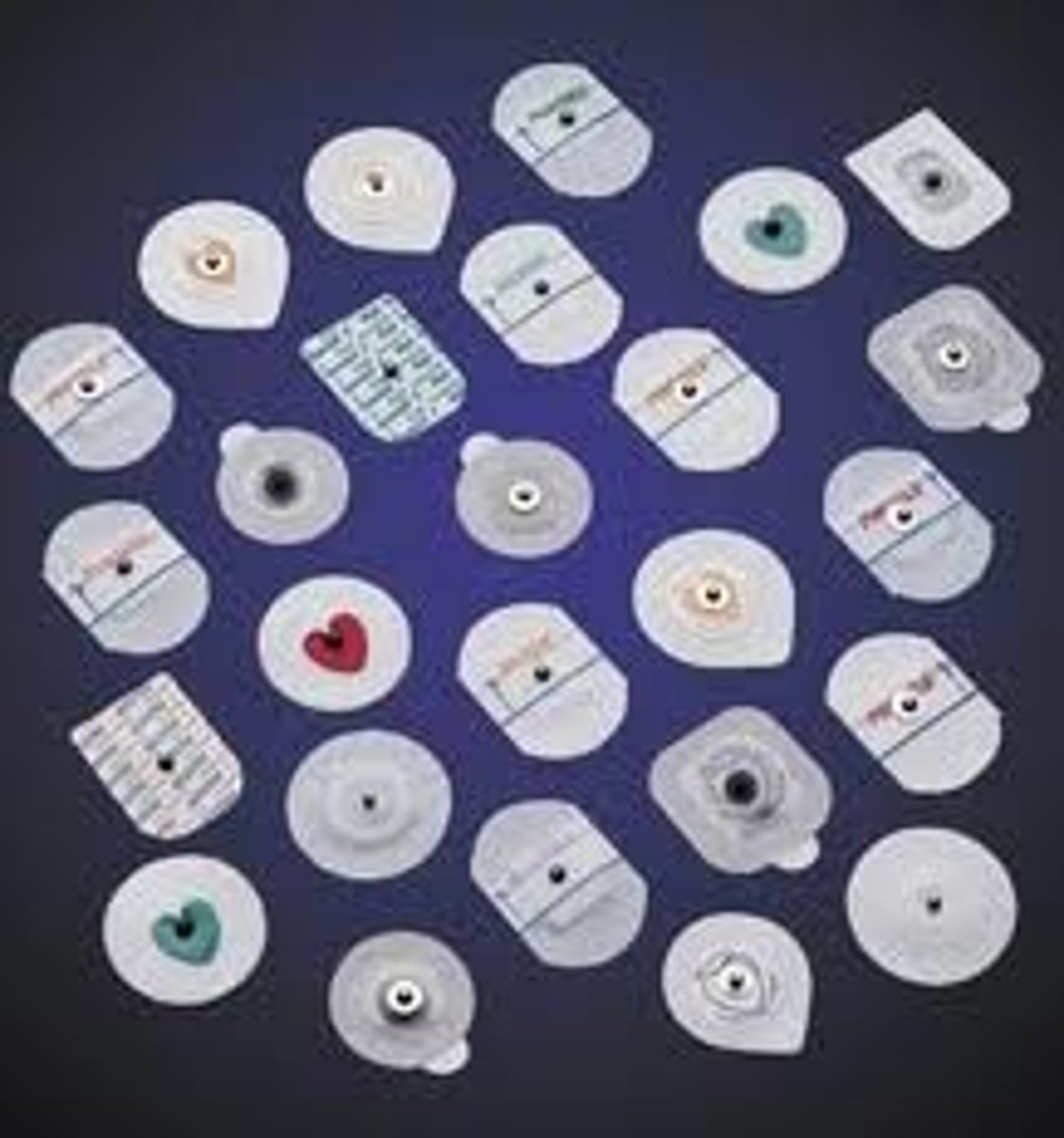
Electrode
Skin sensor that interfaces btwn the ♡ & EKG
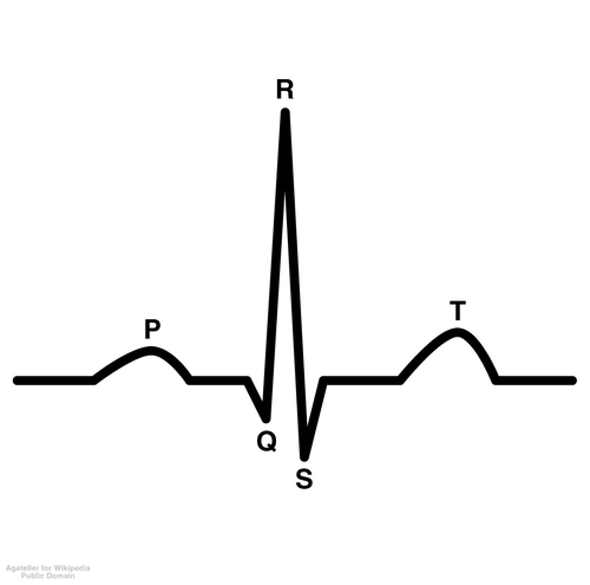
PQRST waves
Standard waveforms on EKG tracing, each corresponding 2 a specific event in 1 full electrical ♡ cycle
Stenotic
Narrowed/constricted
Diaphoresis
Excessive sweating
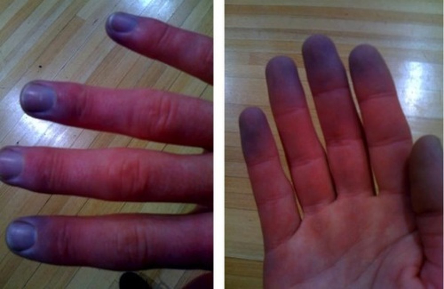
Cyanosis
Skin, lips, toes/fingers blue/discolored
Syncope
Fainting
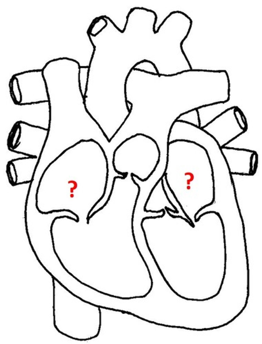
Atrium
A small muscular pouch-like structure that fills ventricles w/ blood
Septum
A dividing wall, like the 1 btwn the atria or ventricles
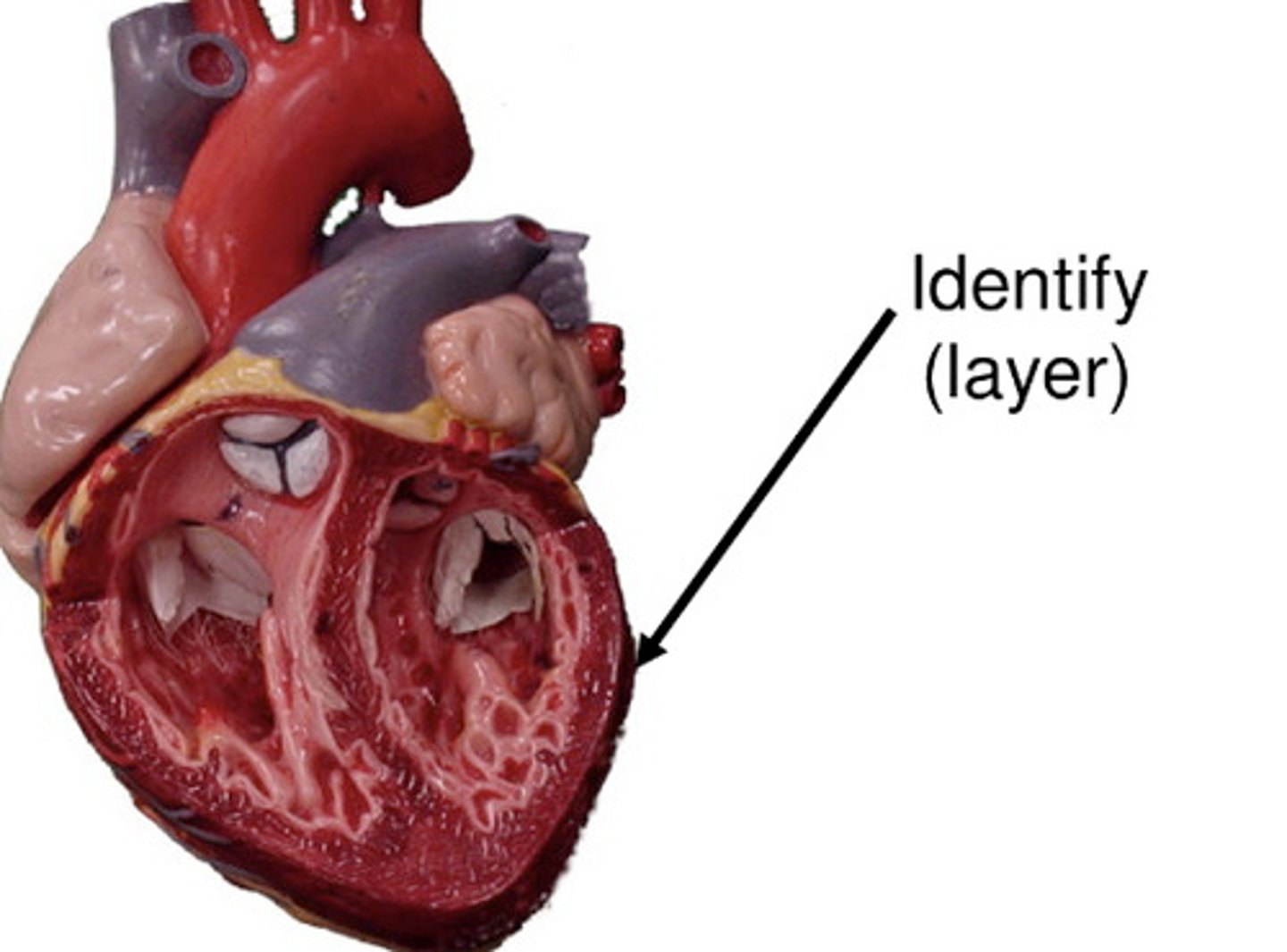
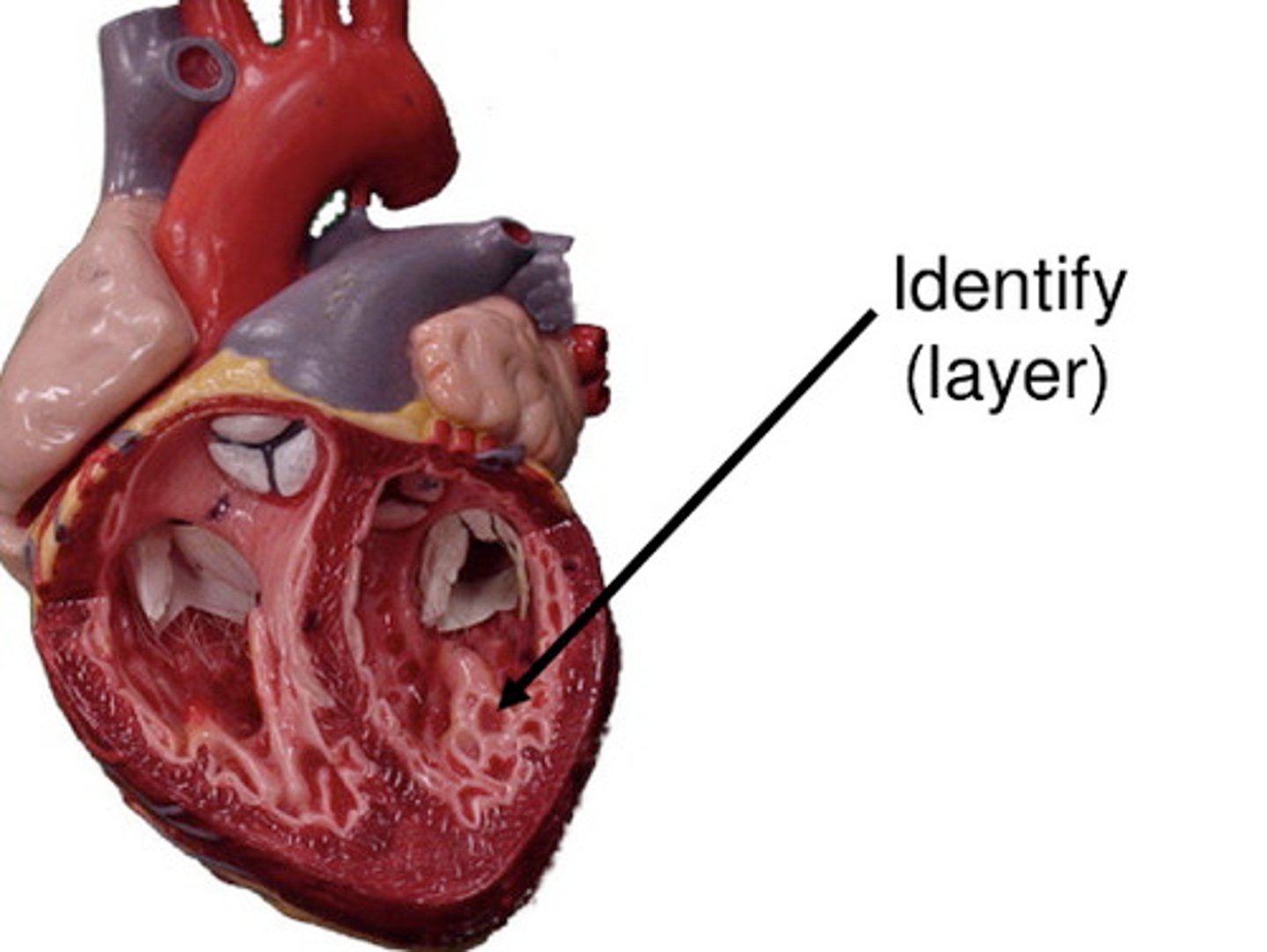
Epicardium
The outer ♡ layer
Pericardium
A 2 layered sac that holds the ♡, filled w/ fluid that prevents friction w/ ♡ muscular action
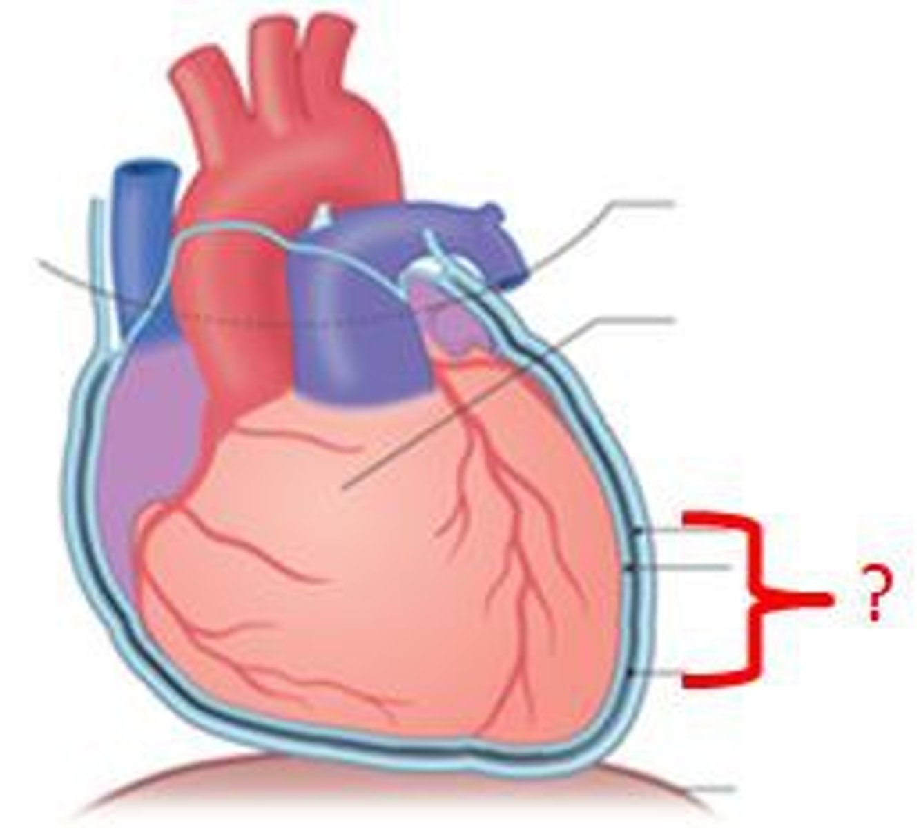
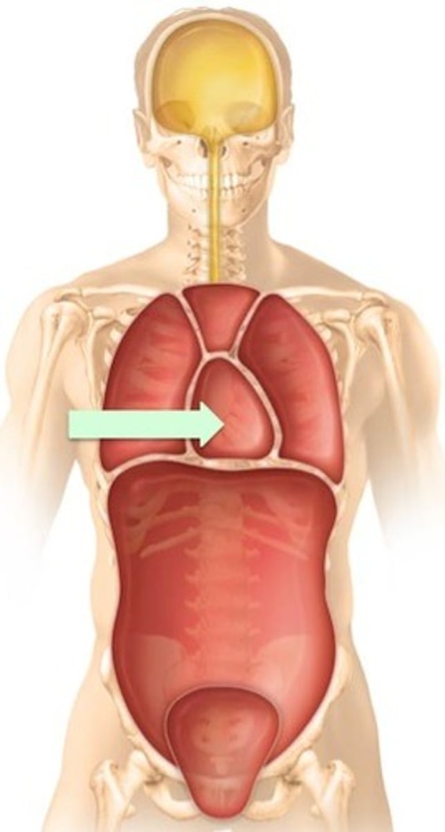
Mediastinum
1 of 3 chest compartments; holds the ♡ & great vessels
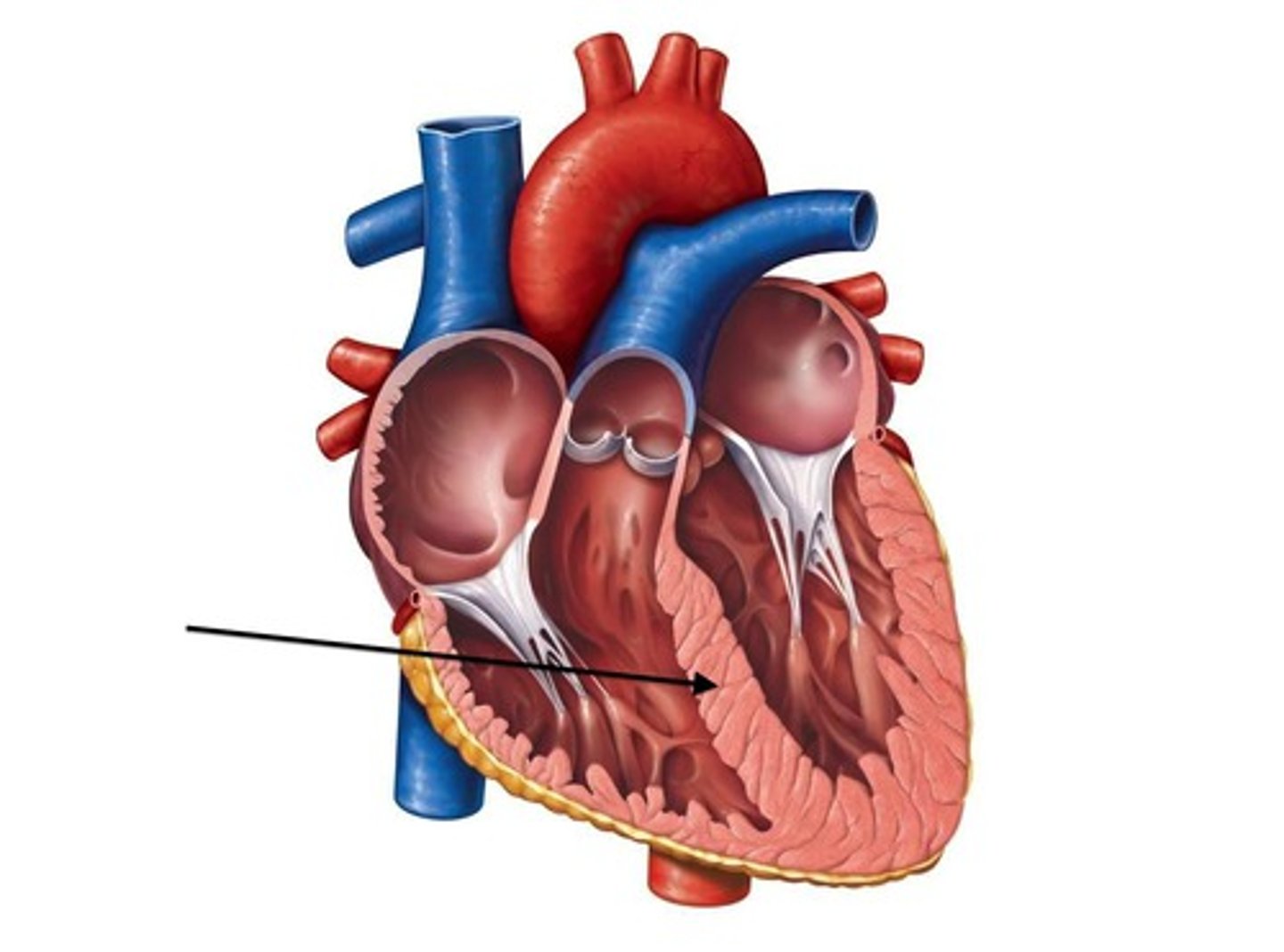
Myocardium
The mid muscular contracting ♡ layer
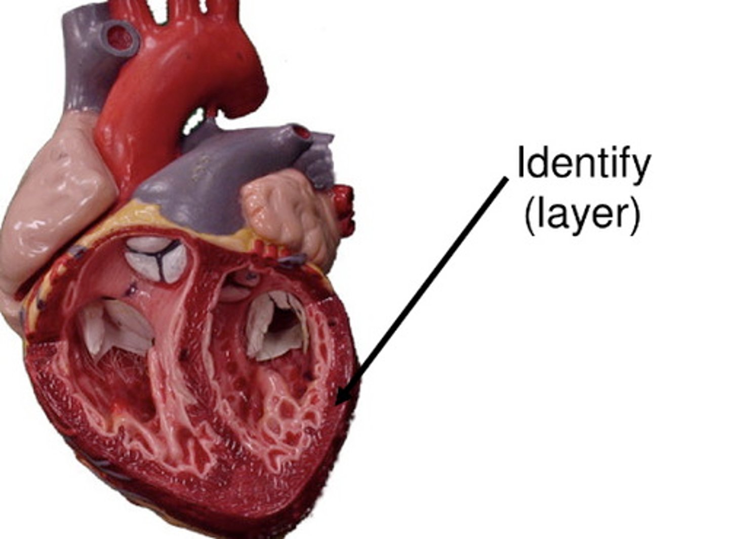
Endocardium
The inner ♡ layer
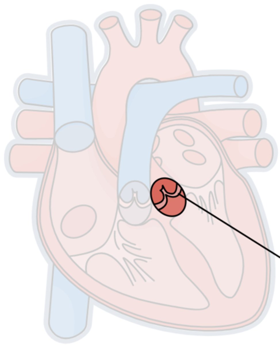
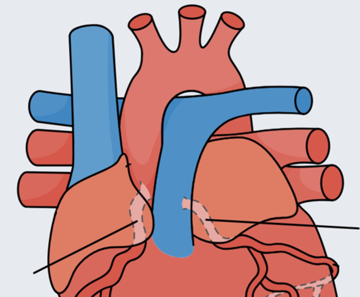
Tricuspid valve
Btwn the right atrium & right ventricle
Bicuspid (mitral) valve
Btwn the left atrium & left ventricle
AV valves (tricuspid & bicuspid)
Btwn the atria & ventricles
Pulmonary valve
Btwn the right ventricle & pulmonary arteries
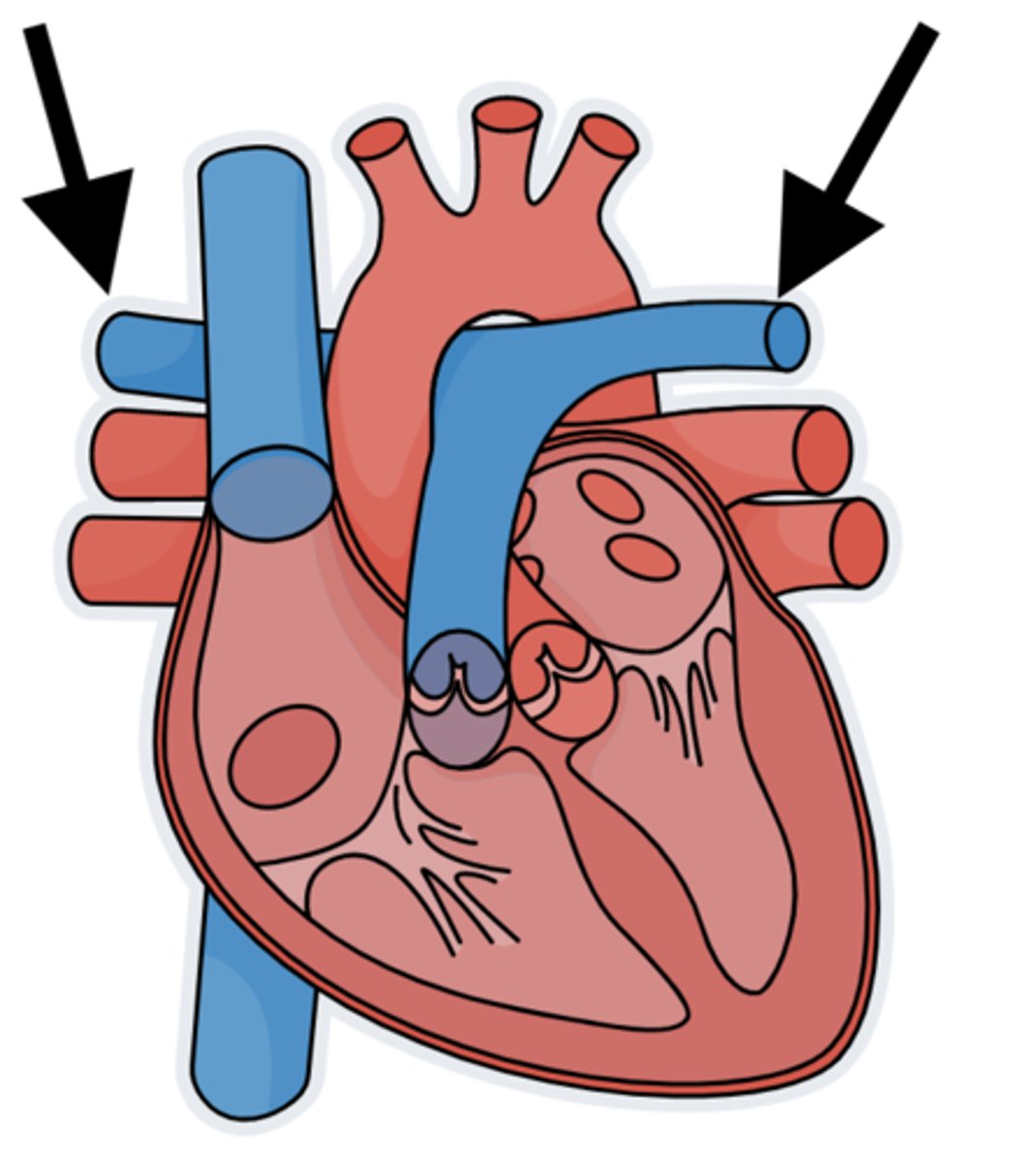
Pulmonary arteries
The only arteries that carry deoxygenated blood. These arteries transport blood from the right ventricle 2 the lungs
Aortic valve
Btwn the left ventricle & aorta
Coronary arteries
Arteries that give oxygenated blood 2 the myocardium
Perfuse
Give blood
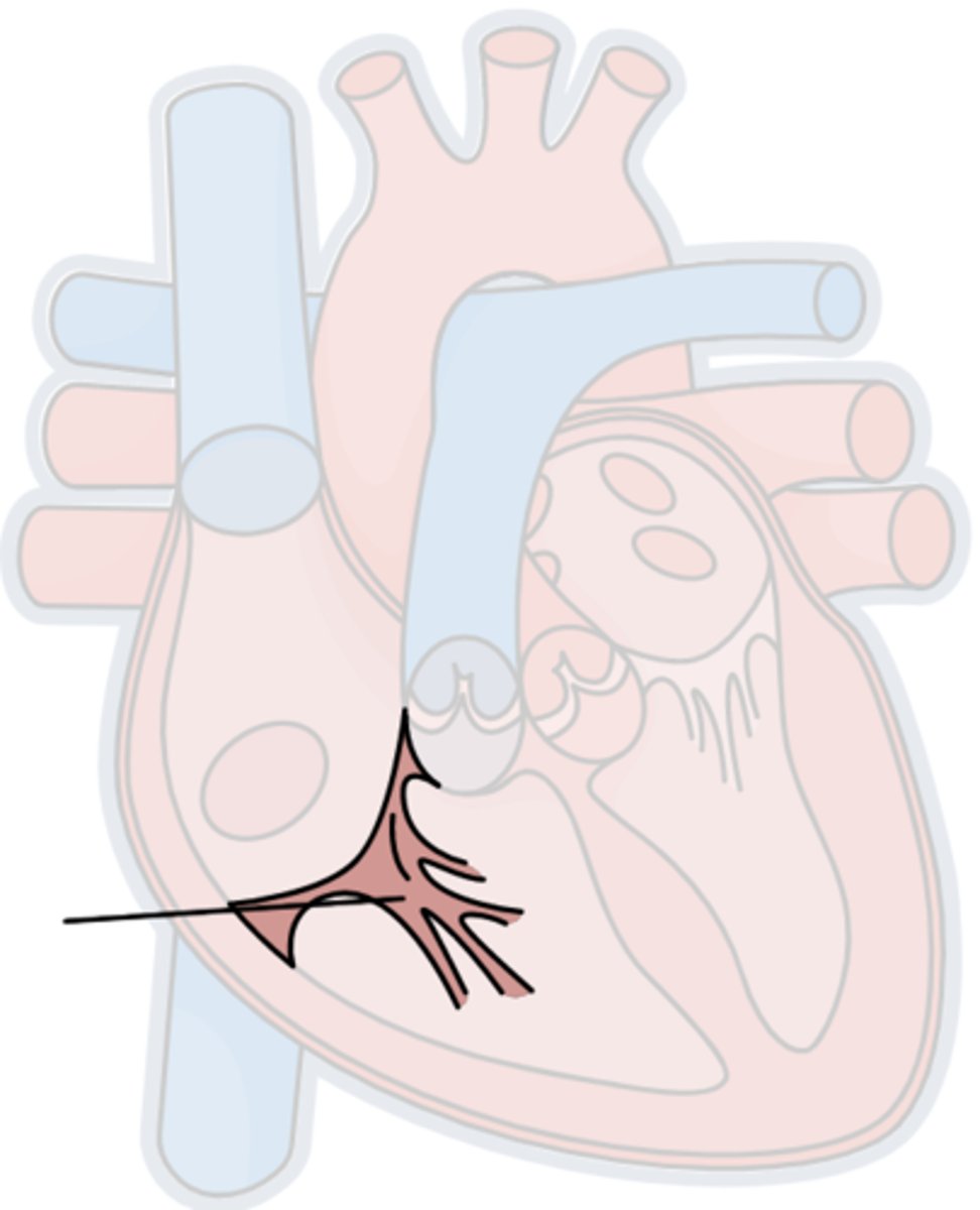
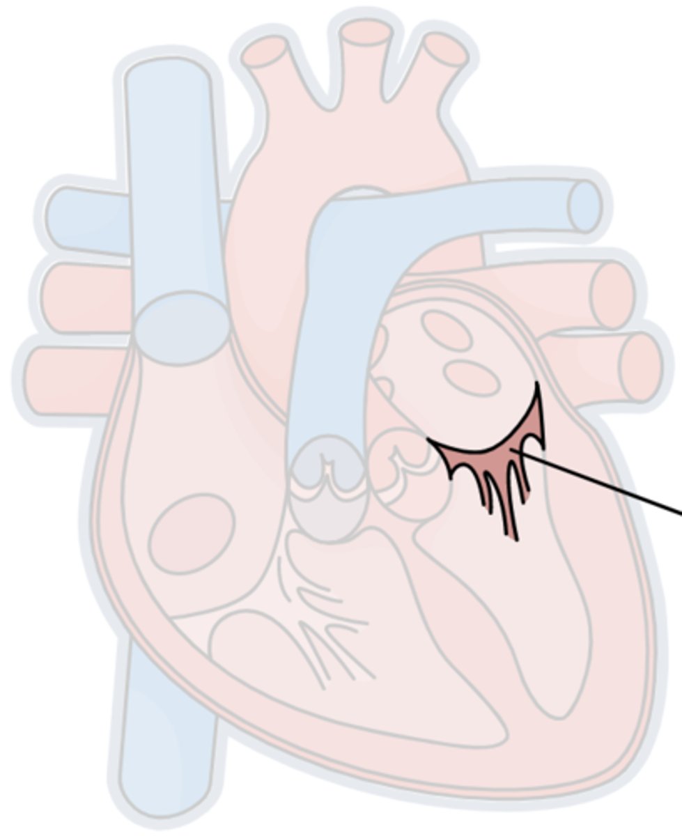
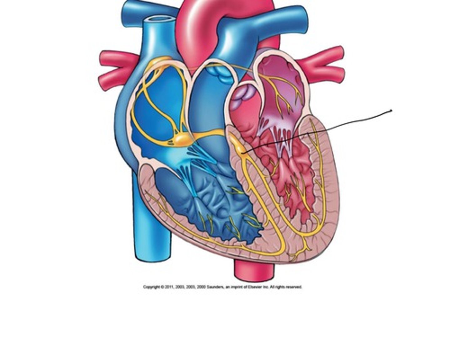
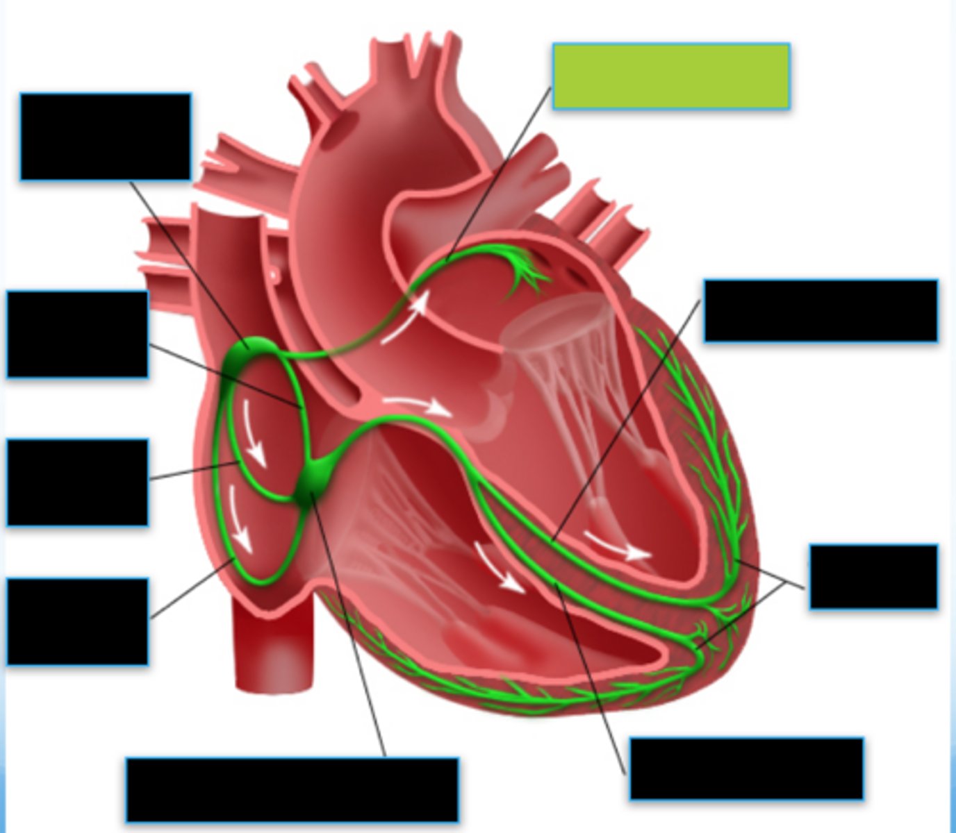
SA node
A small tissue mass, in the right ↑er atrium, the ♡'s main pacemaker. Depolarization of this makes P-waves
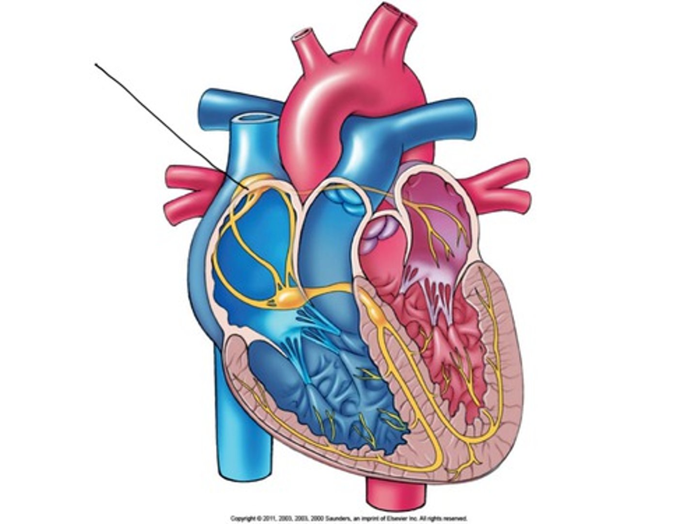
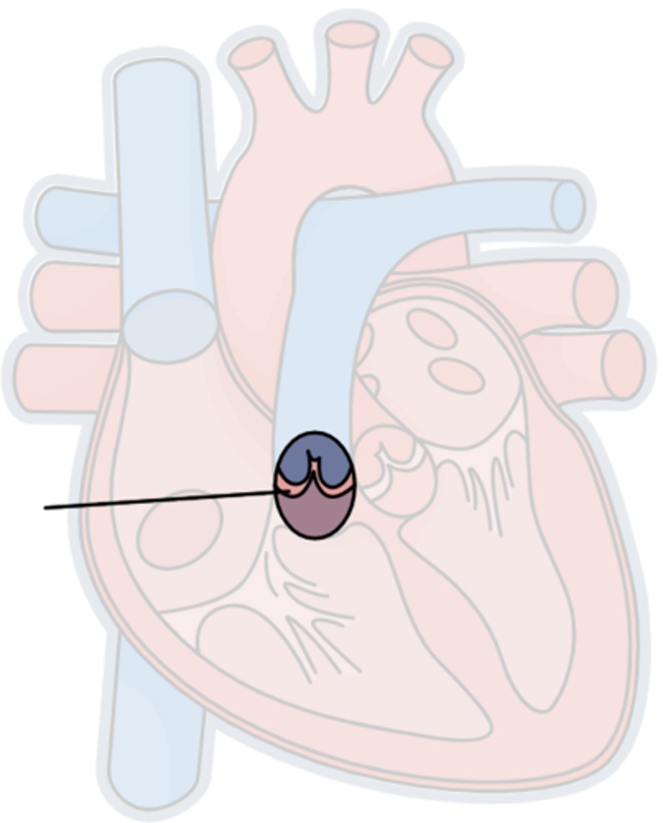
AV node
Specialized tissue that regulates the impulses btwn the atria & ventricles
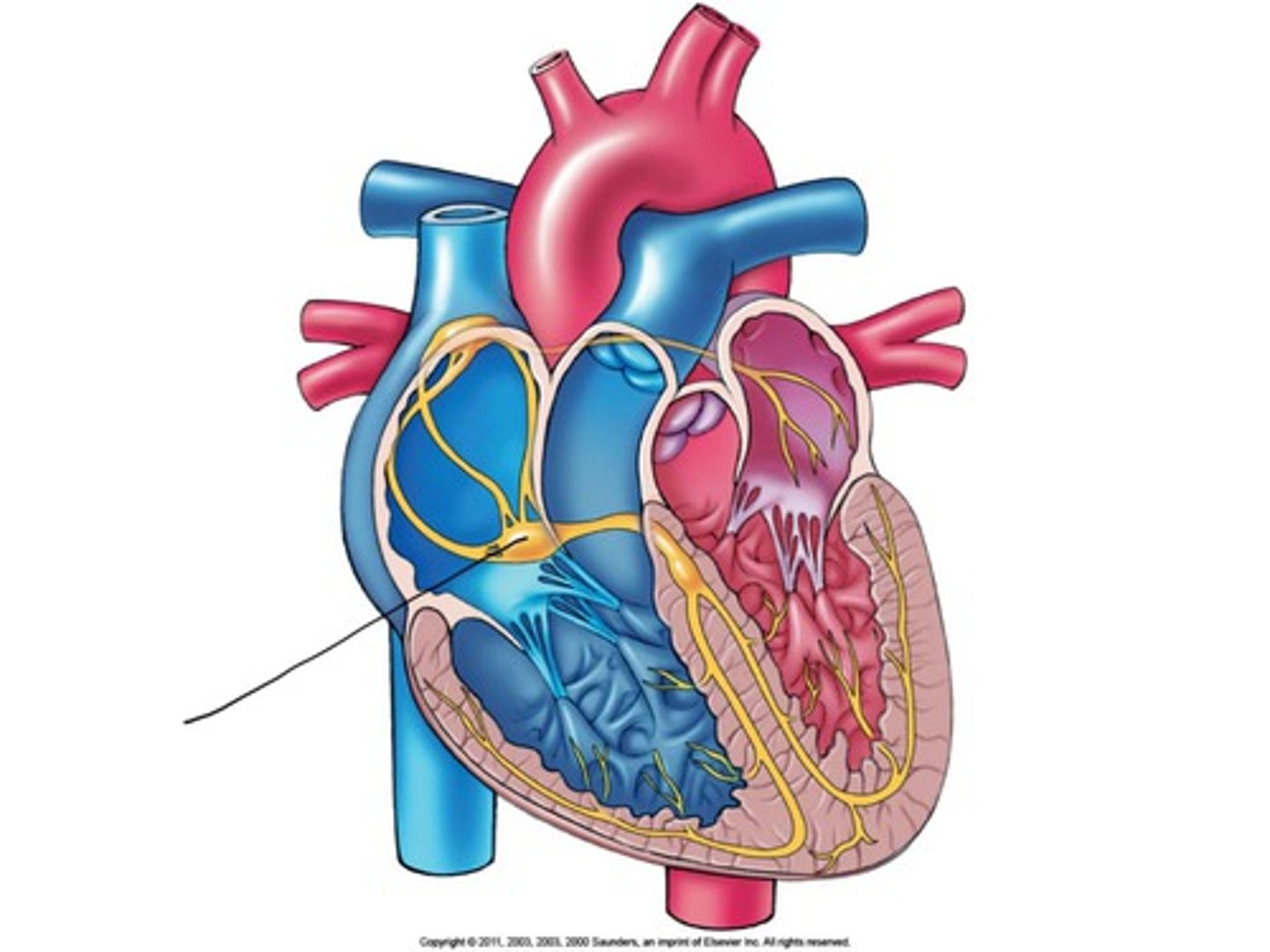
Depolarization
Process involving the Na & K channels opening on the cardiac cell surface resulting in the polarity loss. Causes the muscle 2 contract
Bradycardia
Slow HR
Bundle of his
The conduction system part that receives electrical impulses from the AV node & transmits the impulses 2 the bundle branches
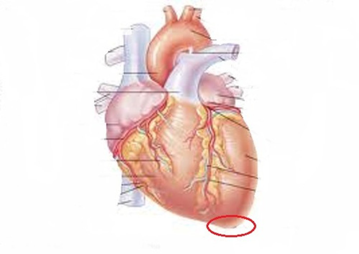
Apex
The ↓er, pointed ♡ end
Bundle Branch
The conduction system part that transmits electrical impulses from the AV node 2 the purkinje fibers 2 trigger ventricular depolarization

Purkinje fibers
Fibers that conduct electrical impulses thru both ventricles causing ventricular depolarization
Automaticity
Cardiac cell's ability 2 spontaneously make electrical activity
Ectopic
Starting sumwhere besides the SA node
Bachmann's bundle
The ♡'s electrical conduction system part that ensures both atria depolarize simultaneously
Precordial leads
6 EKG leads put on the anterior chest 2 record the ♡'s unipolar electrical activity @ a specific place
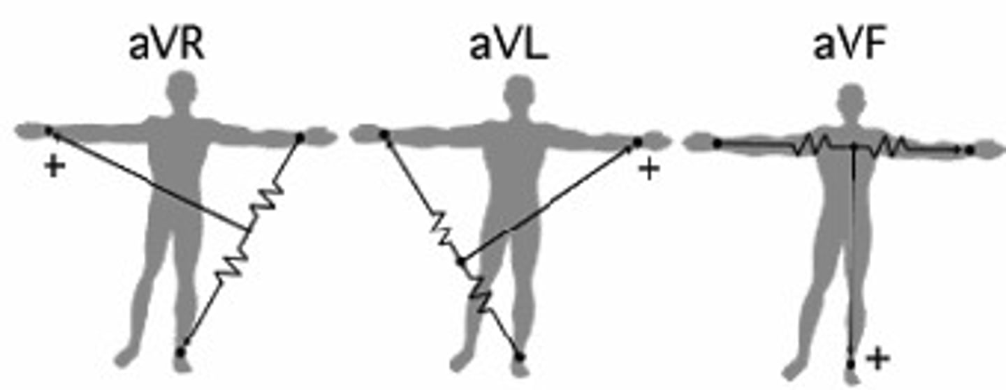
Augmented leads
Unipolar leads made by combining 2 of the 3 limb leads 2 make a + electrode
Artifact
Electrical/magnetic interference that ∆s the tracing
Tachycardia
Fast HR
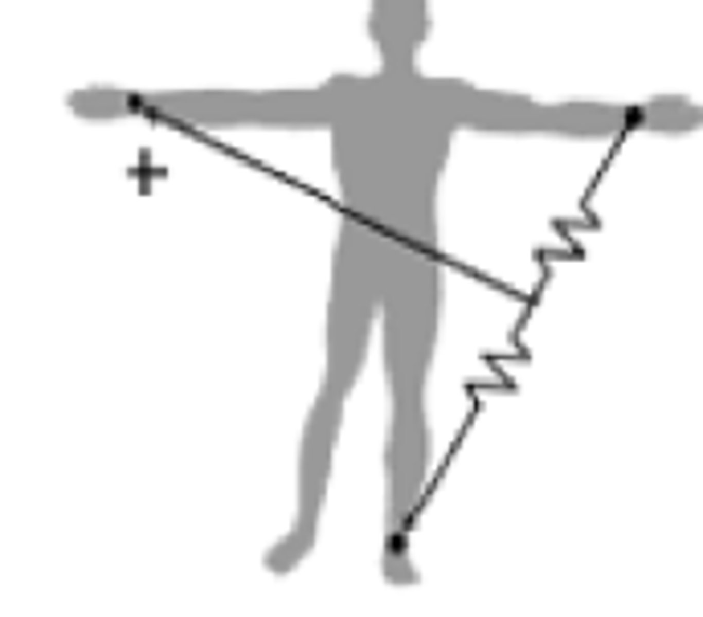
Lead I
Records impulses btwn the left & right arms
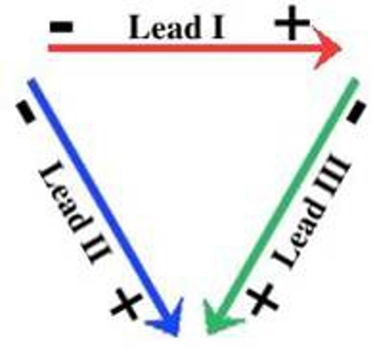
Lead II
Records impulses btwn the right arm & left leg

Lead III
Records impulses btwn the left arm & left leg

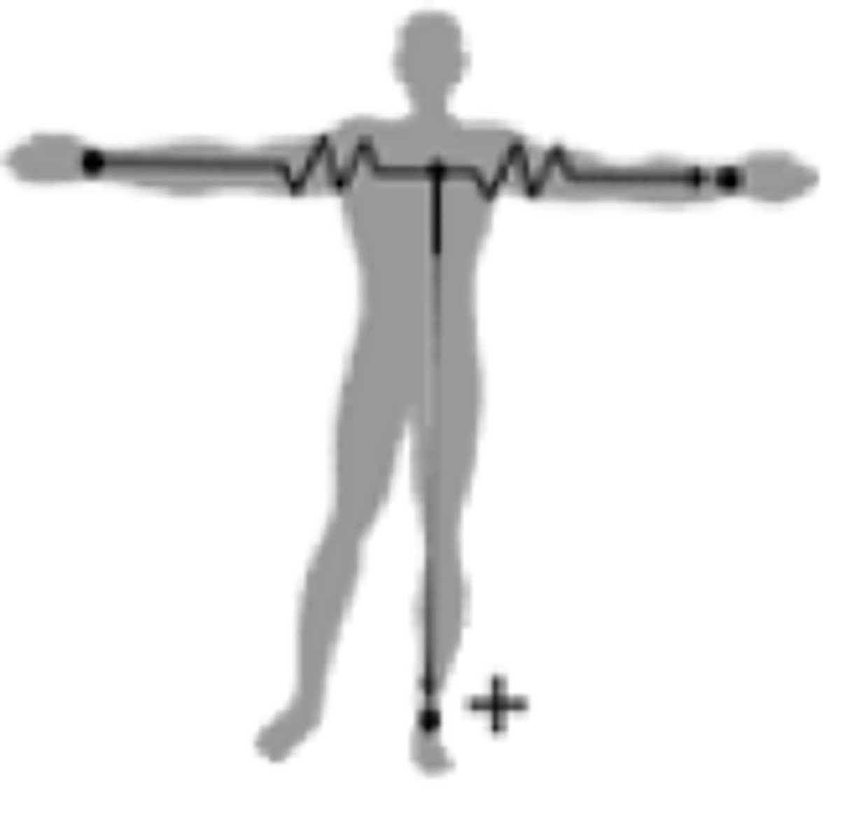
aVL
The left leg & right arm help w/ the left arm tracing
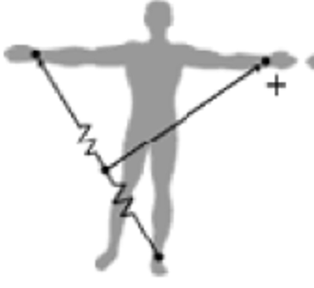
aVR
The left arm & left leg help w/ the right arm tracing
aVF
The right & left arms help w/ the left leg tracing
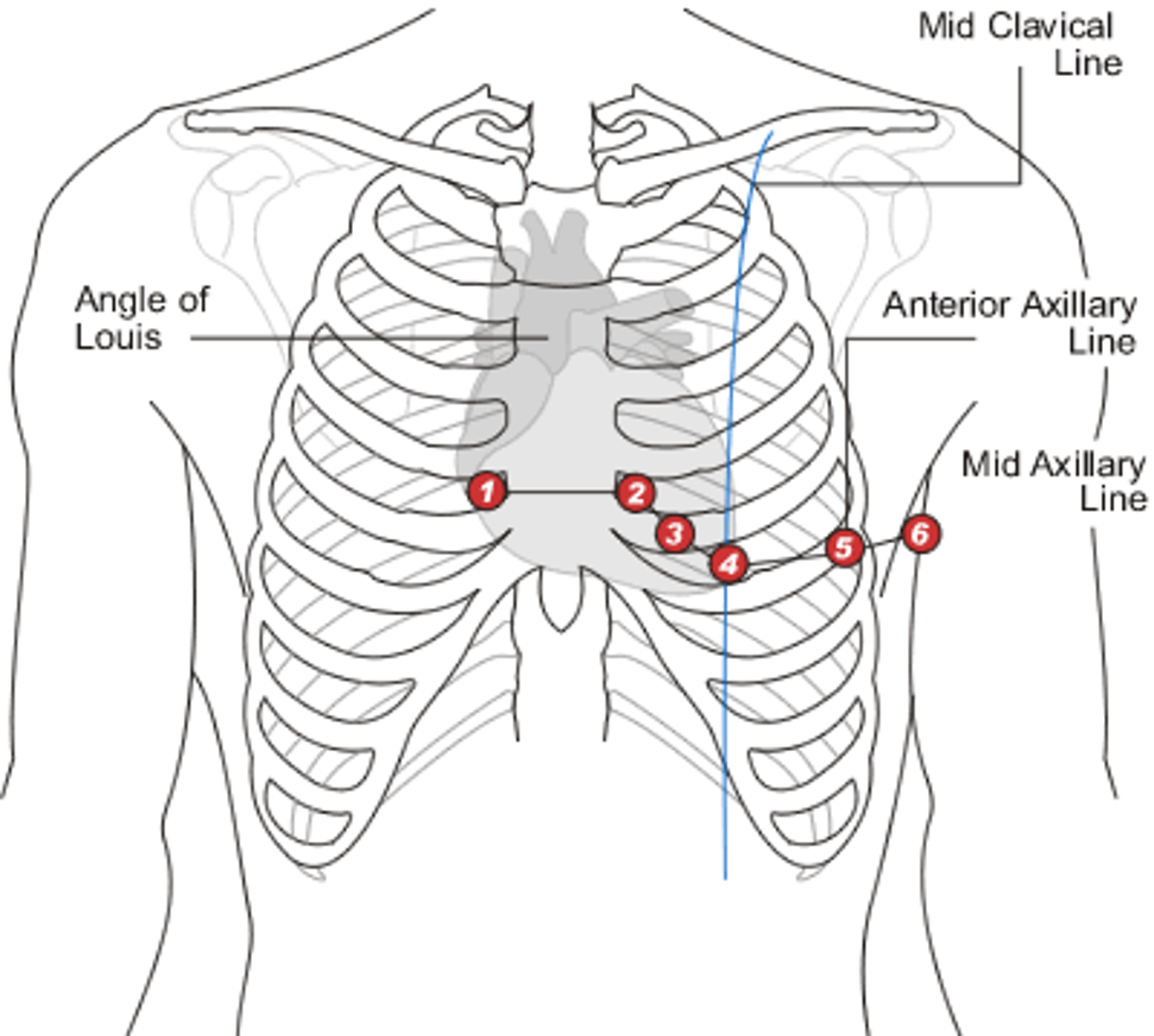
Intercostal
Btwn the ribs
V1
4th ICS, right of sternum
V2
4th ICS, left of sternum, across from V1
V3
Mid btwn V2 & V4
V4
5th ICS, mid-clavicular line
Mid-clavicular
An imaginary line thru the clavicle mid extending vertically
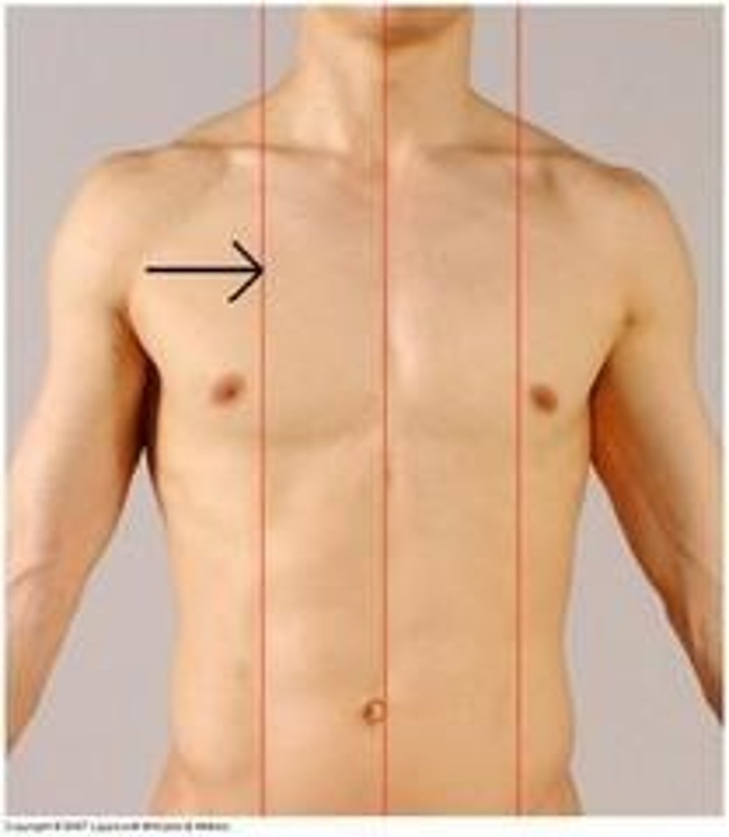
V5
5th ICS, mid btwn V4 & V6, @ the anterior axillary line
V6
5th ICS, @ the mid-axillary line
Mid-axillary
An imaginary line thru the axillary area that divides the bod front & back

3-lead EKG white lead
Right shoulder below clavicle
3-lead EKG black lead
Left shoulder below clavicle
3-lead EKG red lead
Below left pec @ the ♡ apex
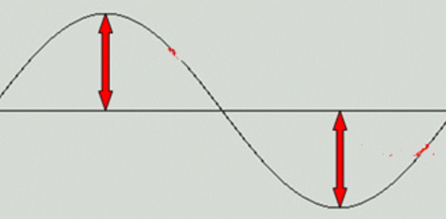
+ deflection
The waveform part in an EKG tracing above the isoelectric line
- deflection
The ↓ward waveform presentation on the tracing that's below the isoelectric line
Repolarization
The relaxation ♡ phase that preps the ♡ 4 another depolarization
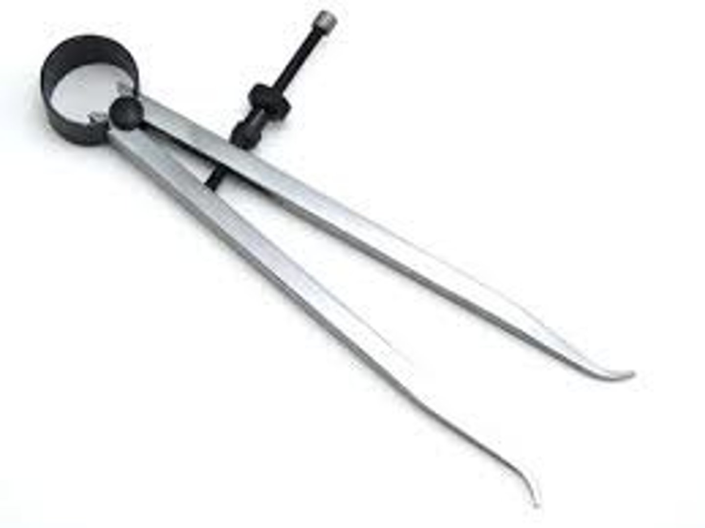
Caliper
An instrument 4 measuring distances on a tracing
Amplitude
Voltage/gain
Ischemia
↓ in tissue oxygenation bc of poor blood flow
Isoelectric line
The straight line on the tracing btwn cardiac cycles where there's no ♡ electrical activity or deflection
Flutter
Fast wave-like contractions
Fibrillation
Quivering state w/o organized contraction
Atrial kick
The atrial excitement w/ a ↑ in internal pressure 2 forcefully push blood n2 the ventricles
Unifocal PVC
Single early PVC implies 1 irritable area
Multifocal PVC
PVC w/ multi shapes implies >1 irritable area
Interpolated PVC
PVC occurs w/o interruption in the norm rhythm