Structure & function of the heart
1/19
There's no tags or description
Looks like no tags are added yet.
Name | Mastery | Learn | Test | Matching | Spaced | Call with Kai |
|---|
No analytics yet
Send a link to your students to track their progress
20 Terms
The Cardiovascular system
Responsible for the transport of substances throughout the body and plays a vital role in maintaining homeostasis
Consists of 3 main components:
The heart
Blood vessels
Blood
Function of Cardiovascular system - Transport
Delivers O2, nutrients, and hormones to body cells
Removes metabolic wastes such as CO2
Function of Cardiovascular system - protection
Circulates white blood cells, antibodies, and complement proteins to defend against pathogens
Enables. bblood clotting to prevent excessive blood loss following injury
Function of Cardiovascular system - Regulation
Helps regulate body temperature
Maintains pH balance
Controls fluid, H2O content of cells
Structure & function of the heart
The heart is a muscular pump responsible for generating pressure to circulate blood throughout the body
It is located in the mediastinum, between the lungs, slightly to the left of the mifline
The adult human heart : Is approximately the size of a clenched fist , cone-shape
Coverings the heart ( pericardium )
The heart is enclosed by a double-walled sac called the pericardium
It consists of 2 layers with fluid between them
The fluid allows the layers to slide smoothly over each other as the heart beats, reducing friction
Functions of pericardium
Holds the heart in position within the chest cavity
Prevents the heart from overexpanding when it fills with blood
Reduces friction as the heart contracts and relaxes
Provides protection from infection and physical damage
Heart wall
3 layers: outer layer, middle layer, inner layer
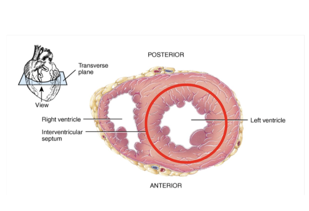
The middle layer ( myocardium ) is made of cardiac muscle and varies in thickness:
Thicker in the ventricles than the atria
Thickest in the left ventricle due to higher pressure requirements
Cardiac muscle
Specialised muscle found only in the heart
It is a specialised to continuosly and repeatedly contract, providing circulation of blood throughout the body
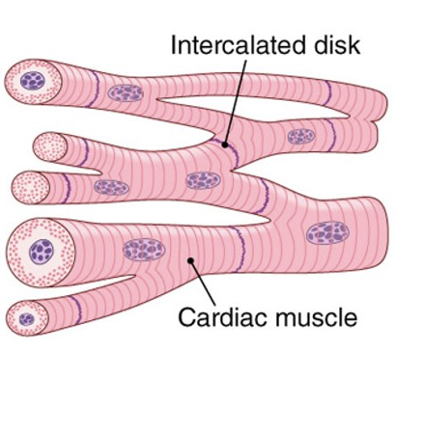
Cardiac muscle
Striated muscle fibres
Cells are branched and interconnected
Typically contain a single, centrally located nucleus
Designed for continous, rhythmic contraction
Adjacent cardiac muscle cells are connected by intercalated discs, which locks cells together to allow for rapid transmission of electrical impulses
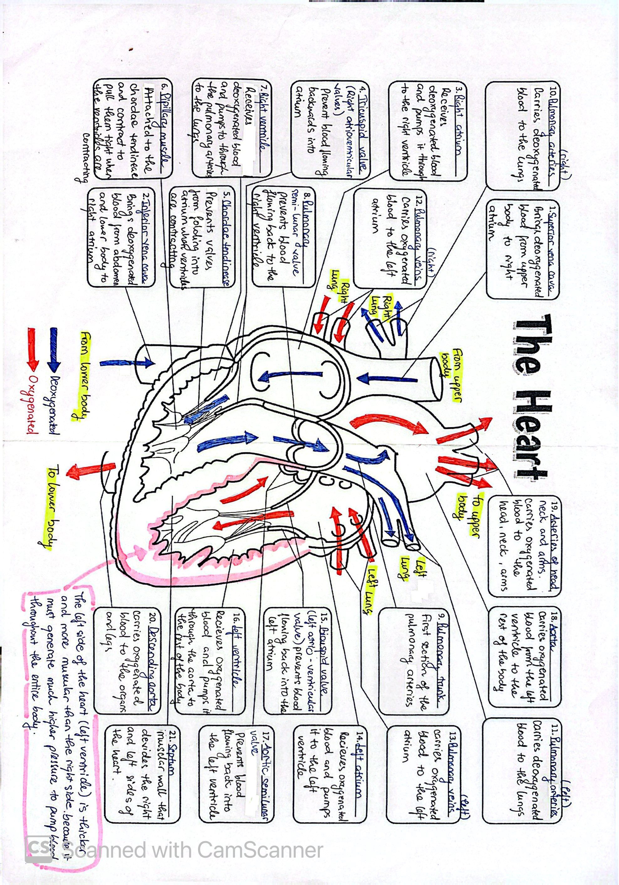
Pathway of blood around the body
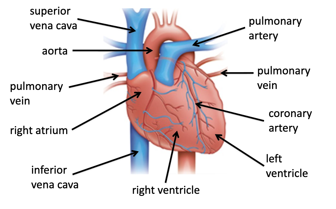
External view of the heart
Chordae tendineae and papillary muscles
Trong connective tissue cords that attach AV valves to papillary muscles
During ventricular contraction:
-Papollary. muscles contract
Tension is applies to the chordae tendineae
This prevents valve cusps from inverting and stops backflow
Semilunar valves
Have 3 cusps
Lack chordae tendineae and capillary muscles
Function of Semilunar Valves
consist of pockey-shaped cusps reinforced with connective tiisue
whne ventricles relax:
blood flows back toward the heart
cusps fill with blood
valves close, preventing backflow into the ventricles
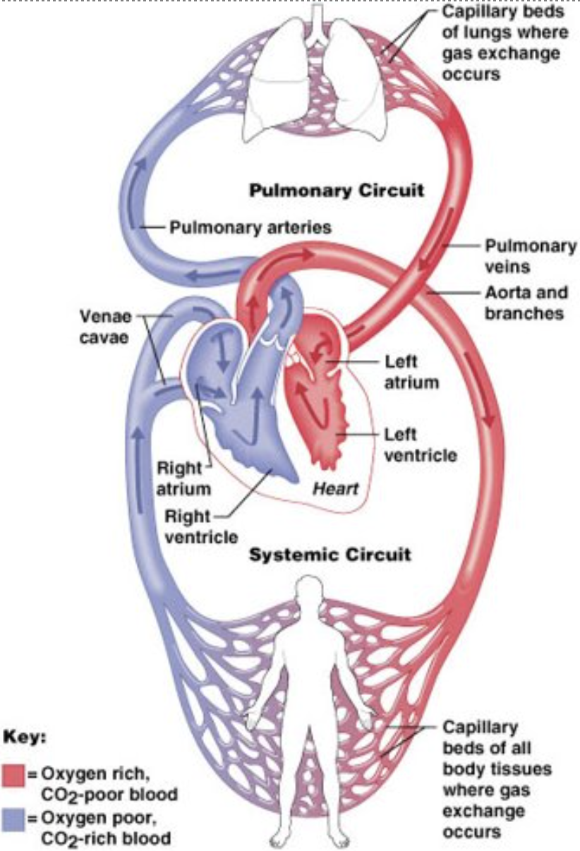
double circulatory system
Humans have a double circulatory system, meaning blood passes through the heart tiwce per complete circut
Pulmonary circulation: Heart→ Lungs →heart
Systemic circulation: heart→ body→ heart
This allows efficient O2 delivery at high pressure
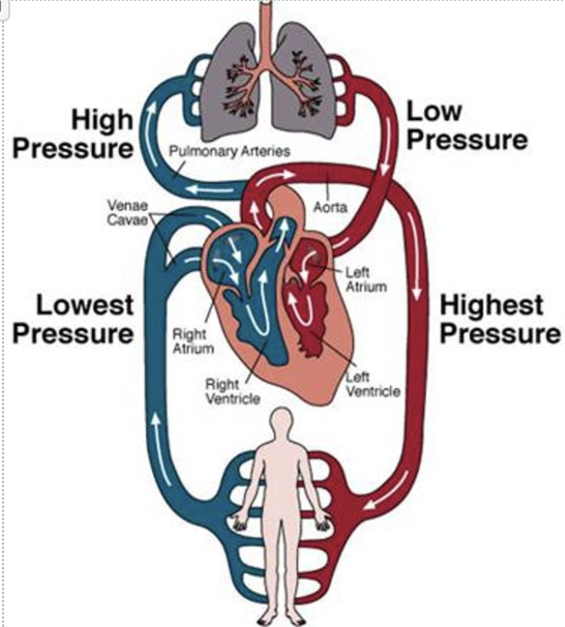
What causes blood to flow through the heart
Blood flows due to pressure differences created by the contraction and relaxtion of heart chambers
Blood moves from areas of high pressure to low pressure
Pressure changed result from:
Systole ( contraction )
Diastole ( relaxtion )
One complete sequence of events is called the cardiac cycle
Electrical control of the heart
The sinoatrial ( SA ) node:
A cluster of specialised cardiac cells in the right atrium
Acts as the heart’s natural pacemaker
Initiates electrical impluses causing atrial contraction
Electrical control of the heart
The atrioventricular ( AV ) node:
delays the electrical impluse
Ensures atria fully contract before ventricles
Transmits the impulse to the ventricles via specialised conducting fibres