ANAT 100 - Module 9
1/89
There's no tags or description
Looks like no tags are added yet.
Name | Mastery | Learn | Test | Matching | Spaced | Call with Kai |
|---|
No analytics yet
Send a link to your students to track their progress
90 Terms
What does the respiratory system do?
Facilitates breathing and allows the body to acquire oxygen, a molecule necessary for metabolic function and cell survival, from the air.
What do the organs in the respiratory system contribute to?
The conduction of air to and from the lungs, as well as gas exchange within the lungs.
Breathing involves two cyclic phases called
Inspiration (inhalation) and expiration (exhalation)
Inspiration
Draws oxygen rich air into the lungs
Expiration
Forces oxygen poor air out of the lungs
Functions of the respiratory system
Gas exchange
Gas conditioning
Sound production
Olfaction
Defense
Gas exchange
Involves the movement of gases across membranes.
External respiration and internal respiration
External respiration
Refers to the exchange of gases between the air and blood.
Inspired oxygen moves across the cellular membranes of the alveolus of the lung and its associated capillaries into the blood, while waste, carbon dioxide, moves in the opposite direction and out of the body through expiration.
Internal respiration
Similar mechanism that exchanges gases (O2 and CO2) between the blood and cells of the body
Gas conditioning
Gases entering the lungs need to be conditioned, or warmed an cleansed, in order to prevent damage to the lungs
Where does gas conditioning occur and how?
In the nasal cavities and paranasal sinuses where air is swirled around to become warmed and humidified. Inhaled air is cleansed of particulate matter through contact with the mucosal lining of respiratory epithelium
Sound production
Production of sound, such as singing or speech, occurs by forceful expiration of air through the vocal cords in the larynx, causing them to vibrate
Different tensions of the vocal cords produce different sounds with help from the teeth, lips and tongue
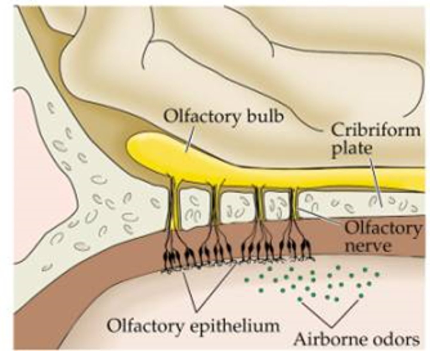
Olfaction
The olfactory epithelium covers the top of the nasal cavity. The receptors for the sense of smell are located within this epithelium
How does olfaction work?
When air is inhaled into the nasal cavity, airborne molecules dissolve in the mucus which lines the cavity and stimulates the receptors. Signals from these receptors travel to the brain through the olfactory nerve (CN1), resulting in a sense of smell
Defense
There are many airborne molecules and microbes that can cause disease. As such, the respiratory system has a line of defense against these molecules that can cause infection
What mechanisms are in place to prevent infection?
The coarse hairs of the nostrils, the ciliated cells of the respiratory epithelium, and the mucus lining help to trap particles and microorganisms from entering the nose and respiratory system
Respiratory tract epithelium
Pseudostratified ciliated columnar epithelium
Goblet mucus cells
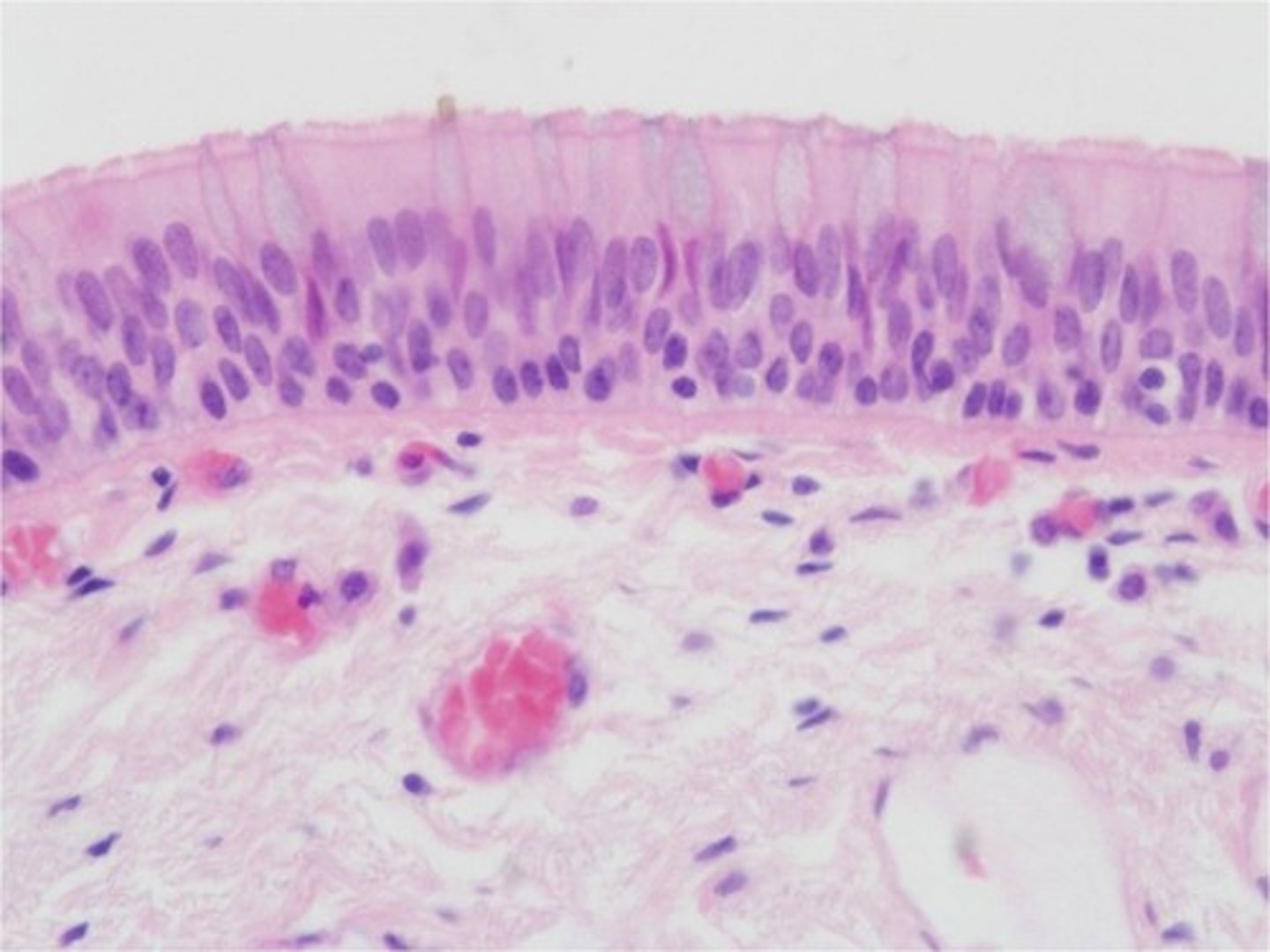
What does pseudostratified mean?
All cells are attached to the basal lamina but only some reach the surface
What is the apical surface of the epithelium covered in?
Cilia, which are small fingerlike projections extending from the cell that provide an increased surface area for conditioning air.
What else do the cilia do?
Also function to trap inhaled particles and microorganisms caught in mucus, and sweep them back up the respiratory tract and out through the nose and mouth.
Where can goblet mucus cells be found and what do they produce?
Found interspersed throughout the pseudostratified epithelium.
These cells produce mucus, causing them to stain lightly in a histological image.
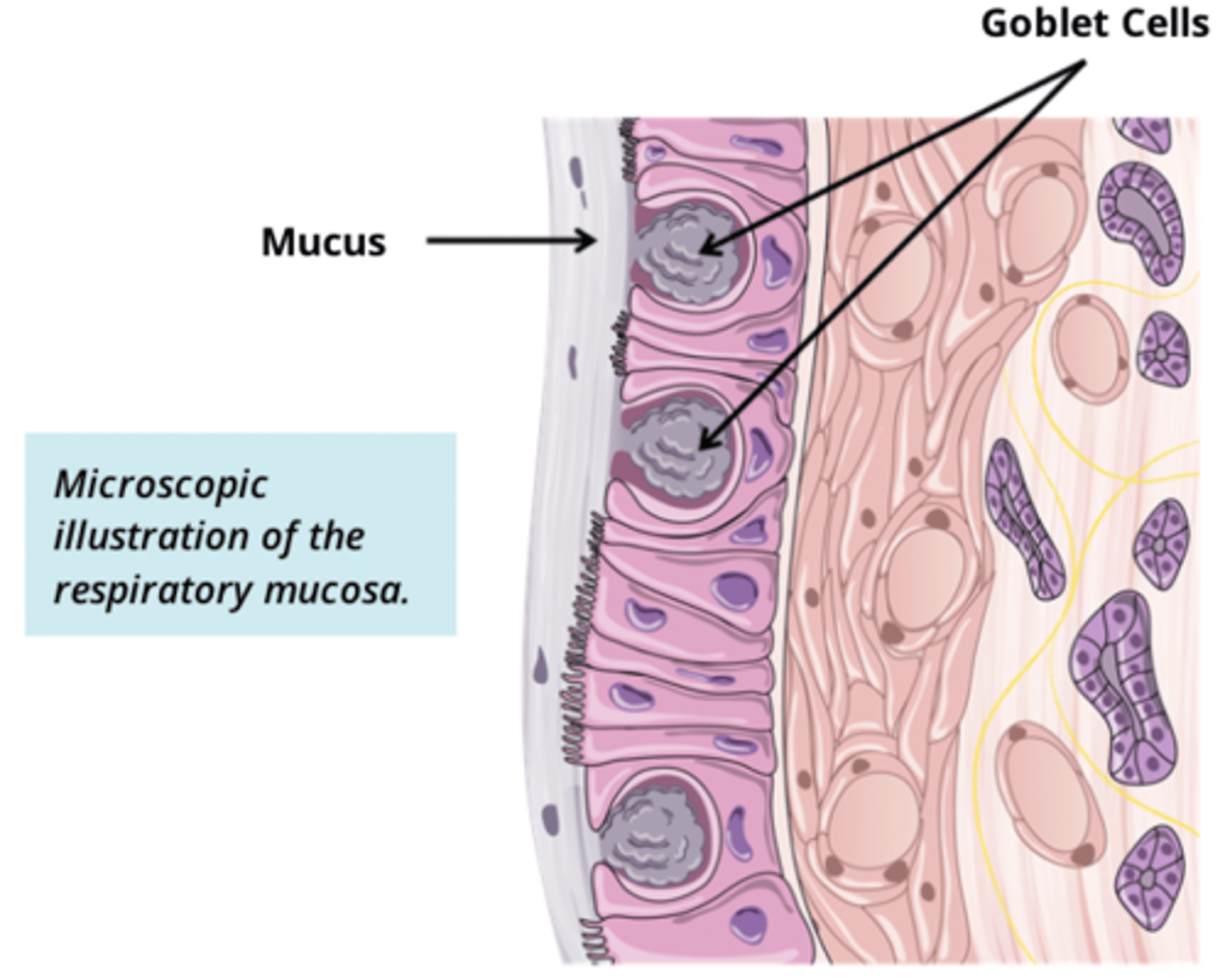
What does the mucous from goblet cells do?
Forms a protective layer over the epithelium and traps particulate matter or microorganisms that may be inhaled. It also provides moisture to humidify the air before it reaches the lungs.
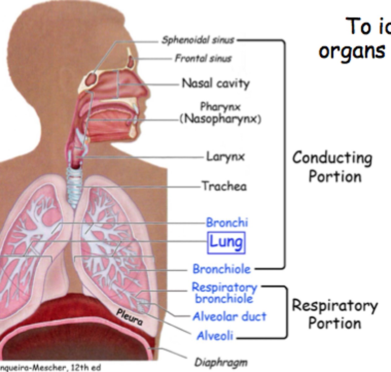
What is the function of the conducting portion of the respiratory system?
Functions to transfer or 'conduct' inhaled air from the outside world to the lung tissue. This portion also conducts air from the lungs to the outside world.
What occurs in the conducting portion?
Humidification and trapping of debris occurs. No oxygen is absorbed into the blood in this region as the walls of the organs are too thick.
What are the structures of the conducting portion?
Nose and nasal cavity
Paranasal sinuses
Pharynx
Larynx
Trachea
Primary, secondary, and tertiary bronchi
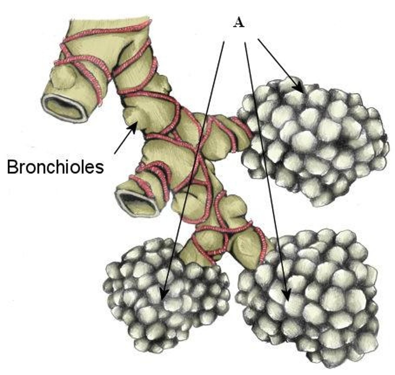
Terminal bronchioles
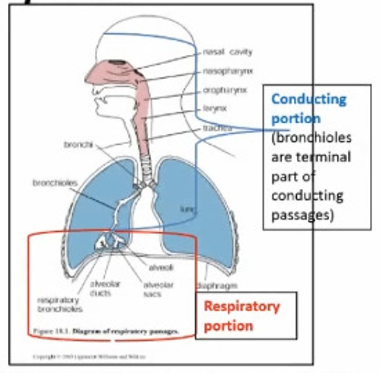
Function of the respiratory portion of the respiratory system
Functions to transfer gases between the lungs and pulmonary capillaries. The pulmonary capillaries are the terminal structures within the lungs that have walls thin enough to facilitate the movement of gases from air to blood and vice versa.
What are the structures that make up the respiratory portion?
Respiratory bronchioles
Alveolar ducts
Alveolar sacs
Alveoli
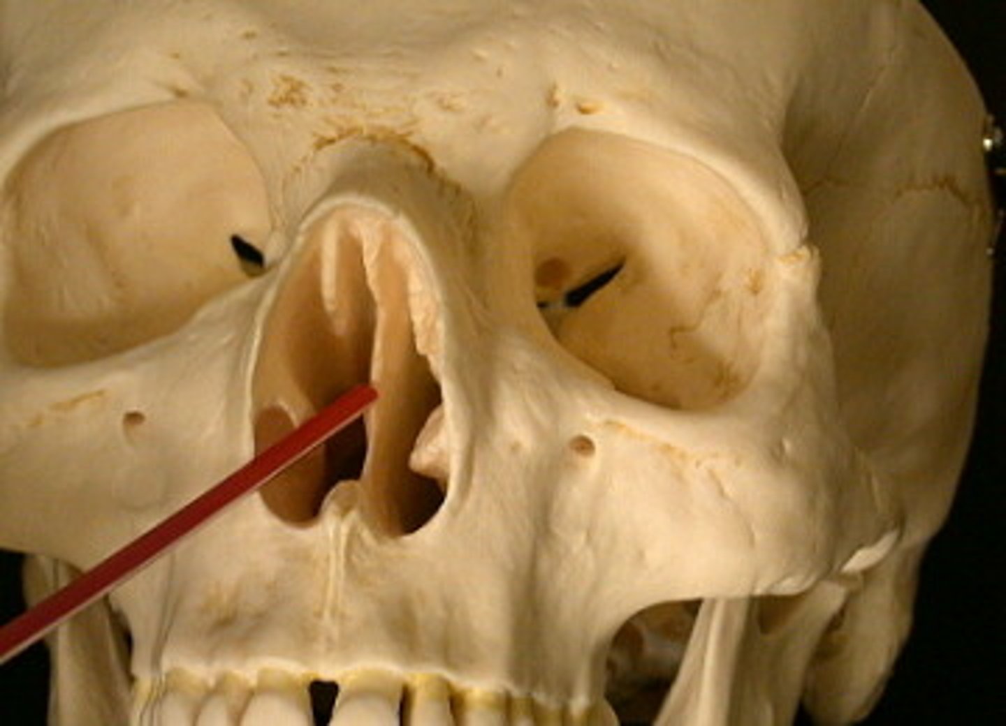
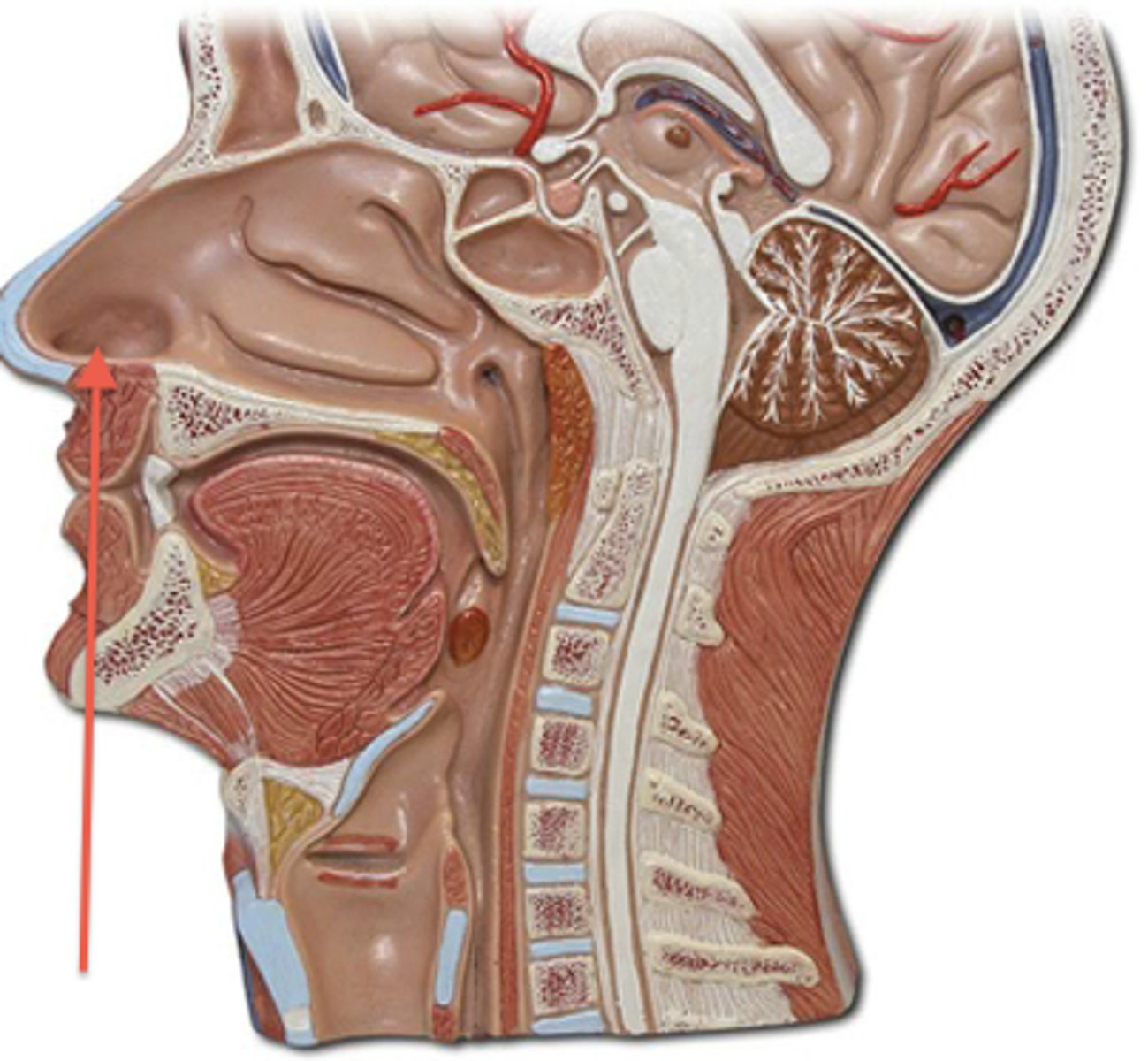
What are the paranasal sinuses?
Collection of air filled spaces within the bones of the skull communicating with the nasal cavity. The sinuses aid in the conditioning of air (warming and humidifying), defense against pathogens, and act as resonance chambers for speech
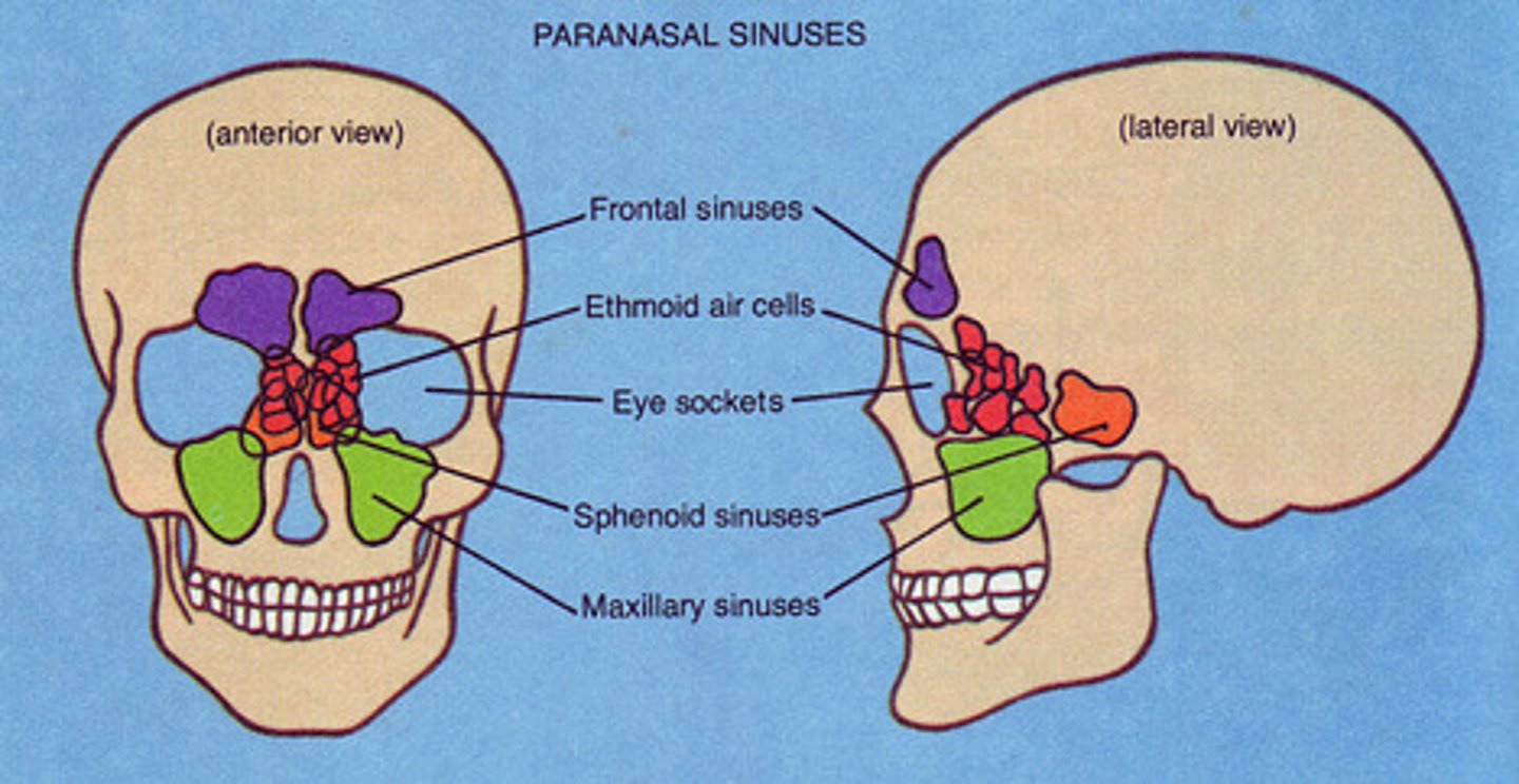
What are the different paired sinuses and what are they named after?
Named after the bones of the skull that contains them
Frontal sinus (2)
Maxillary sinus (2)
Ethmoid sinus (2)
Sphenoid sinus (2)
What are the paranasal sinuses lined with?
Respiratory tract epithelium, again contributing to its function in defense and conditioning
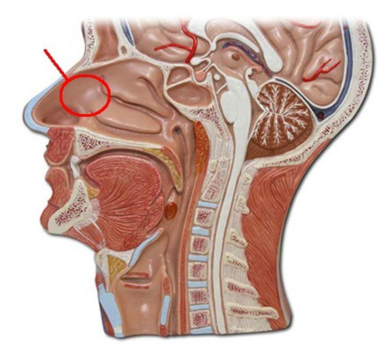
Nose and nasal cavity
First line of defense against invading pathogens and debris, trapping them in coarse hairs and mucus.
Air enters the vestibules (openings) of the nostrils and is passed into the nasal cavity where it is conditioned
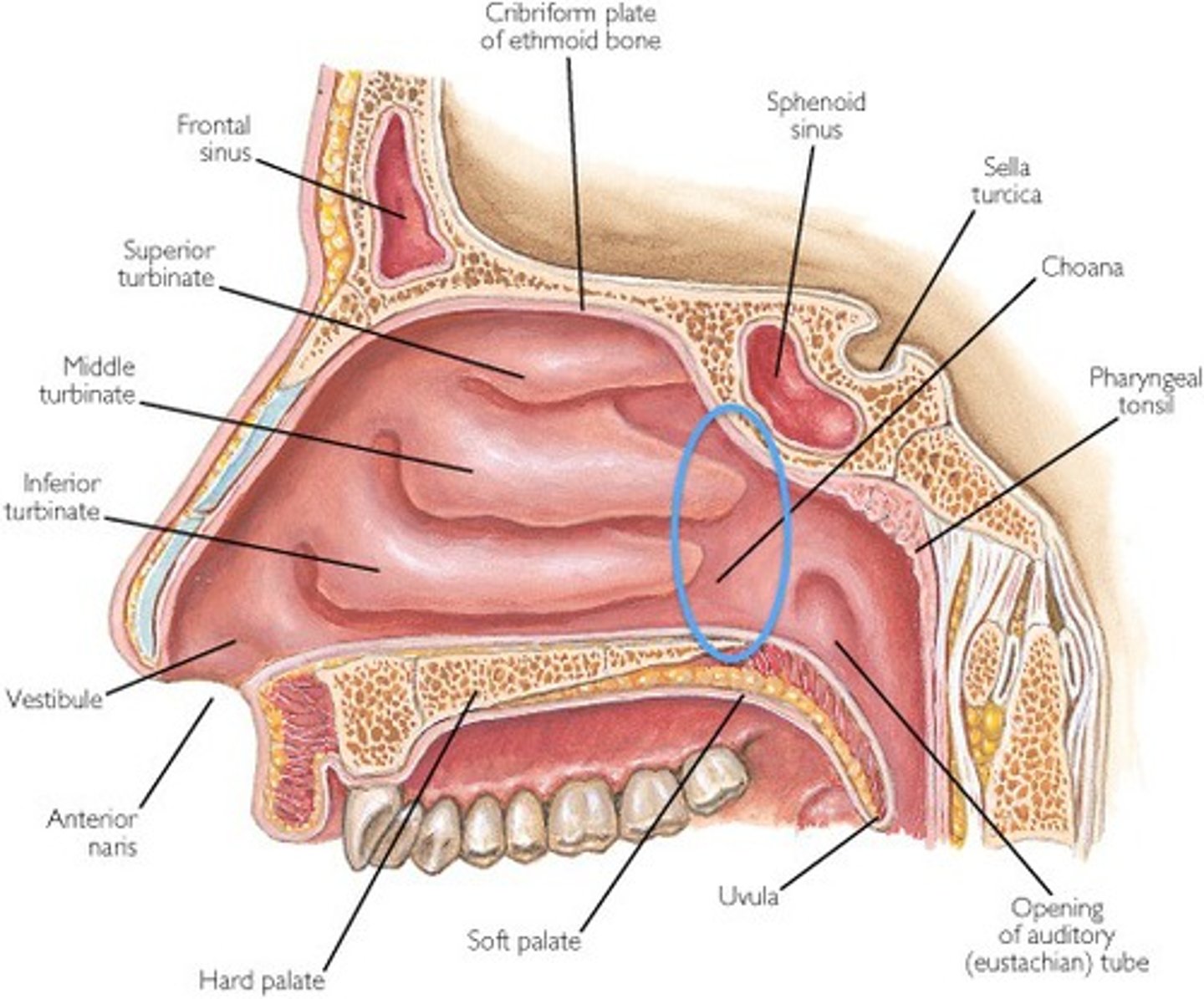
Nasal cavity boundaries
Roof
Floor
Medial wall
Lateral walls
Anterior border
Posterior border
Roof of nasal cavity
Bone in the skull called the ethmoid
Floor of nasal cavity
Composed of the hard palate (roof of the mouth)
Medial wall of nasal cavity
Makes up the nasal septum.
The septum is composed of the vertical bones in the skull, and separates the two halves of the nasal cavity
Lateral walls of nasal cavity
Contain structures known as the nasal conchae.
These structures create turbulence in the air as it passes through the cavity, allowing for conditioning and catching debris.
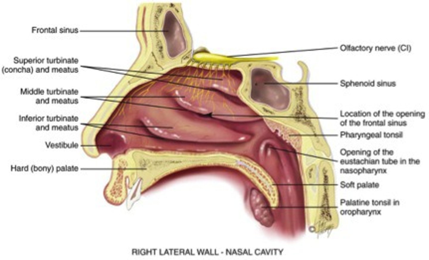
Anterior border of nasal cavity
Made up by the nares, which is the opening between the nose and nasal cavity
Posterior border of nasal cavity
Opening to the nasopharynx (choanae), where the nasal cavity connects to the pharynx
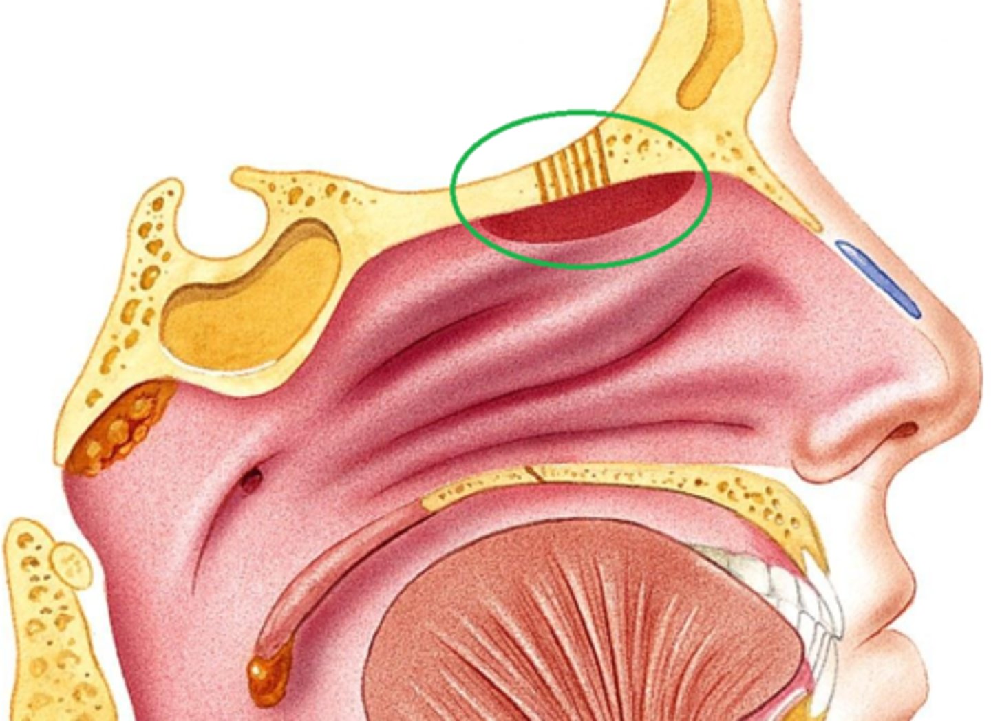
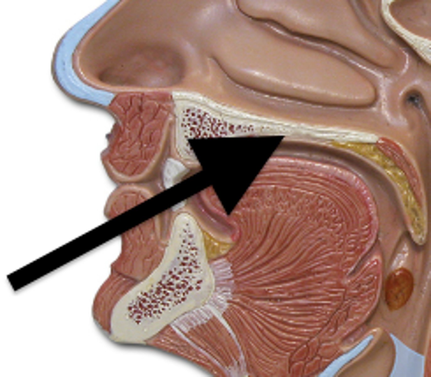
Nasal cavity histology
Respiratory tract epithelium (RTE) and olfactory epithelium
Nasal cavity histology: RTE
Mostly covered in RTE because of its role in protection from airborne debris and microorganisms, as well as the conditioning of air.
Nasal cavity histology: olfactory epithelium
Roof of the nasal cavity is lined with olfactory epithelium, which contains sensory receptors for smell.
The olfactory epithelium is composed of pseudostratified ciliated columnar epithelium and bipolar olfactory receptor neurons
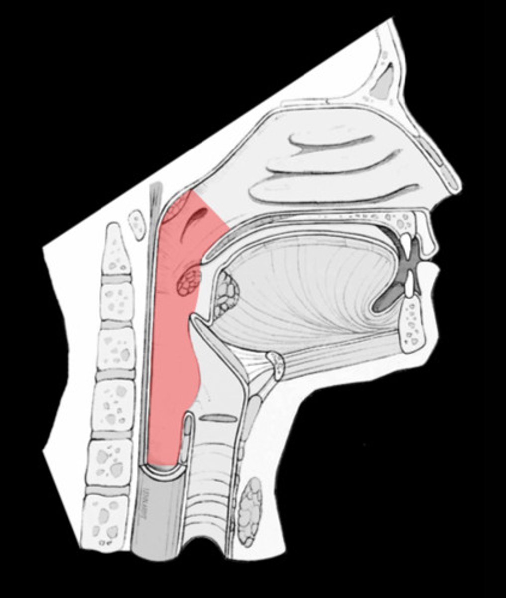
What is the pharynx and what are its functions?
Muscular tube that connects the nasal cavity and the larynx in the respiratory system.
It also functions to connect the oral cavity with the esophagus in the digestive system
Components of the pharynx
Nasopharynx
Oropharynx
Laryngopharynx
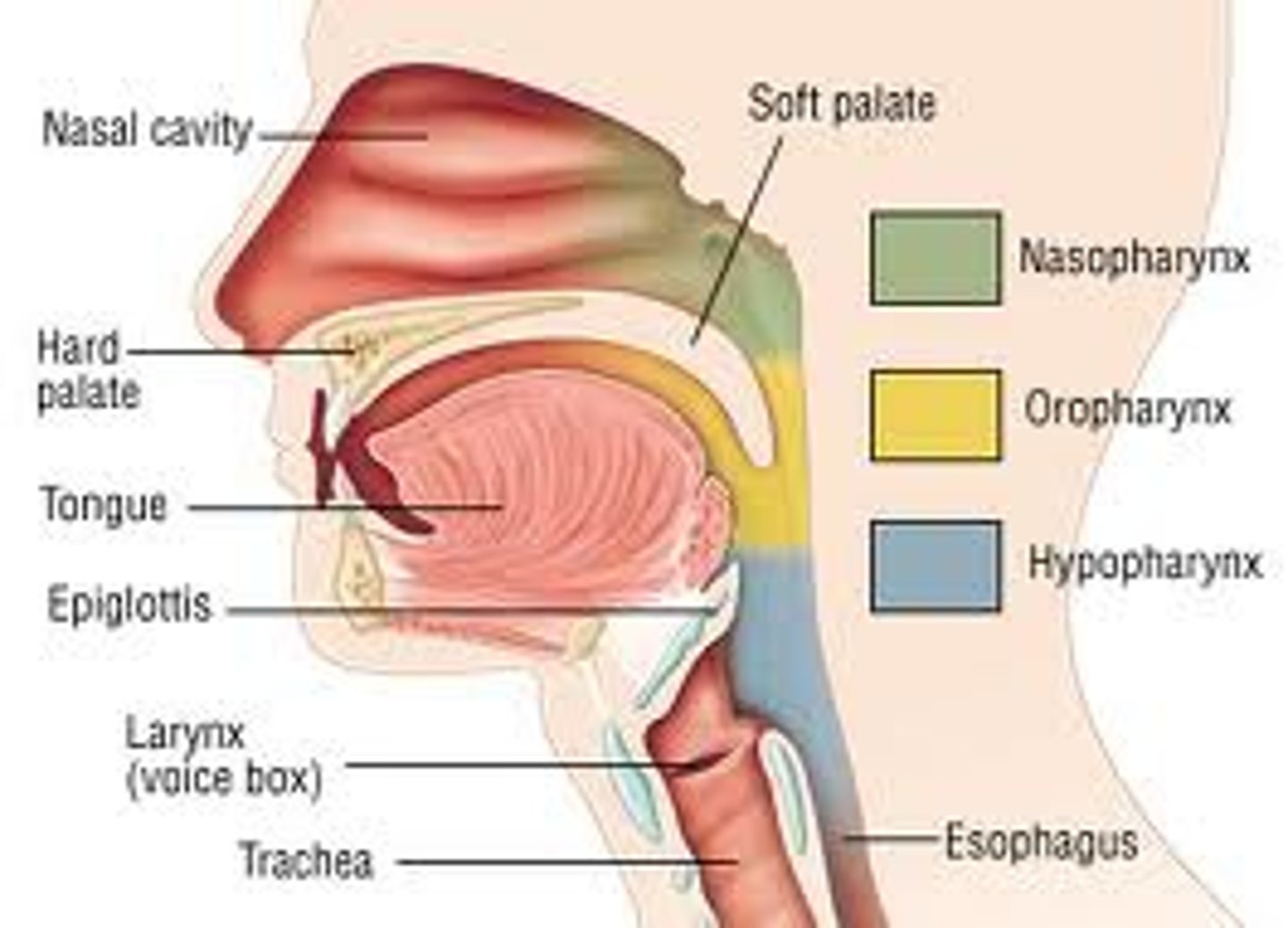
Nasopharynx
Most superior aspect of the pharynx
Oropharynx
Middle aspect of the pharynx. It's part of both the respiratory and digestive system. This muscular tube passes air from the nasopharynx and food from the oral cavity into the laryngopharynx
Laryngopharynx
Most inferior aspect of the pharynx.
It is also involved in both the digestive and respiratory systems, passing both food and air into the respective systems
Is the histology of the pharynx the same throughout?
No, regions of the pharynx have different histology, due to their varying functions
What is the histology of the nasopharynx?
Mostly RTE because it has mainly respiratory functions
What is the histology of the oropharynx and laryngopharynx?
Lined with stratified squamous epithelium. This is due to the need for durability when swallowing food.
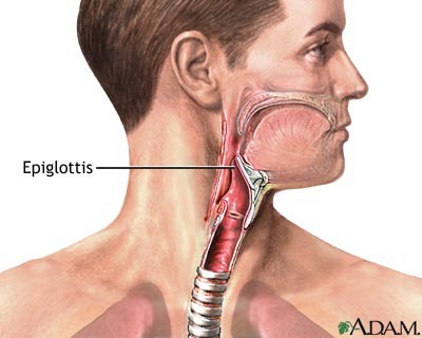
Larynx
Organ that produces sound, also known as the voice box.
It's made up of a variety of cartilages, ligaments, and associated muscles in order to serve that purpose
Location of the larynx
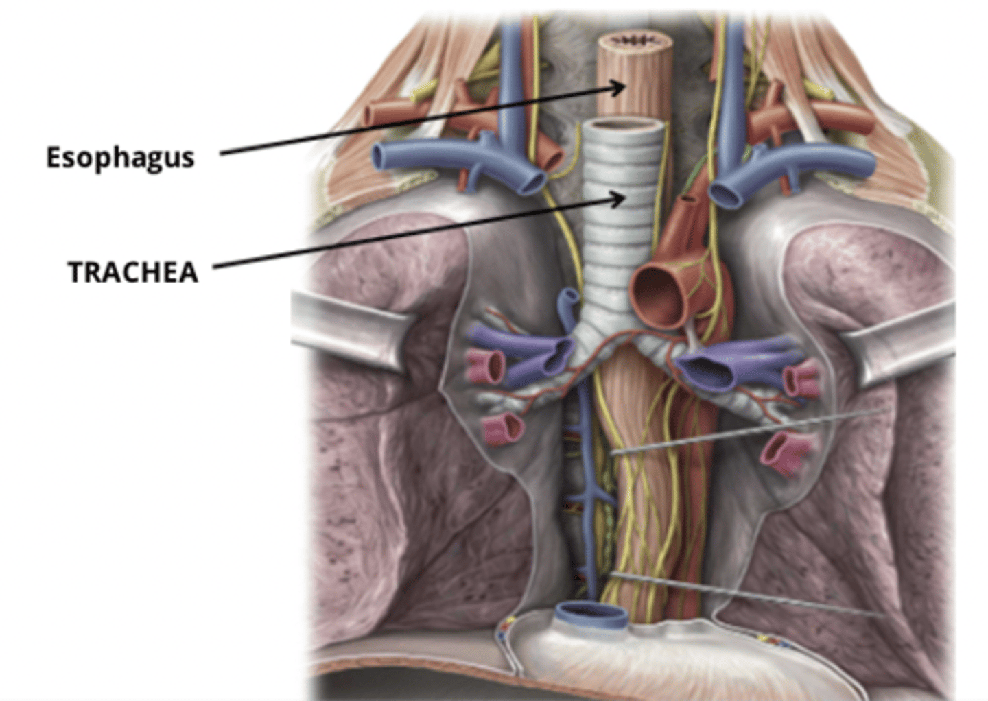
This organ sits anterior to the esophagus, connecting the pharynx with the trachea and preventing food from entering the trachea
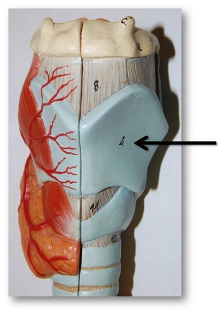
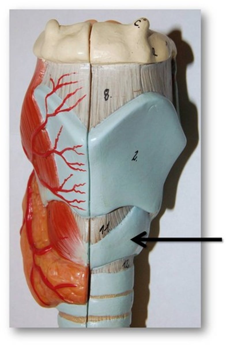
Cartilage of the larynx
Epiglottis, thyroid cartilage, cricoid cartilage
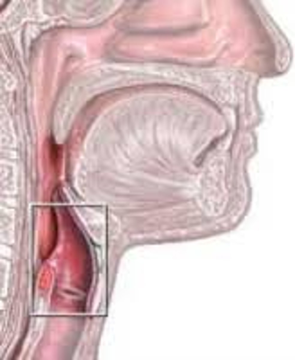
Epiglottis
Large spoon shaped elastic cartilage that functions to prevent food from passing into the trachea (windpipe).
During swallowing, the epiglottis flips downwards and covers the opening of the trachea
Thyroid cartilage
'Shield shaped' hyaline cartilage that provides attachment for many muscles as well as the vocal cords
Cricoid cartilage
Complete ring of hyaline cartilage. It is narrow anteriorly and broad posteriorly. This structure functions as an attachment for muscles and the vocal cords
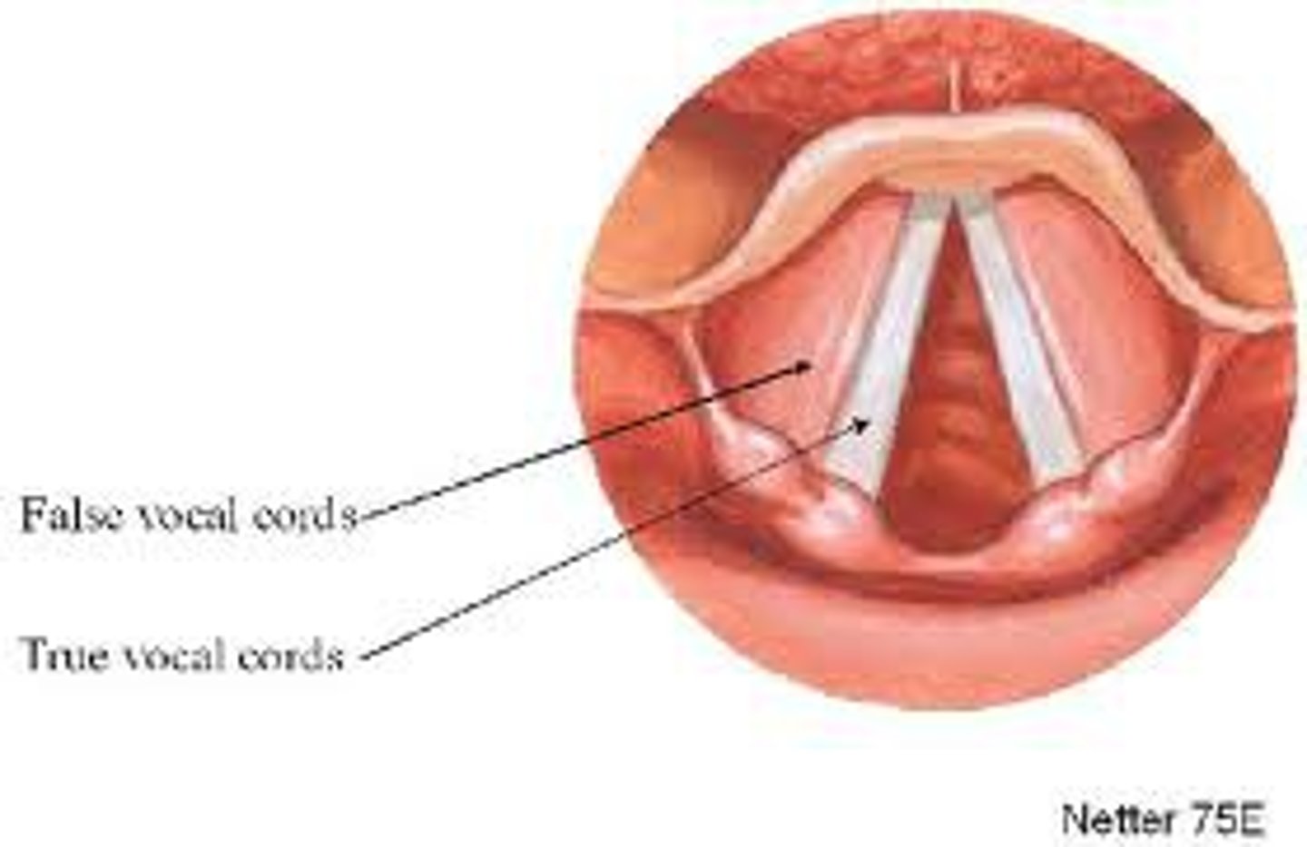
Vocal cord
Small ligaments attached to the laryngeal cartilages that vibrate when air is forced out of the lungs. When these cords vibrate, sound is produced. Different sounds can be made by altering the tension on the cords.
What is the difference between true and false vocal cords?
True vocal cords are the ligaments that produced sound
False vocal cords are a membranous flap that protect the true vocal cords
Trachea location and function
Extends from the larynx to approx. the level of T4/T5 where it splits at a junction called the carina.
The main function of the trachea is the conduction of air to the lungs.
What is the trachea made up of, what is their function and what are they connected by posteriorly?
15-20 C shaped cartilaginous rings, which are incomplete posteriorly.
The rings function to keep the airway open.
The cartilaginous rings are connected posteriorly by the trachealis muscle.
Histology of the trachea
Mucosa
Submucosa
Adventitia
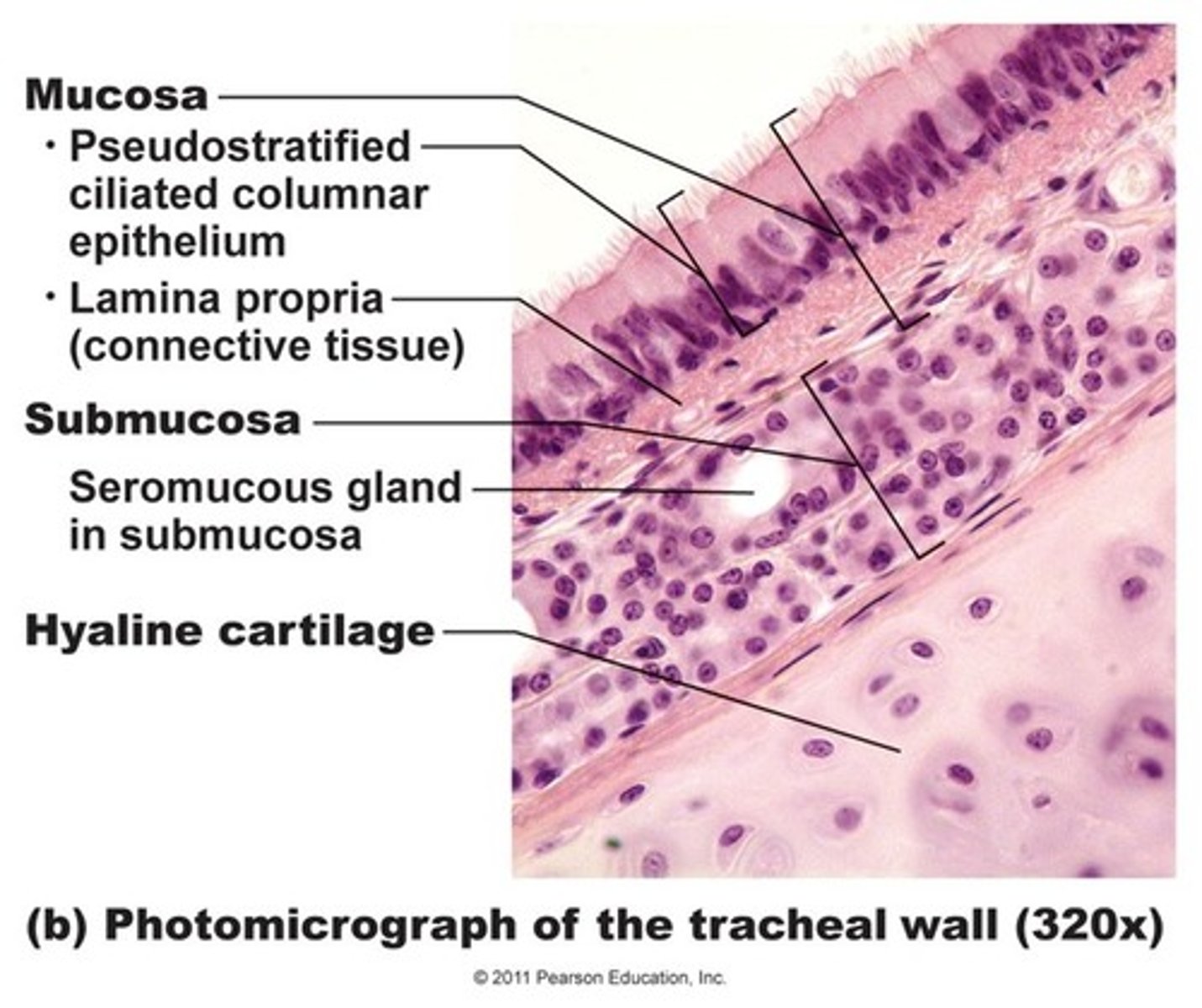
Mucosa of the trachea
The trachea is lined with RTE in order to clear any debris or pathogens that make it into the lower respiratory tract
Submucosa of the trachea
Lies between the mucosa and adventitia.
Made up of loose (areolar) connective tissue, containing larger vessels and nerves, as well as mucus secreting glands
Adventitia of the trachea
Outer layer of connective tissue surrounding the trachea. This layer also encloses the C-shaped cartilaginous rings made of hyaline cartilage.
These rings make the trachea flexible and durable, which is important as the organs in the thoracic cavity shift with the movements of breathing
Primary bronchi
Splitting of the trachea results in 2 primary bronchi, left and right, which have the same functional and histological features as the trachea
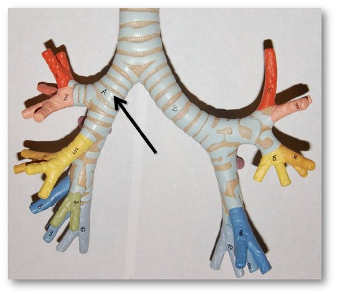
Where does each bronchus enter?
Enters its respective lung on the medial side of the hilus
What is different between the right and left primary bronchi?
The right primary bronchus is wider, shorter, and more vertical than the left primary bronchus (this means that foreign objects traversing the trachea are more likely to enter the right main bronchus)
What is the end of the conducting portion characterized by?
Characterized by bronchi dividing into smaller and smaller tubes. Moving from the large diameter, thick walled tube to a smaller diameter, thin walled tube for gas exchange
There is also corresponding change in histology as you move down the tree
How do the bronchi split?
Primary bronchi --> secondary bronchi --> tertiary bronchi --> terminal bronchioles
Why is the respiratory portion important?
Cells in the body need to absorb oxygen and expel carbon dioxide to survive.
At this level of the bronchial tree, how thick are the walls of the remaining structures?
One cell layer thick, and gases are able to cross the membrane easily
What are the lungs and where are they located?
The respiratory portion makes up a majority of the lung tissue within both lungs.
The lungs are located on either side of the heart within the thoracic cavity.
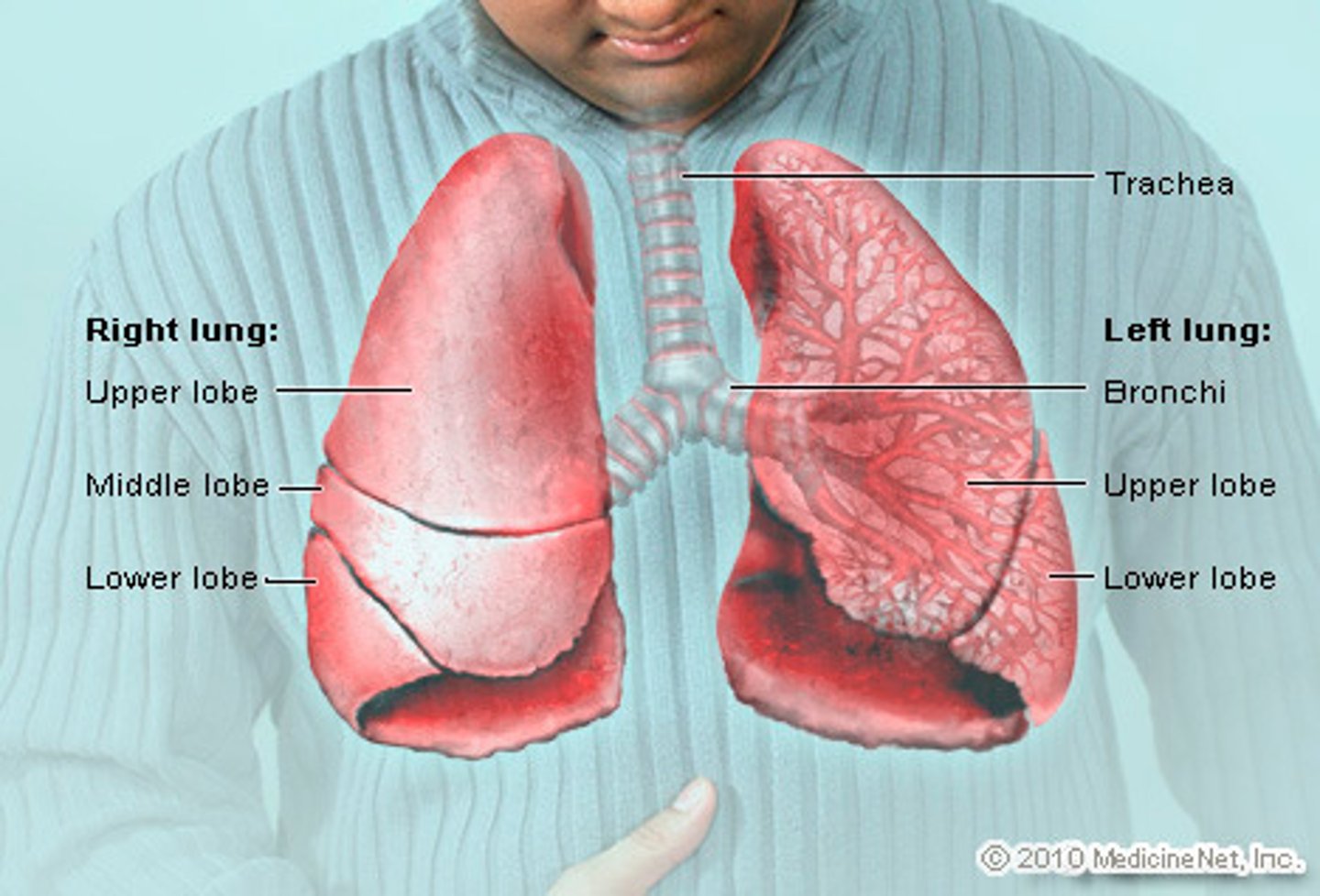
Are the lungs the same size?
No. The right lung is slightly larger than the left due to the positioning of the heart
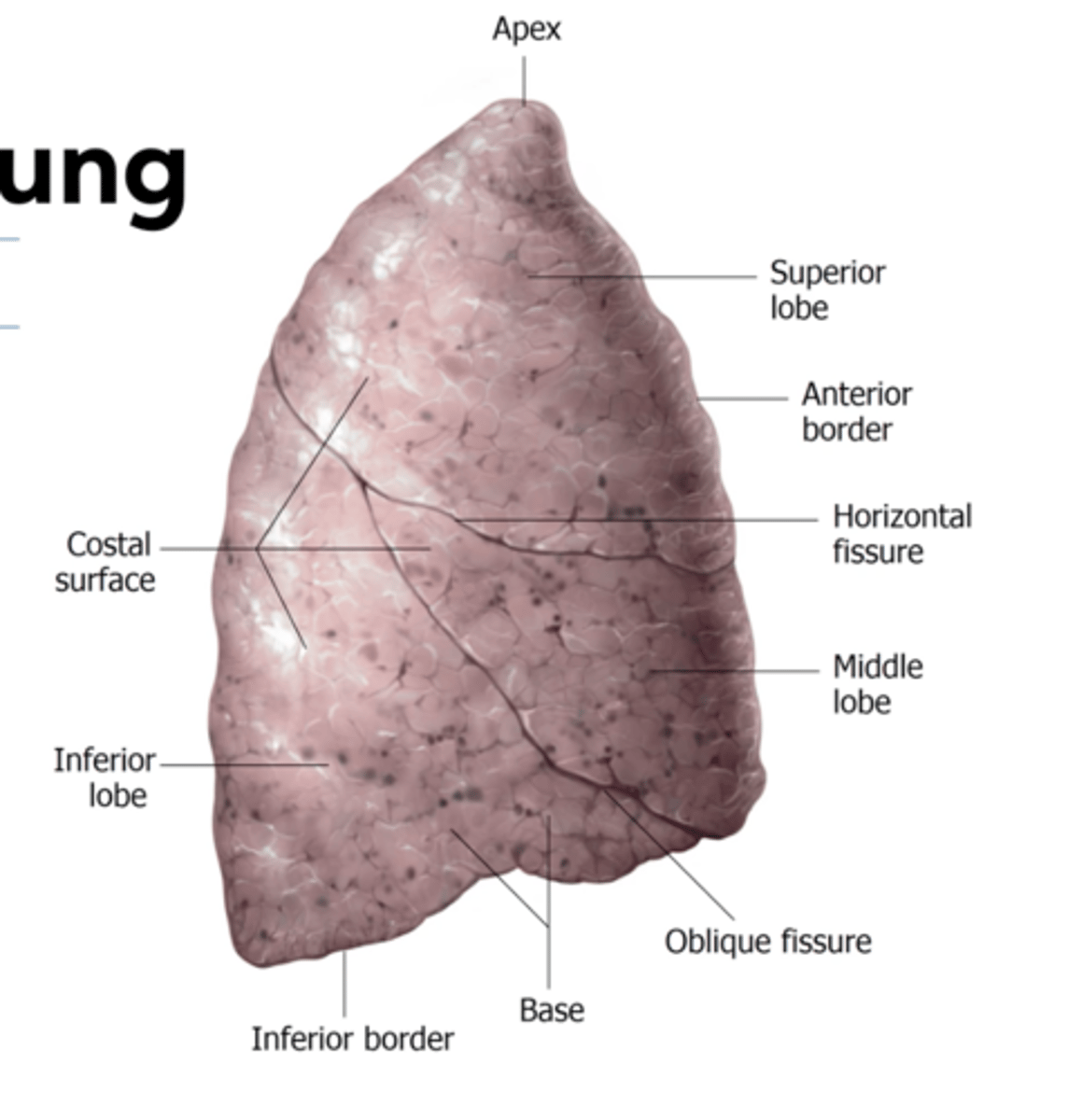
Right lung
3 lobes: superior, middle and inferior
2 fissures that separate the lobes: horizontal and oblique
Left lung
2 lobes: superior and inferior
1 fissure: oblique fissure that separates the lobes
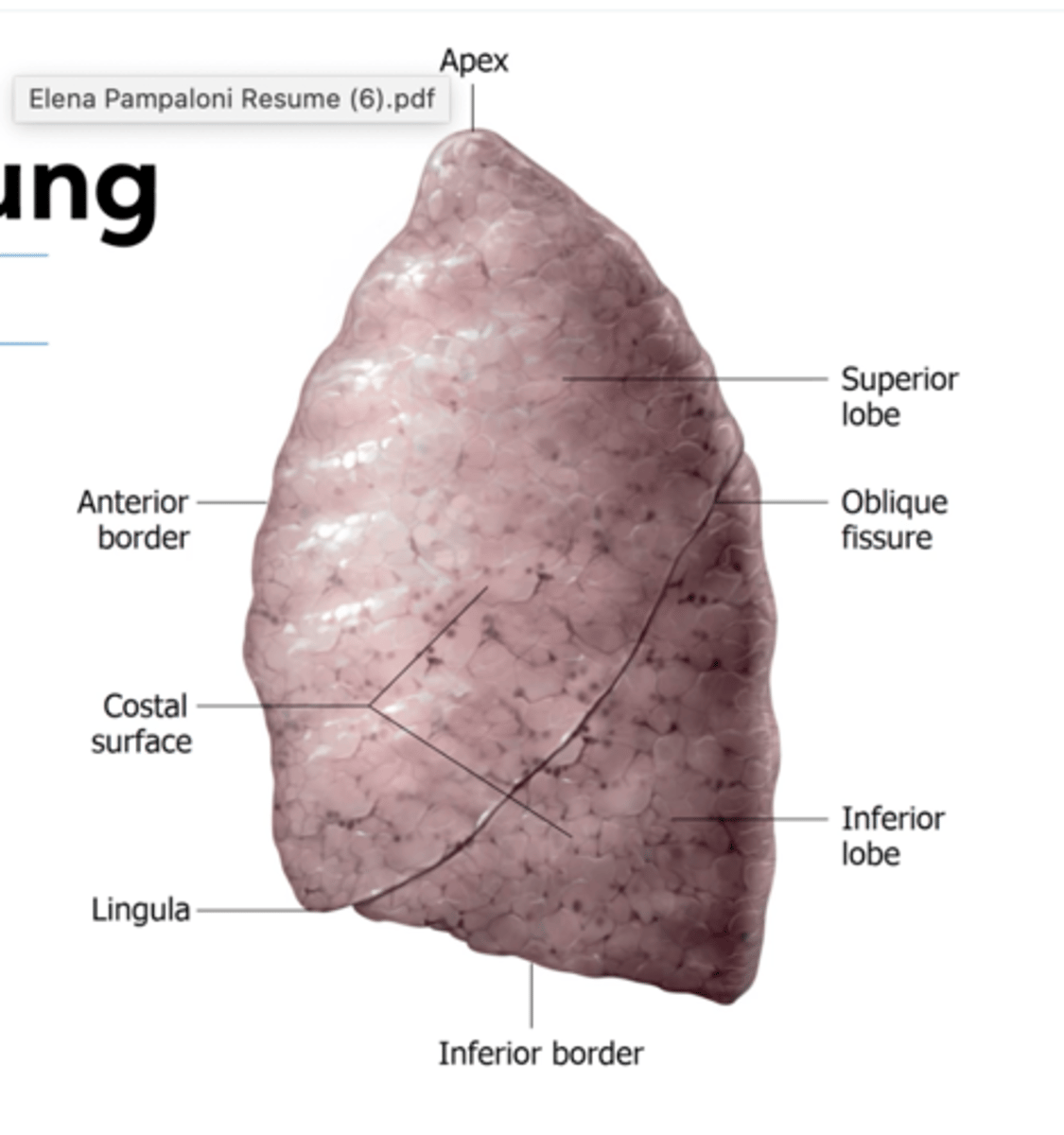
What is found on the superior lobe of the left lung?
The cardiac notch
Small outward facing process called the lingula, which covers the heart
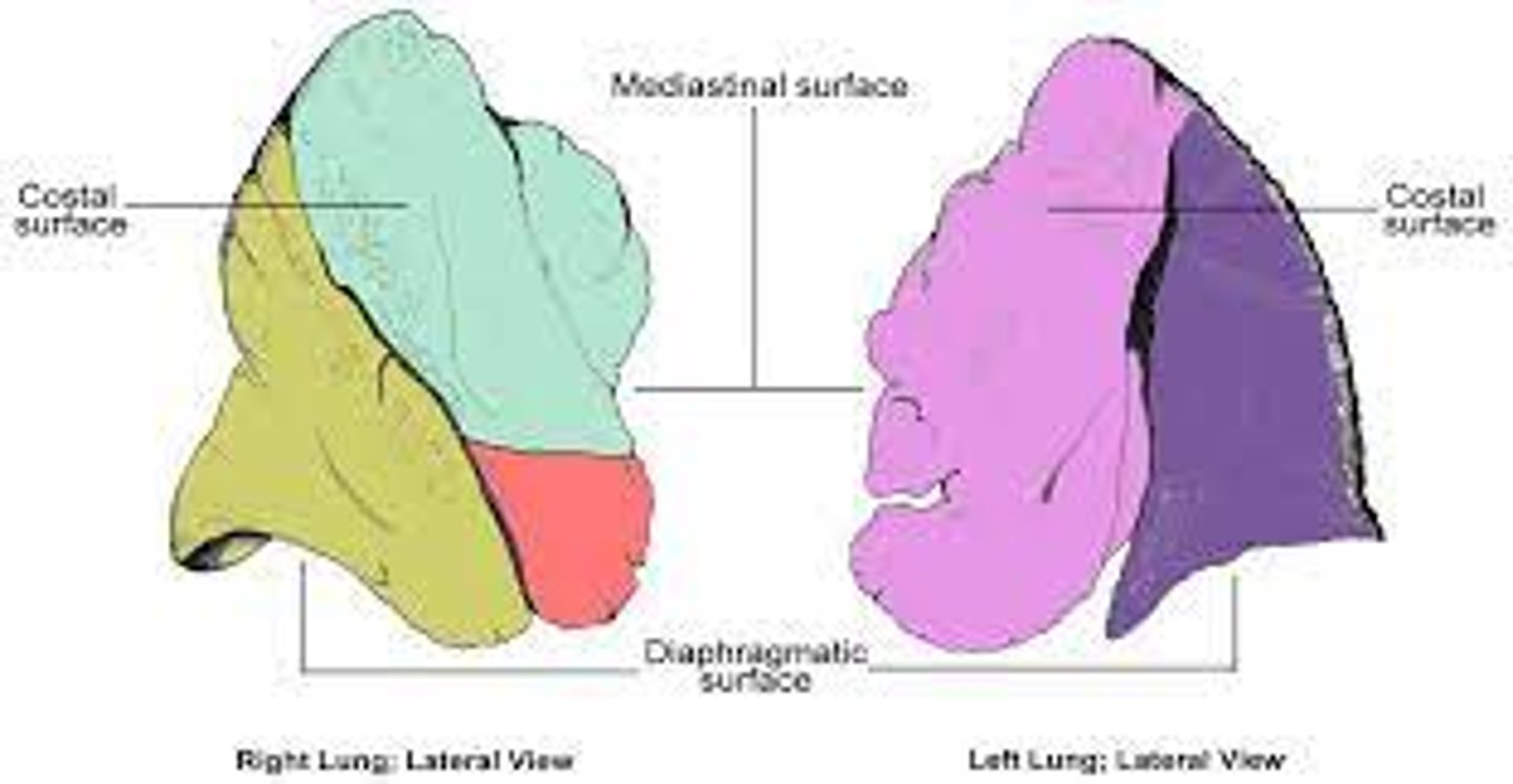
What creates the surfaces of the lungs?
Within the thoracic cavity, the lungs are in close contact with their surrounding structures. This creates the surfaces of the lungs, often named after structures they contact
What are the different surfaces of the lungs?
Apex
Diaphragmatic
Costal
Mediastinal
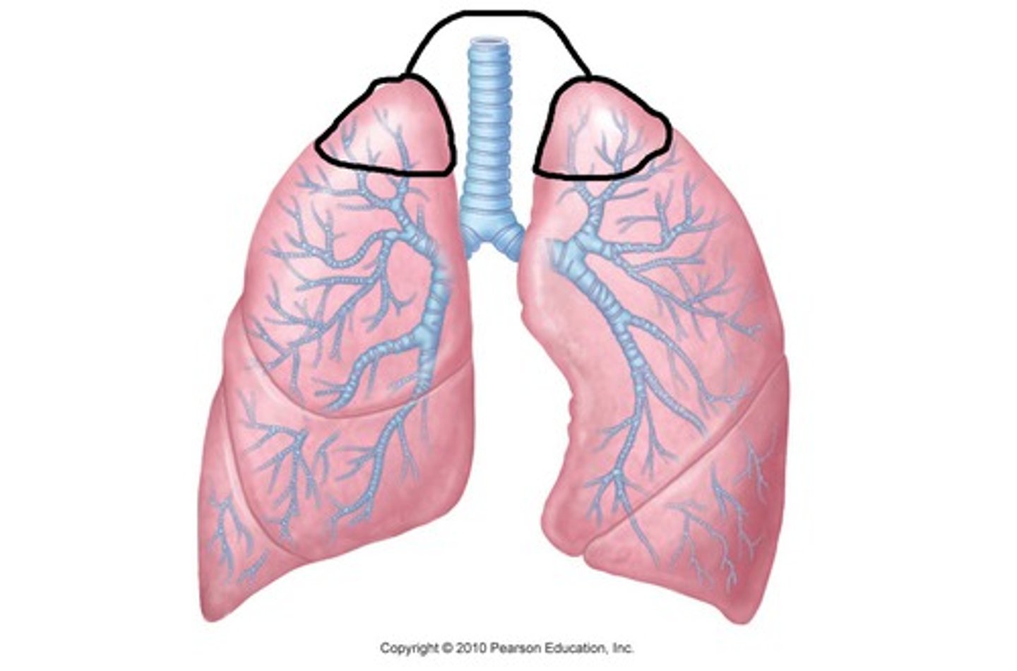
Apex surface
Most superior point of this organ. In the body, it sits just above the first rib
Diaphragmatic surface
The lung sits superiorly to the diaphragm, a dome-shaped skeletal muscle.
The base of the lung is known as the diaphragmatic surface. This surface rests on the diaphragm
Costal surface
Named after the adjacent ribs
This surface curves around the lateral aspect of the lung
Mediastinal surface
Medial surface
It contains the entry and exit points for all vessels and airways at a structure known as the hilus
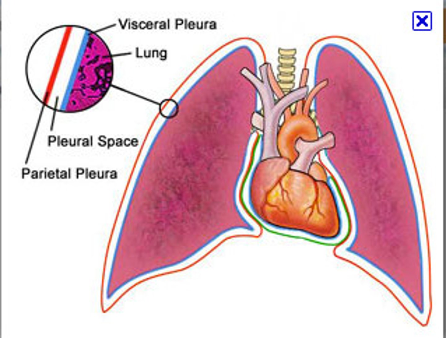
What are the pleural membranes and what do they do?
The lungs are important organs for gas exchange, and therefore need to be protected.
The pleura consists of 2 continuous membranes that form a sac around each lung
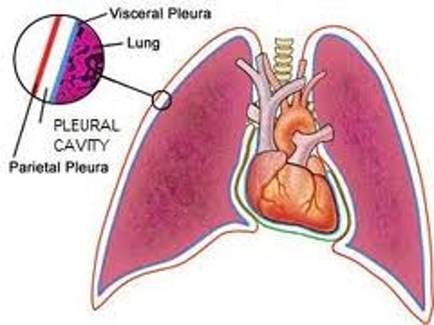
What do the pleura secrete and what is its purpose?
Pleural fluid to fill the pleural space between the 2 membranes.
This fluid acts as a lubricant, allowing the visceral pleura of the lung to slide freely on the parietal pleura of the thoracic wall during inflation and deflation
Parietal pleura
Similar to the parietal membrane of the pericardium, the parietal pleura is an outer serous membrane attached to the walls and floor of the thoracic cavity around the lungs
The parietal pleura is continuous with the visceral pleura at the hilus of the lung where it's reflected inwards
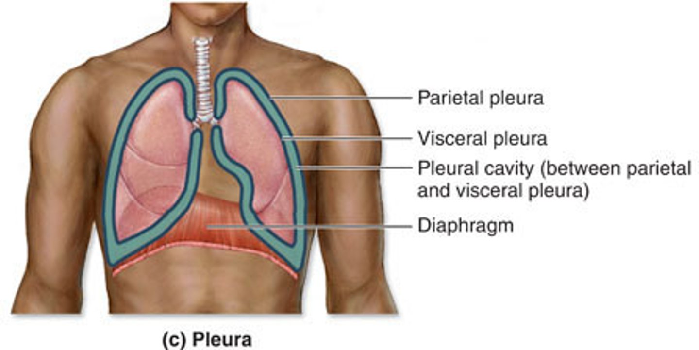
Visceral pleura
A serous membrane attached to the surface of the lung that is continuous with the parietal pleura at the hilus
Structures of the respiratory portion
Respiratory bronchioles
Alveoli
Respiratory bronchioles
Branch from the terminal bronchioles (the last structure in the conducting portion)
They are the first structure to contribute to gas exchange in the lungs
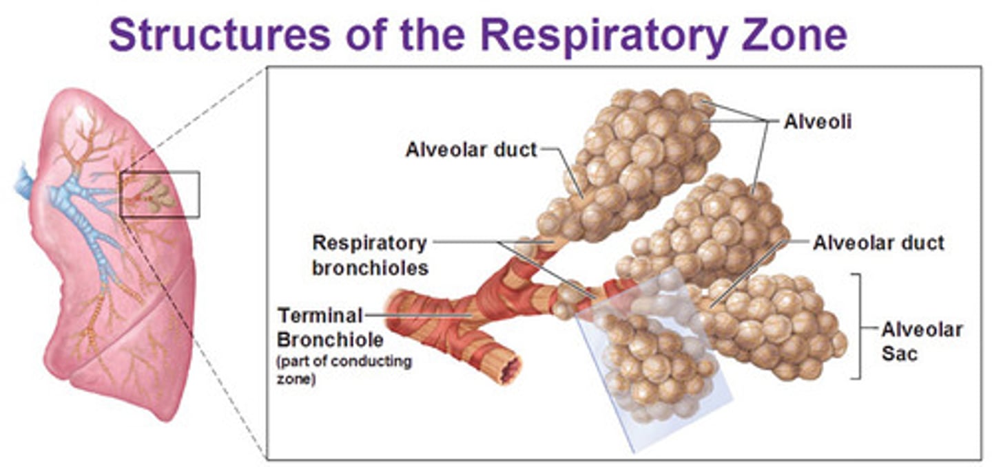
Histology of respiratory bronchioles
Thin walled ducts with simple ciliated cuboidal epithelium
They continue to branch, ending with alveoli
Alveoli
The alveolus is the functional unit of the lung, where gas exchange takes place.
Each alveolus is surrounded by capillaries in order to maximize the amount of oxygen going into the body and carbon dioxide leaving the body
Causes of COPD
Smoking
Second hand smoke
Air pollution
Dust or workplace fumes
Biomass exposure (i.e. wood smoke)