visual neuroscience midterm
1/151
There's no tags or description
Looks like no tags are added yet.
Name | Mastery | Learn | Test | Matching | Spaced |
|---|
No study sessions yet.
152 Terms
visual neuroscience
a section of neuroscience that focuses on the neural mechanisms that underly visual perception
photoreceptors
light sensitive cells in the eyes called cones and rods
cells differ in their light absorption properties
perception
requires a stimulus and a visual system capable of responding to the stimulus
it makes assumptions/predictions to interpret the visual world
light
electromagnetic radiation in the visual spectrum that is a “carrier” of visual information
made up of photons/quanta (packet of energy)
wave-particle duality
wave-particle duality
light is both a particle (massless) and a wave
wavelength (lambda) = 1/ frequency
electromagnetic spectrum
a range of radiation/light sorted by short to long lambda (wavelengths)
visible light ranges 400-700
ultraviolet (uv) sight: birds, reindeer
infrared (iv) sight: snakes, fish, frog, mosquitos
3 (physical) properties of light
wavelength/frequency, amplitude, and wave-particle duality
light refractory
light can bend when it passes through materials
lenses bend light in a controlled way
most photons that enter out eye is reflected from surfaces
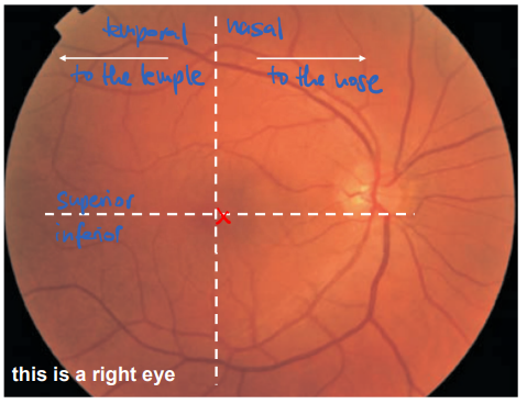
eye anatomy
retina, cornea, lens, pupil, fovea, optic nerve
pupil and iris: structures in the eye that make sure enough light gets into the eye
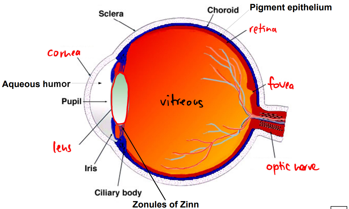
image formation
eye collects light that enters our lens and is flipped upon the lens onto the retinal surface
cornea is static and lens is dynamic
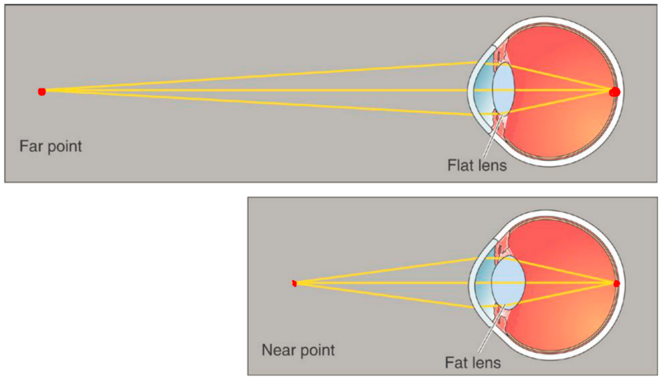
accommodation
process of focusing on objects at varying distances (like how a camera lens will adjust to bring an image to focus)
ciliary body
ciliary body
controls the shape of the lens to focus the visual image of the retina (accommodation)
changing lens shape allows extra focusing
pupil size
controlled by the iris, regulated by ambient light that is modulated by the autonomic nervous system
pupil diameter reduces as light goes up
eye types
compound eyes (multiple lens/ fly) and non-compound eyes(one lens/ jumping spider/ simple eye)
stereoscopic vision
combination of a single perception of slightly different images received from each eye, that results in depth perception
binocular: area/overlap seen by both eyes
monocular: area seen by one eye
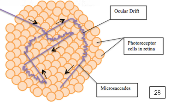
eye movement
catch images and holds them steady through the oculomotor nerve
saccadic and smooth pursuit eye movements
fixational eye movements
used to try and hold an image steady on the retina, but it is not perfect as the retina image is always in motion
perception is active as the brain directs eyes to areas to take in info as needed of the visual field
lenses
change color with age (get more yellow)
and cataracts (bottom row)
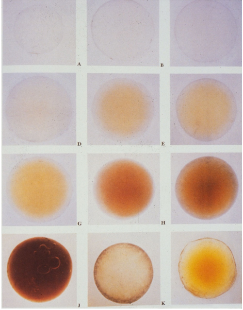
cataract
clouding of the lens which can lead to blurry/hazy vision
painting like monet’s can reveal how visual disorders work
commonly due to age
optical dysfunction
image not properly focused on the retina either due to the shape of the eyeball or shape of the lens
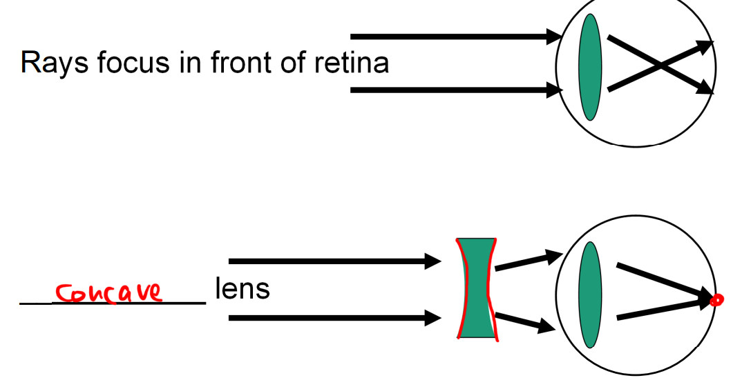
myopia (“nearsightedness”)
lens focus the rays of light together before they reach the retina due to development shape of eyeball/lens
can be fixed by glasses with a concave lens
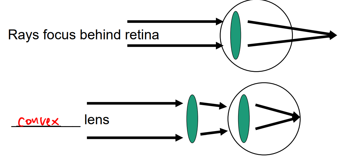
hyperopia/hypermetropia (“farsightedness”)
lens do not focus the light sharply enough to bring to focus on the retina due to development shape of eyeball/lens
can be fixed by glasses with a convex lens
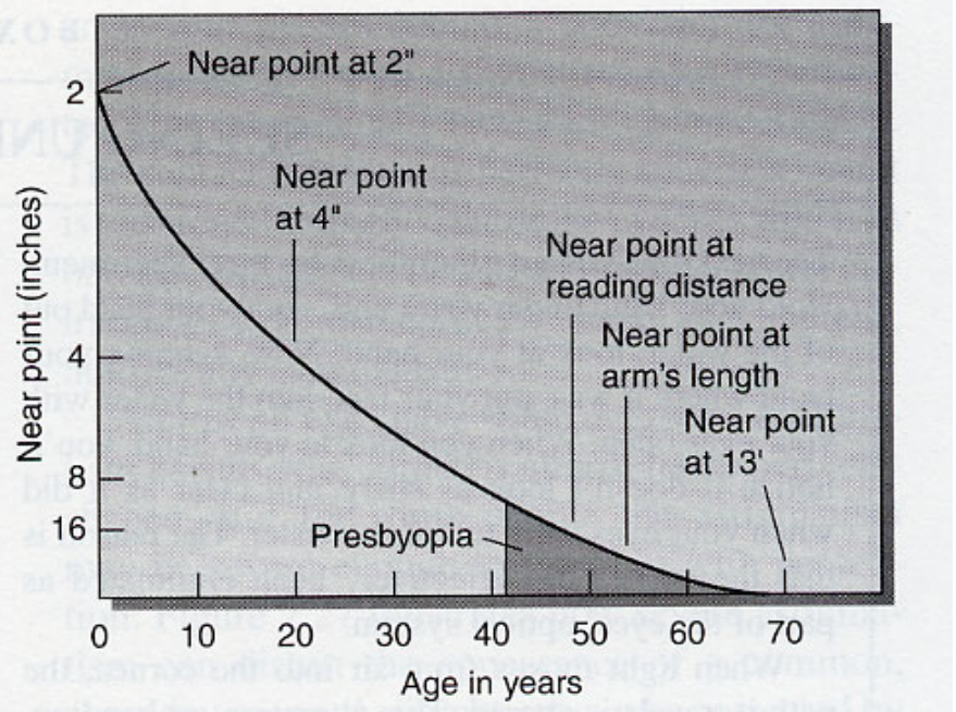
presbyopia (“old eye”)
throughout aging the distance of the near point increases due to a mix of the hardening of the lens and weakening of ciliary muscles
corrective lenses needed for close activities like reading
kinda like hyperopia that happens due to old age
astigmatism (“double vision”)
an irregularly shaped cornea bends rays of light unevenly causing a blurred image on the retina
corrected by a cylindrical lens
strabismus (“lazy eye(s)”)
two eyes are not properly aligned
corrected by a prism lens
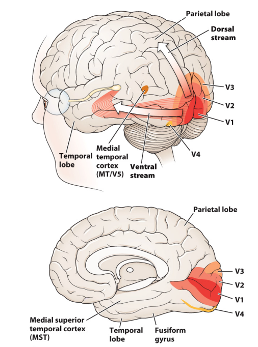
two-stream hypothesis
ventral stream: “what” pathway that focuses on object recognition
dorsal stream: “where” pathway that focuses on spatial location and action
inner and outer retina
refers to the orientation with regards to the inside of the eye; light passes through the inner retina to the outer where it first hits photoreceptors
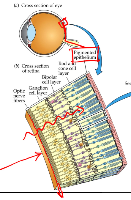
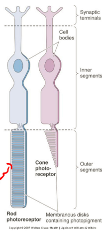
photoreceptors
cells that respond to light; inner segment keeps cells alive while outer is light sensitive
transducers: convert one form of energy into another
photous → chemical → electrical
they are presynaptic to bipolar and horizontal cells
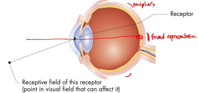
fovea
small region of the retina where the center of the field of vision is focused, has a lot of cones while the peripheral of this area has many rods
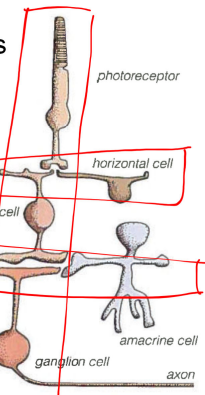
visual information flow
light flows from inner to outer where it first hits photoreceptors; then it transmitted:
direct (verticle) pathway: photoreceptors → bipolar → ganglion
lateral processing: horizontal cells and amacrine cells
it is a radial pathway in the retina that transmits signals about light to the brain
receptive field
angle of the visual field from which light evokes a response from a cell
the area/point in the visual field that a single receptor in our eye will respond to, that area is the receptor’s receptive field
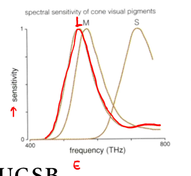
spectral sensitivity of cones
each cone has a different peak sensitivity, this is the frequency at which its sensitivity is greatest
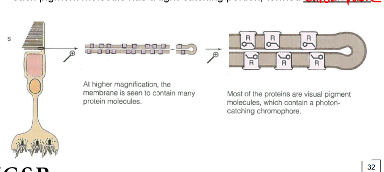
photopigment molecules
located in the disk membrane of a cone’s outer segment; they absorb photons
each pigment molecule has a light-catching portion called a chromophore
around ½ of cone outer segment consists of infoldings of cell membrane (contains protein molecule which are mostly visual pigments)
rod distribution
the retina, away from the fovea, is dominated by rods that peak at about 5mm eccentricity (18 degrees of visual angle)
when we fixate eccentrically (from the side) we can see a dim star that disappears as soon as we look directly at it
foveal pit
the cross section of the fovea that is a pit in the retina where the outer layers are pushed aside, maximizing the spatial resolution
contains all cones
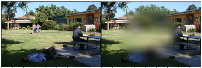
blind spot
place where the optic nerve leaves the eye
we don’t see this spot in our vision since one eye covers the blind spot of the other, it is located at the edge of the visual field, and the brain “fills in” the spot
age-related macular degeneration (AMD)
caused by defects within the RPE (leads to dry AMD) or neo-vascularization and leakage (wet AMD)
advanced versions leads to loss of central vision, but many people are not aware cause the brain guesses whats in the blank (perceptual fill-in)
effects the retina by destroying the fovea and surrounding area that creates a blind spot on the retina
most common in older individuals and is genetic/environmental
retinitis pigmentosa
a genetic disease that effects the retina by firstly destroying rods and then can attack foveal cones
severe cases result in complete blindness
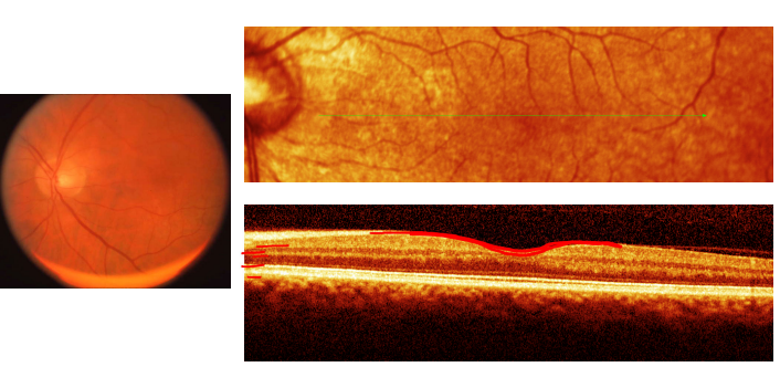
optical coherence tomography (OCT)
permits a non-invasive imaging through the depth of the retina
usually the side view photo not the one of the fundus
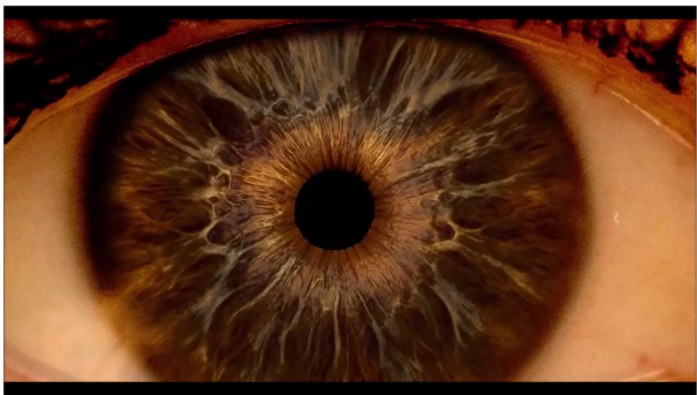
ophthalmoscope
can view the inside of the eye directly (also called a funduscope)
usually done as a part of an eye exam and pupil dilation (mydriasis) via eye drops can make it easier to inspect the structures behind the pupil
looks the things doctors use to shine light in your eye/ear through a glass plate
fundus imaging/ fundus photography
the back inner surface of the eye is called the fundus
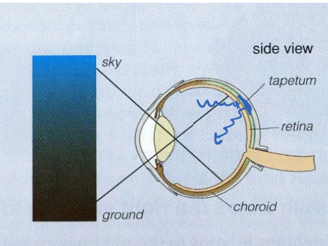
tapetum
a reflective surface behind the retina shown in some species like cats and lemurs (sometimes have reflective eyes in photos)
increases sensitivity by reflecting photons back through the retina
retinal nourishment
retinal vessels nourish the inner retina, though spare the fovea, through a dense capillary network contained in the choroid
retinal disease can cause disruptions in the RPE permitting serum to leak beneath the retina
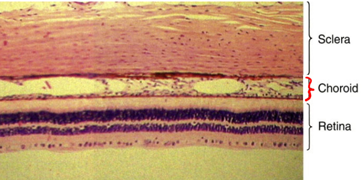
retinal pigment epithelium (RPE)
is between the choroid and the photoreceptors of the neural retina; multiple functions:
absorption of light (melanin) to prevent furthering scattering within the eye
nutrient transport from blood vessels to choroid to photoreceptors
recycling visual pigments
waste removal (old cellular components/debris)
regulation of ion and fluid balance
overall supports the health and function of the retina, and any dysfunction of this layer can lead to vision problems or even blindness
bipolar cells
classifiable by the depth of termination of their axon terminals in the IPL (there are 9 different cone bipolar cell types and 1 rod bipolar cell)
pool inputs from different photoreceptors
in the central fovea: there is 1 photoreceptor per ganglion/bipolar cell; this allows the ability to discriminate details but a con is the cones need more light to respond
in the peripheral: there are many photoreceptors per cell; allows a higher ratio of rods to cones → rods take less light to respond but cannot distinguish details
amacrine cells
there are many types found in the INL or GCL:
starburst ama. cells: have radial dendrites that look like fireworks that are presynaptic to other cells
dopaminergic: sparse dendritic fields with extensive axonal processes
ganglion cells
more than 17 types in the primate retina
magnocellular (M) cells: large and respond to motion (no color)
parvocellular (P) cells: small and responds to color (red-green)
koniocellular (K) cells: respond to color (blue-yellow)
each cell type is thought to create tiles on the the visual scene
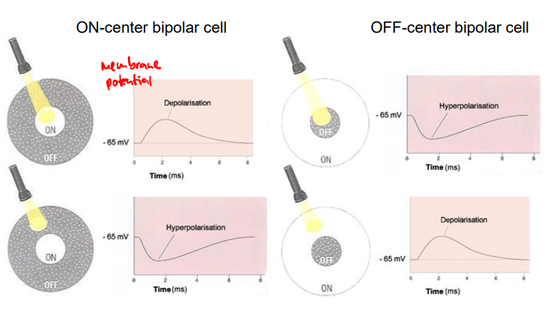
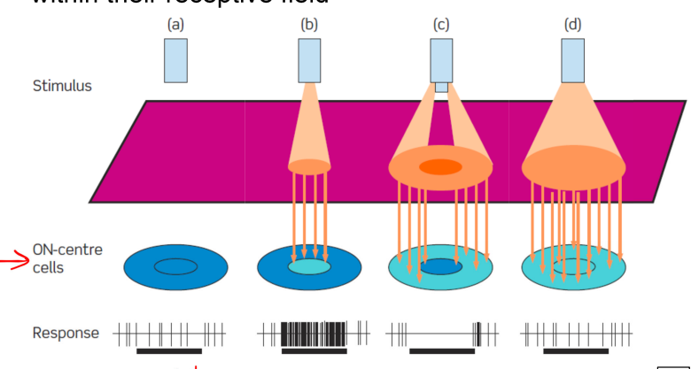
ganglion cells ON/OFF receptive fields
cells respond to differences in intensity (contrast) within their receptive field
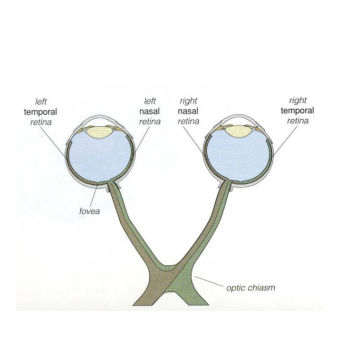
retinal ganglion cells (RGCs)
ganglion cells send their axons to the brain via the optic nerves, chiasm, and tracts
saccades
rapid, jerky eye movements that shift the eyes to a new point of fixation
smooth pursuit
slower, continuous eye movement that keeps the eyes fixed on a moving object
photons and stars
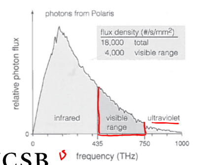
almost all photons we see comes from stars as they convert matter too energy and dissipate energy as photons
flux: total amount of light per second (18000 photons /s)
density: concentration (per square millimeter)
photons from stars have a wide range of frequencies; most are infrared (we cannot see them but they will warm us when absorbed by the skin); some are in the visible range that are photoreceptors are capable of catching
photon
a fundamental particle of physics
frequency (v): c/lambda
polarization
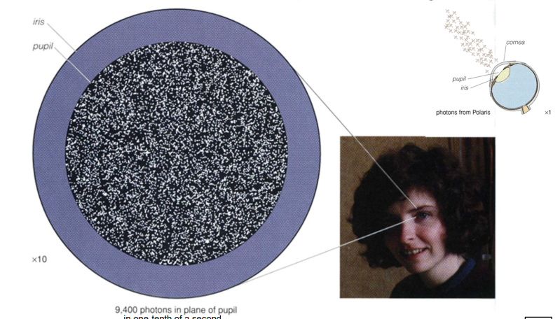
number that hits photoreceptors is limited by the atmosphere, cornea, lens, and vitreous humor
photon flux density (PFD)
the number of photons passing through a specific area per unit time
#/s/mm²
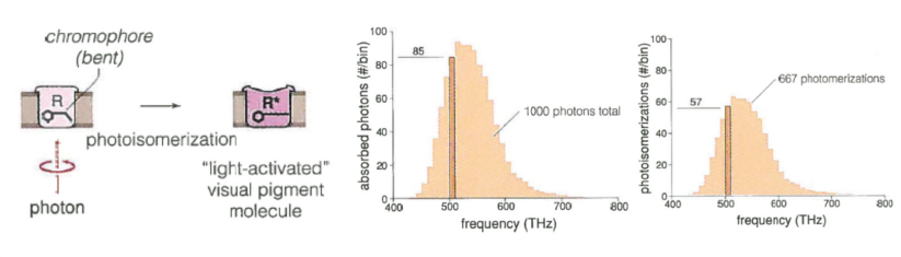
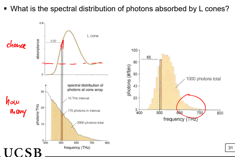
principle of univariance
each photons that photoisomerizes a visual pigment molecule has the same effect as any other photon that does so
a single photoreceptor has no means of distinguishing changes in wavelength and intensity, it can only register changes in the overall level of light (# photons absorbed) (not a specific color since it has no others to compare to)
isomers
2 molecules that differ only in shape; but a change in shape of a (portion of a) molecule is called isomerization
a change brought upon by a photon is called photoisomerization
quantum efficiency factor: only 2/3 of photons photo isomerize a visual pigment molecule
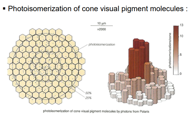
photoisomerization
a photon hits a chromophore (that is bent) causing it to undergo photoisomerization that unbends it and changes it to a “light-activated” visual pigment molecule
R → R*
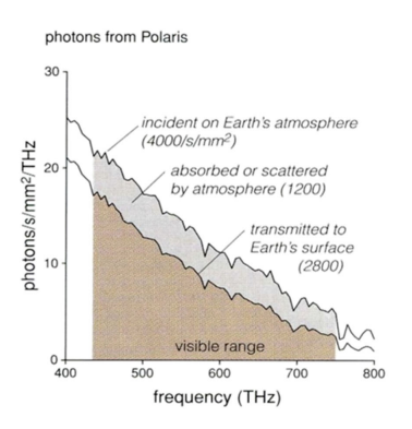
photons limited by earth atmosphere
some photons are absorbed by ozone molecules, water droplets, and other particles in the air when coming from stars into the earth atmosphere
others are scattered by the atmosphere so when they do reach earth, they reach in a different direction from which they entered at
flux density reduced by 22%
simple: absorption and scattering
optical density
the product of some constant (unique for every medium passed through) X path length
total for a series of media is simple the sum of their individual optical densities
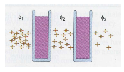
photons limited by cornea
cornea will reflect photons; photons must pass through cornea in order to be seen
photons limited by lens vitreous
contains light-absorbing pigments that reduces nearly all photons in the UV region
chromatic aberration: vitreous humor is water-like; higher-frequency photons in water are focused in front of other photons in the visible spectrum → removing high-frequency photons sharpen the image
optical distortion caused by different wavelengths focusing at different depths
also protects the retina as high-freq. photons have more energy thus more likely to damage the visual pigment molecules
vitreous light-scattering and lens absorption (+macular pigment)
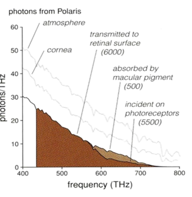
macular pigment absorption
lutein & zeaxanthin in the foveal region absorb violet to blue light (high-frequency) (limits photons)
this causes the red appearance around the macula (fundus red)
color also caused by choroid’s rich vascular layer
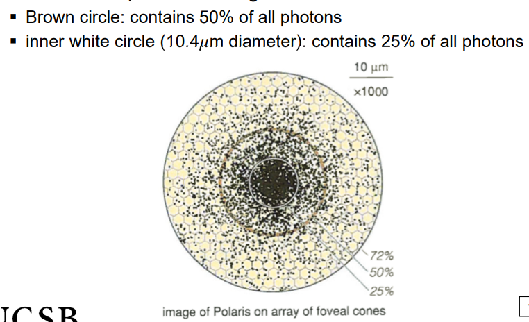
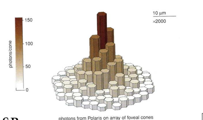
point spread function
an image of a point of light is spread over many cones (scattered over a local region of the fovea)
2% of photons lie farther than a millimeter from the foveal center (gray circle)
scattering due to close encounters with molecules
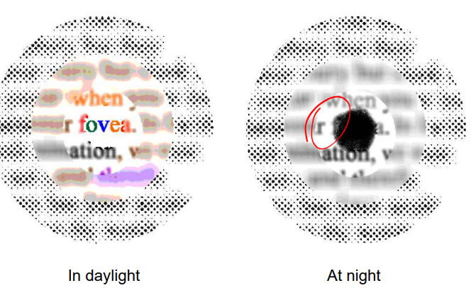
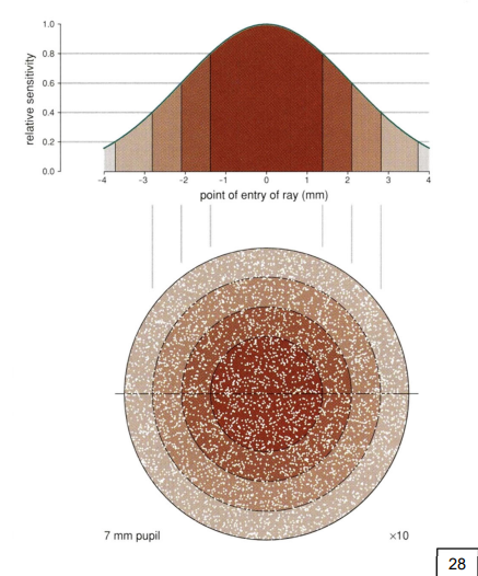
point spread function and optical elements
function’s distribution is determined by optical elements in the eye
night: pupil is fully open therefore spread is mainly due to the optical imperfection of the eye
day: the pupil is small therefore spread is mainly due to photon diffraction (intrinsic nature of how photons go from place to place)
point spread peak
the focus of photons from a point source of light has a peak approximately the size of a single cone inner segment
peak/hill is constantly moving due to eye movements
this distribution shape blurs the image projected on the retina
cone photoactivation
when photons arrive at cones some will be absorbed and others will be reflected/transmitted, but must pass through photoreceptors to reach light-catching pigments
absorption probability depends on the direction of arrival, frequency of the photons (wavelength), and the cone type
photon direction cone absorption
photons best captured when the path of the photons is parallel to the long axis of the cone (inner segment helps funnel photons into the outer segment
does NOT depend on the frequency of the photon
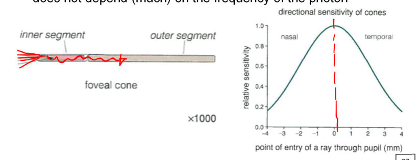
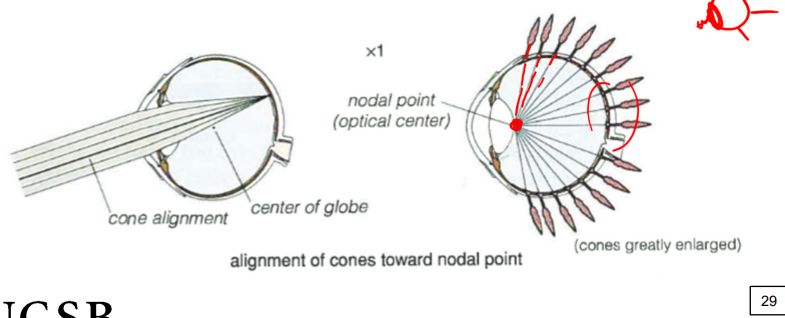
cone alignment
in the peripheral retina the long axes of the cones are not directed toward the center of the eyeball but instead with the direction of arriving rays
nodal point: optical center of this projection; imaginary point near the back surface of the lens
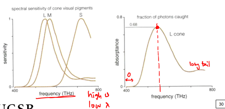
cone sensitivity
each type of cone has a different peak sensitivity, which is the frequency at which it’s sensitivity is the greatest
photopigment molecules are, themselves, spectrally sensitive
dark adaptation (threshold intensity)
in the first minutes of being in the dark your cones are not activated. first 5-10 mins your cones will be active/sensitive before your rods (cone-dominated)
overtime in the dark your rods will actually drop below the cones (your rods can see better in the dark; rod-dominated since your rods are more sensitive in the dark)
threshold intensity: the limit of what we can see (lowest amount of light we can see)
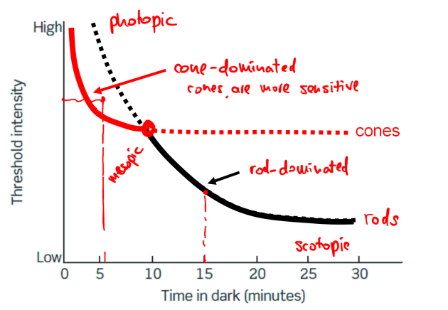
photopic
when your cones are dominating (more sensitive) your vision
happens during the day since cones are good for seeing color
scotopic
when your rods are more dominating in your visions
happens in the dark as your rods are more sensitive at night and better at detail
rods in retina positioned away from fovea (rods peak at about 5mm eccentricity, (~18 (15) degrees of visual angle)
in this area cones are larger than rods but more disperse than they are in the fovea, rods are small but numerous
mesopic
range of vision where cones and rods are equally activated in the eye (occurs in moderate-to-low ight conditions
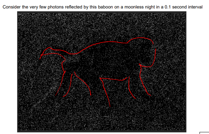
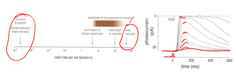
photons in day and night
in the day photoreceptors catch 300,000 photons every second, while at night photoreceptors catch 1 photon every 30 seconds (that is not enough to stimulate cones)
dim light conditions
dim lights are noisy; the random nature of arrival of individual photons becomes significant in seeing under dim conditions
since under bright conditions (100-1000 photons/s), we can ignore random photons and treat light as continuous
photons will arrive 1 by 1, creating a relatively long-lasting image, allowing us to see better at night
probability of any 2 photons being in eye at same time is very small (0.001%)
rods are more sensitive to single photons than cones
photoisomerization of a dim star: one sample, 28 photons caught (not the same every time but around this number) 0.1s shutter time
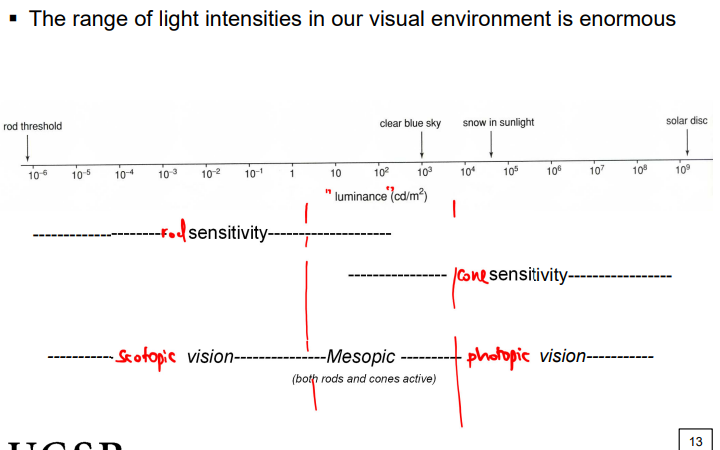
photopic and scotopic vision
our visual system splits up the workload, in brighter areas we use cones and in darker we use our rods (first approach to dealing with range is to split it up)
since we have an enormous range of light in our visual field
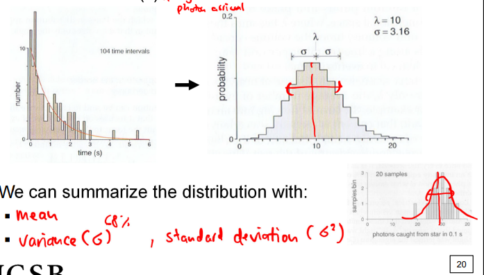
poisson process
a sequence of a random event that has a predictability to the behavior of the group
only property we can ascribe to this process is it’s average rate since times of occurrence are independent of one another (one time of arrival does not predict the next)
but the shape of distribution changes with the average number of events (lambda)
we can summarize the distribution with the mean and the variance (or standard deviation); reliable way to describe these random processes
key: random but independent events → especially in low-light conditions
mean = variance
eigengrau
spontaneous background activity in the visual system under low-light conditions
contributes to noise making detection of stimuli near threshold more difficult
can reduce effect by looking at a point for longer → easier for us to make sense of noise (like how in dark time mode on your camera makes you hold it still for a couple of seconds)
rod photoactivation
the sensitivity of rod vision is much lower than individual rods (for some reason we loose some sensitivity from an individual rod)
psychophysical sensitivity is 100x lower than that of rods (consistent with ganglion cell sensitivity)
through most of the scotopic range of our vision, increases in photon catch rate within the shutter time never affects the same rod
rods catch rate is reliable across the range and does not fluctuate much
increases in photon catch rate translate into greater bipolar cell responses due to convergence (trick in space rather than time)
rod signals get pooled to maximize visual sensitivity
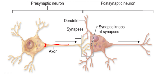
neurons
composed of:
dendrites: fibers that project out of the cell body, receiving info from other neurons
cell body (soma): : contains nucleus and other biologic essentials to keep cell alive
axon: transmits signals through the neuron
axon terminal: end of the axon; send messages to a different neuron
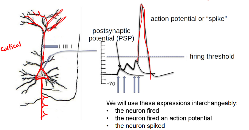
action potentials
not all neurons produce action potentials; integrates input currents and ire action potentials (aka spike)
firing threshold; EPSP’ PSP
depolarization of the cell membrane leads to the generation of a spike → going down after is called hyperpolarization leading to the refractory period
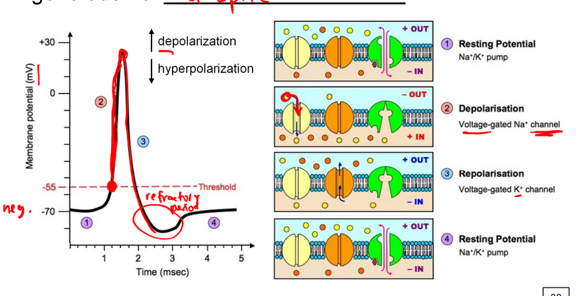
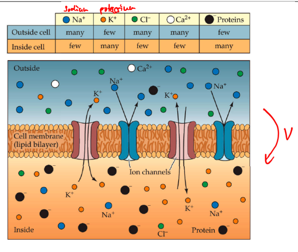
ion channels
how Na+, K+, Cl-, Ca2+, and proteins move through the neuron during a spike
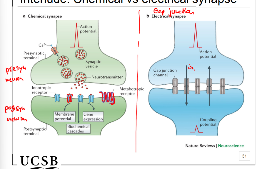
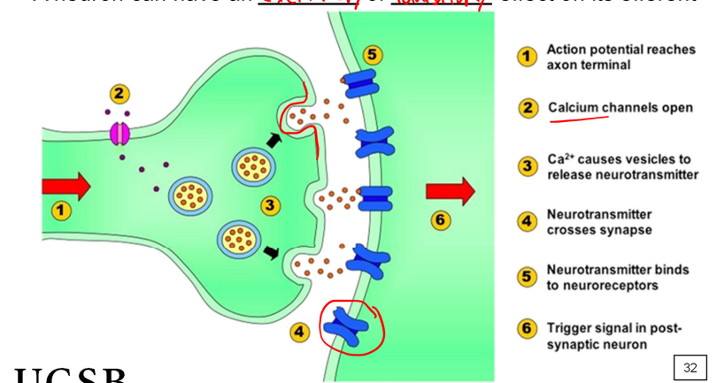
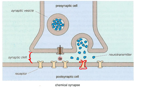
chemical vs electrical synapse signaling
a spike arrives at the synapse leading to neurotransmitter release → excitatory or inhibitory effect on efferent
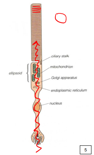
rod physiology
inner segment works as light guides
also contain metabolic and protein synthetic machinery’
the ellipsoid is the portion of the inner segment packed with mitochondria
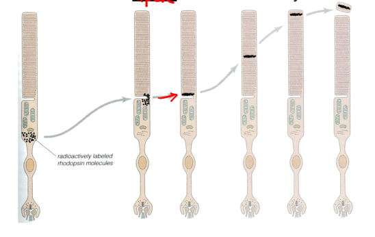
how photoreceptors work
new disc membrane is assembled at the base of the outer segment and shed at the apex on a circadian cycle
RPE phagocytoses shed disk membranes; peaking shedding occurs at dawn; entire OS disks are renewed every 10 days
photoactivation of visual pigment molecules sets in motion a cascade of events that ultimately reduces the release of glutamate from the terminal:
photoactivation → biochemical cascade → electronic spread → synaptic deactivation → decrease in glutamate release
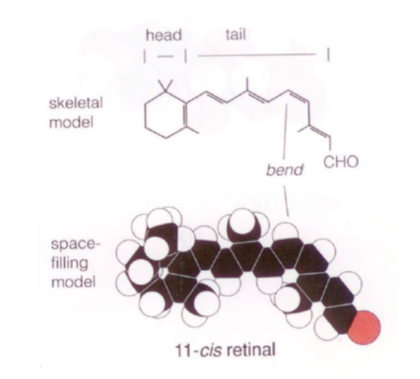
11-cis retinal
a form of retinal, which is derived from vitamin A (retinol); specifically the cis-isomer (a kink/cis double bond) at the 11th carbon in its carbon chain
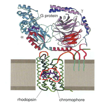
rhodopsin
visual pigment that uses 11-cis retinal as its chromophore
protein portion of the rhodopsin is called an opsin
light-absorbing portion is called chromophore
G protein lies above a rhodopsin molecule
overall is a photopigment molecule that is a 7 transmembrane-spanning G-protein receptor (tail of chromophore attaches to FFAK → a protein code at a lysine in the 7th alpha helical domain)
photoactivation
“to catch/capture a photon” which refers to:
the absorption of a photon by a visual pigment molecule
the photoisomerization of its chromophore
these result in the activation of the molecule
3 fs to absorb a photon and 200 fs to photoisomerize
photoactivation of rhodopsin
a single photon activates the rhodopsin molecule, where most of the photons energy is required to do this
it could also happen spontaneously through molecular collisions and shape modifications take 0.25-0.5 ms
R: deactivated rhodopsin molecule and R* is photoactivated rhodopsin molecule
retinitis pigmentosa (RP)
caused by numerous point mutations in the rod opsin gene
progressive photoreceptor degeneration is characteristic of mutations in the rhodopsin gene
biochemical cascade of photoisomerization
rhodopsin molecule in a disk membrane is activated → G proteins are activated → phosphodiesterase (PDE) is activated → cG-gated ion channels close → a photocurrent is activated
biochemical cascade in the dark
in the dark a steady flow of Na+ and Ca+ cations from outside the cell come in (the photoisomerization of the cascade will eventually lead to these closing)
in the dark, an outer segment contains small molecules called guanosine 3’,5’ cyclic monophosphate (cG)
and due to random collisions these molecules move rapidly around the cytoplasm of the outer segment (membranous discs act as partial barriers/ concentration of these cG molecules plays a critical role in vision)
this is also one of the 4 molecules involved in phototransduction (others are GMP, GDP, GTP)
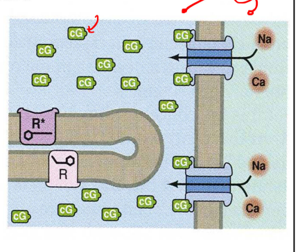
cG-gated channels
need cG to stay open and let the Na+/Ca++ in
during the cascade a photon will turn R → R* (continues to diffuse), the R* bumps into G proteins and activates them, G proteins trap cG molecules and converts them to GMP
this path overall leads to a reduced concentration of cG, and the cG-gated channels start to close
each channel composed of 4 subunits with pore at center where cations can pass
each subunit contains binding spot for cG molecules (so there are 4 potential binding spots)
the detailed structure of these channels are not known
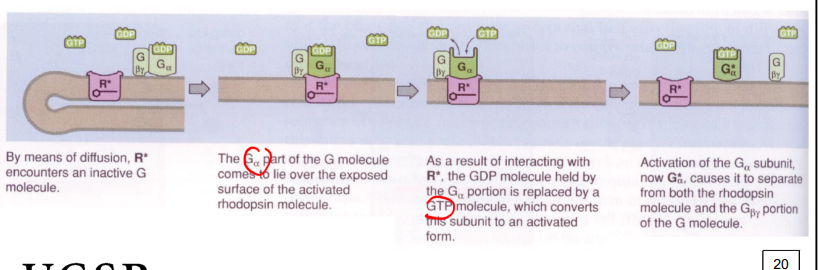
G-proteins
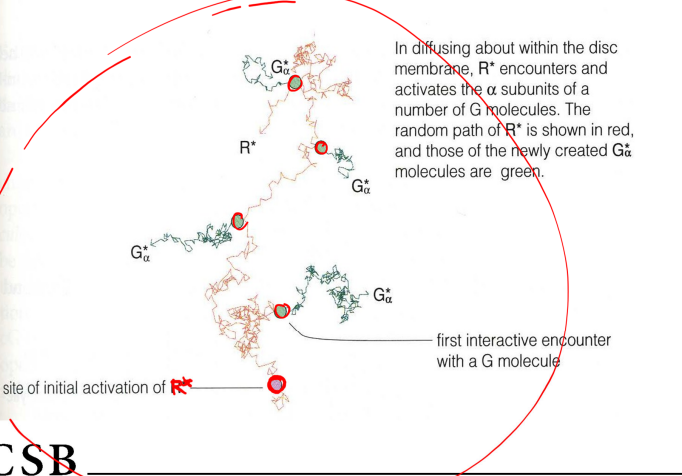
a single activated rhodopsin molecule can activate many Gα subunits
within 100 ms a single R* molecule will have created about 700 G*a subunits
activation of these subunits causes it to seperate causes it to seperate from rhodopsin and other G portion
it then goes onto bind to PDE
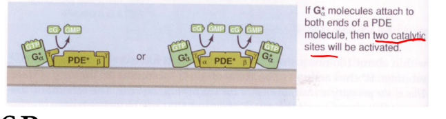
phosphodiesterase (PDE)
PDE contains 2 symmetric portions who catalytic site can act on cG, but it is usually blocked by PDEy → G*a subunit will displace the PDEy and expose the catalytic site
the exposed catalytic site means it is activated and can break a specific bond in a cG molecule, converting it into a GMP molecule
GMP molecules are relatively inert and unable to attach to cG-gated ion channels
synaptic deactivation
neurotransmitter release occurs at specialized membrane sites located just below the synaptic ribbon
glutamate is released from synaptic vesicles into the invagination of the photoreceptor terminal (regulated by calcium concentration)
photocurrent hyperpolarizes the cell => closes voltage-gated Calcium channels in the terminal => reduces glutamate release

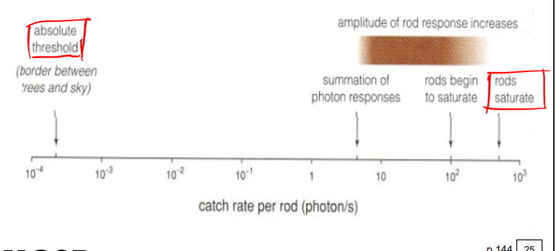
rods response to numerous photons
simple rods begin to swim up single photon events in the mesopic range, and continue to do so over 2 log unit range until their response saturated
for dim flashes the response is proportional to number of photoisomerization, but amplitude grows more slowly at greater stimulus intensifies (until about 200 photoisomerizations
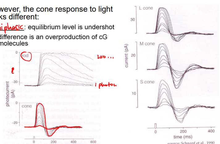
cone biochemical cascade
similar to that described of rods but they mostly occur on infoldings of the plasma membrane rather than on disks
one part looks different: it is biphasic (equilibrium level is undershot)
difference is an overproduction of cG molecules
electronic spread
changes in the transmembrane potential (generated in the outer segment) spread to the synaptic terminal
this is a physical process: membrane tries to balance out the charge inequalities; thus a change in charge displacement in one region spreads rapidly across the membrane
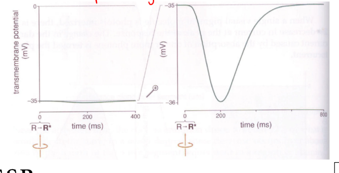
photocurrent
the change in dark current caused by the absorption of one pr more photons
when a single visual pigment molecule is photoisomerized, there is a 2% decrease in current at the peak of the response
when cG channels close, inward movement of Na+ and Ca2+ is reduced about 1pA (but outward flow of K+ continues; thus there is a net loss of positive charge in a rod
results in hyperpolarization of thee membrane by about 1mV
change in transmembrane potential called photovoltage