Ovarian Neoplasms ~ Epithelial Tumors
1/49
There's no tags or description
Looks like no tags are added yet.
Name | Mastery | Learn | Test | Matching | Spaced |
|---|
No study sessions yet.
50 Terms
Gynecologic tumors that arise from the surface epithelium and covers the ovary and the underlying stoma?
Surface epithelial - stromal tumors
70% of epithelial tumors are _________, whereas 30% are _________.
Benign, Malignant
The two most common types of epithelial tumors?
Serous
Mucinous
Benign or low-malignancy potential form =
Adenoma
Malignant form =
Adenocarcinoma
The prefix ‘cyst’ is added if the lesion is _______.
Cystic
The prefix ‘fibroma’ is added if the tumor is more than ___% ________.
50% fibrous
Mucinous Cystadenoma is the ________ form.
Benign
___ - ___% of Mucinous Cystadenoma’s are benign.
80-85%
A type of epithelial tumor that is lined by the mucinous elements of the endocervix and bowel.
Mucinous Cystadenoma
Mucinous Cystadenoma is most common with what age group?
13-45 age range
What is the sonographic appearance of Mucinous Cystadenoma?
Multiloculated cyst-like appearance
Internal compartments may differ in echogenicity
LARGE 15-30 cm
Unilateral
Mucinous Cystadenocarcinoma is the __________ form.
Malignant
Mucinous Cystadenocarcinoma is most common with what age group?
40-70 age range
Mucinous Cystadenoma makes up ___% of all malignant primary ovarian tumors.
10
Mucinous Cystadenocarcinoma are _______ and have a ________ incidence of rupture.
Large, higher
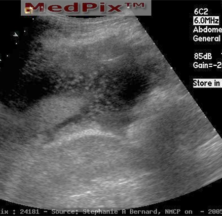
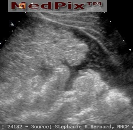
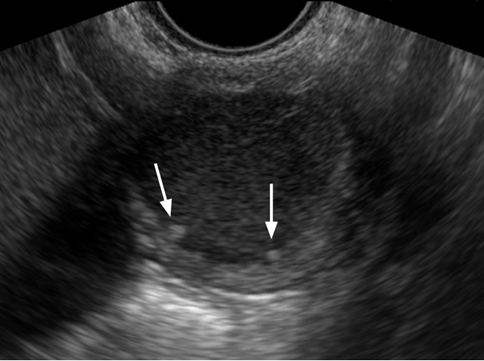
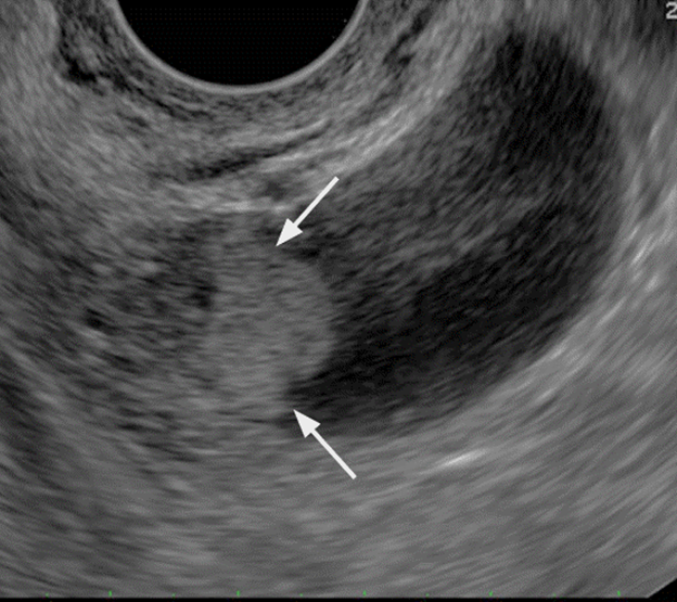
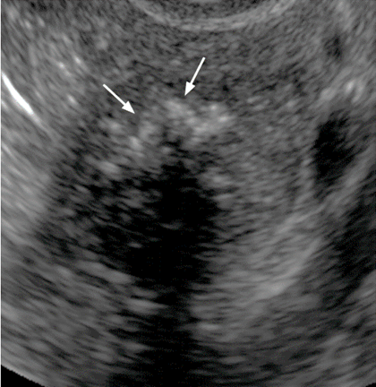
What is the sonographic appearance Mucinous Cystadenocarcinoma?
Large with thick irregular walls
May have papillary projections
Ascites – w/ bright punctate echoes
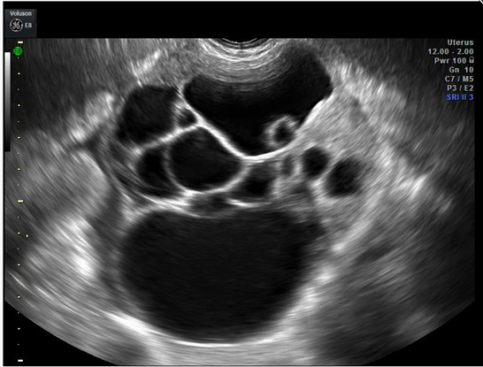
What is this US image?
Mucinous Cystadenocarcinoma
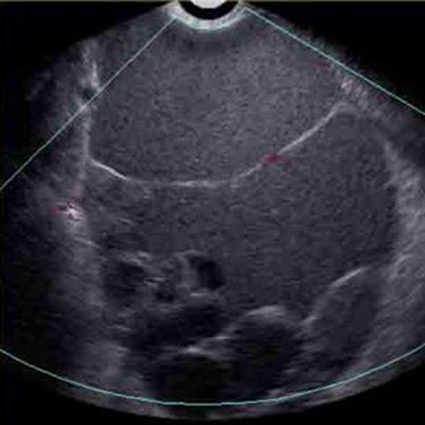
What is this US image?
Mucinous Cystadenocarcinoma
What is this US image?
Mucinous Cystadenocarcinoma
What is Pseudomyxoma Peritonei?
Spread of ruptured contents
Adhesions of abdominal contents
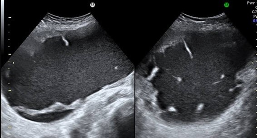
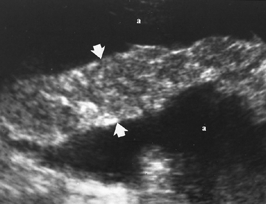
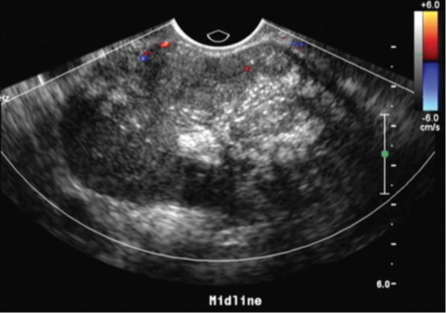
What is the sonographic appearance of Pseudomyxoma Peritonei?
Fills much of the abdomen
Mass-like effect
Loculated ascites
Multiple clusters of anechoic spaces
What is this US image?
Pseudomyxoma Peritonei
What is this US image?
Pseudomyxoma Peritonei
What is this US image?
Pseudomyxoma Peritonei
2nd most common benign ovarian tumor.
Serous Cystadenoma
Serous Cystadenoma are normally _______ & _________ than mucinous cystadenomas.
Unilateral, smaller
What is the sonographic appearance of Serous Cystadenoma?
Multiloculated
Papillary projections
Septations
Serous Cystadenocarcinoma constitutes ___-___% of all ovarian carcinomas?
60-80%
More than half of Serous Cystadenocarcinoma’s are found __________.
Bilaterally
What is the sonographic appearance of Serous Cystadenocarcinoma?
Multiloculated
Irregular borders
Internal papillary projections
Ascites
May have calcifications
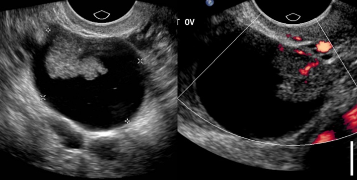
What is this US image?
Serous Cystadenocarcinoma
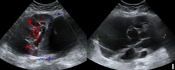
What is this US image?
Serous Cystadenocarcinoma
Endometrioid tumors are almost always ________, and 50% are found _________.
Malignant, bilateral
Endometrioid tumors are most common with what age group?
50-60 age range
What is the histologically appearance of Endometrioid tumors?
Tissue is similar to the endometrium
What is the sonographic appearance of Endometrioid tumors?
No specific distinction from other ovarian tumors
What is this US image?
Endometrioid Tumor
Endometrioid tumors are almost always ________, and 20% are found _________.
Malignant, bilateral
Clear Cell Adenocarcinoma is considered a clear _________, and a variant of ___________ CA.
Cytoplasm, Endometrioid
Clear Cell Adenocarcinoma are most common with what age group?
50-70 age range
What is the sonographic appearance of Clear Cell Adenocarcinoma?
Usually present as nonspecific complex, predominantly cystic
What is this US image?
Clear Cell Adenocarcinoma
What is another name for Brenner Tumor?
Transitional Cell
Brenner Tumor is _________ {<2%}, and the majority are ___________.
Uncommon, benign
Brenner Tumors are most common with what age group?
40-70 age range
Brenner tumors are _______, ________ fibrous stroma.
Solid, Dense
What is the sonographic appearance of Brenner Tumor?
Small & Solid
Hypoechoic
May have calcification
Cystic areas may be present with coexistent cystadenoma
What is this US image?
Brenner Tumor
What is this US image?
Brenner Tumor