Inhibition in the brain
1/35
There's no tags or description
Looks like no tags are added yet.
Name | Mastery | Learn | Test | Matching | Spaced | Call with Kai |
|---|
No analytics yet
Send a link to your students to track their progress
36 Terms
What is the most abundant inhibitory neurotransmitter in the brain?
GABA (y-Aminobutyric acid)
How is GABA synthesised?
GABA is synthesized from glutamate
Enzyme involved: Glutamic Acid Decarboxylase (GAD)
Neurones that release GABA are called..
GABAergic neurones
Types of GABAergic Neurones
1. GABAergic Interneurones
Make synaptic connections with nearby neurones = control the activity of large groups of neurones = synchronise neuronal firing
Very diverse (~20 types) with different shapes and locations
2. GABAergic Projection Neurones
Make synaptic connections with neurones located outside of the region
Example: Medium spiny neurones of the striatum
GABAergic Synaptic Transmission
What is the presynaptic element of GABAergic synapses?
The ends of axons of GABAergic neurones
GABAergic Synaptic Transmission
How is GABA released into the synaptic cleft?
Action potential arrives at GABAergic axon terminal
Terminal membrane depolarises
Voltage-gated Ca²⁺ channels open
Ca²⁺ enters the terminal
Ca²⁺ binds to specific proteins which triggers fusion of GABA-containing vesicles with the plasma membrane
GABA is released into the synaptic cleft
GABAergic Synaptic Transmission
What happens after GABA is released into the synaptic cleft?
GABA binds to GABAᴀ receptors
GABAᴀ receptors are ligand-gated ion channels. They are permeable to mainly: Cl⁻ and HCO₃⁻
Cl⁻ enters the postsynaptic neuron
Membrane becomes more negative (hyperpolarisation)
Membrane potential drops below resting potential (−70 mV). This change is called an IPSP
IPSPs close together in time and space summate, resulting in further hyperpolarisation
Leads to stronger inhibition
Where are GABAergic synapses found?
Dendrites
Soma
Axonal Initial Segment (AIS)
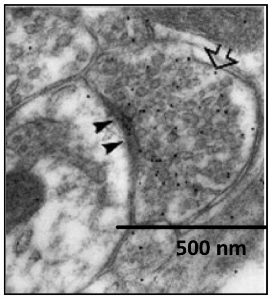
In this electron micrograph, what do the black dots represent?
Specific antibodies labelled with gold particles used to detect the presence of GABA
Why so inhibition at the Axon Initial Segment (AIS) especially powerful?
Can strongly inhibit the initiation of action potentials
Bicuculline Experiment
What is bicuculline?
A competitive antagonist of GABAA receptor
Blocks the binding of GABA to GABAA receptors
Bicuculline Experiment
What does this experiment prove?
Fast IPSPs are inhibited by Bicuculline
» Shows that fast IPSPs are mediated by GABAA receptors
However, a slow hyperpolarising response is still detected
» Caused by activation of GABAB receptors which are not blocked by bicuculline
» Produce slow IPSPs
2 types of GABA receptors on post-synaptic neurones
GABAA receptors
GABAB receptors
GABAA Receptors
Ionotropic (ligand-gated)
Fast inhibition
Blocked by bicuculline
Produce fast IPSPs
GABAB Receptors
Metabotropic (G-protein coupled):
Activate second messenger cascades
Open voltage-gated K⁺ channels
K⁺ exits the neurone → hyperpolarisation
Produce slow IPSPs
Not blocked by bicuculline
How is the Na+/K+ concentration gradient maintained across the neuronal plasma membrane?
Na+/K+ ATPase
Maintains ion gradients using ATP
Pumps: Na⁺ out, K⁺ in
Results in:
High Na⁺ outside (150 mM)
High K⁺ inside (110 mM)
How is the Cl- concentration gradient maintained across the neuronal plasma membrane?
Why is this gradient essential for GABAA-mediated inhibition?
Maintained by K⁺–Cl⁻ cotransporter (KCC2)
Pumps Cl⁻ out of the neuron
Results in:
High Cl⁻ outside (~130 mM)
Low Cl⁻ inside (~8–10 mM)
Without it, Cl⁻ would not enter the neurone when GABAA receptors are activated by GABA and IPSPs would not be generated
Prevalence of GABA receptors in the brain
Second most abundant neurotransmitter receptors in the brain after glutamate receptors
2 main classes of GABA receptors
Ionotropic GABA receptors - fast inhibition
Metabotropic GABA receptors - slow inhibition
Ionotropic GABA Receptors
What are they?
These receptors are ligand-gated ion channels.
When GABA binds, the channel opens and allows:
Cl⁻ ions to enter
HCO₃⁻ ions to leave
This makes the inside of the neurone more negative (hyperpolarised) → neurone is less likely to fire an action potential
2 types
GABAA Receptor
GABAC Receptor
Ionotropic GABA Receptors
GABAA Receptors
Where are they found?
Speed of synaptic transmission
Which disorders are these receptors involved in?
Drugs which target these receptors?
Controlled by?
Expressed in all neurones the brain
Fast synaptic inhibition
Involved in disorders such as:
Anxiety
Epilepsy
Panic disorders
Insomnia
Drugs which target GABAA
Benzodiazepines
Barbiturates
Anaesthetics
Alcohol
Also modulated by stress hormones and neurosteroids
Ionotropic GABA Receptors
GABAA Receptors
Structure
Pentamer (made up of 5 subunits)
There are 16 different genes which code for the GABAA receptor subunits
Based on similarity in amino acid sequence, these subunits are further classified into 6 different groups: α, β, γ, δ, ε, θ, π
Typical GABAA receptor composition:
2 α + 2 β + 1 γ (or δ/ε/θ/π)
Ionotropic GABA Receptors
GABAA Receptors
What does subunit composition determine?
GABA affinity
Channel properties
Drug sensitivity
Where the receptor is expressed
Subcellular localisation
Ionotropic GABA Receptors
GABAC Receptors
Where are they expressed?
Mainly expressed in the retina
Less widespread than GABAA
Ionotropic GABA Receptors
GABAC Receptors
Structure
Pentamer (made of 5 subunits)
Forms from ρ subunits (ρ1–3)
Metabotropic GABA Receptors
What are they?
G-protein-coupled receptors
Main type are GABAB receptors
Produce slow inhibitory responses (slow IPSPs).
Work via Gi/o proteins:
α subunit → inhibits adenylyl cyclase → ↓ cAMP → ↓ PKA activity
βγ subunits → open K⁺ channels → hyperpolarisation
Also inhibit Ca²⁺ channels → ↓ neurotransmitter release
Metabotropic GABA Receptors
GABAB Receptors
Specific agonist of GABAB receptors
Baclofen
Metabotropic GABA Receptors
GABAB Receptors
Structure
GABA_B receptors are dimers:
GABAB1 → GABA binds to the extracellular domains of the GABAB1 subunit
GABAB2 → G-proteins bind to the intracellular domains of the GABAB2 subunit
Metabotropic GABA Receptors
GABAB Receptors
Where are they located?
Postsynaptic membrane → slow IPSPs
Presynaptic GABA terminals → inhibit GABA release (presynaptic autoreceptors)
Presynaptic glutamate terminals → inhibit glutamate release (presynaptic heteroreceptors)
What is the main inhibitory neurotransmitter in the spinal cord and brainstem
Glycine
What are glycine receptors (GlyRs)
Ligand-gated Cl- channels
Structure of glycine receptors
Heteropentamer of α and β subunits
4 α isoforms and 1 β isoform
What happens when glycine receptors are activated?
Influx of Cl-
Postsynaptic membrane becomes hyperpolarised
Reduced firing of action potentials
Competitive antagonist for the glycine receptor
Strychnine
Causes over-excitation
Leads to pain, muscle cramps, exaggerated startle
Apart from in the brain and spinal cord, where else does glycine mediate inhibitory neurotransmission?
Via glycinergic amacrine cells in the retina