Therapy and protozoan diseases
1/6
There's no tags or description
Looks like no tags are added yet.
Name | Mastery | Learn | Test | Matching | Spaced | Call with Kai |
|---|
No study sessions yet.
7 Terms
Therapy of fish diseases
Therapeutic baths of eggs and fish
Baths
Water OFF, 15–30 min
Most effective
Best for hatcheries, low stocking density, high O₂
Targets ecto-/endoparasites, fungal & bacterial skin/gill diseases
Substances: formalin, NaCl, antibiotics, metronidazole, praziquantel, mebendazole, anesthetics
Rules: starve 24 h, treat at lowest temperature, monitor fish, test one tank first, start with low dose
Flush treatments
Water ON
For large ponds/tanks
High stocking density, low oxygen
Easy but less effective
Dips
Short exposure
Small number of large fish
Effective for large parasites
❌ Causes handling stress
Medicated feed
Pellets / granulated feed
Drugs: antibiotics, sulphonamides, anthelmintics
Best for intestinal/systemic diseases
Individual treatment
Probe (into GIT): few fish only
Injections: vaccines, precise dosing
Local treatment: focal lesions
Total Protection Strategy
Combination of immersion + injection + oral treatment
Includes Ergosan (immunomodulator feed)
Provides early, broad protection
Always performed in hatcheries
Exam memory tip
👉 Bath = off / Flush = on
👉 Dip = few big fish
👉 Feed = internal
👉 Injection = precise
Piscioodiniosis:
Pathogen
Susceptible hosts
Localization
Life cycle
Clinical signs
Pathological changes
Prevention and therapy
Diagnosis
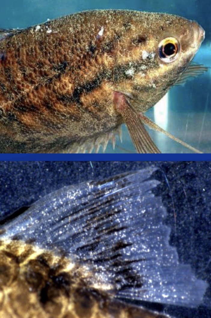
Freshwater Velvet Disease
Causative agent:
Piscinoodinium pillulare (ectoparasitic dinoflagellate protozoan)
Hosts:
Non-specific – aquarium fish; tropical & temperate species (e.g. common carp)
Localization:
Skin, fins, gills
Life cycle:
Trophont (on fish) → Tomont (encysted, off fish) → Dinospore (infective)
⏱ ~2 weeks, trophonts contain chloroplasts
Clinical signs:
Golden/yellow velvet sheen, excess mucus, dark skin, gill hyperplasia
Pathogenesis:
Most severe in young fish, rapid spread, possible mass mortality
Diagnosis:
Clinical signs ± skin/gill scraping
Prevention / Therapy:
↑ Temp 24–27 °C, remove fish for 2 weeks
NaCl, formalin, methylene blue
Ichthyophthiriosis
Pathogen
Susceptible hosts
Localization
Life cycle
Clinical signs
Pathological changes
Prevention and therapy
Diagnosis
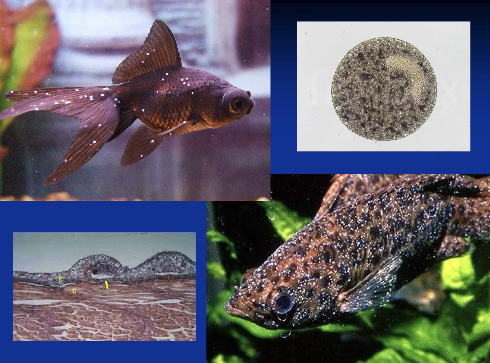
Freshwater White Spot Disease (Ich)
Causative agent:
Ichthyophthirius multifiliis (ciliate protozoan)
Hosts:
Virtually all freshwater fish
High risk: scaleless fish (e.g. catfish)
Outbreaks linked to stress
Localization:
Skin and gill epithelium
Life cycle:
Trophont (feeding stage inside nodules on skin/gills) → leaves host → Tomont (encysted, asexual division) → Tomites → motile Theronts (infective stage invading new host)
Clinical signs:
White/grey nodules ≤1 mm, fin & gill damage, respiratory distress, suffocation, rubbing the body against objects
Pathogenesis:
Heavy infections → up to 100% mortality
Diagnosis:
Typical white spots (± microscopy)
Prevention / Therapy:
Treat theronts
↑ Temp >30 °C for ~7 days, formalin, toltrazuril, UV
Daily vacuuming; cysts killed by drying
Chilodonellosis; Trichodinosis
Pathogen
Susceptible hosts
Localization
Life cycle
Clinical signs
Pathological changes
Prevention and therapy
Diagnosis
Chilodonellosis
Causative agents:
Chilodonella piscicola
Chilodonella hexasticha (less common; mainly older fish, higher temperatures)
Hosts:
All freshwater fish (especially fingerlings); also brackish water fish
Localization:
Skin and gills
Life cycle:
Trophont (feeding on epithelium) → Tomont (asexual division in environment) → Theront (infective)
Clinical signs:
Excess mucus, whitish/blueish sheen, gill necrosis, dyspnea, skin ulcers
Pathogenesis:
Direct epithelial feeding → tissue damage
Heavy infections → secondary bacterial disease, high mortality
Diagnosis:
Microscopy of skin and gill
Prevention / Therapy:
Formalin bath and immersion, acetic acid bath, salt baths, potassium permanganate immersion, copper immersion
Trichodinosis
Causative agents:
Trichodina jadranica (+++)
Trichodinella, Tripartiella, Dipartiella
Hosts:
Low host specificity; freshwater and marine fish
Localization:
Skin and gills
Some species: urinary bladder, oviducts, GIT, kidneys
Life cycle:
Trophont → Tomont → Theront
Clinical signs:
Grey/blue coating, frayed fins, epithelial erosion, suffocation
Pathogenesis:
Usually mild–chronic disease
↑ losses in young fish, secondary bacterial infections
Diagnosis:
Microscopy of skin and gill scrapings
Prevention / Therapy:
Formalin bath. Acetic acid, salt baths (FW only)
Freshwater baths (marine fish)
Copper, potassium permanganate
extra comparing:
Both are ciliate ectoparasites of skin and gills with similar life cycles and treatment, but Chilodonella feeds directly on epithelium and is more pathogenic, while Trichodina mainly causes mechanical irritation and is usually chronic
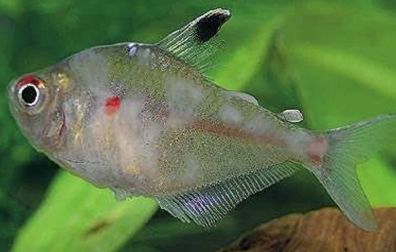
Ichthyobodosis
Pathogen
Susceptible hosts
Localization
Life cycle
Clinical signs
Pathological changes
Prevention and therapy
Diagnosis
Causative agent:
Ichthyobodo necator complex(flagellate protozoan)
Hosts:
Freshwater and saltwater fish
Mostly young or stressed adults
Localization:
Skin and gills
Life cycle:
Motile stage – free-living, off the fish
Pyriform stage – feeding stage attached to epithelium
Binary fission for reproduction
Clinical signs:
Grey/blueish or whitish film on body
Epithelial hyperplasia, increased mucus
Respiratory distress in severe cases
Pathogenesis:
Feeds and penetrates the epithelium → disrupts osmoregulation
Dangerous for young or stressed fish
Diagnosis:
Microscopic examination of skin and gills
Prevention / Therapy:
Formalin baths, potassium permanganate
Raise temperature to shorten parasite cycle
Oral metronidazole in severe cases
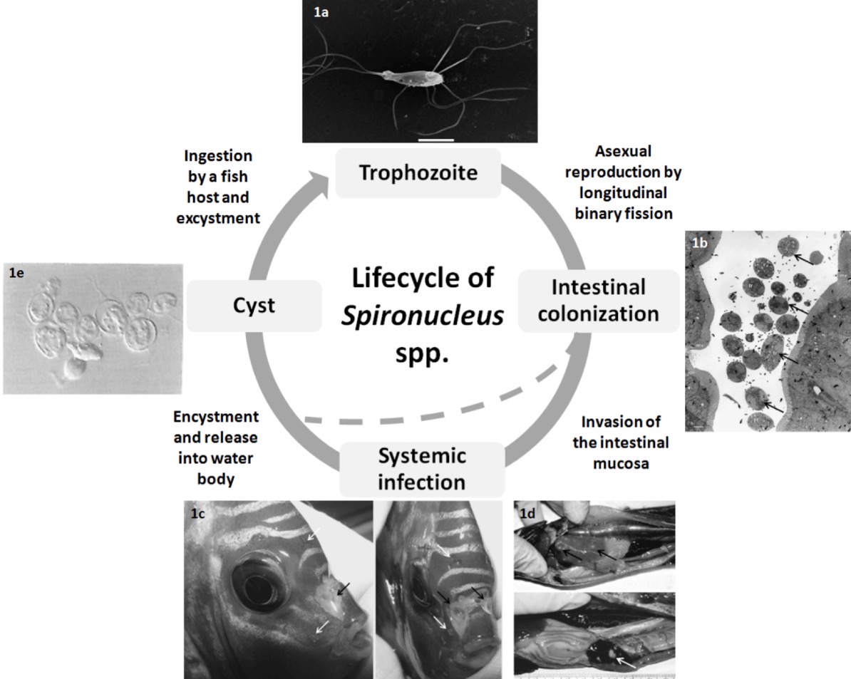
Spironucleosis
Pathogen
Susceptible hosts
Localization
Life cycle
Clinical signs
Pathological changes
Prevention and therapy
Diagnosis
Causative agents:
Spironucleus salmonicida, S. salmonis, S. barkhanus, S. torosa, S. vortens (flagellated protozoans)
Hosts:
Salmonids (freshwater, seawater, cage-cultured)
Aquarium fish
Localization:
Intestine (primary)
Gall bladder, stomach
Some species: internal organs, muscle, skin
Life cycle:
Trophont – reproduces by binary fission in intestinal mucosa
Encysted stage – released with feces
Ingestion by fish – cysts excyst in stomach → trophonts colonize intestine
Clinical signs:
Inappetence, dark color, emaciation (“pinheads”)
Ascites, pale stringy feces, enteritis
Exophthalmos
Chronic mortality
Pathogenesis (species-specific):
S. salmonis: intestines → abdominal distension, emaciation
S. barkhanus: internal organs, muscle, skin
S. vortens: cachexia, gastroenteritis, peritonitis, “hole head vs lateral line” syndrome
Diagnosis:
Fecal exam (trophonts or cysts)
Necropsy more accurate → assess intensity (treatment if 15–30 organisms/fish)
Prevention / Therapy:
Metronidazole (oral or immersion)
Magnesium sulfate (oral)
Raise water temperature to 35 °C for 7 days
Gill Amoebic Infestation; Nodular Gill Amoebic Infestation
Pathogen
Susceptible hosts
Localization
Life cycle
Clinical signs
Pathological changes
Prevention and therapy
Diagnosis
Amoebic Gill Disease (AGD)
Causative agent:
Neoparamoeba perurans
Hosts:
Marine fish, especially Atlantic salmon (other salmonids)
Localization:
Gills → may become systemic in chronic cases
Life cycle (important exam clarification)
Exists mainly as a trophozoite (trophont)
Divides by binary fission
Can survive freely in seawater
No well-defined cyst → theront cycle like Ich or Velvet
👉 Examiners usually accept:
“Direct life cycle with free-living and parasitic trophozoites”
Clinical signs:
Lethargy, ↓ swimming, ↓ feeding & growth
Respiratory distress
Progressive epithelial hyperplasia and ↑ mucus on gills
Pathogenesis:
Chronic but rapidly progressive
Severe gill hyperplasia → impaired respiration, osmoregulation & circulation
High mortality (up to 85%)
Diagnosis:
Macroscopic gill lesions
Microscopic examination of gill scrapings
Prevention / Therapy:
Freshwater baths, hydrogen peroxide baths
Management: functional feeds, selective breeding
No effective commercial vaccine
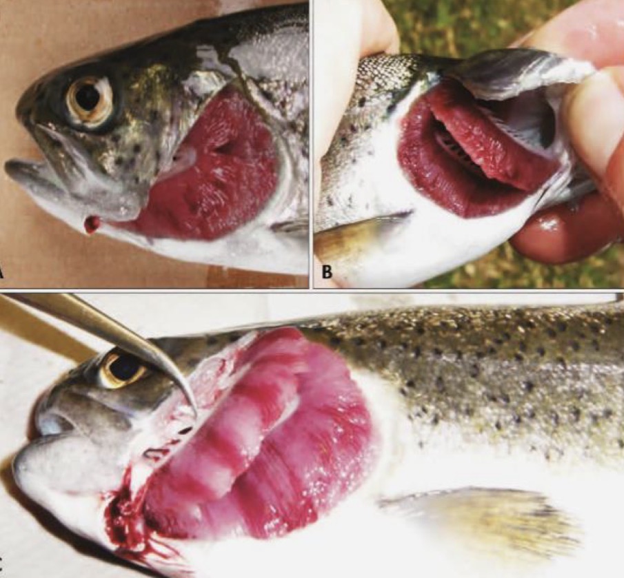
Nodular Gill Amoebic Infestation (NGD)
Causative agents:
Various amoebae (not Neoparamoeba perurans)
Hosts:
Freshwater fish, especially rainbow trout
Localization:
Gills
Life cycle
Direct life cycle
Trophozoites divide by binary fission
Free-living and parasitic forms
No obligate cyst stage required for transmission
Clinical signs:
Reduced activity, abnormal swimming
Respiratory distress
Reduced growth
White nodular lesions on gill filaments
Increased mucus
Pathogenesis:
Chronic course
Epithelial hyperplasia → partial or complete lamellar fusion
Mortality occurs weeks after onset
Diagnosis:
Wet mount examination of gills
Prevention / Therapy:
Formalin treatments
Salt baths
extra for comparing: Amoebic gill disease is caused by Neoparamoeba perurans in marine salmonids and is highly lethal, whereas nodular gill disease affects freshwater fish, is caused by other amoebae, and is usually more chronic.