Biology 151 Exam 3
1/72
There's no tags or description
Looks like no tags are added yet.
Name | Mastery | Learn | Test | Matching | Spaced | Call with Kai |
|---|
No analytics yet
Send a link to your students to track their progress
73 Terms
Meiosis
Cell division that produces reproductive cells (gametes)
Sperm & egg
Daughter cells are genetically different from parent cells
Genetic material is halved
Gamete
A sex cell (sperm or egg)
Mitosis
Cell division that produces body cells (somatic cells)
Cytokinesis
The division of the cytoplasm into two daughter cells
Occurs in mitosis and meiosis
Occurs during Telokinesis
Cleavage furrow in animal cells
Cell plate in plant cells
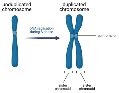
Chromosome
A single long double helix of DNA wrapped around histone proteins
Gene
A particular region of DNA in a chromosome
Codes for specific RNA / proteins
Sister chromatids
Identical copies of a duplicated chromosome, attached by a centromere
Seperated during Anaphase
Each half goes to a daughter cell

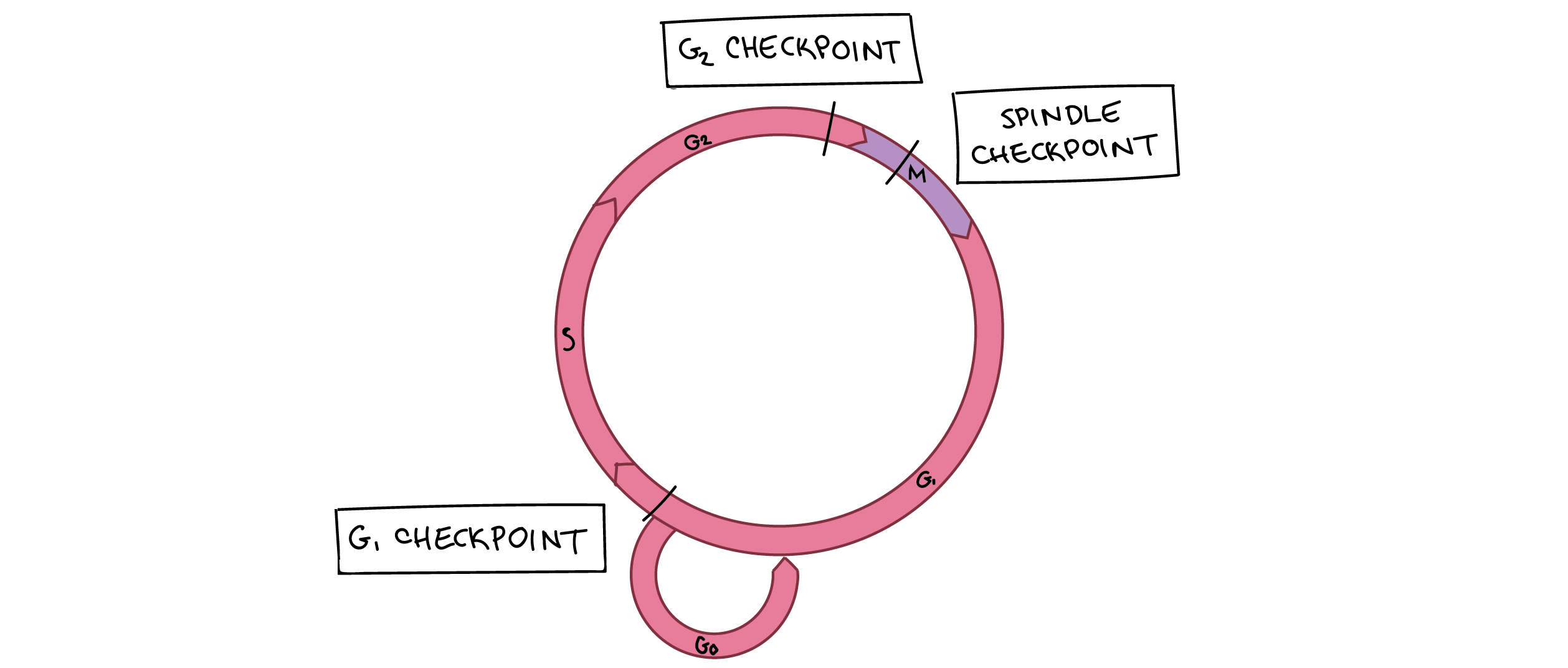
Cell cycle phases
The cell alternates between Interphase and M Phase
G0 phase - Not dividing
Interphase - Preparation to divide
G1 phase - Cell grows and duplicates organelles
S phase - Cell duplicates chromosomes and centrosome
G2 phase - Cell grows more and reorganizes its organelles
M phase - Division
Mitosis - Replicated chromosomes are separated by centrosomes
Forms two daughter nuclei
Cytokinesis - The cytoplasm divides
Forms two daughter cells
Interphase
The cell prepares to divide
G1 phase - Cell grows and duplicates organelles
S phase - Cell duplicates chromosomes and centrosome
G2 phase - Cell grows more and reorganizes its organelles
Centrosome
A microtubule structure that helps separate sister chromatids in anaphase
Mitotic (M) Phase
The cell is dividing
Mitosis - Replicated chromosomes are separated by centrosomes
Forms two daughter nuclei
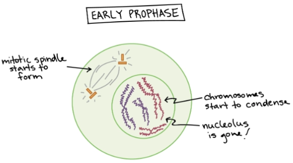
Prophase - Chromosomes condense, miotic spindle forms, nucleolus breaks down
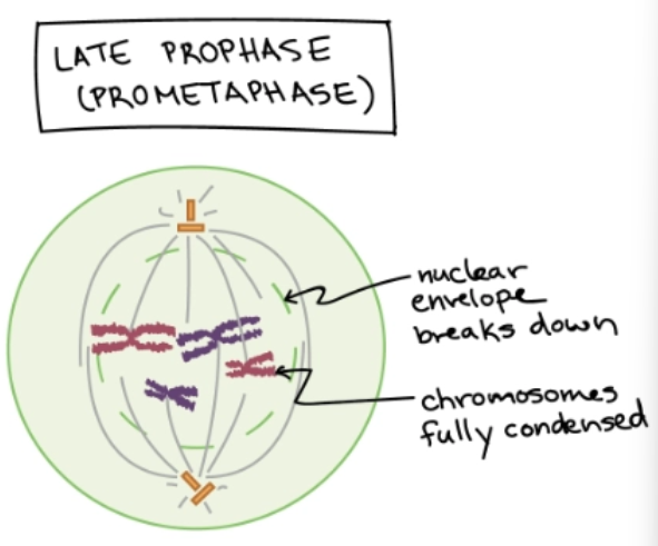
Prometaphase - Miotic spindle captures & organizes chromosomes
Metaphase - Miotic spindle lines chromosomes in the middle of the cytoplasm
Anaphase - Sister chromatids are seperated and pulled to opposite poles
Telophase - The cell restablishes its structures
Cytokinesis - The cytoplasm divides
Forms two daughter cells
Occurs during Telophase
Prophase
Mitosis - Step 1
Chromosomes condense
Miotic spindle forms
Nucleolus breaks down
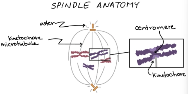
Miotic spindle
A microtubule structure that organizes and moves chromosomes during mitosis
Grows between centrosomes
Formed during prophase
Nucleolus
The region of the nucleus where ribosomes are made
Broken down during Prophase
Prometaphase
Mitosis - Step 2
Chromosomes condense more
Nuclear envelope breaks down, chromosomes are released
Miotic spindle grows and captures chromosomes
Kinetochore
A patch of proteins at the centrosome of each sister chromatid where miotic spindle grabs
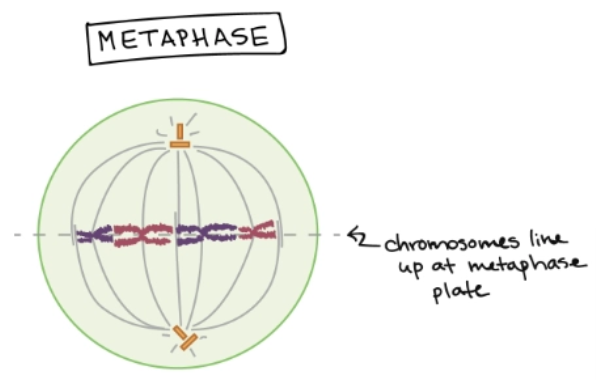
Metaphase
Mitosis - Step 3
Miotic spindle line chromosomes up at the metaphase plate (middle of cell)
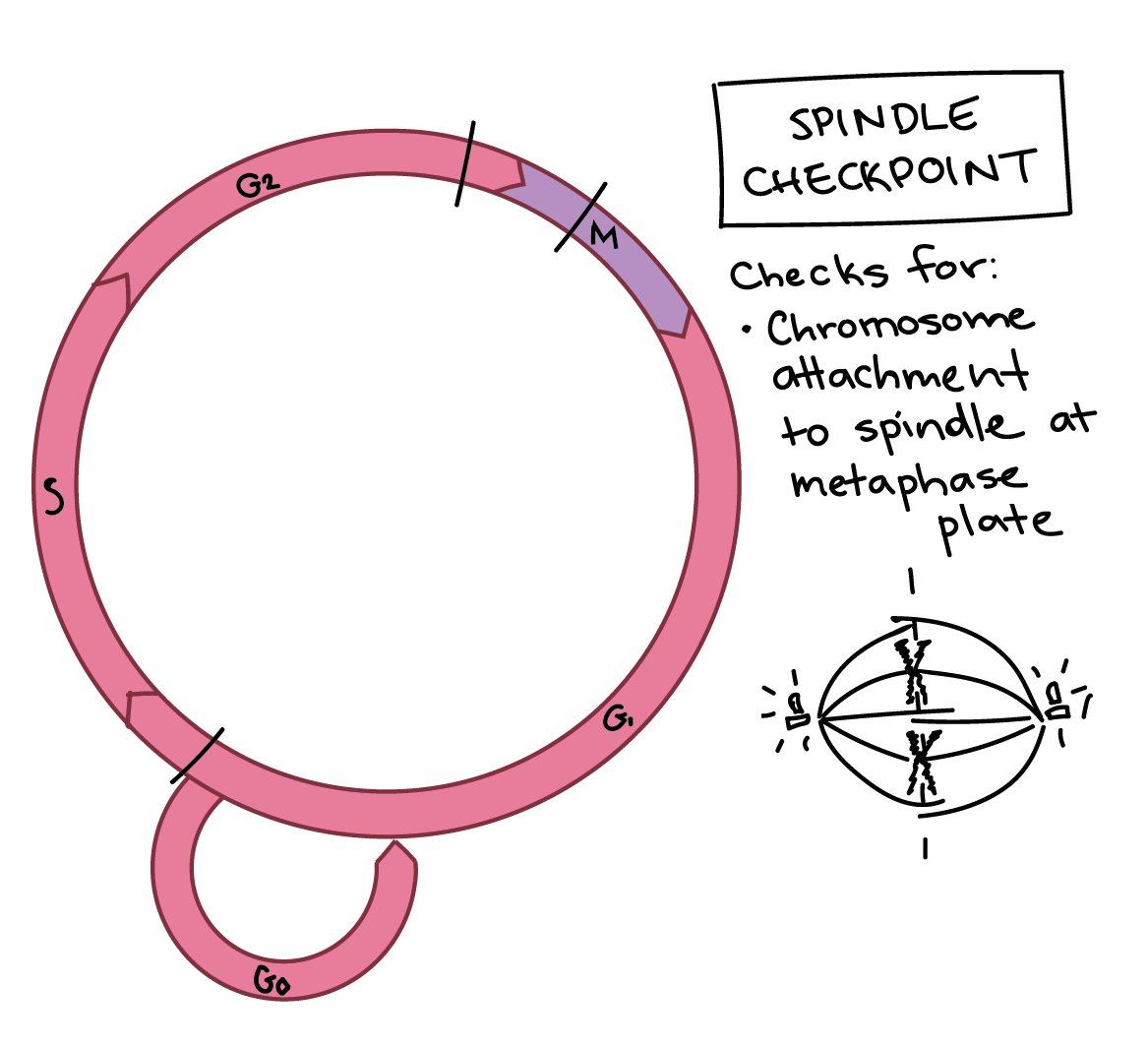
Spindle Checkpoint - Ensures that everything is attached correctly. The cell will not move on the the next phase if not
Spindle checkpoint
Occurs during metaphase
The cell checks that:
Chromosomes are correctly lined up
Each chromosome has two correctly attached kinetochores
All kinetochores are correcly attached to the miotic spindle
Division will halt if the checkpoint is failed
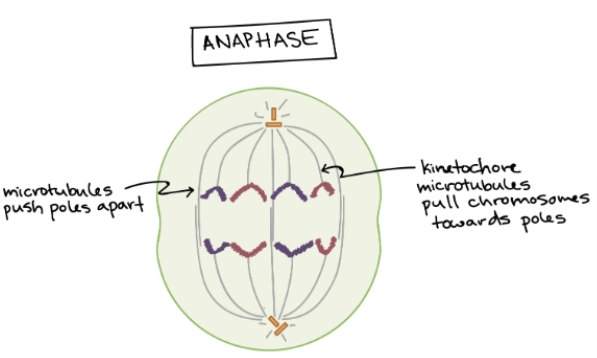
Anaphase
Mitosis - Step 4
Sister chromatids separate into individual chromosomes
Each new chromosome is pulled to opposite ends of the cell
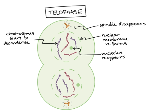
Telophase
Mitosis - Step 5
Mitotic spindle breaks down
Two new nuclei form
Chromosomes decondense
Simultaneously, cytokinesis occurs
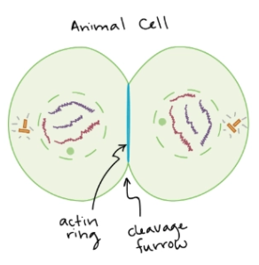
Cleavage furrow
Cytokinesis in animal cells
Actin filaments pinch across the cytoplasm to split the cell into two
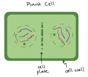
Cell plate
Cytokinesis in plant cells
A new cell wall forms down the middle of the cell, splitting it into two
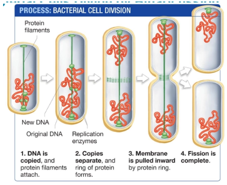
Binary fission
Cell division and reproduction in bacteria
DNA is copied and protein filaments attach to either pole of the cell
DNA copies separate, a protein ring forms in the middle of the cell
The cell membrane is cleaved by the protein ring
Cyclin-CDK complex
Protein complexes that push the cell towards the next phase when activated
Often requires an activating kinase to phosphorylate the complex
Cyclin-dependent kinase (CDK)
A kinase that binds to cyclin to produce a protein complex that pushes the cell towards the next phase
Cyclin
A protein that binds to CDK to produce a protein complex that pushes the cell towards the next phase
Concentrations cycle through the cell cycle
Specific to the phase transition
Kinase
Proteins that add phosphate groups to molecules (phosphorylate)
M-Phase promoting factor (MPF)
A cyclin-CDK complex that pushes the cell into M-phase
Inhibited by phosphorylation
Cell cycle checkpoints
Regulatory points throughout the cell cycle in which the cell decides whether to proceed with division
Cancer is caused by checkpoint failures
Tumor
A growth caused by cells that divide without regulatory control at checkpoints
M-phase checkpoint
Checkpoint conditions
Sister chromatids have attached to the mitotic spindle
Chromosomes have properly seperated
M-phase promoting factor is absent
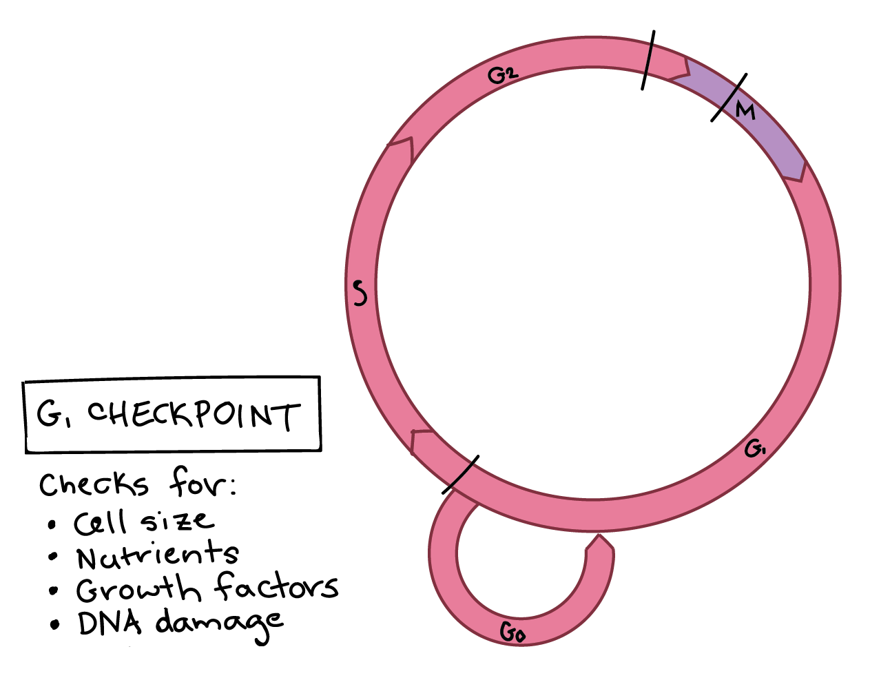
G1 checkpoint
Checkpoint conditions
Cell size is adequate
Nutrients are sufficient
Social signals are present
DNA is not damaged
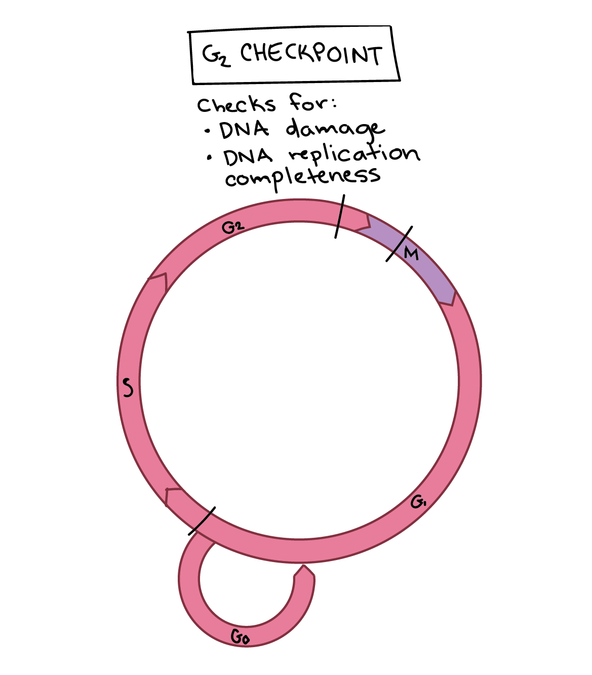
G2 checkpoint
Checkpoint conditions
Chromosomes have successfully replicated
DNA is not damaged
M-phase promoting factor (MPF) is present and active
p53
A regulatory protein that stops the cell cycle if DNA is damaged
The protein binds to the DNA, which produces a cyclin-CDK inhibitor
Oncogenes
Overexpressed genes that promote constant cell division
Benign tumor
A noncancerous and noninvasive tumor
Malignant tumor
A cancerous and invasive tumor
Spreads through the body and creates more tumors
E2F
A protein that triggers the expression of genes required for S phase
Deactivated by Rb protein
Rb
A tumor suppressor that binds to E2F to deactivate it and inhibit the cell from moving into S phase
Causes of cancer
Permanently activated CDK
The cell is constantly dividing
Defective Rb
Cannot bind to E2F, so S phase is falsely started
Mutated p53
Cannot halt division if DNA is damaged
Membrane proteins
Proteins that regulate transport across the plasma membrane
Attach to interior cytoskeletal structures and exterior matrix structures
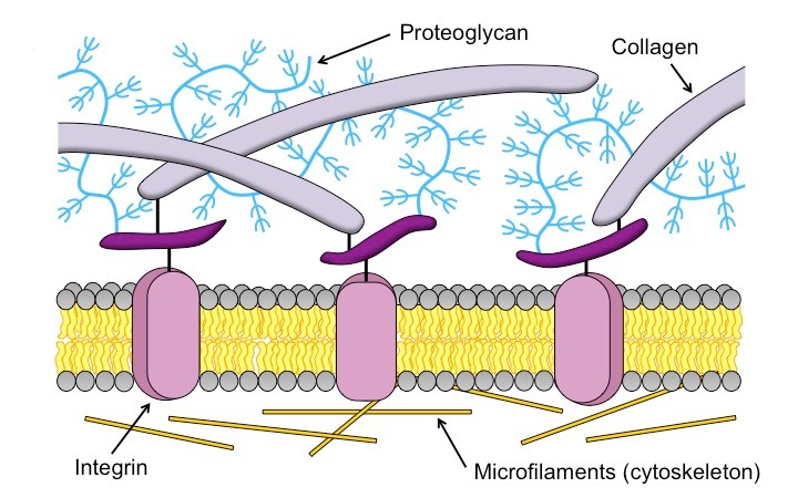
The extracellular matrix (ECM)
A protective layer or wall that forms just beyond the plasma membrane
Defines cell shape
Attaches cells to other cells (junctions)
First defense system
Components
Collagen fibrils
Proteoglycans
Integrin proteins
Cytoskeleton microfilaments
Plant extracellular matrix
Primary cell wall - Located to the very exterior of the cell
Secondary cell wall - Located between the primary cell wall and the plasma membrane
Contains waxes in leafs
Contains lignin in wood
Pectin
A gelatinous sugar embedded between cellulose microfibrils in the primary cell wall that store moisture
Collagen
The most abundant structural protein in the animal extracellular matrix
Form fibrils that provide support and flexibility
Proteoglycans
The gelatinous ground substance of the extracellular matrix
Composed of many polysaccharides attached to a core protein
Give cartilage its rubbery consistency
Stores water
Tissue
Similar cells that function as a unit
More ECM than actual cells
Integrins
Membrane proteins that bind to cross-linking proteins in the extracellular matrix
Laminins - Anchors the ECM to the plasma membrane and cytoskeleton
Laminins
A type of integrin (transmembrane protein) that anchors the ECM to the plasma membrane and cytoskeleton
Cell-cell attachments
Materials and structures that allow cells to connect, communicate, and transfer materials
Middle lamella - Glues plant cells together
Tight junctions - Stitch animal cells together via proteins
Desosomes - Anchor cells together via cytoskeleton intermediate filaments
Gap junctions - Attach animal cells with gaps for small molecule transfer
Plasmodesmata - Attach plant cells with gaps for small molecules transfer
Epithelia
The tissues that line organs
Require very strong cell-cell attachments
Middle lamella
An indirect cell-cell attachment that glues plant cells together
Made of gelatinous pectins
Continuous with the primary cell wall
Tight junction
A cell-cell attachment ta stitches animal cells together
Watertight seal
Chains of proteins line up and bind to each other
Usually found in epithelia (Organ-lining cells)
Can loosen to permit transport or in response to environmental changes
Desosomes
Strong cell-cell attachments common in animal epithelial and muscle cells
Intermediate filaments inside of the cell link to cadherin proteins
Cadherins extend across the ECM and bind to the cadherins of adjacent cells
Cadherins only bind to cadherins of the same type
Gap junctions
Cell-cell attachments in animal cells that allow ions and small molecules to flow between cells
Communication portals - Channels that help adjacent cells coordinate activities
Plasmodesmata
Cell-cell attachments in plant cells that allow ions and small molecules to flow between cells
Symplast - A continuous network of cytoplasm
Apoplast - A continuous network of cells walls and ECMs
Filters water and nutrients before they enter the cell
Signaling molecules
Molecules that deliver messages between cells by binding to receptor proteins
Lipid-insoluble - Hydrophilic, do not cross the plasma membrane
Processed by signal transduction
Lipid-soluble - Hydrophobic, diffuse across the plasma membrane
Processed directly in the cells cytoplasm
Receptor proteins
Membrane proteins that change shape and activity after binding to a signaling molecule
Hormones
Information-carrying signaling molecules
Often trigger changes in gene expression
Secreted from cells
Circulate the body
Act on target cells far away
Signal transduction
The conversion of an extracellular hormone into an intracellular signal
Required for hydrophilic, lipid-insoluble signaling molecules
Steps
Signal reception - The signaling molecule binds to a receptor protein outside of the plasma membrane
Signal transduction - The extracellular signal is converted into an intracellular signal
The signal may be amplified
Signal response - The signal may lead to changes in protein activity or gene expression
Direct signal processing
A signal diffuses across the plasma membrane and is processed by receptors in the cytoplasm
Used for hydrophobic, lipid-soluble signaling molecules
Steps
Signal arrival - A carrier protein transports the hormone to the cell surface
Signal entry - The hormone diffuses across the plasma membrane into the cytosol
Signal reception - The hormone binds to a receptor protein, inducing conformational change
Direct signal response - The hormone receptor complex binds to the DNA, inducing an change in gene expression
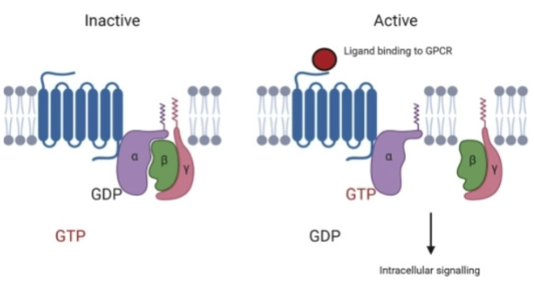
G-protein-coupled receptors (GPCRs)
Receptor proteins that initiate the production of second messengers inside the cell
Required for hydrophilic, lipid-insoluble signals that cannot cross the plasma membrane
Amplify and diversify the signal
Active when bound to GTP
Inactive when bound to GDP
Second messengers
Amplified signals triggered by an activated G-protein-coupled receptor (GPCR)
Diffuse rapidly throughout the cell
Produced quickly in large quantities
May activate protein kinases, which can activate / inactivate other proteins
Protein kinases
Proteins that activate or deactivate other proteins by phosphorylating them (adding phosphate groups)
Enzyme-linked receptors
Transmembrane signal receptors that phosphorylate proteins inside of the target cell
Directly catalyze intracellular reactions
Receptor tyrosine kinases (RTKs) - The best known group of this receptor
Receptor tyrosine kinases (RTKs)
Enzyme-linked receptors that autophosphorylate (phosphorylate themselves
Signal transduction steps
A hormone binds to 2 RTK subunits, they form a dimer
RTK uses ATP to autophosphorylate
Proteins build a bridge between RTK and Ras protein
Activates Ras
Activated Ras phosphorylates protein kinase
Triggers a phosphorylation cascade of protein kinases
Mitogen-activated protein kinases (MAPKs)
Signaling molecules that activate cell division by phosphorylating proteins
Phosphatases
Proteins that remove phosphate groups from proteins
Regulate signaling pathways
Opposite of kinases
Crosstalk
Signaling pathways interact to form a complex signaling network
Pathways can inhibit and stimulate each other
Quorum sensing
Unicellular signaling pathways that respond to population density
Allows bacteria to coordinate activities
Can occur through G-protein coupled receptors (GPCRs)
May cause free-living cells to aggregate (form a collective body)
Adenylyl cyclase
A membrane-bound enzyme that amplifies signals
Converts ATP into cAMP (a second messenger)
Triggered by G-protein coupled receptors
Photosystems
Proteins complexes of chloroyphyll and accessory pigments in the thylakoid membrane
Exterior - Light-harvesting complex
Interior - Reaction center
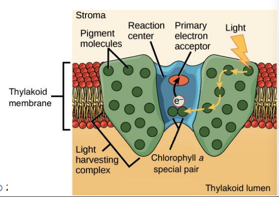
Photosystem reaction center
Where electromagnetic energy from sunlight is transformed into chemical energy