SSS Week 1
1/28
There's no tags or description
Looks like no tags are added yet.
Name | Mastery | Learn | Test | Matching | Spaced | Call with Kai |
|---|
No analytics yet
Send a link to your students to track their progress
29 Terms
Functions of the skin
thermoregulation (evaporation of sweat, in hot or cold weather, cutaneous blood vessels dilate or constrict to give off/conserve heat, arrector pili contraction produces heat (goosebumps))
protective function and repair
sensation
immunological response
metabolic (vit d synthesis)
psychological and communication (aesthetic appearance, apocrine gland secretion - odour)
Important notes on epidermis
stratum corneum - anuclear
(stratum lucidum)
stratum granulosum
stratum spinosum
stratum basale - melanocytes, keratocytes (where it develops but migrates up) and merkle cells
+basement membrane zone and then dermis
epidermis is avascular
cells of epidermis
keratinocytes: develops from basal layer and then migrates upwards to the stratum corneum where it is shed
langerhan cells - APCs found in epidermis
melanocytes produce pigment, protect cells of epidermis and dermis from sun damage
Merkles cells - (sensory mechanoreceptors) transducers associated with fine touch via fine unmyelinated nerve fibres — only present in thick skin
epidermis has an undulating surface with cross-crossing ridges and valleys, with invaginations due to follicles and sweat duct ostia (what we see as fingerprints)
Important notes on dermis (major fibres, major cells)
fibrous connective tissue in skin
major fibres
collagen: provide the skin with strength and toughness. Collagen bundles are small in the papillary dermis and form thicker bundles in the reticular dermis (deeper).
Elastin: provides the properties of elasticity and pliability to the skin.
dermis also contains nerves, blood vessels, lymphatics, epidermal adnexal structures, arrector pili muscle and cells.
major cells
fibroblasts
macrophages
mast cells
t and b lymphocytes
What are the physiological changes of the skin in aging
thinning of epidermis
flattening of dermal-epidermal junction
hyperpigmentation
loss of melanocytes
degradation of collagen and elastin
Hypodermis
Adipocytes organised into lobules (between these lobules are septa) septa contain nerves, larger blood vessels, fibrous tissue and fibroblasts — cna form cellulite
function
cold insulation
site of fat and energy storage
epidermal crosstalk
immune surveillance
Epidermal appendages - hair types, hair cycle
on all body except glabrous skin (lips, glans penis, labia minora, palms and soles)
provides protection, sensation and social communication
three types
lanugo - fine long hair of fetus that is shed 1 month prior to birth
vellus - fine short hair found all over body
terminal - thick hair on scalp, beard, axilla and genital area
hirsutism - vellus to terminal
male pattern alopecia - terminal to vellus
hair growth cycle
anagen - long growth phase
catagen - where active hair growth stops and the follicle begins to regress
telogen - ‘resting’ follicle shrinks and atrophies, and new hair follicle begins to grow
exogen - hair shed
Epidermal appendages - nails
specialised plates of hard keratin that develop from epidermis overlying small bones at ends of fingers and toes
stratching
grooming
picking up ifne objects
psycho-sexual communication
weapon
paronychium - skin around nail
lunula - white area of base of nail
Types of glands - epidermal appendages
sebaceous glands
associated with hair follicles
secreted onto skin through pilosebaceous canal-discharge into hair follicle ducts
antibacterial and antifungal action
eccrine sweat glands
all over body
merocrine
opening directly into skin
cholinergic innervation
watery (hypotonic) secretion
sweating/thermoregulation
onset of activity: birth
apocrine sweat glands
axilla, groin, mammary area, umbilicus
apocrine
opens into hair follicle (like sebaceous glands)
adrenergic innervation
viscous secretion
body odor
onset of activity: puberty (androgen dependent)
Macule
flat area of altered colour ≤ 1cm
Patch
flat area of altered colour > 1cm
Papule
elevated, solid, palpable lesion that is ≤ 1cm in diameter.
may be
acuminate (pointed)
dome shaped (ronded)
filiform (thread like)
flat-topped
oval or round
pedunculated
sessile
umbilicated
verrucous
Nodule
elevated, solid, palpable lesion > 1cm
Cyst
papule or nodule that contains fluid or semi-fluid material so is fluctuant
Plaque
circumscribed, palpable lesion > 1cm, most plaques are elevated, with a flat top
Vesicle
raised clear fluid-filled lesion ≤ 0.5cm
Bullae
raised clear fluid-filled lesion > 1cm
Pustule
pus-containing lesion < 0.5cm
Abscess
localised accumulation of pus in dermis or subcutaneous tissue (u can’t rlly see pus that much compared to a pustule)
Wheal
transient-raised lesion due to dermal oedema —> indicates urticaria
Comedone
papule due to blocked sebaceous follicle (occurs in acne) —— open (blackhead) or closed (whitehead)
What is diascopy
test where a clear glass slide is pressed against a skin lesion to assess if it blanches
helps differentiate between telangiectasia and petechiae
Petechiae is caused by blood leaking out from capillaries into surrounding tissue, whereas telangiectasia is the blood is still in the vessel but the capillaries are just dilated which means the blood can be pushed aside in the vessel —> blanching
Auspitz sign
peeling off surface scale to reveal areas of pinpoint bleeding (typical in psoriasis)
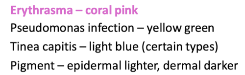
Woods light
fluorescence in visible range when applied to certain skin conditions
dermographism
where skin is stroked and a wheal is formed (indicative of tendency to urticaria)
nikolski’s sign
shearing stress of skin causes separation of skin resulting in traumatic bulla
koebner phenomenon
localisation of a non-infective skin disorder to area of trauma eg. psoriasis developing at site of a scar
dariers sign
rubbing of area of mastocytosis causes an intense urticarial reaction
pathergy
penetrating injury to skin causes area of pustulation 72 hours afterwards
bullous diseases types
subcorneal —> impetigo (children)
intraepidermal —> pemphigus vulgaris (middle aged), herpes zoster (older adults)
subepidermal —> bullous pemphigoid (elderly), dermatitis herpetitiformis (adults)
subcorneal - thin roof, not usually intact
intraepidermal - thin roof, rupture easily and leaves oozy denuded surface
subepidermal - thick roof so tending to be tense and intact - may contain blood