18. Surgical diseases of the pylorus and spleen. Aetiology, diagnosis and therapy. Pyloroplasty. Splenectomy.

0.0(0)
studied byStudied by 0 people
learnLearn
examPractice Test
spaced repetitionSpaced Repetition
heart puzzleMatch
flashcardsFlashcards
Get a hint
Hint

What is the location of the spleen?

Get a hint
Hint

Left side of the cranial abdominal cavity, parallel to the abdominal wall, between the abdominal wall and stomach (curvatura major).

Get a hint
Hint

With what ligament is the spleen associated with the stomach?

Get a hint
Hint

Ligamentum gastrosplenicum.

Card Sorting

1/60

encourage image

There's no tags or description

Looks like no tags are added yet.

Study Analytics
Name
Mastery
Learn
Test
Matching
Spaced

No study sessions yet.

61 Terms

1
New cards

What is the location of the spleen?

Left side of the cranial abdominal cavity, parallel to the abdominal wall, between the abdominal wall and stomach (curvatura major).

2
New cards

With what ligament is the spleen associated with the stomach?

Ligamentum gastrosplenicum.

3
New cards

With which other organ's blood supply is the spleen's associated?

The pancreas (pancreatic arteries).

4
New cards

What are the functions of the spleen?

Immunological and haematological: blood reservoir, blood filtration, IgG and cytokine synthesis, RBC maturation, platelet reservoir, removal of old platelets, and production of B and T lymphocytes and IgM.

5
New cards

What are the surgical diseases of the pylorus?

  1. Stenosis

  2. Obstruction

  3. Neoplasia

6
New cards

What is the aetiology of pyloric stenosis?

  1. Congenital in brachycephalic breeds.

  2. Acquired in response to infection

7
New cards

What are clinical signs of pyloric stenosis?

Vomiting several hours after eating

8
New cards

How is pyloric stenosis diagnosed?

X-ray with contrast, endoscopy, biopsy (to exclude neoplasia)

9
New cards

What is the treatment for pyloric stenosis?

Surgical (pyloroplasty)

Medical treatment of oesophagitis, acid-base abnormalities and dehydration should be instituted before surgery

10
New cards

What is a common cause of pyloric obstruction?

Foreign bodies.

11
New cards

What is a common clinical sign of pyloric obstruction?

Vomiting.

12
New cards

How is pyloric obstruction diagnosed?

X-ray with or without contrast and endoscopy.

13
New cards

How is pyloric obstruction due to a foreign body treated?

Surgical removal of the foreign body.

14
New cards

What is a less common surgical disease of the pylorus?

Neoplasia.

15
New cards

What are examples of pyloric neoplasia?

  1. Benign: adenoma

  2. Malignant (more common): lymphoma (cats), adenocarcinoma, leiomyosarcoma

16
New cards

What are general clinical signs of pyloric disease?

Anorexia, vomiting, weight loss, decreased appetite with water intake only.

17
New cards

How are pyloric diseases diagnosed generally?

History, endoscopy, and barium contrast radiography.

18
New cards

What is pyloroplasty?

Surgery to widen the pyloric opening.

19
New cards

What are indications for a pyloropasty?

  1. Recurrent gastric dilatation

  2. Neoplasia

  3. Hepatic or pancreatic abscesses

  4. Specific inflammatory processes

  5. Gastroduodenal ulcers

  6. Congenital pyloric stenosis with hypertrophy of pyloric musculature

20
New cards

What are three common methods of pyloroplasty?

  1. Fredet-Ramstedt pyloromyotomy
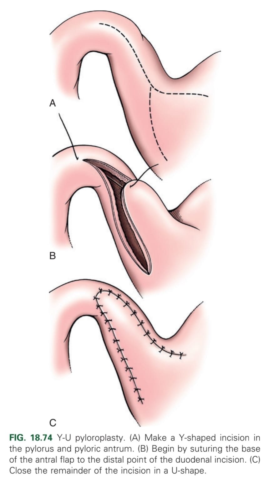
  2. Y-U shaped

  3. Heineke-Mikulicz.

21
New cards

What is the Fredet-Ramstedt pyloroplasty?

Performed by making longitudinal incision through serosa & muscularis of ventral pylorus. Only the serosa and muscularis should be incised, not mucosa (partial thickness incision)

<p>Performed by making longitudinal incision through serosa &amp; muscularis of ventral pylorus. Only the serosa and muscularis should be incised, not mucosa (partial thickness incision)</p>
22
New cards

What are the indications for Fredet-Ramstedt pyloroplasty?

Stenosis and chronic antral mucosal hypertrophy.

23
New cards

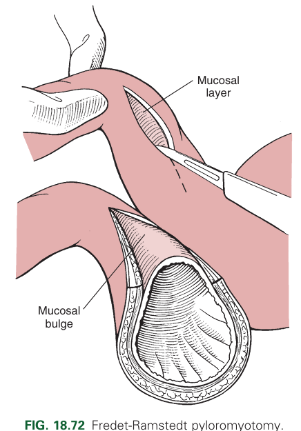
What is a limitation of the Fredet-Ramstedt pyloroplasty?

Limited visualisation into the lumen.

24
New cards

What is the Y-U shaped pyloroplasty?

  1. Making a full-thickness Y-shaped incision

    1. Y-base: antimesenteric aspect of the duodenum and pyloric sphincter

    2. Y-arms: pyloric antrum

  2. Advancing the U-shaped flap and suture to the base of the Y.

<ol><li><p>Making a full-thickness Y-shaped incision </p><ol><li><p>Y-base: antimesenteric aspect of the duodenum and pyloric sphincter</p></li><li><p>Y-arms: pyloric antrum</p></li></ol></li><li><p>Advancing the U-shaped flap and suture to the base of the Y.</p></li></ol><p></p>
25
New cards

What is the Heineke-Mikulicz pyloroplasty?

A full-thickness longitudinal incision of the ventral pylorus closed transversely with an appositional interrupted pattern.

<p>A full-thickness longitudinal incision of the ventral pylorus closed transversely with an appositional interrupted pattern.</p>
26
New cards

What is the advantage of the Heineke-Mikulicz pyloroplasty?

It is good for beginners.

27
New cards

What are surgeries for when pyloric outflow obstruction cannot be treated with a routine pyloroplasty?

Billroth I: Pylorectomy with end-to-end gastroduodenostomy

Billroth II: Resection of pylorus, antrum, and proximal duodenum with anastomosis of proximal jejunum and stomach

28
New cards

What are the surgical diseases of the spleen?

  1. Torsion

  2. Rupture/trauma

  3. Neoplasia.

29
New cards

In which animals is splenic torsion more common?

Large/giant breed dogs that are deep-chested.

30
New cards

What is the pathophysiology of splenic torsion?

Occlusion of venous drainage, causing congestion, enlargement, and cyanosis.

31
New cards

With which other condition may splenic torsion occur?

GDV.

32
New cards

What are the clinical signs of splenic torsion?

Abdominal pain, vomiting, abdominal distension, inappetence, PU/PD.

33
New cards

How is splenic torsion diagnosed?

X-ray and USG.

34
New cards

What are radiographic findings in splenic torsion?

Missing splenic silhouette or a C-shaped appearance.

<p>Missing splenic silhouette or a C-shaped appearance.</p>
35
New cards

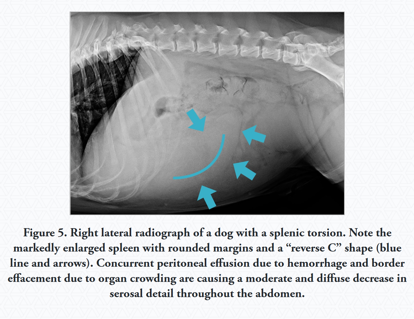
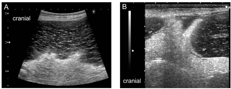
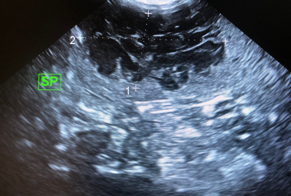
What is the most useful diagnostic tool for splenic torsion?

USG (detects splenomegaly and a lacy pattern).

<p>USG (detects splenomegaly and a lacy pattern).</p>
36
New cards

How is splenic torsion treated?

  1. Supportive therapy

  2. Total splenectomy

  3. Prophylactic gastropexy with partial splenectomy.

37
New cards

What are common causes of splenic rupture/trauma?

Penetrating foreign bodies (sticks, fence material, bite wounds, gunshot) or iatrogenic lacerations.

38
New cards

What are the clinical signs of splenic rupture/trauma?

Haemorrhage into the abdominal cavity, distended abdomen, pale mucous membranes, hypotension, acute abdomen, depression, and enlarged spleen.

Small bleedings may stop spontaneously. Large bleeding may lead to haemorrhagic shock.

39
New cards

How is splenic rupture/trauma diagnosed?

Abdominocentesis, X-ray, USG, and exploratory laparotomy.

40
New cards

How is splenic rupture/trauma treated?

Partial or total splenectomy and supportive therapy (blood transfusion).

41
New cards

What are common types of splenic neoplasia?

Haemangiosarcoma and haemangioma.

42
New cards

What is a characteristic of haemangiosarcoma and haemangioma?

They are "bleeding tumours" with widespread metastasis potential.

43
New cards

What are the clinical signs of splenic neoplasia?

Nonspecific signs, acute abdomen, abdominal distension, lethargy, and pale mucous membranes.

44
New cards

How is splenic neoplasia diagnosed?

X-ray and USG (showing fluid-filled cavities).

<p>X-ray and USG (showing fluid-filled cavities).</p>
45
New cards

How is splenic neoplasia treated?

Patient stabilisation, total or partial splenectomy followed by chemotherapy.

46
New cards

What are examples of splenic surgical interventions?

  1. Splenorrhaphy

  2. Splenectomy

47
New cards

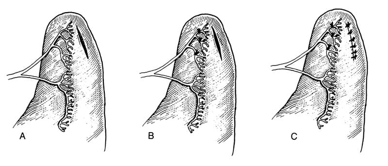
What is splenorrhaphy?

Surgical repair of small splenic lacerations or punctations with interrupted pattern.

<p>Surgical repair of small splenic lacerations or punctations with interrupted pattern.</p>
48
New cards

What suture material and pattern are used in splenorrhaphy?

4-0 or 5-0 rapidly absorbable monofilament in an interrupted pattern.

49
New cards

What is splenectomy?

Surgical removal of the spleen.

50
New cards

What is a major consideration regarding splenectomy?

The spleen is a blood reservoir, so its removal will have related repercussions.

51
New cards

What are indications for splenectomy?

Haemangiosarcoma, GDV, splenic torsion, severe trauma, generalised infiltrative disease

52
New cards

What are the types of splenectomy?

Partial (rare) and total.

53
New cards

What is an advantage of partial splenectomy?

Preservation of some splenic function.

54
New cards

What is recommended for vessel ligation during splenectomy?

Double ligation.

55
New cards

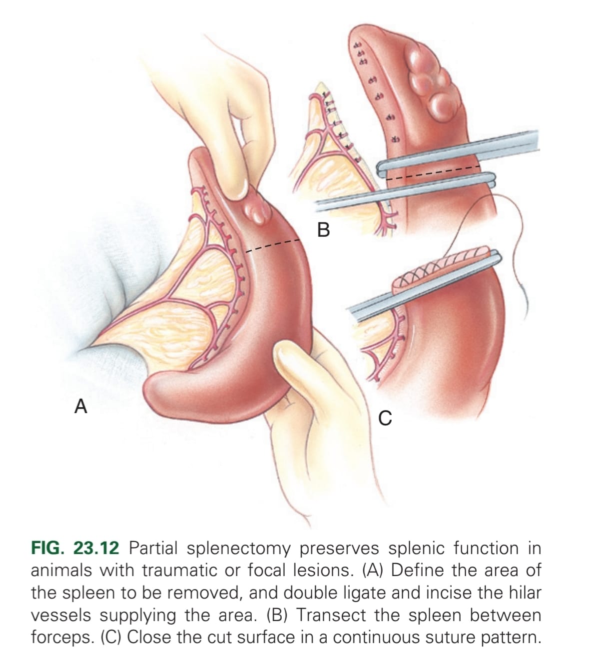
What is the method for a partial splenectomy?

Elevate spleen from abdomen → Ligate or seal hilar vessels in area → may use automatic stapling device to separating parenchyma, or digital pressure (not to destroy capsule) → haemostatic clamp across separated parenchyma & second clamp 1-2 cm distally, & transect midway between clamps → Appose capsule w/ continuous absorbable material. A second line of continuous or interrupted mattress may be used to gain a more haemostatic effect.

<p>Elevate spleen from abdomen → Ligate or seal hilar vessels in area → may use automatic stapling device to separating parenchyma, or digital pressure (not to destroy capsule) → haemostatic clamp across separated parenchyma &amp; second clamp 1-2 cm distally, &amp; transect midway between clamps → Appose capsule w/ continuous absorbable material. A second line of continuous or interrupted mattress may be used to gain a more haemostatic effect.</p>
56
New cards

What are methods for separating splenic parenchyma during partial splenectomy?

Automatic stapling devices or digital pressure.

57
New cards

What is the surgical approach for total splenectomy?

Midline celiotomy from xiphoid to pubis.

58
New cards

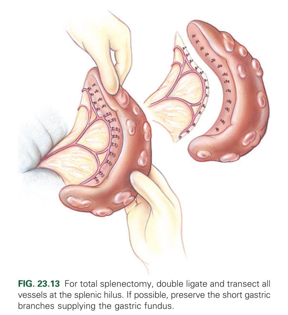
What is the method for a total splenectomy?

Elevate by gentle manipulation out of abdomen → double ligation of each vessel that remains in abdomen (if possible, preserve the short gastric branches supplying the gastric fundus) → Resect spleen. If spleen has ruptured at the time of surgery, lavage the abdominal cavity to prevent neoplastic cells entering.

<p>Elevate by gentle manipulation out of abdomen → double ligation of each vessel that remains in abdomen (if possible, preserve the short gastric branches supplying the gastric fundus) → Resect spleen. If spleen has ruptured at the time of surgery, lavage the abdominal cavity to prevent neoplastic cells entering.</p>
59
New cards

How can an animal be tested to see if pyloroplasty is indicated?

When contrast agent is not evacuated from the stomach 12-24 hours after administration

60
New cards

Which breeds are more predisposed to requiring pyloroplasty?

Miniature and brachycephalic breeds

61
New cards

Which vessels are ligated in a complete splenectomy?

All vessels at splenic hilus (or splenic artery distal to the branches supplying left limb of the pancreas; pancreatic artery)