Infratemporal Fossa
1/18
There's no tags or description
Looks like no tags are added yet.
Name | Mastery | Learn | Test | Matching | Spaced | Call with Kai |
|---|
No analytics yet
Send a link to your students to track their progress
19 Terms
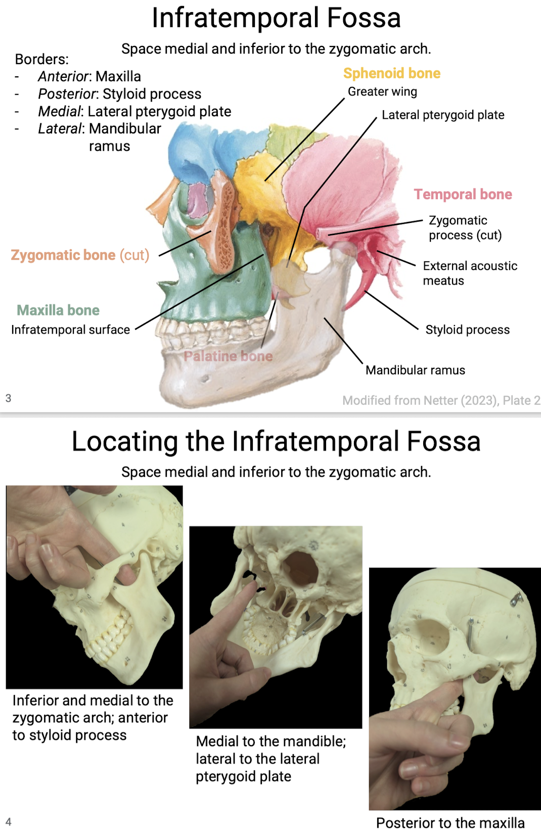
What is the infratemporal fossa and what are the borders?
Space medial and inferior to the zygomatic arch.
Borders: Infratemporal surface of maxilla (anterior), styloid process (posterior), lateral pterygoid plate (medial), mandibular ramus (lateral).
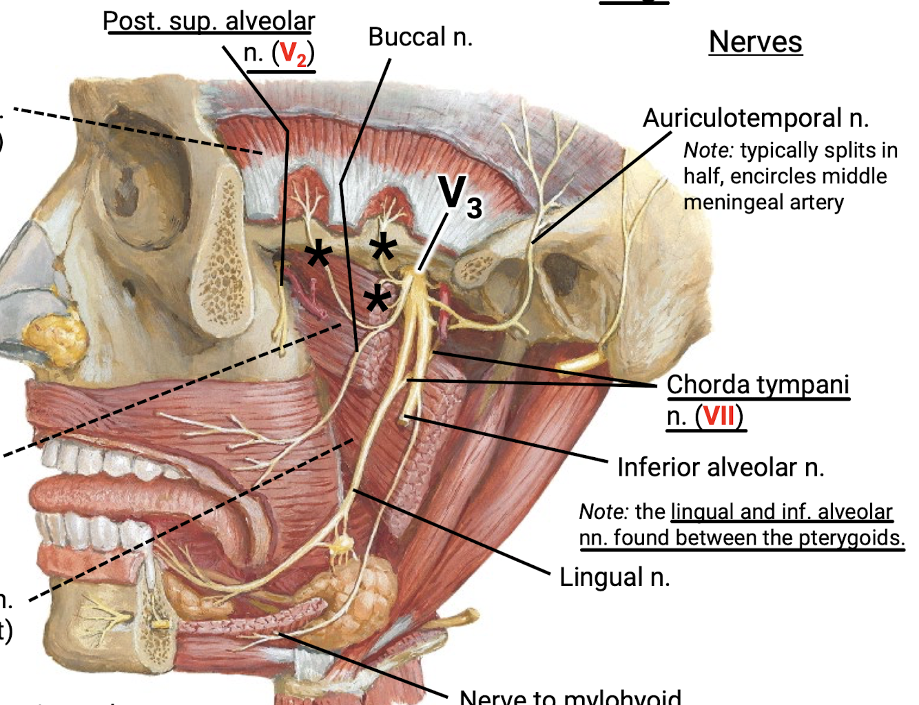
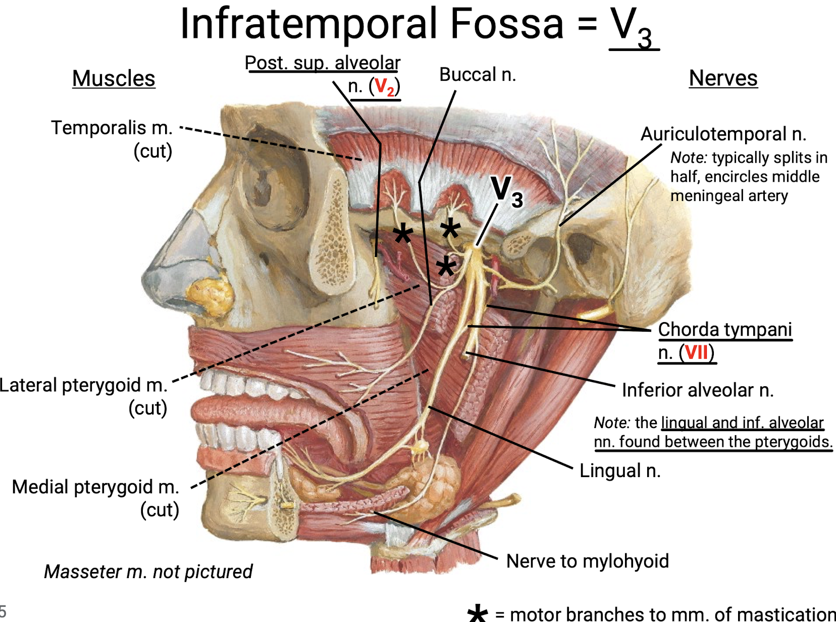
What nerves run through the infratemporal fossa?
Mainly V3, including auriculotemporal n. (which typically splits in half to encircle the middle meningeal artery), inferior alveolar n. (found between the pterygoids with…), lingual n., n. to mylohyoid, and buccal nerve.
Nerves NOT from V3 include the posterior superior alveolar nerve (V2) and chorda tympani n. (VII).
What muscles are found in the infratemporal fossa?
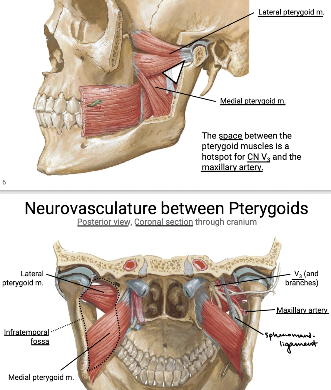
Temporalis m., lateral pterygoid m., and medial pterygoid muscle.
What can be found in the space between the pterygoid muscles? (3)
Hotspot for CN V3 and the maxillary artery. Also sphenomandibular ligament.
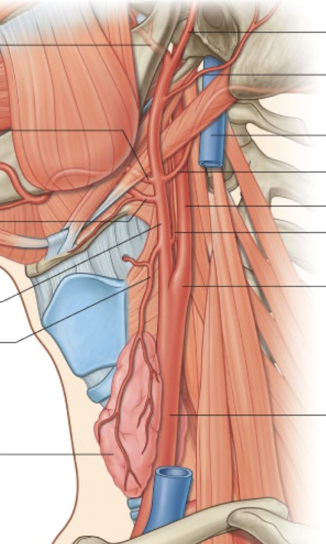
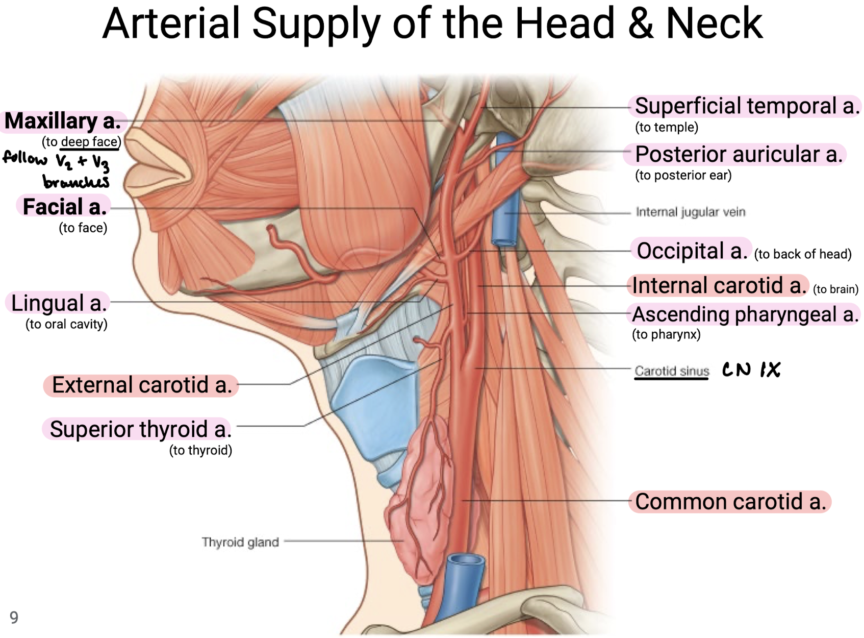
Describe the branches off of the external carotid artery (8) and where they supply.
Superior thyroid artery (to thyroid)
Ascending pharyngeal artery (to pharynx)
Lingual a. (to oral cavity)
Occipital a. (to back of head)
Facial a. (to face)
Posterior auricular a. (to posterior ear)
Maxillary a. (to deep face)
Superficial temporal a. (to temple)
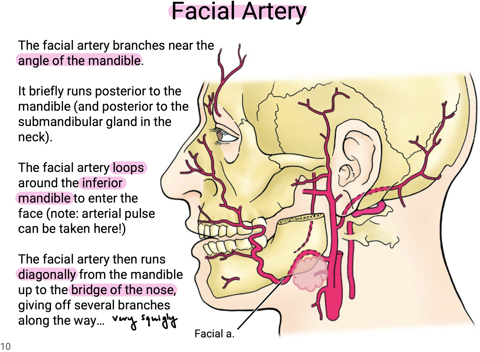
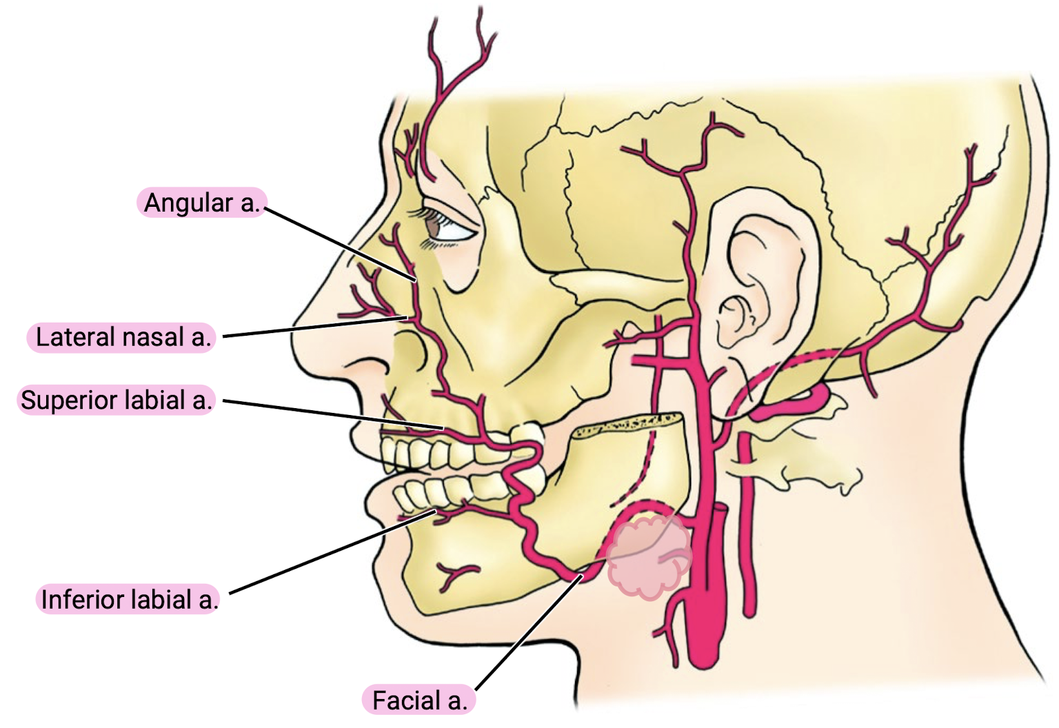
Where does the facial artery branch? Where does it enter the face? Where does it run from there?
The facial artery branches near the angle of the mandible. It briefly runs posterior to the mandibule (and posterior to the submandibular gland in the neck). The facial artery loops around the inferior mandible to enter the face (note: arterial pulse can be taken here!). The facial artery then runs diagonally from the mandible up to the birdge of the nose, giving off several branches along the way.
What are the branches off of the facial artery?
Inferior labial a.
Superior labial a.
Lateral nasal a.
Angular a.
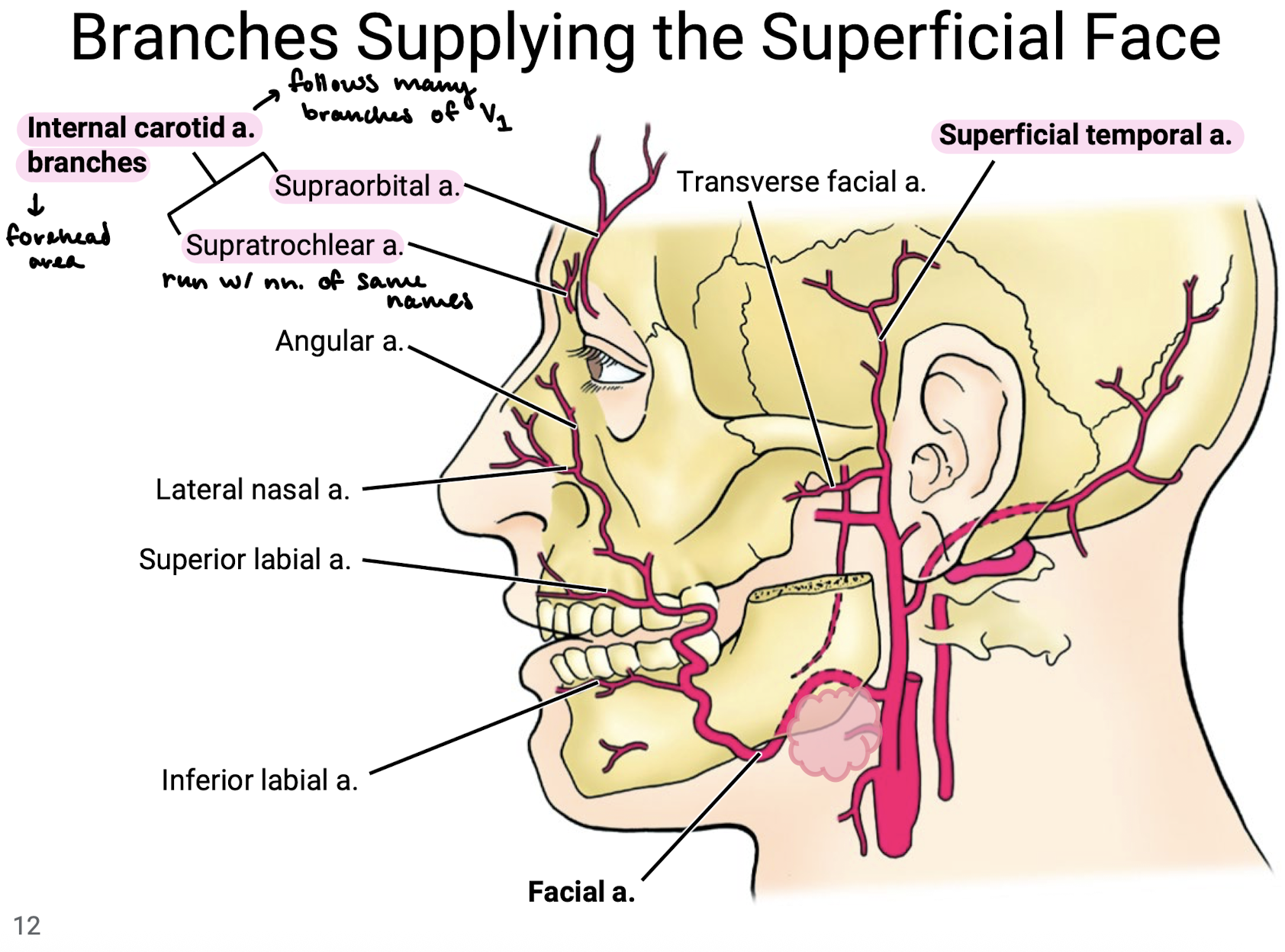
What arterial branches supply the superficial face (from either internal or external carotid)?
Superficial temporal a. and facial a. from external carotid a.
Internal carotid a. branches including supraorbital a. and supratrochlear a. (that run with the nn. of the same name) to supply the forehead area.
NOTE: Many branches of internal carotid a. follow the branches of V1.
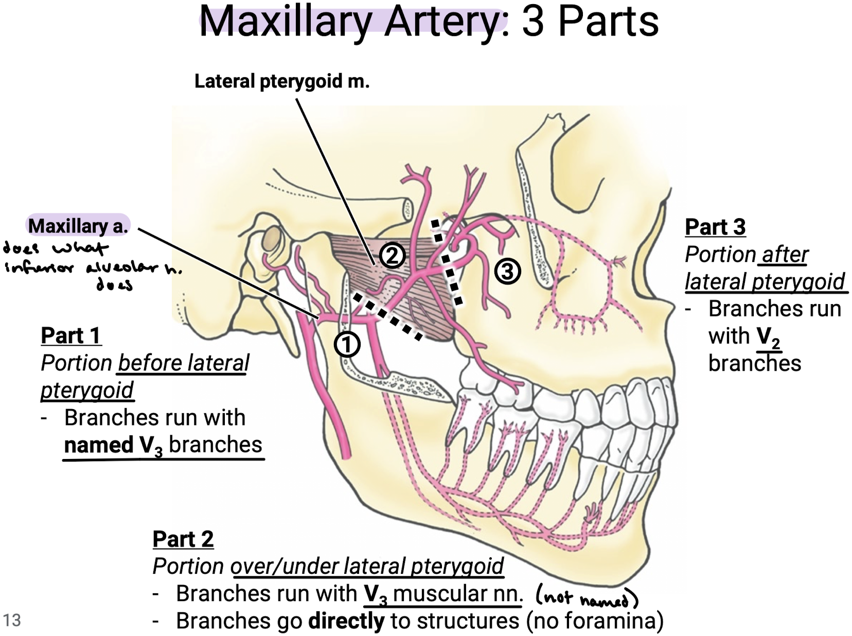
What are the different parts to the maxillary artery? Describe the general trends of branches from each part.
3 Parts:
Part 1: Portion BEFORE lateral pterygoid, whose branches run with named V3 branches
Part 2: Portion OVER/UNDER lateral pterygoid, whose branches run with V3 muscular nn. (not named) that go directly to structures (no foramina).
Part 3: Portion AFTER lateral pterygoid, whose branches run with V2 branches.
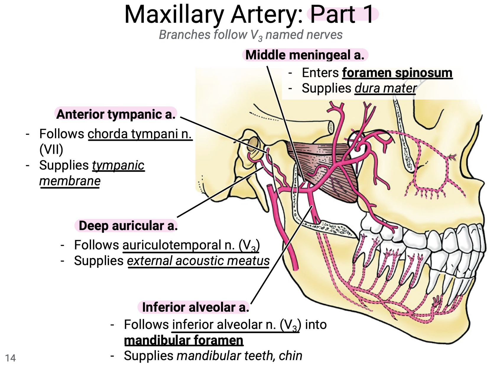
What are the four branches off of Part 1 of the maxillary artery? Know what nerve they follow and what area they supply.
Deep auricular a. follows auriculotemporal n. (V3) and supplies the external acoustic meatus.
Anterior tympanic a. follows chorda tympani n. (VII) to supply the tympanic membrane.
Middle meningeal a. enters foramen spinosum to supply the dura mater.
Inferior alveolar a. follows the inferior alveolar n. (V3) into the mandibular foramen to supply the mandibular teeth and chin.
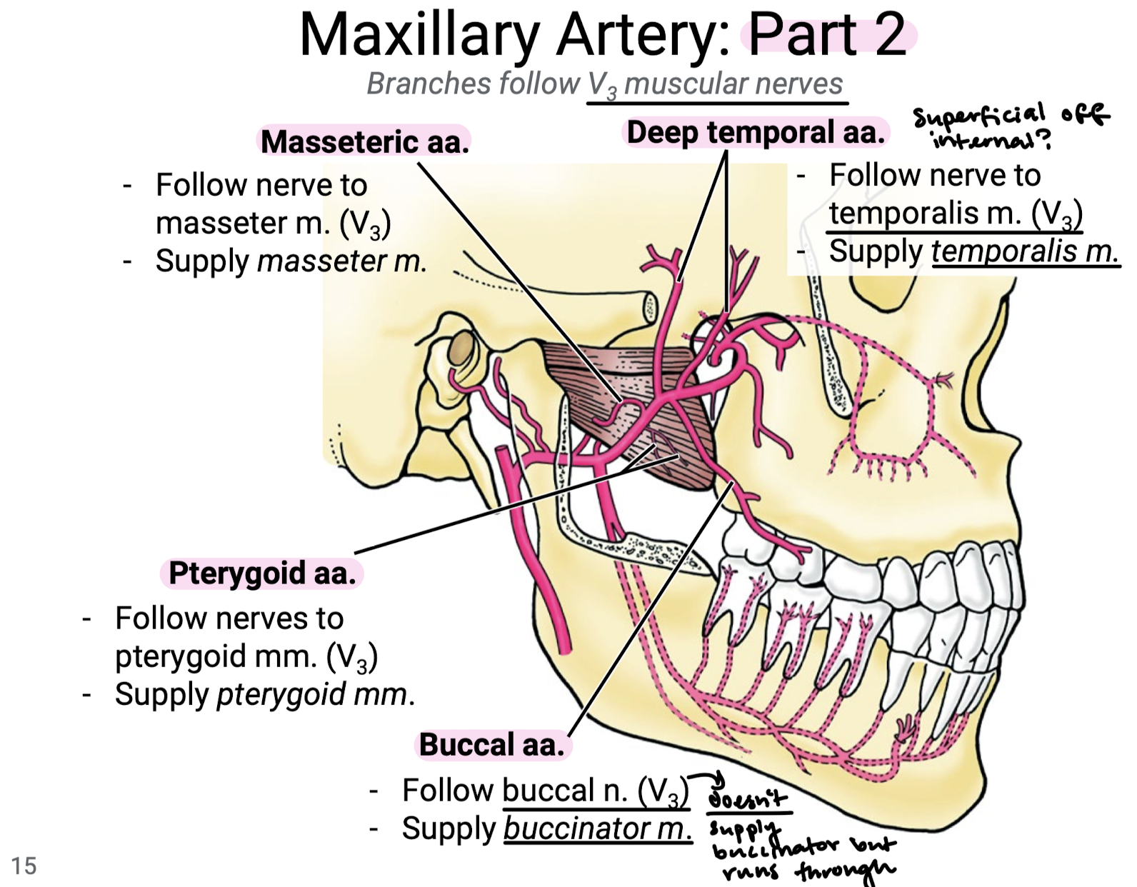
What are the four branches off of Part 2 of the maxillary artery? Know what nerve they follow and what area they supply.
Pterygoid aa. follow nerves to pterygoid mm. (V3) to supply the pterygoid mm.
Masseteric aa. follow the nerves to masseter m. (V3) to supply the masseter m.
Deep temporal aa. follow nerve to temporalis m. (V3) to supply temporalis m.
Buccal aa. follow the buccal n. (V3) to supply buccinator m., but remember the buccal n. of V3 only runs through buccinator and does not supply it.
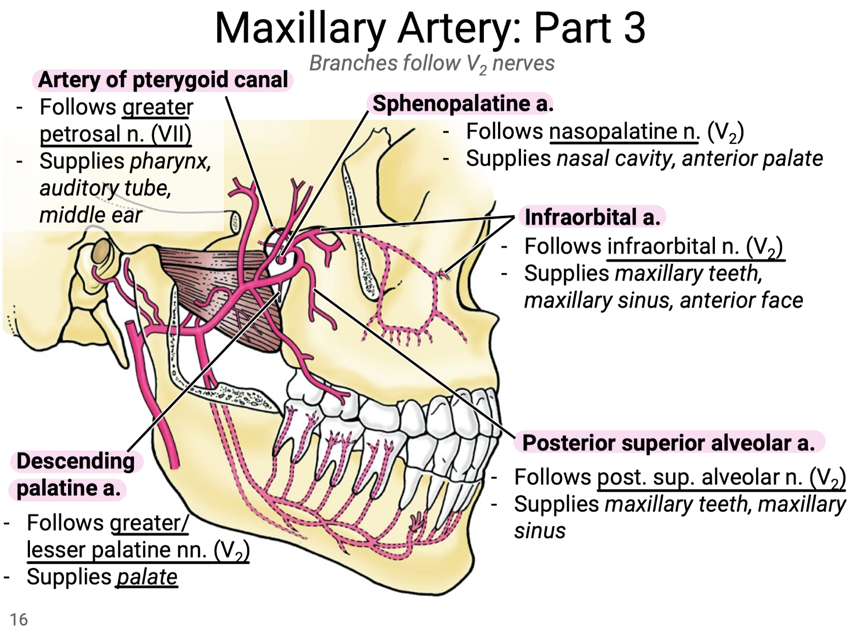
What are the five branches off of Part 3 of the maxillary artery? Know what nerve they follow and what area they supply.
Descending palatine a. follows the greater/lesser palatine nn. (V2) to supply the palate.
Posterior superior alveolar a. follows the post. sup. alveolar n. (V2) to supply the maxillary teeth and maxillary sinus.
Infraorbital a. follows the infraorbital n. (V2) to supply the maxillary teeth and sinus and anterior face.
Artery of pterygoid canal follows the greater petrosal nerve (VII) to supply the pharynx, auditory tube, and middle ear.
Sphenopalatine a. follows the nasopalatine n. (V2), supplying the nasal cavity and anterior palate.
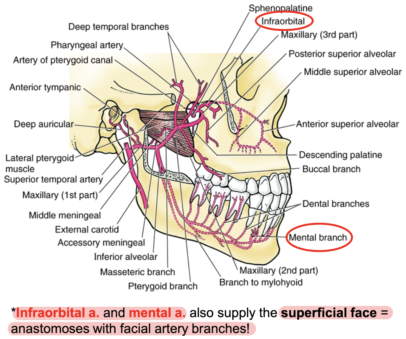
Which of the maxillary artery branches also supply the superficial face and anastomose with facial artery branches?
Infraorbital a. (part 3) and mental branch (part 1 from inferior alveolar a.).
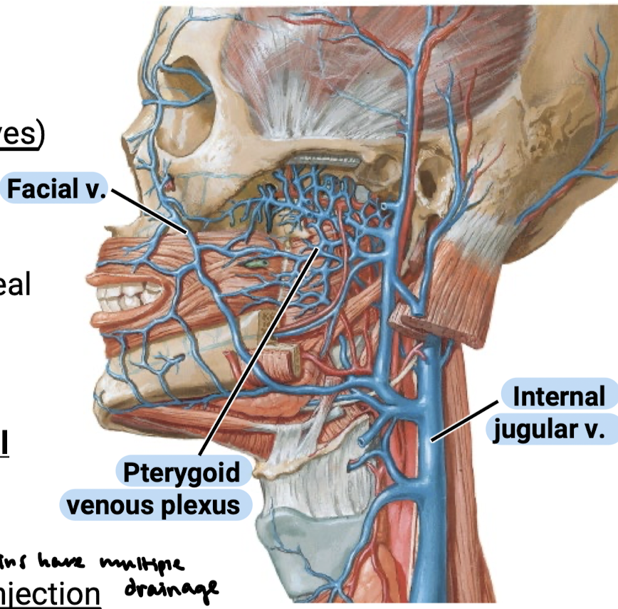
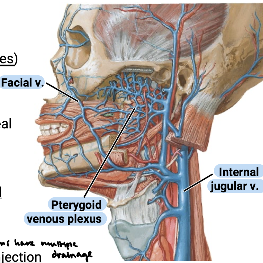
What is the main venous return method of the infratemporal fossa? In what four ways do these veins drain?
The pterygoid venous plexus. The plexus and facial veins have multiple draining pathways including:
Down to the internal jugular veins
Back to the retromandibular veins
Up to the ophthalmic veins
Up to the cavernous sinus (meningeal blood space)
What is the clinical significance of infratemporal vasculature?
Blood/nutrient supply to the teeth
Spread of infection (especially so because veins have multiple drainage)
Vascular injury during anesthetic injection
Inadvertent intravascular injection of anesthetic
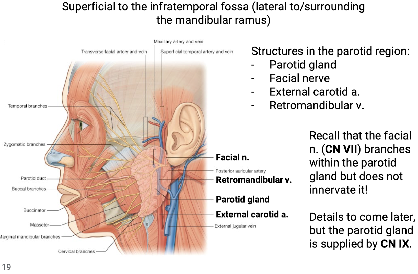
What is the parotid region? What are the structures found in the parotid region?
Superficial to the infratemporal fossa (lateral to/surrounding the mandibular ramus). Structures in the parotid region:
Parotid gland
Facial nerve (recall that the facial nerve does not innervate the parotid but branches within it)
External carotid a.
Retromandibular v.
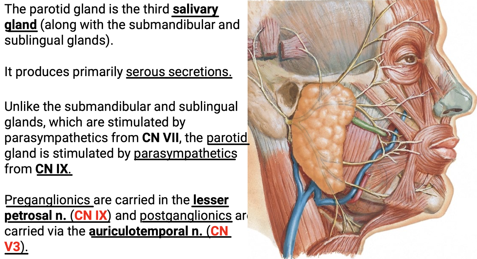
What are the primary secretions of the parotid gland? Explain the parasympathetics of the partoid gland.
Serous secretions. Stimulated by parasympathetics from CN IX (lesser petrosal n.) carrying the preganglionics with CN V3 carrying the postganglionics (auriculotemporal n.).
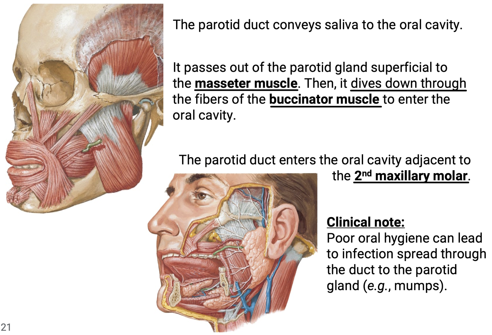
Where does the parotid duct travel to enter the oral cavity? Where in the oral cavity does the parotid duct enter? What is the clinical significance?
It passes out of the parotid gland superficial to the masseter muscle. Then it dives down through the fibers of the buccinator muscle to enter the oral cavity. The parotid duct enters the oral cavity adjacent to the 2nd maxillary molar.
Clinical Significance: Poor oral hygiene can lead to infection spread through the duct to the parotid gland (e.g., mumps).
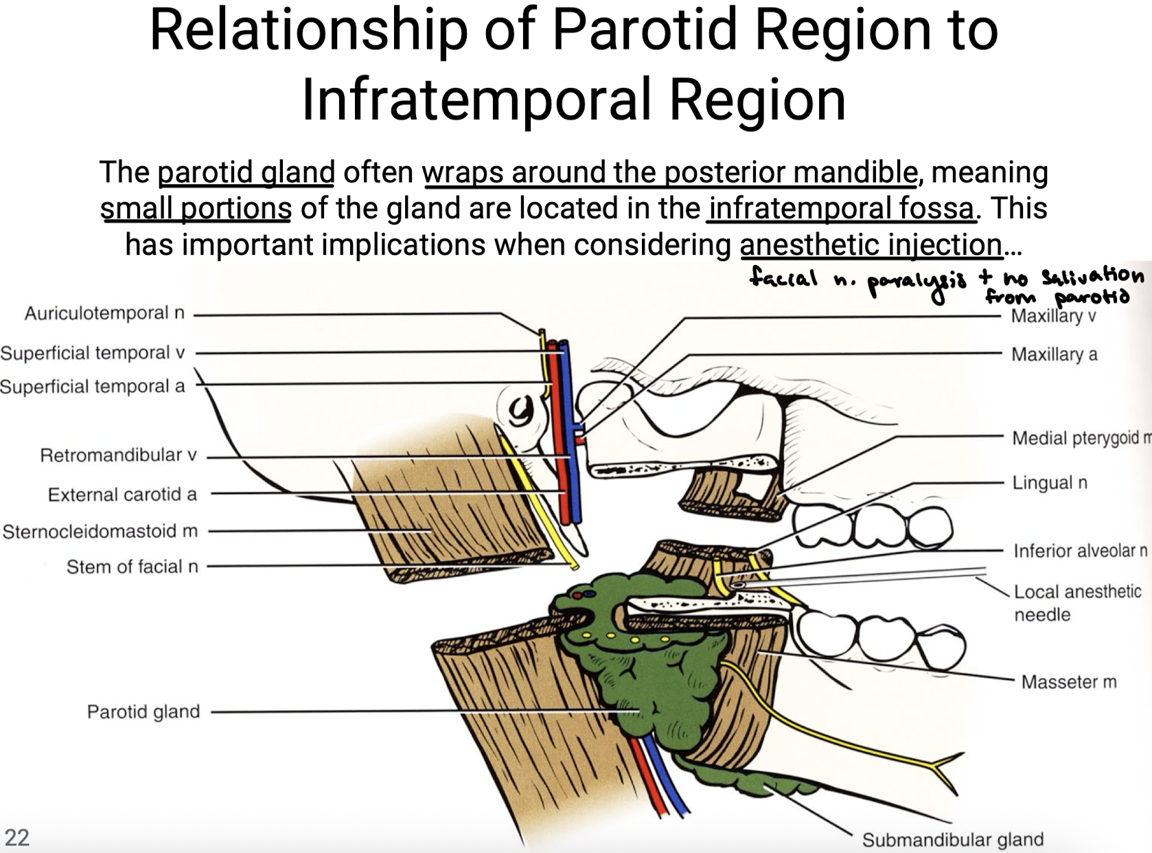
What is important to remember about the parotid region with respect to the infratemporal region?
The parotid gland often wraps around the posterior mandible, meaning small portions of the gland are located in the infratemporal fossa. This has important implications when considering anesthetic injection because accidental injection of the parotid would lead to facial n. paralysis and no salivation from parotid.