UTWB Lesson 21: Basic Organs and Structures in the Abdominal and Pelvic Cavity, The Kidneys
1/68
There's no tags or description
Looks like no tags are added yet.
Name | Mastery | Learn | Test | Matching | Spaced | Call with Kai |
|---|
No analytics yet
Send a link to your students to track their progress
69 Terms
When estimating the size of the kidneys in dogs or cats on radiographs, what lumbar vertebrae is used as the reference
L2
Where is the right kidney located in dogs?
Ventral to the last thoracic and first 2-3 lumbar vertebrae
Where is the left kidney located in the dog?
Below the first/second to fourth lumbar vertebrae
Where is the R kidney located in relation to the L kidney in the canine?
R kidney is cranial to the L kidney
Why is the R kidney more restricted in the canine?
Deeply recessed within the caudate lobe of the liver and is related medially to the right adrenal gland and caudal vena cava
What is the clinical consideration of the R kidney in the canine?
More difficult to visualize the R kidney for imaging (radiology) as well as it is more difficult to access for surgery
What does retroperitoneally mean?
Related to the peritoneum of the abdominal cavity
Where are the kidneys located in the canine?
Paired kidneys lie retroperitoneally pressed against the dorsal abdominal wall on either side of the vertebral column
They are usually ventral to the lumbar vertebra and extend cranially under the last ribs
What is the shape of the kidneys in cats and dogs?
Bean shaped
What is present on the kidney surface in cats?
Capsular veins
What is the renal capsule and what does its ability to be easily peeled off mean?
Thin sheet of connective tissue that surrounds the kidney, if there is difficulty peeling off the renal capsule during a necropsy it usually signifies that there was a problem with the kidney
In a healthy kidney, the renal capsule should be easily peeled off
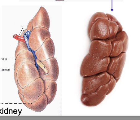
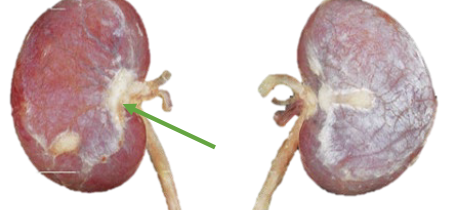
What is the kidney hilus?
Concave indent where the renal artery, vein, and ureter enter the kidney
What are the 2 main internal structures of the kidney, and what are their appearances?
Cortex - granular appearance
Medulla - Striated appearance
Structure of the cortex and medulla in bovine
Cortex is unfused and lobulated
Medulla is also unfused and form medullar projections called renal/medullar pyramids
What does the cortex of the kidney contain?
Renal corpuscles and convoluted parts of the tubules
What does the medulla of the kidney contain?
Contains the collecting ducts
What is the renal crest?
Inner margin of the medulla where the ducts empty into the renal pelvis
What species have medullary pyramids?
pigs, cows, and humans
What are medullar pyramids/papilla?
Medullary zones divided by inter-lobar vessels
What is the renal pelvis?
Wide, funnel-shaped structure that collects the urine, it is the terminal dilated part of the ureter within the kidney
What is the renal/pelvic recess?
Depressions caused by extensions of the renal pelvis into the medulla on both sides of the crest
What is the renal sinus?
fat-filled space surrounding the renal vessels and the ureter
What are the 5 radiographic densities, and what colors do they show up in radiographs?
Air- black
Fat- Lighter black
Soft tissue- Dark gray
Bone- light gray
Metal- white
How are the kidneys viewed on radiographs?
Kidneys are clearly seen in about 50% of plain studies of the abdomen of the dog
Kidneys are more clearly identifiable in cats
What is used to make the kidneys more visible in detailed studies?
Contrast radiography using intravascular perfusion with a contrast agent
What views are usually performed to visualize the kidneys in radiographs?
Lateral and ventral views, R lateral more often than L lateral
What is the normal radiologic appearance of the kidneys?
Bean shaped, smooth, homogenous, soft tissue opacity
Often partially superimposed on one another
What is the normal length of the canine kidney in radiographs?
Appx 2.5-3.5 x the length of the body of the second lumbar vertebra
What is the normal length of the feline kidney in radiographs?
Appx 2.4-3 x the length of the body of the second lumbar vertebra
Where is the R kidney located on radiographs in the dog and cat?
Dog- T13-L2
Cat- L2-L4
Where is the L kidney located on radiographs in the dog and cat?
Dog- L1-3
Cat- L3-L5
What is the normal contrast radiologic appearance of the kidneys?
Both kidneys should be well visualized, with the renal cortices diffusely opaque
Smooth in outline
Medulla should be more radiolucent because of the renal pelvis and pelvis recess
What is anechoic?
Black imaging on ultrasonography correlating to blood/fluid
What is Hypoechoic?
Darker gray imaging on ultrasonography correlating to the renal cortex and liver
What is isoechoic?
Lighter gray than hypoechoic imaging on ultrasonography correlating to fat and the spleen
What is hyperechoic?
White imaging on ultrasonography correlating to bone/gas and connective tissues
Ultrasonography of the kidneys
Echotexture is fine, granular, and markedly hypoechoic
Renal cortex is hypoechoic or isoechoic as compared with the liver
Renal medulla is hypoechoic or anechoic in relation to the renal cortex
What is the corticomedullary junction defined by on ultrasonography?
Presence of bright hyperechoic specks that represent the arcuate vessels
Why is the renal pelvis hyperechoic?
Because of the presence of fat and fibrous tissue
Why does renal length vary in dogs but less in cats?
Because of body size variation
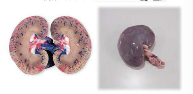
What are some significant features of the bovine kidney?
Lobated
No renal crest or renal pelvis
R kidney is flattened and oval and related ventrally to the colon and descending duodenum
R kidney’s cranial pole embeds in the liver
L kidney slightly twisted and displaced to the right by the rumen
What kidney does this species belong to?
Bovine
What are pyramids drained by in the bovine?
Minor renal calices
What do the minor renal calices drain into in the bovine kidney?
Drained by major calyces (cranial and caudal collecting ducts) that join to form the ureter
How is the ureter structured in regards to calices in the bovine kidney?
The ureter divides into 2 and subsequently into 18-25 calices draining the renal papillae
What do the small ruminant kidneys resemble?
Canine and feline kidneys
Features of small ruminant kidneys
Fusion of the cortex and medullary region
Bean shaped with a renal hilus
Medullary region fuses to form a single renal pyramid/renal crest
Features of the cortex and medulla of the porcine kidney
No external lobation because the cortex is fused and smooth
Internal evidence of lobation
Medullary region comprise separate renal pyramids
Dorsoventrally flattened
What is unique about the porcine kidneys?
Only domestic mammal whose kidneys are at virtually same level (no contact between right kidney and liver)
Internal features of the porcine kidney
up to 10 minor calyces draining same number of medullary pyramids
2 major calyces at both cranial and caudal poles that drain the minor calyces into the renal pelvis
It is multi pyramidal
How is the porcine kidney associated with the pancreas?
Cranial poles of both kidneys associated with the left lobe (left pole) and body (right lobe) of the pancreas
What species does this kidney belong to?
Porcine
What species does this kidney belong to?
Small ruminant
What species do these kidneys belong to?
Equine
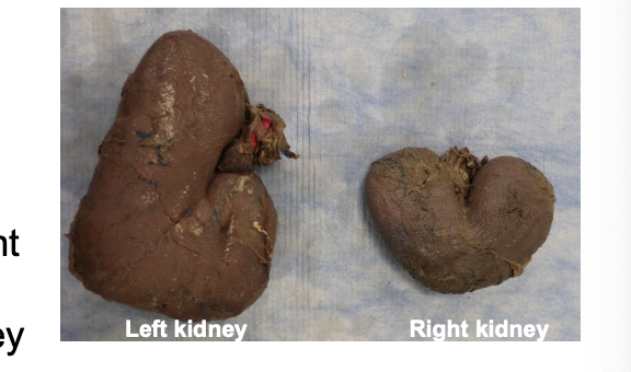
Features of the equine R kidney
Retroperitoneal
Heart shaped
Cranial pole makes a renal impression on caudate process of liver
Where is the equine R kidney located?
Ventral to last 2 or 3 ribs and 1st lumbar transverse process
Cranial to the liver
Ventral to the descending duodenum, pancreas, and base of cecum, coils of small colon and small intestine
Medially to the R adrenal gland and aorta
Features of the equine L kidney
More caudal than right kidney
Retroperitoneal (less than R kidney)
Flattened and bean-shaped (number 6)
Where is the equine L kidney located?
Ventral to T17-L2
Cranial to the spleen and stomach
Medial to the aorta and small colon and coils of small intestine
Caudal pole may be just within reach in rectal palpation
Ventrally related to small colon and small intestines
Internal features of the equine kidney
Unipyramidal type with a common renal crest
Renal pelvis
2 polar terminal recesses
Papillary ducts
What is unique about the equine kidney?
Has 2 polar terminal recesses that extend from the poles of the kidneys towards the renal pelvis
Papillary ducts open into the terminal recesses
Where does the kidneys main blood supply come from?
Renal artery
What does the renal artery divide into?
Interlobar arteries
What do interlobar arteries branch into?
Arcuate arteries (reside in the corticomedullary junction)
What do the arcuate arteries give rise to?
Interlobular arteries that radiate to the cortex
What happens after the interlobular arteries?
Afferent arterioles leave the interlobular arteries to enter the renal corpuscles which will divide to form the capillary loop of the glomerulus
What happens after the blood leaves the glomerulus?
The glomerular capillary loops will then recombine to form the efferent arteriole
What does the efferent arteriole do?
Provides a second capillary system which branches into peritubular capillaries that surround the tubules
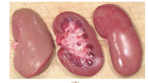
What species does this kidney belong to?
Canine
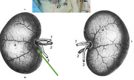
What species does this kidney belong to?
Feline