ANAT 100- Module 10
1/77
There's no tags or description
Looks like no tags are added yet.
Name | Mastery | Learn | Test | Matching | Spaced | Call with Kai |
|---|
No analytics yet
Send a link to your students to track their progress
78 Terms
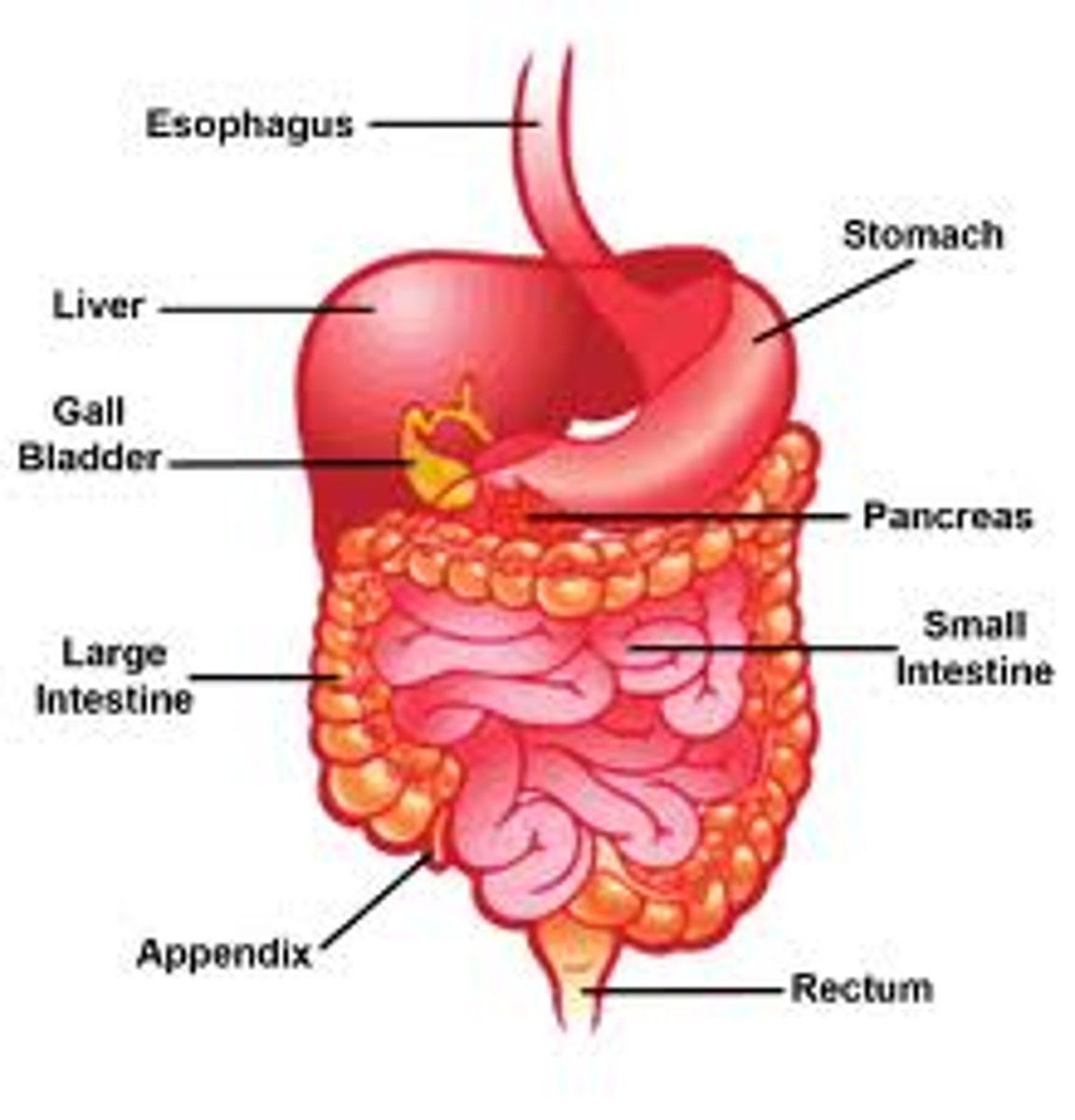
components of the digestive system (organs are divided into 2 groups)
1. Digestive Tract (Alimentary Canal)
- mouth, oral cavity, pharynx, esophagus, stomach, small intestine, large intestine, and anus
2. Accessory Digestive Organs
- tongue, teeth, salivary glands, liver, biliary ducts and gallbladder, and pancreas
Functions of the Digestive Tract
Digestion
Absorption
Secretion
- secreting acid into the stomach
Motility
- muscles push food down
Elimination of Waste
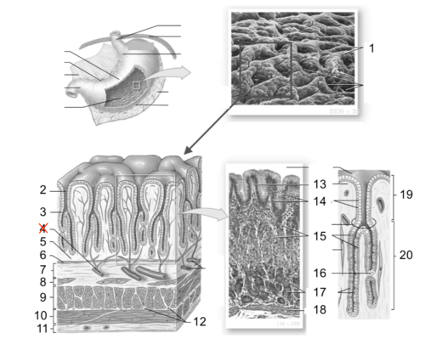
General Histology Organization of Digestive Tract Wall
Mucosa
Submucosa
Muscular Externa
Serosa/Adventitia
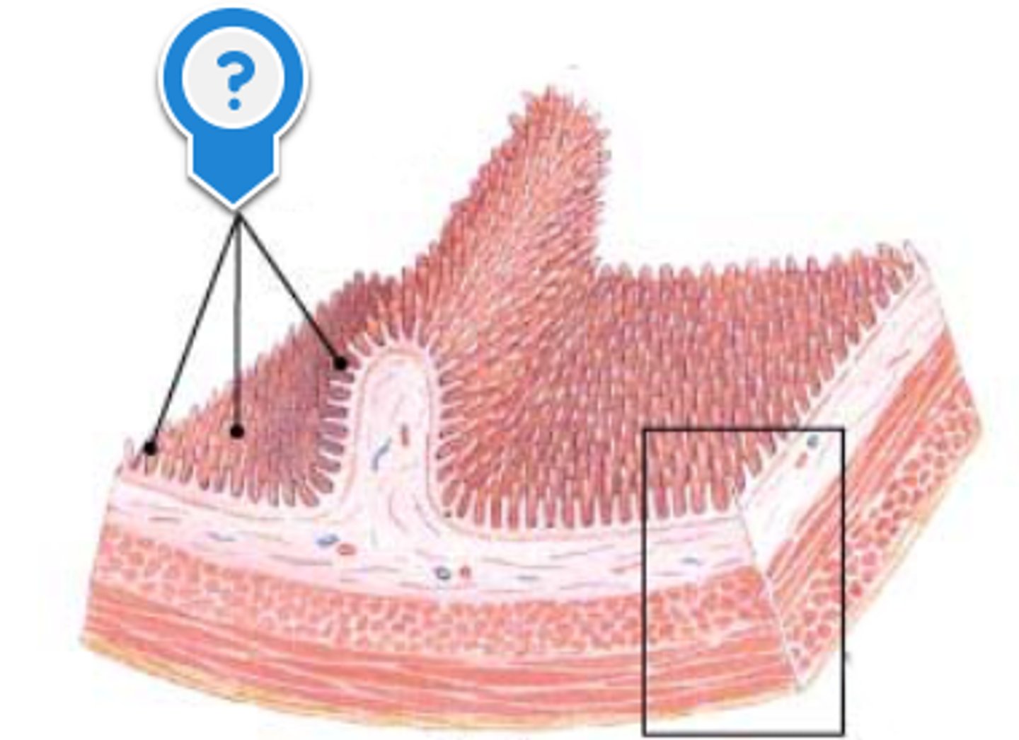
General Histology Organization of Digestive Tract Wall: Mucosa
- the innermost layer of the digestive tract wall
- composed of 3 main elements:
surface epithelium
lamina propria
muscularis mucosa
(look at image in notes)

mucosa: surface epithelium
- the type of epithelial layer reflects the expected function of the organ
- examples of function include secretion, absorption, and protection
mucosa: lamina propria
- this is a layer of loose connective tissue under surface epithelium
mucosa: muscularis mucosa
- this layer is composed of smooth muscle fibres under the lamina propria
General Histology Organization of Digestive Tract Wall: Submucosa
- layer below the mucosa
- composed of dense irregular connective tissue, and contains blood vessels, lymphatics, glands, and nerve plexuses
(look at image in notes)

General Histology Organization of Digestive Tract Wall: Muscular Externa
- composed on circular and longitudinal layers of smooth muscle with nerve plexuses between the layers
- the nerve plexuses are responsible for muscular contraction to propel food through the digestive tract
(look at image in notes)
General Histology Organization of Digestive Tract Wall: Serosa/Adventitia
- Serous membrane: single layer of thin flat cells that create a membrane sheet is secrete lubricating fluid
- Adventitia: loose connective tissue
Oral Cavity parts
(first part of the digestive tract, and consists of 2 parts )
VESTIBULE
ORAL CAVITY PROPER
Oral Cavity: Vestibule
- the space between the cheeks and lips and the gums and teeth
(look in notes for better picture)
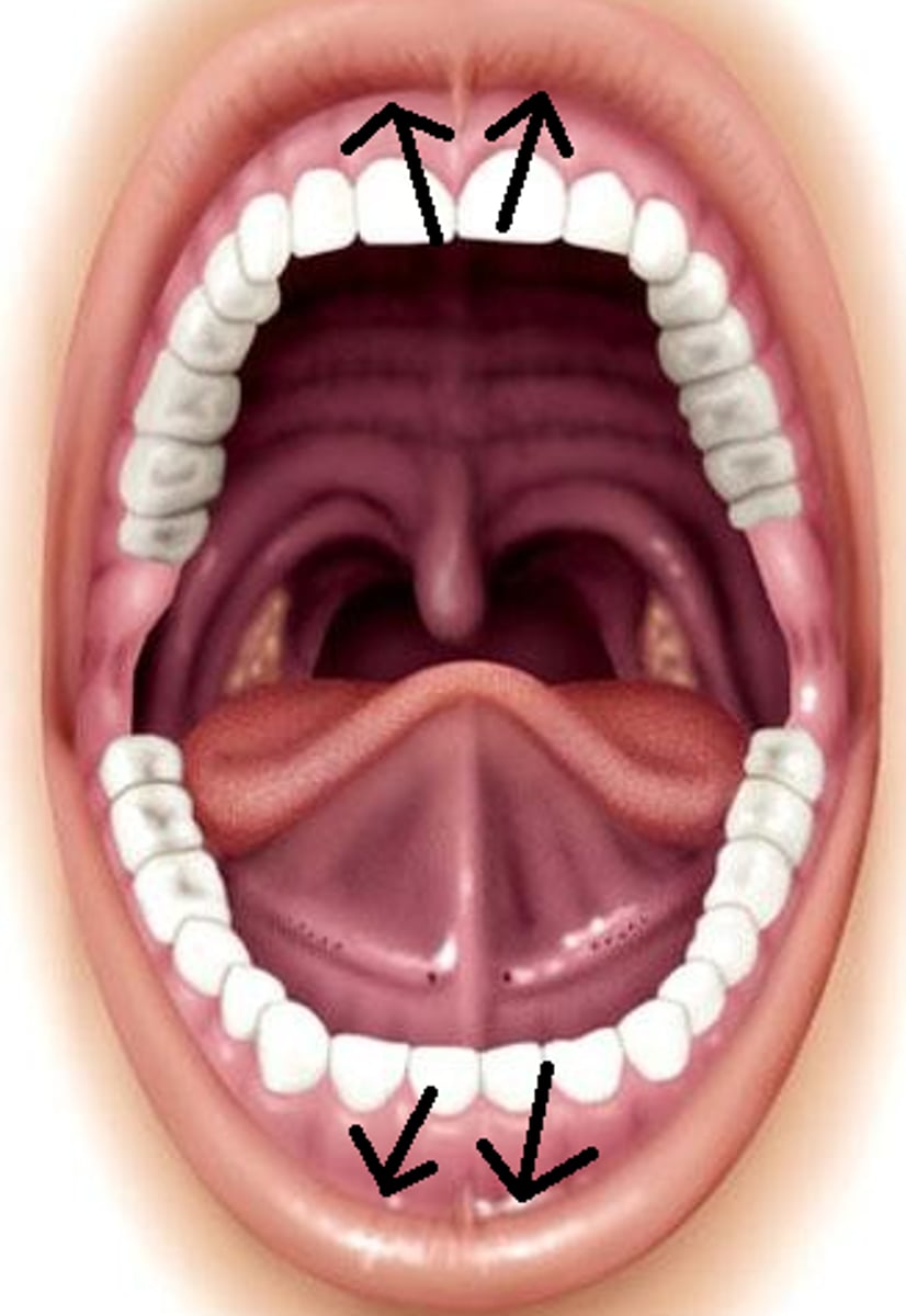
Oral Cavity: Oral Cavity Proper
- all other areas of the mouth
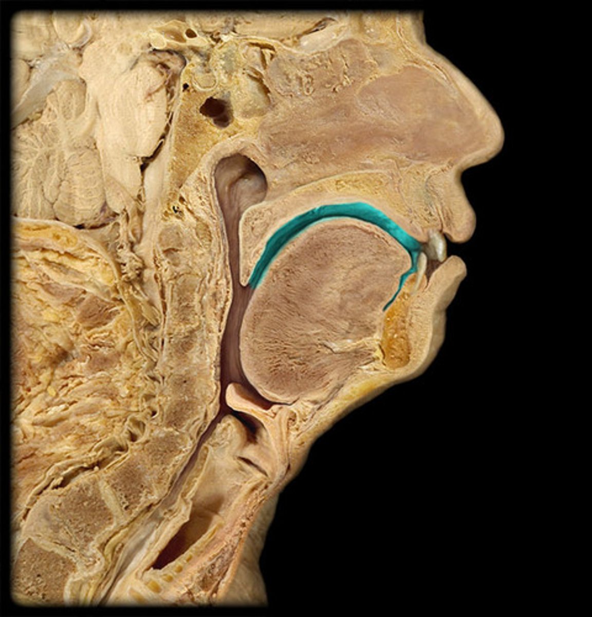
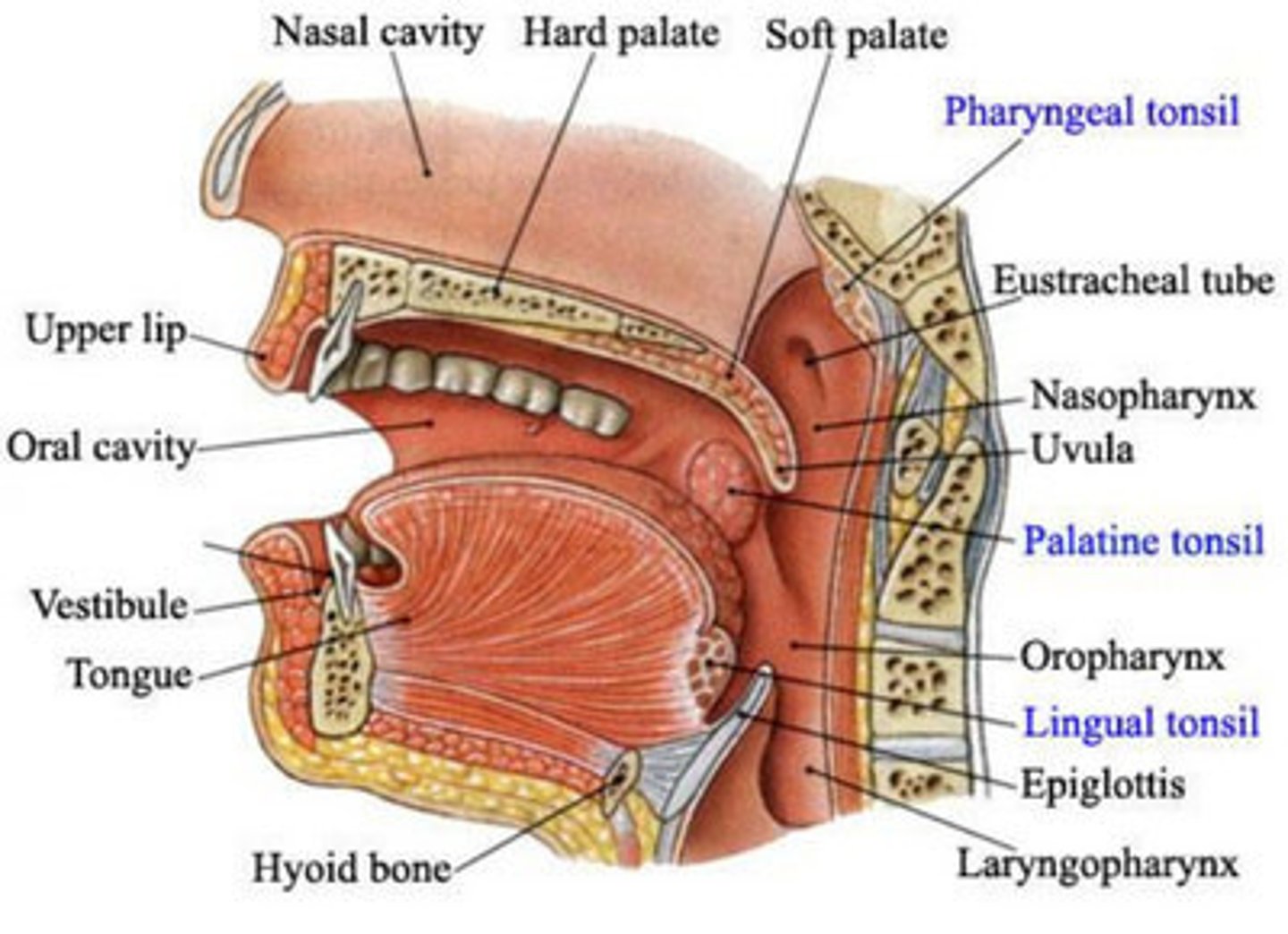
Oral Cavity: Palate
- makes up the superior border or the oral cavity
- divided into hard palate (bone) and soft platelet (muscle)
- the posterior extension of the soft palate is called the uvula
hard palate
soft palate
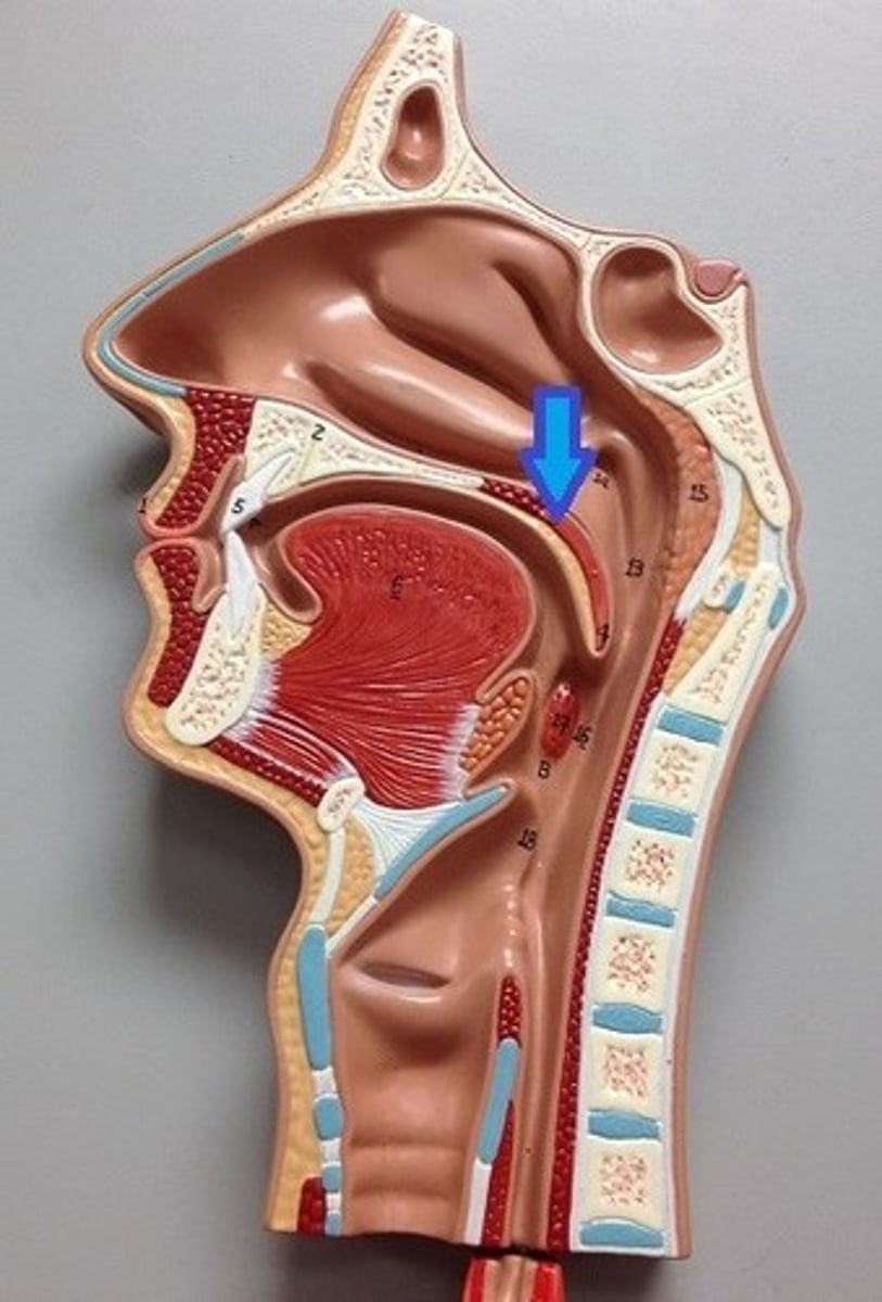
uvula
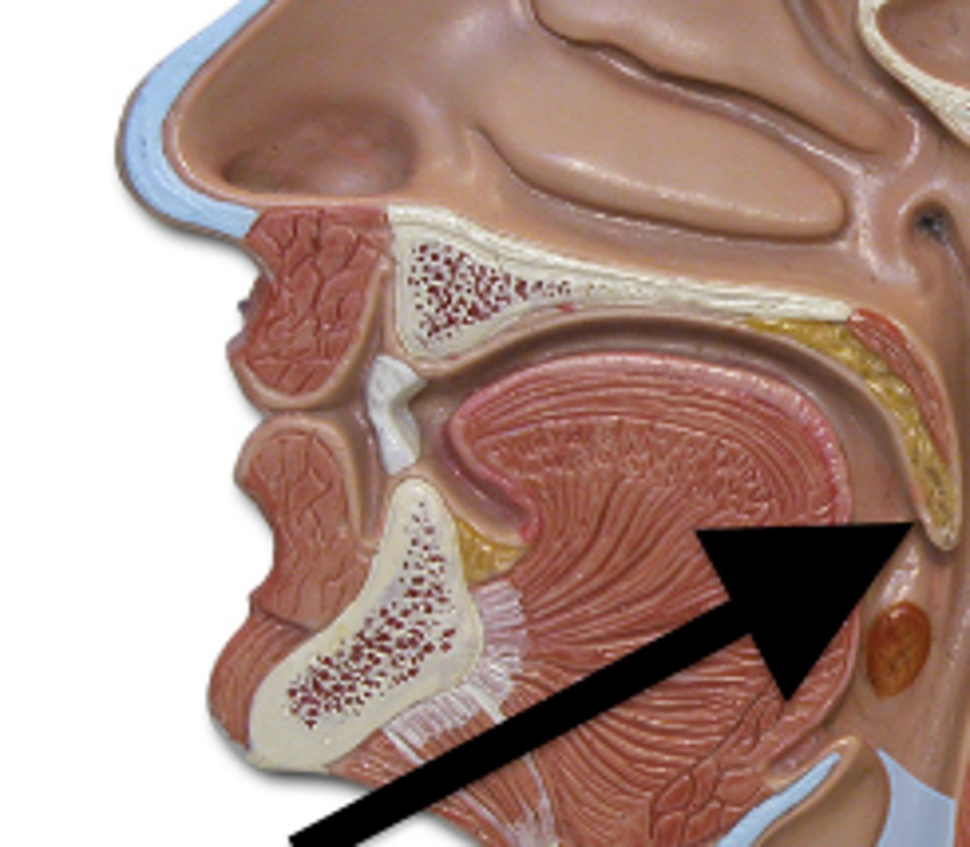
Oral Cavity: Tongue
- muscle associated with speech, taste, and mechanical manipulation of food
- the tongue is made up of muscles that control its shape (intrinsic muscles) and muscles that move the tongue during chewing and speech (extrinsic muscles)
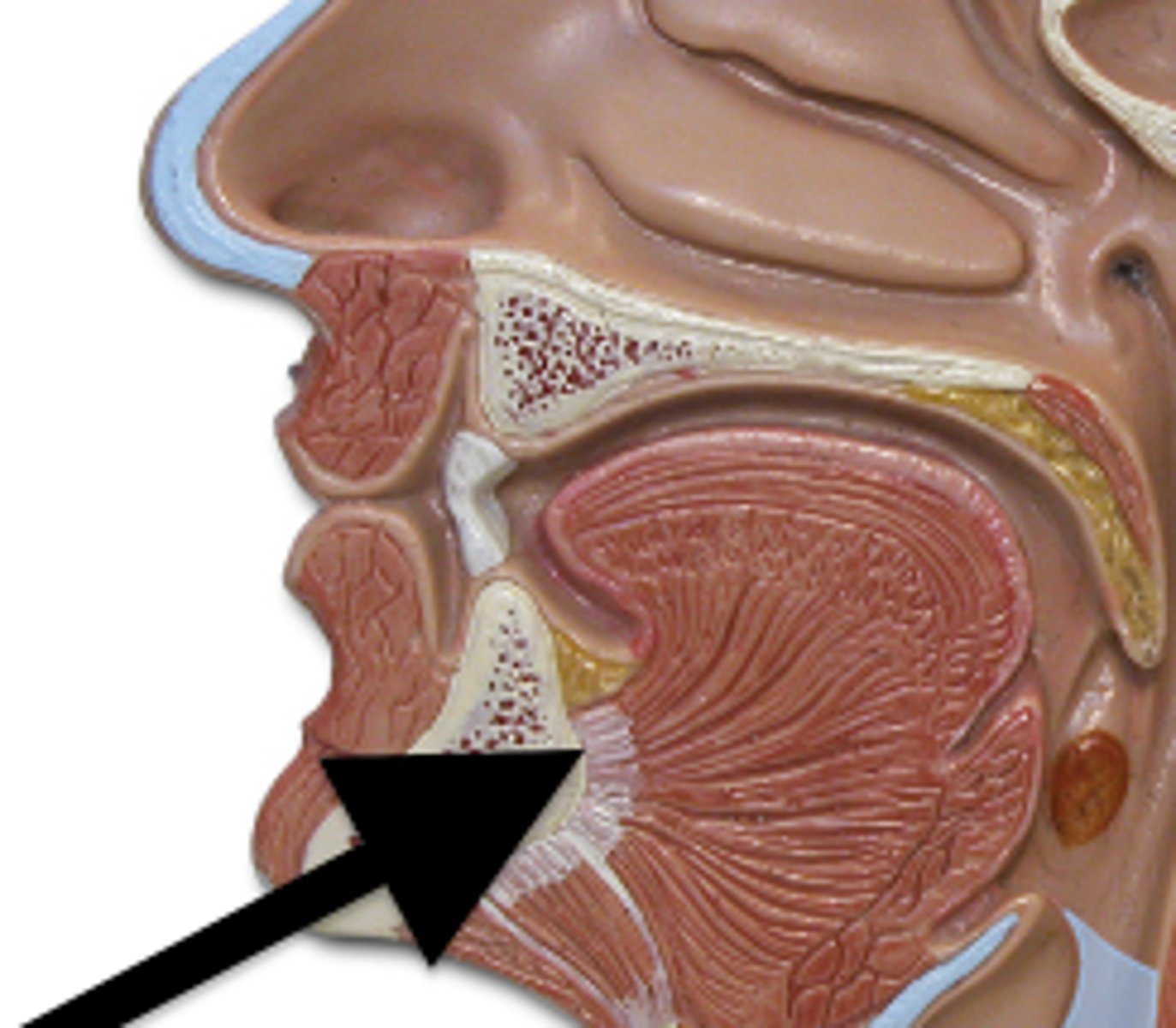
- on the inferior of the surface of the tongue is the frenulum, which anchors the tongue to the bottom of the mouth
frenulum
Papaillae of the tongue
- the superior and lateral surfaces of the tongue are covered in papillae (bumps on the tongue)
- there are 4 types papillae (some containing taste buds)
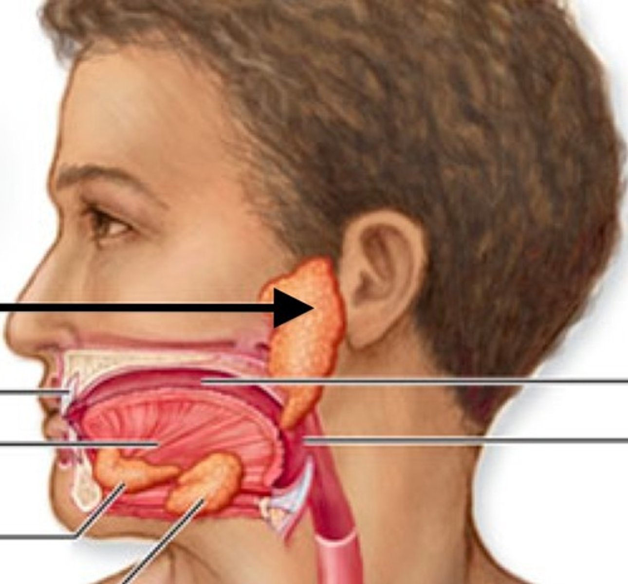
Oral cavity: salivary Glands
-secrete digestive enzymes to help break down food while chewing
- 3 major glands with different secretions:
parotid
submandibular
sublingual
Salivary Glands: parotid
- secretes serous (watery) fluid and is located anterior and inferior to the external ear opening
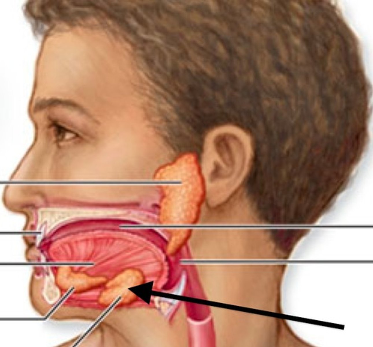
salivary glands: Submandibular
- secretes serous and mucous (viscous) fluid and is inferior to the mandible
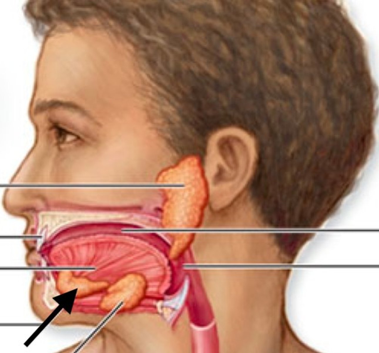
salivary glands: sublingual
- secretes mucus and is inferior to the tongue
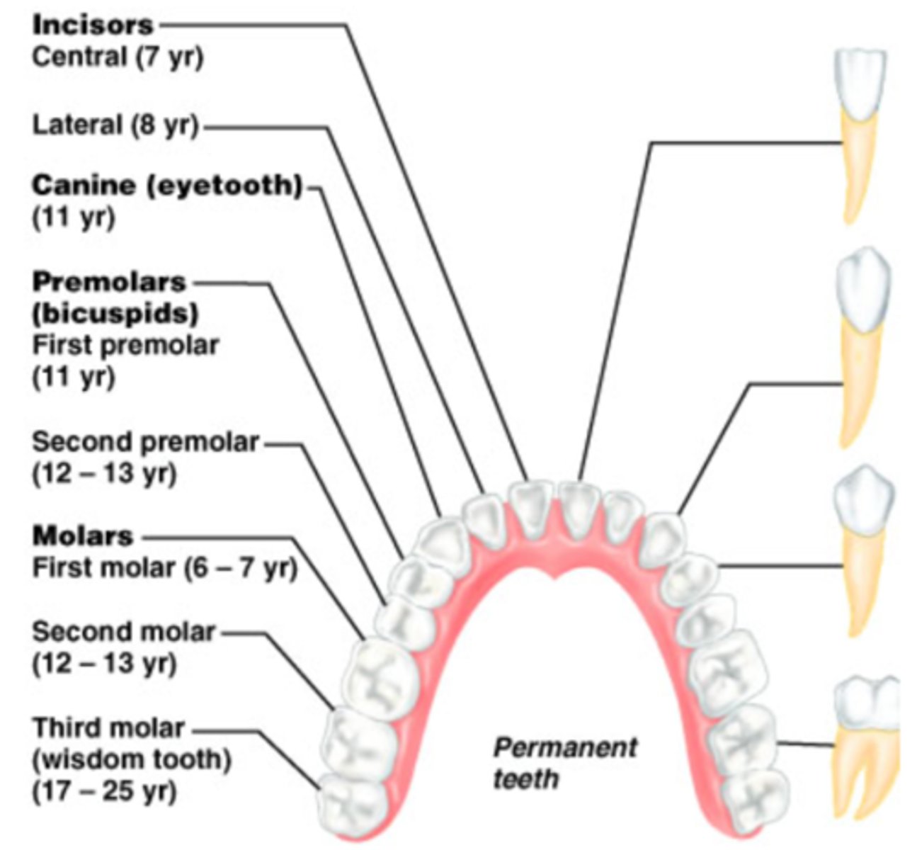
Oral Cavity: Teeth
- in a lifetime, humans have 2 sets of teeth:
deciduous teeth
permentant teeth
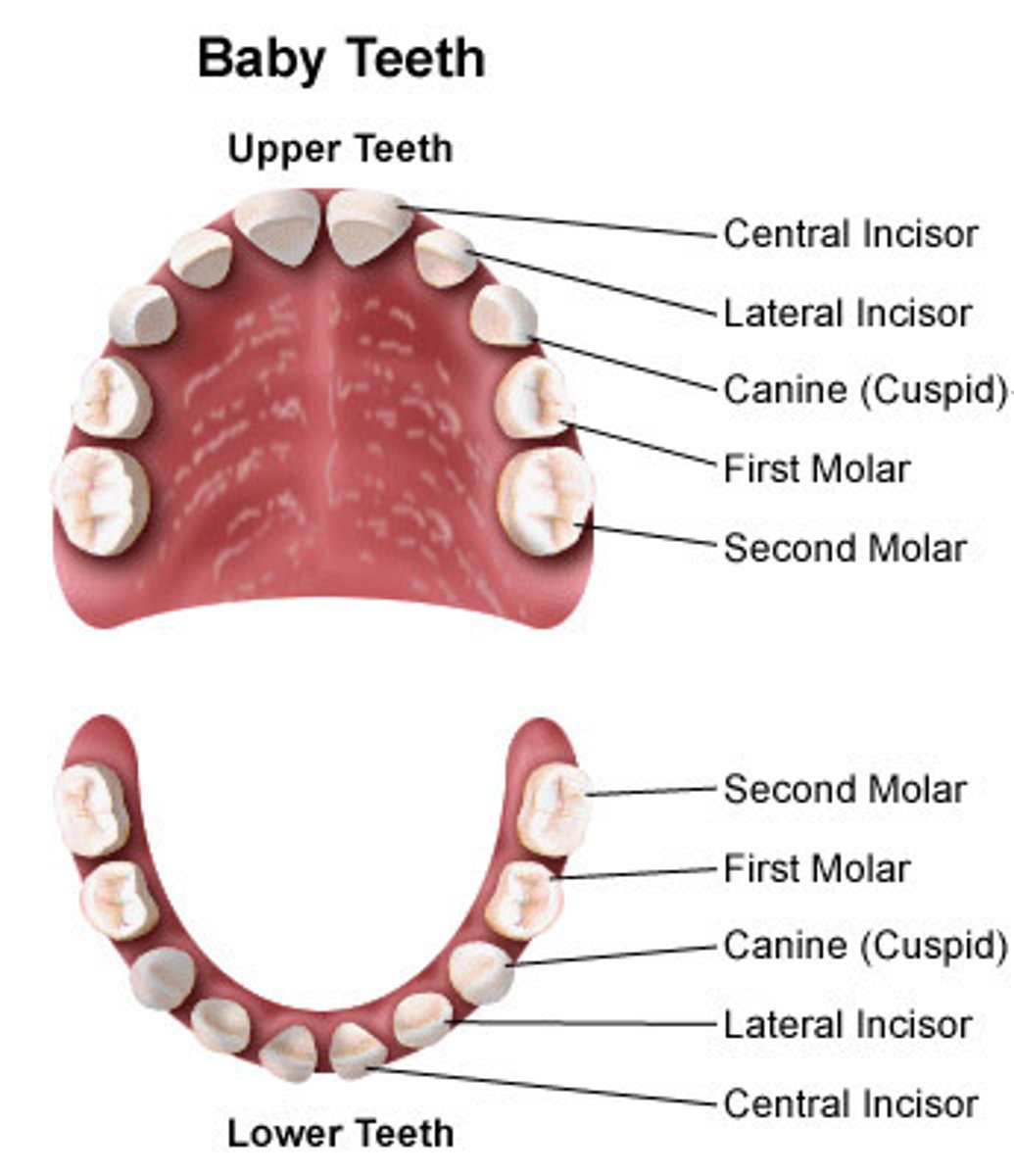
Deciduous Set of teeth
- first set (baby teeth)
- consist of:
2 incisors, 1 canine, and 2 molars in each quadrant of the jaw
- total of 20 teeth in the whole mouth
Permanent set of teeth
- consists of 2 incisors, 1 canine (cuspid), 2 premolars (bicuspids), and 3 molars in each quadrant of the jaw
- total of 32 teeth in the whole mouth
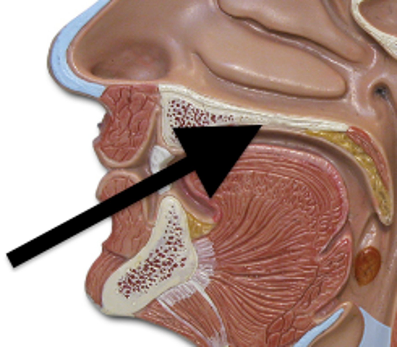
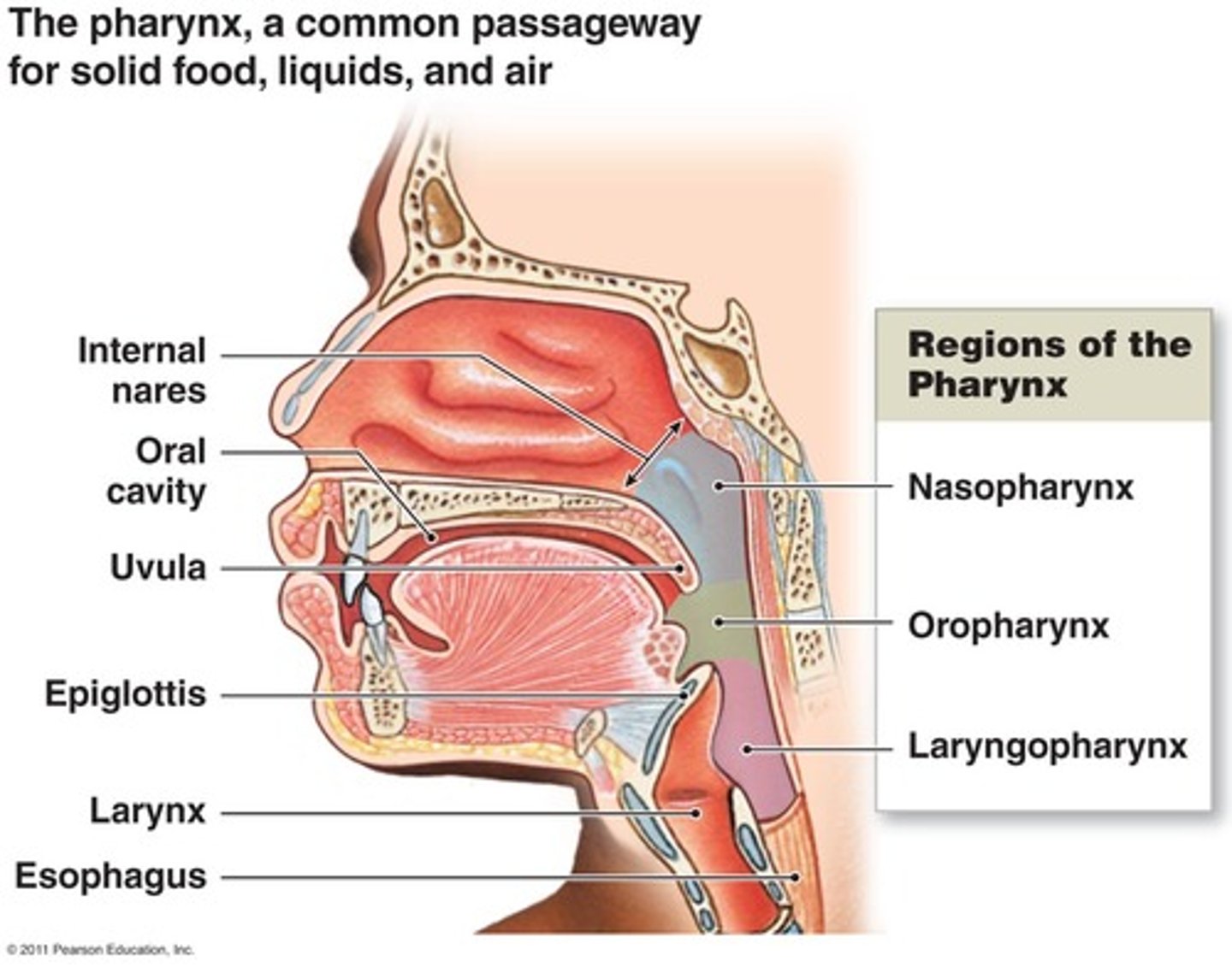
Oral Cavity: Pharynx
- part of both the digestive and respiratory system
Oral Cavity: Tonsils
- collection of lymphoid tissue found in areas of the pharynx
- they play a role in immune system
- there are 3 tonsils (palatine, pharyngeal and lingual tonsils)
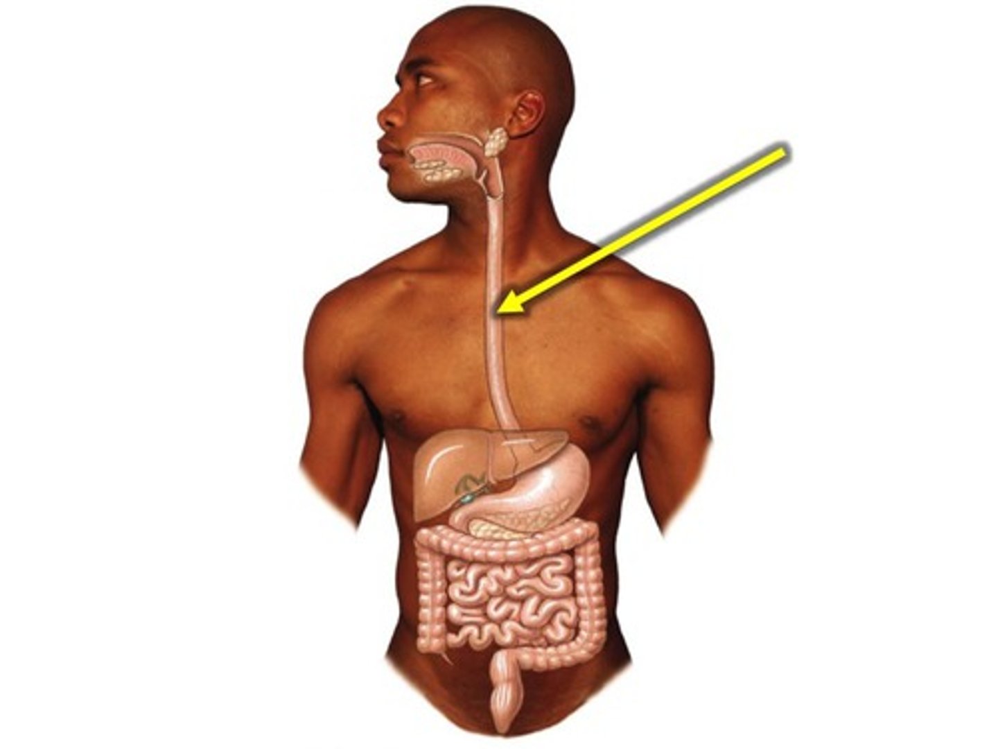
Esophagus: Gross Anatomy
- muscular tube
- 25cm in length
- extending from the pharynx to the stomach
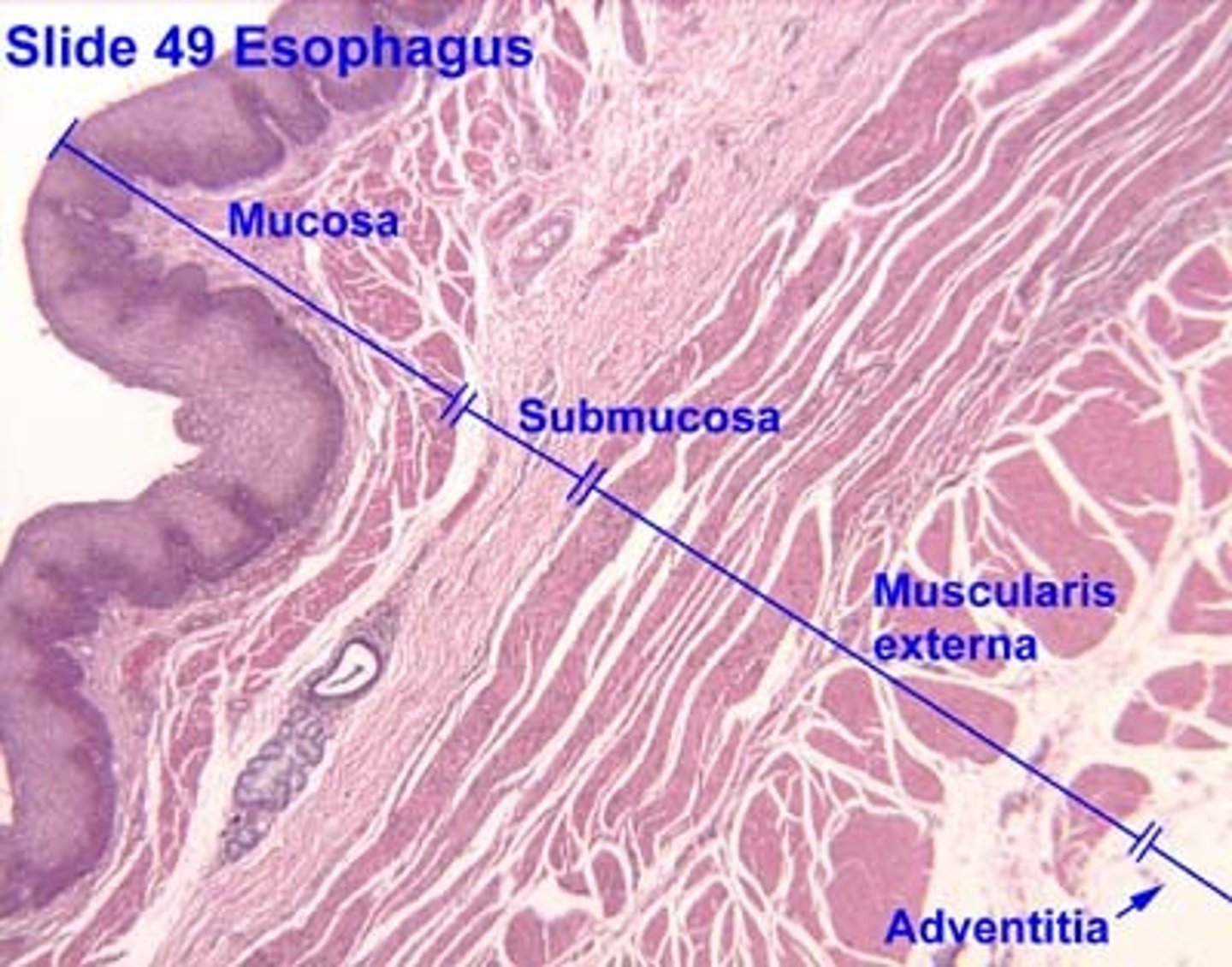
Esophagus Histology
- there are again 4 layers:
mucosa
submucosa
muscularis Externa
serosa/adventitia
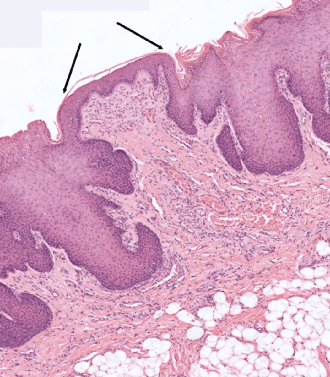
esophagus histology: mucosa
- the stratified squamous epithelium protects the esophagus from friction as food travels from the oral cavity down to the stomach
esophagus histology: submucosa
- contains mucus secreting glands
- the mucus is secreted through a duct to the lumen of the esophagus (lubricate the tract to allow food to pass)
esophagus histology: muscularis externa
- typically it contains only smooth muscle BUT in the esophagus, this layer includes both smooth muscle and skeletal muscle
- the upper 1/3 is skeletal muscle, the middle 1/3 is mixed, and the lower 1/3 is smooth muscle
esophagus histology: serosa/adventitia
- the major of the esophagus is covered by adventitia, which is composed of loose connective tissue
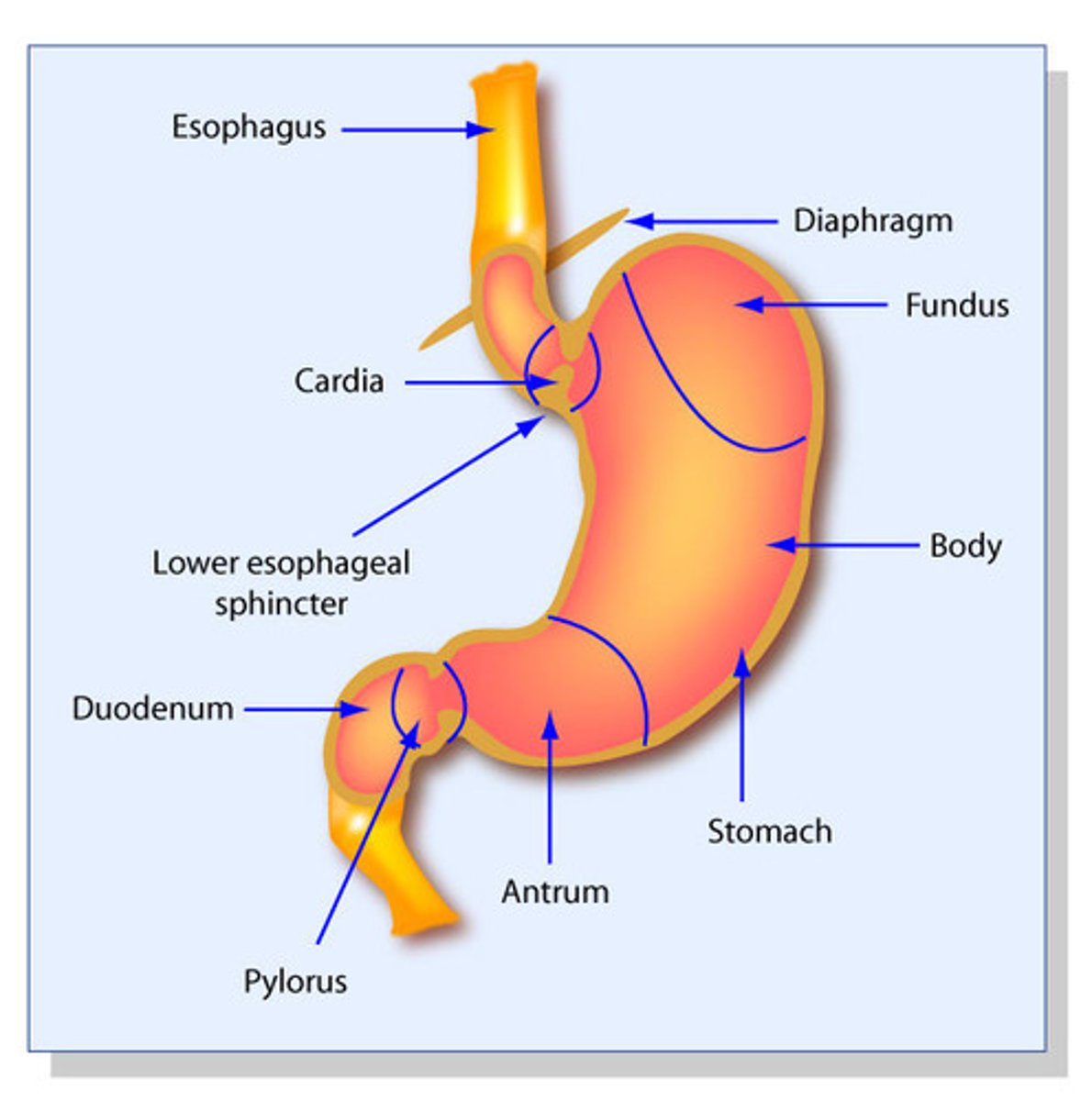
Regions of the stomach:
1. fundus
2. body
3. antrum
(the antrum is continuous with the duodenum of the small intestine)
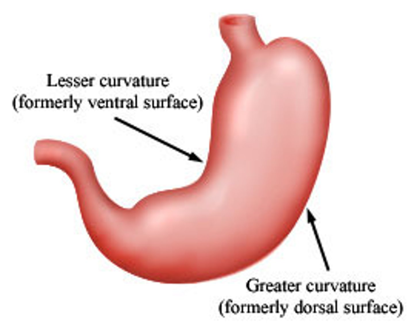
Stomach: Gross anatomy
- lesser and greater curvatures
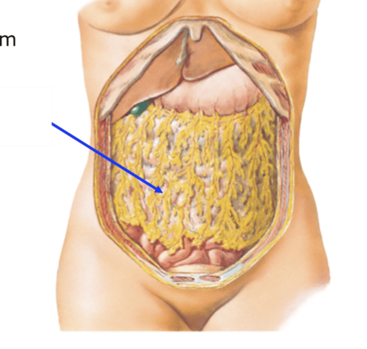
- greater omentum
lesser and greater curvature
greater omentum
- a structure that hangs off the greater curvature of the stomach
- it is an apron like structure that covers and protects the abdominal viscera
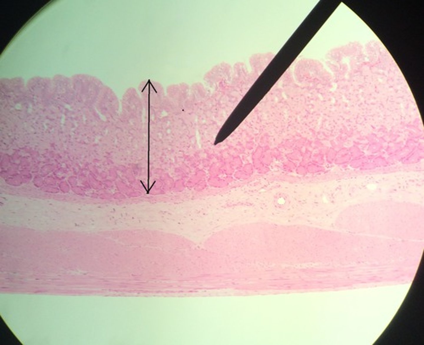
Stomach: histology
- the histology of the stomach is different from the esophagus because it has a different function
mucosa
submucosa
muscularis externa
serosa/adventitia
stomach histology: mucosa
- folded into ridges called RUGAE (allows the stomach to expand after ingestion of food or liquid)
- the epithelium is simple columnar and inward folds extend down into the LAMINA PROPORIA to form GASTRIC GLANDS
- the epithelium secretes mucus to help protect the stomach from acids secreted by gastric glands (that help digest food)
LOOK AT PICTURE IN BOOK
stomach histology: submucosa
- contains blood vessels, lymphatics, glands and nerve plexuses, which supply the stomach tissue with oxygen and control the contraction of the muscle
LOOK AT PICTURE IN BOOK
stomach histology: muscularis externa
- has layers of smooth muscle arranged in different directions
an outer longitudinal
middle circular
an inner oblique layer
- the stomach has 3 layer of muscle while the esophagus only has to because the stomach needs to churn and push out food
LOOK AT PICTURE IN NOTES
stomach histology: serosa/adventitia
- the stomach is covered by serosa
LOOK AT PICTURE IN NOTES
differences in the histological layers between the esophagus and the stomach
- the type of muscle in the esophagus changes the lower you get (skeletal to smooth)
- the stomach has 3 layers of muscle, the esophagus only has 2 layers of muscle
- there are differences in mucosa (esophagus has stratified squamous, stomach has simple columnar)
differences in mucosa of the esophagus and the stomach
- the stratified squamous of the esophagus allows for protection of the friction from passing food
- the simple columnar allows the secretion of acid, and mucus
2 types of intestines
- small intestine
- large intestine
Small intestine: gross anatomy
- approximately 6m in length and is divided into 3 portions
- it is attached to the posterior abdominal wall
Sections:
1. duodenum
2. jejunum
3. ileum
1. duodenum
- c-shaped and encloses the head of the pancreas
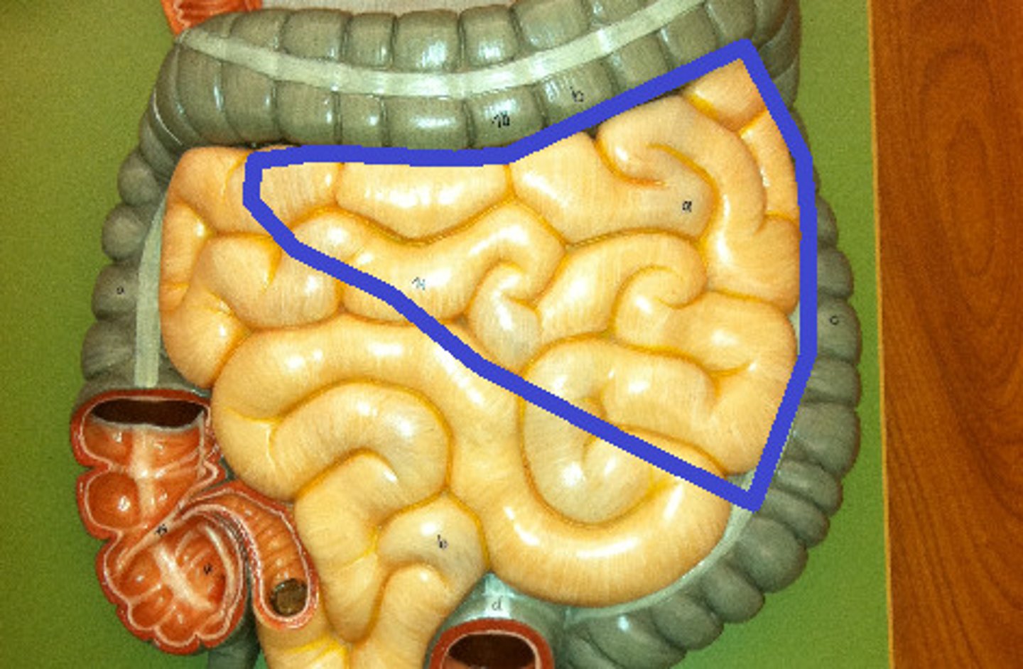
2. jejunum
- most of the jejunum lies in the upper left quadrant of the abdomen
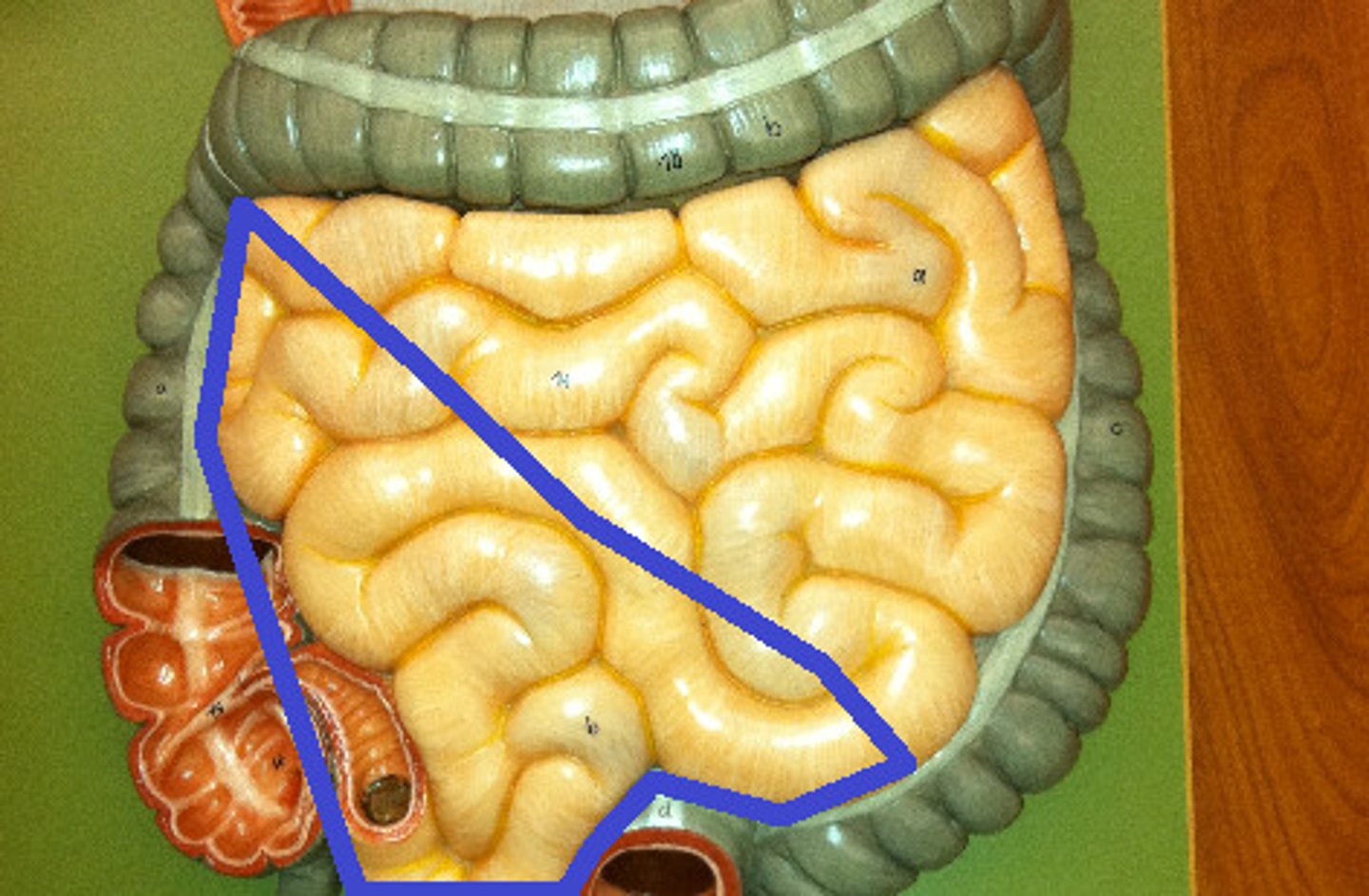
3. Ileum
- most of the ileum lies in the lower right quadrant of the abdomen
Small Intestine: histology
1. Mucosa
(villi)
(Epithelium)
(Lamina Propria)
(intestinal glands)
(plicae circulares)
2. Submucosa
(duodenal glands)
3. Muscularis Externa
(circular and longitudinal layers)
4. Serosa
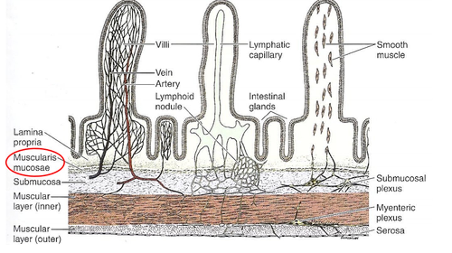
Small Intestine Histology: Mucosa (Villi)
- finger like projections that extend into the limen of the small intestine
Small Intestine Histology: Mucosa (Epithelium)
- the epithelium has villi
- simple columnar with many absorptive cells (whohas microvilli aka brush border)
- between are goblet cells, which secrete mucus to lubricate the passage of food
- Endocrine cells found in the epithelium that secrete hormones involved in the regulation of safety, blood sugar levels, and growth of epithelial cells
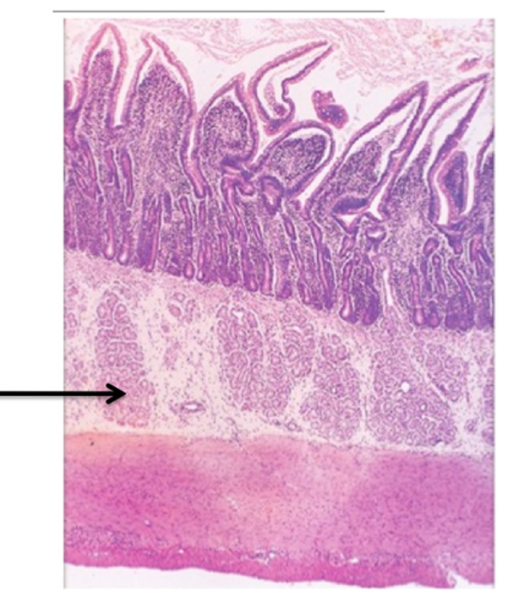
Small Intestine Histology: Mucosa (lamina propria)
- forms the core of each villus and contains blood capillaries and lymphatic capillaries
- there are collections of lymphatic tissue located in the lamina propria known as PETER'S PATCH which are primarily abundant in the ileum
LOOK AT PICTURE IN NOTES
Small Intestine Histology: Mucosa (intestinal glands)
- these glands are deep folds of the mucosa between the villi and secrete intestinal juices
LOOK AT PICTURE IN NOTES
Small Intestine Histology: Mucosa (Plicae circulares)
- the mucosa and submucosa of the small intestine form PLICAE CIRCULARES which are permanent transverse fold that help increase the surface area for absorption and causes the material to spiral through the small intestine
LOOK AT PICTURE IN NOTES
Small Intestine Histology: Submucosa
- has DUODENAL (BRUNNER'S ) GLANDS that secrete alkaline mucus
- this mucus protects the small intestine from stomach acids that still might be in the digested food
- these glands are only found in the duodenum portion
Small Intestine Histology: Muscularis Externa
- the small intestine has 2 smooth muscle layers which are organized into CIRCULAR AND LONGITUDINAL LAYERS
- a nerve plexus is located between these 2 layers of smooth muscle
Small Intestine Histology: Serosa
- the outermost layer of the small intestine is the serosa
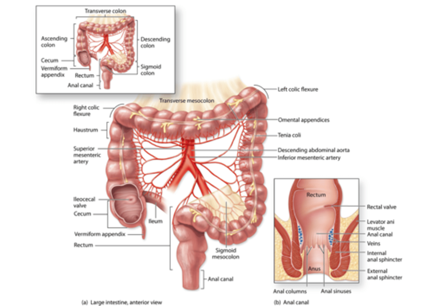
Large Intestine: Gross anatomy
- 3 sections:
1. cecum
2. colon
3. rectum
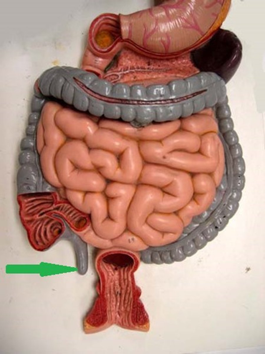
1. cecum
- forst portion of the large intestine
- has an extension off of it called the VERMIFORM APPENDIX
(the image points to the vermiform appendix)
2. colon
- second part of the large intestine
- 4 sections:
Ascending colon
transverse colon
descending colon
sigmoid colon
ascending colon
extends from the cecum up the right side of the abdomen to joint he transverse colon just below the liver

transverse colon
- extends from below the liver, crossing the abdomen to join the descending colon just below the spleen

descending colon
- extends down the left side of the abdomen from the splenic end of the transverse colon to the sigmoid colon
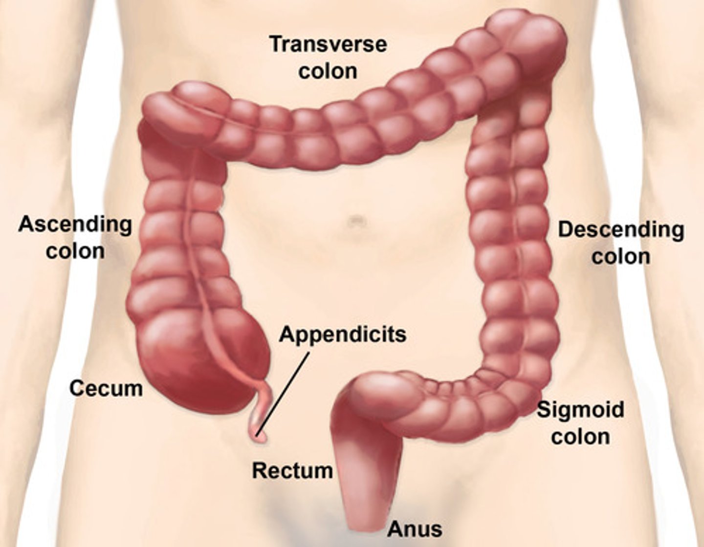
sigmoid colon
- the s-shaped terminal portion of the colon that leads to the rectum

3. rectum
- extends from the sigmoid colon to the anal canal
- it is the last potion of the large intestine

Large Intestine histology: Mucosa
(Epithelium)
(similar to the small intestine, however does not contain plicae circulares or villi)
- the epithelium is simple columnar and contains an increasing amount of goblet cells as you move towards the anus
(in the anal canale, a change from simple columnar to stratified squamous epithelium takes place)
LOOK AT PICTURE IN NOTES
Large Intestine histology: Mucosa (intestinal glands)
- like in the small intestine, intestinal glands are also present in the large intestine
- also, accumulations of LYMPHATIC TISSUE are present in the lamina propria (and submucosa)
LOOK AT PICTURES IN NOTES
Large Intestine histology: Submucosa
- contains blood vessels, lymphatics, and nerve plexuses
LOOK AT PICTURE IN NOTES
Large Intestine histology: Muscularis Externa
- depending on the location of the large intestine, the muscualris externa has a unique arrangement
Large Intestine histology: Muscularis Externa (Cecum and colon)
- the outer longitudinal layer forms 3 longitudinal branches (TENIAE COLI) that contract and bunch up the wall of the large intestine, forming sac-like structures called HAUSTRA
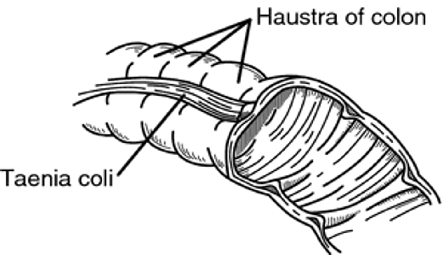
Large Intestine histology: Muscularis Externa (rectum)
- in the rectum, the 3 teniae coli merge to form the continuous longitudinal muscular layer
(in the image, the less bumpy part)
Large Intestine histology: Muscularis Externa (anal canal)
- the circular muscle layer thickens into the internal anal sphincter, which is involved with waste excretion
Large Intestine histology: adventitia/serosa
- the large intestine can be covered in either adventitia or serosa
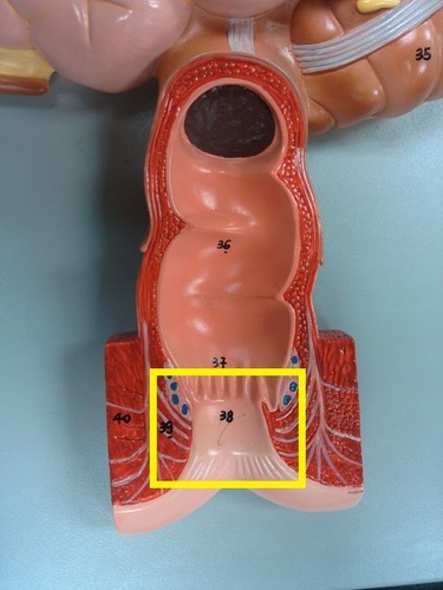
Anal Canal
- last portion of the digestive tract
Anal Canal : Gross anatomical features
ANAL COLUMN
- longitudinal ridges in the canal
INTERNAL AND EXTERNAL ANAL SPHINCTERS
- these maintain continence, and relax to enable evacuation during defecation
LOOK AT PICTURE IN NOTES