Ch 5: Synaptic Transmission
1/53
There's no tags or description
Looks like no tags are added yet.
Name | Mastery | Learn | Test | Matching | Spaced | Call with Kai |
|---|
No analytics yet
Send a link to your students to track their progress
54 Terms
Define synaptic transmission.
The process of information transfer at a synapse
What is the contribution of Charles Sherrington?
Gave synapses their name
What is the contribution of Otto Loewi?
Did experiments with frogs to provide support for the concept of chemical synapses.
What is the contribution of Furshpan and Potter?
Proved the existence of electrical synapses through experimentation on the nervous systems of crayfish.
Describe a presynaptic cell in a general synapse.
The first neuron in synaptic transmission
Describe a postsynaptic cell in a general synapse.
The target cell in synaptic transmission
Describe a presynaptic cell in the NMJ.
The presynaptic terminal of a motor axon
Describe a postsynaptic cell in the NMJ.
The motor endplate, which contains shallow folds where the presynaptic active zones precisely align with
Describe the structure for a gap junction.
The membranes of two cells are ~3nm apart
ConnexINs (clusters of specialized proteins span the gap)
Six connexIN subunits combine to form a channel called a connexON
Two connexONs (one from each cell) meet and combine to form a gap junction channel
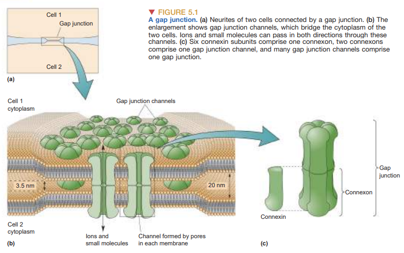
What is the function of the gap junction channel?
It allows ions to pass directly from the cytoplasm of one cell to the cytoplasm of another cell
How large are the pore of most gap junction channels and why is this relevant?
1-2 nm
It is large enough for all the major cellular ions and many small organic molecules to pass through
Describe the synaptic transmission for a gap junction.
Synaptic transmission occurs via electrical current
Cells connected by gap junctions are said to be electrically coupled
An action potential in the presynaptic neuron causes a small amount of ionic current to flow across the gap junction channels into the other neuron
Ionic current causes an electrically mediated postsynaptic potential (PSP) in the second neuron
Most electrical synapses are bidirectional → the generation of an action potential in the second neuron can in turn induce a PSP in the first neuron
It may take multiple PSPs occurring simultaneously to strongly excite a neuron AKA synaptic integration
Describe directionality for a gap junction.
Synaptic transmission is bidirectional due to electrical current
Describe the 7 steps of chemical synaptic transmission.
Presynaptic cell synthesizes neurotransmitter
Presynaptic cell loads neurotransmitter into synaptic vesicles
Vesicles fuse/undergo exocytosis to presynaptic cleft
Neurotransmitter spills into synaptic cleft
Neurotransmitter binds to postsynaptic receptors
Biochemical/electrical response elicited in postsynaptic cell
Leftover neurotransmitter is removed from the synaptic cleft
Describe the 5 types of CNS chemical synapses.
Axodendritic: axon to dendrite
Axosomatic: axon to cell body (soma)
Axoaxonic: axon to axon
Axospinous: Axon to dendritic spine
Dendrodendritic: dendrite to dendrite
Describe the structure for an NMJ.
occurs between the axons of motor neurons of the spinal cord and skeletal muscle
presynaptic terminal contains a large number of active zones
postsynaptic membrane/motor endplate has folds that contain neurotransmitter receptors
This structure ensures that many neurotransmitter molecules are focally released onto a large surface of chemically sensitive membrane
Describe the synaptic transmission for an NMJ.
fast and reliable
an action potential in the motor axon always causes an action potential in the muscle it innervates
Describe directionality for an NMJ.
Neuron to axon to muscle
Describe the 3 types of neurotransmitters and how they are packaged.
Amines → stored in synaptic vesicles
Amino acids → stored in synaptic vesicles
Peptides → stored in secretory granules
Describe neurotransmitter synthesis and storage for peptides.
A long peptide is synthesized in the rough ER
Peptide is split in the Golgi apparatus
One of the smaller peptide fragments is the active neurotransmitter
Secretory granules containing the peptide neurotransmitter bud off from the Golgi apparatus
Those secretory granules are transported to the axon terminal via axoplasmic transport
Describe neurotransmitter synthesis and storage for amino acids and amines.
Neurons contain specific enzymes that synthesize the neurotransmitters from various metabolic precursors
The synthesizing enzymes for both amine and amino acid neurotransmitters are transported to the axon terminal
Enzymes locally and rapidly direct neurotransmitter synthesis in the cytosol of the axon terminal
Neurotransmitters are packed into synaptic vesicles via transporters (special proteins embedded into the vesicle membrane)
Describe the 4 steps of neurotransmitter release.
Vesicle full of neurotransmitter is docked and primed
Intracellular calcium triggers exocytosis of the neurotransmitter
Vesicle membrane is incorporated into presynaptic membrane and neurotransmitter is released into the cleft
Vesicle membrane is recovered by endocytosis
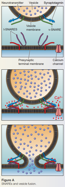
Describe the role of calcium in vesicle fusion.
Calcium binds to synaptotagmin to induce docking of the vesicle to the presynaptic membrane
Describe the role of in synaptotagmin in vesicle fusion.
It is a vesicle protein that sense to Ca2+ to trigger vesicle fusion and neurotransmitter release
Describe the role of v-SNARES and t-SNARES in vesicle fusion.
V-SNARES on the vesicle bind to t-SNARES on the presynaptic terminal membrane
The cytosolic ends of these SNAREs bind very tightly to one another
Allows a vesicle to dock very close to a presynaptic membrane
Describe the role of clathrin in vesicle endocytosis.
A protein that binds to the membrane → causes a conformational change to the membrane to form a “soccerball” vesicle
Describe the role of dynamin in vesicle endocytosis.
Pinches the vesicle off from the membrane
Describe 4 ways neurotransmitter can be removed from the synaptic cleft.
Diffusion of transmitter molecules away form the synapse
Reuptake: NT reenters presynaptic axon terminal
Enzymatic destruction (i.e. acetylcholinesterase) inside the terminal cytosol or synaptic cleft
Desensitization: Receptors become insensitive to NT, despite the presence of NT
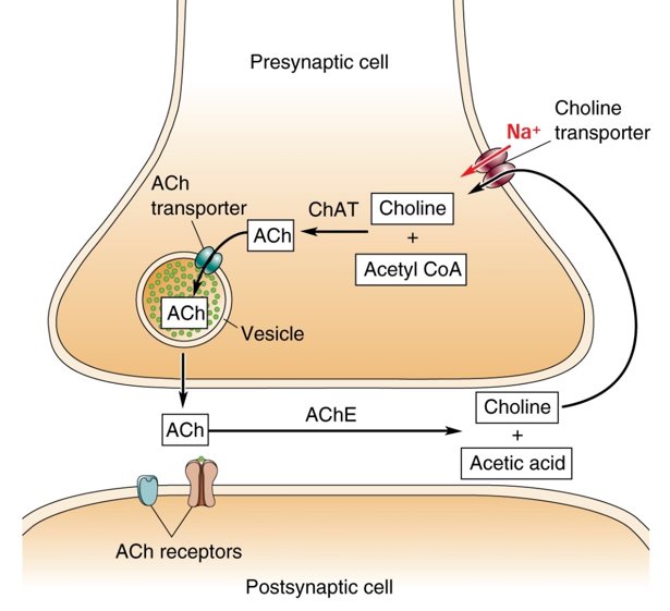
Describe the cycle of acteylcholine.
Neurons release vesicles with acetylcholine
ACh goes into synaptic cleft and binds to ACh receptors in the postsynaptic cell.
AChE in the synaptic cleft degrades ACh into choline and acetic acid
Choline is reuptaken back into the presynaptic cell via a cotransporter → transports both choline and Na+
Choline and Acetyl CoA add together in a chemical reaction catalyzed by choline acetyltransferase (ChAT) to make ACh
ACh is transported into vesicles awaiting exocytosis
Describe the action of the acetylcholine receptor.
Receives ACh from the postsynaptic cleft
Describe the location and role of an autoreceptor.
Presynaptic receptors that are sensitive to the neurotransmitter released by the presynaptic terminal
Located on the presynaptic axon terminal
Functions on negative feedback
Autoreceptor senses release of NT and therefore inhibits further release of NT
Define agonist.
Mimics the action of naturally occurring neurotransmitters
Define antagonist.
Inhibits neurotransmitter receptors
Describe the source and action of alpha bungarotoxin.
From cobra snake
Blocks the AChR channel in a closed position = paralysis (muscle cannot contract)
Describe the source and action of curare.
Naturally occurring poison from plants
Binds and blocks AChR channels from opening
Only prevents movement, needs to be used with an analgesic to numb pain
Describe the source and action of organophosphates.
DTT & herbicides are insecticides (malathion, parathion)
Inhibit AChE → ACh accumulates in the synaptic cleft → AChR becomes desensitized → inability to contract muscles
Describe the source and action of latrotoxin
Produced by the female black widow spider
Causes depletion of synaptic vesicles by stimulating mass exocytosis of of vesicles dependent of calcium increase
Describe the source and action of botulinum toxin.
From Clostridium bacteria
Protease that cleaves the core SNARE complex proteins used for vesicle fusion → prevents vesicles form docking → prevents exocytosis
Describe the source and action of tetanus.
From Clostridium bacteria
Protease that cleaves the core SNARE complex proteins used for vesicle fusion → prevents vesicles form docking → prevents exocytosis
Define EPSP.
Excitatory postsynaptic potential
Presynaptic neurotransmitter release → transient postsynaptic membrane depolarization
Define IPSP.
Inhibitory postsynaptic potential
Presynaptic neurotransmitter release → transient postsynaptic membrane hyperpolarization
Define presynaptic facilitation.
Increase in the amount of neurotransmitter released from postsynaptic neuron
Define quantal analysis.
Used to determine numbers of vesicles that release during neurotransmission
We cannot measure a single neurotransmitter, but we can measure a single vesicle
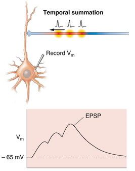
Define temporal summation.
EPSPs generated at same synapse in rapid succession
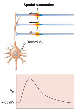
Define spatial summation.
EPSPs generated simultaneous at different sites
Define dendritic length constant.
The distance that a postsynaptic potential can spread along the membrane until it reaches 37% of origin
Define internal resistance.
The resistance to current flowing longitudinally down the dendrite (dependent on diameter)
Define membrane resistance.
Explain how the dendritic length constant changes if the internal resistance increases.
Dendritic length constant increases as membrane potential increases
Explain how the dendritic length constant changes if the diameter increases.
Dendritic length constant decreases as internal resistance/diameter increases
Explain how the dendritic length constant changes if the membrane resistance increases.
Explain how the dendritic length constant changes if more channels are open.
Explain shunting inhibition.
Explain modulation.