Histology - Muscle/Nervous Tissue & Cell Cycle & Integumenary System
1/21
There's no tags or description
Looks like no tags are added yet.
Name | Mastery | Learn | Test | Matching | Spaced | Call with Kai |
|---|
No analytics yet
Send a link to your students to track their progress
22 Terms
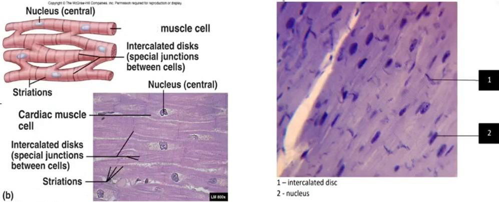
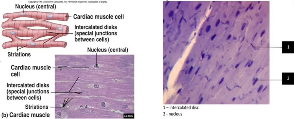
What tissue is this
Cardiac
-has intercalculated discs
-single nucleus per cell
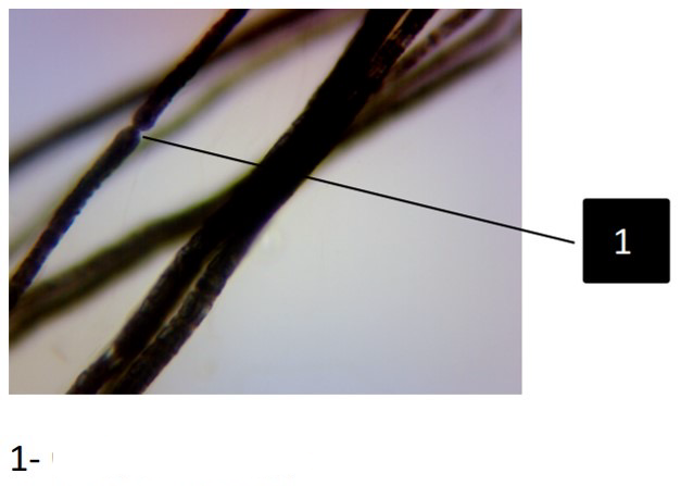
Name trait & which tissue it belongs to
Node of Ranvier; Nervous tissue (breaks up the myelin sheath into sections)
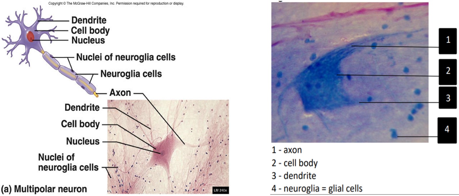
Label all terms
1- axon
2-cell body
3- dendrite
4- neuroglia(glia cells)

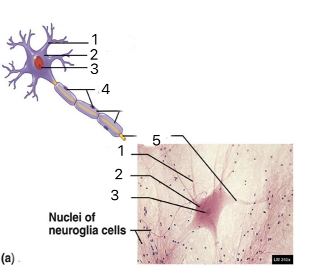
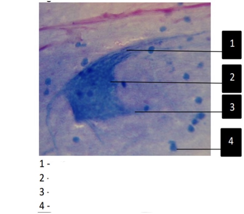
Label all terms
1- Dendrite
2- Cell body
3- Nucleus
4- Nuclei of nueroglia cells
5- Axon
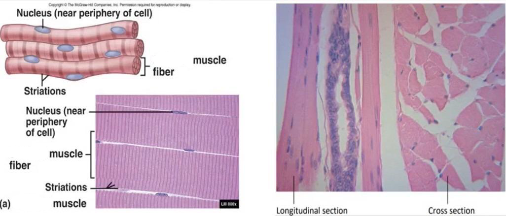
What tissue is this
Skeletal Muscle
-has straitions
-multiple peripheral nuclei
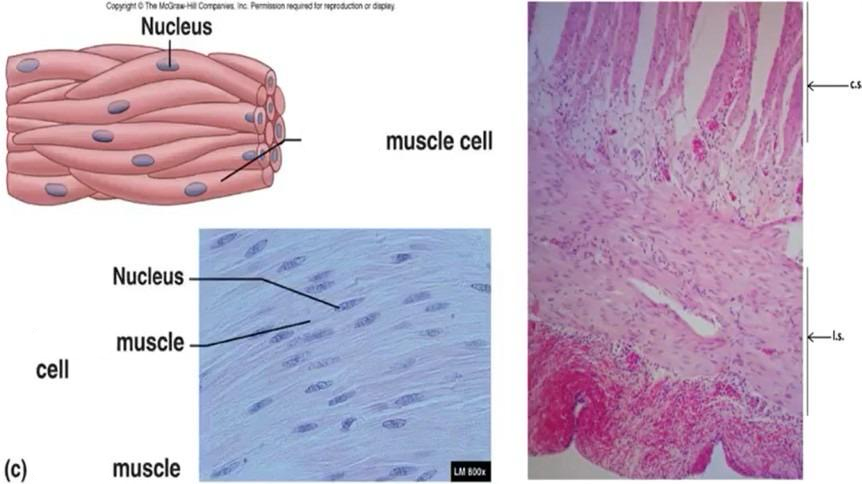
What tissue is this
Smooth muscle
-no straitions
-one nucleus per cell
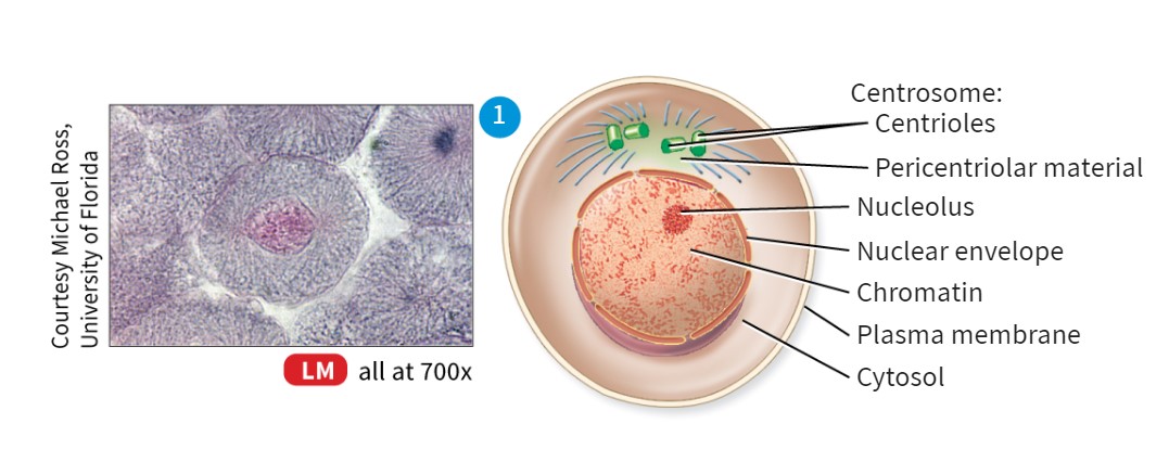
Stages of Interphase
G1 - Cell experiences growth
S - Growth stops, DNA replication occurs
G2 - Cell grows more, metabolic processes increase in preparation for mitosis
Note that interphase’s subdivisions are hard to identify under a microscope, the nucleus will look blurry during this phase
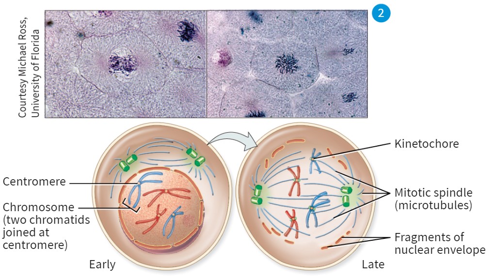
Explain what happens in the prophase stage during mitosis and how it would look
-DNA forms into chromosomes, and the DNA envelope will break down
-Nucleus appearance will look like dark random lines in the center of cell
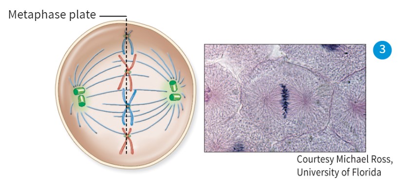
Explain Metaphase and how it would look
-Chromosomes will line up straight in the middle
-Spindle fibers will be on opposite sides
Explain Anaphase and how it would look
Chromosomes are separated into sister chromatids
-spindles are faded/not there
-chromosomes are dark and being pulled apart from the middle
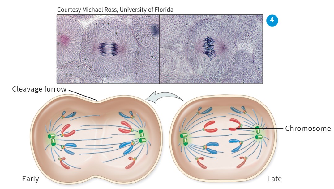
Explain Telophase and how it looks
-DNA condenses back to chromatin
-Clevage burrow forms, nuclear envelope reforms, spindle fibers disappear
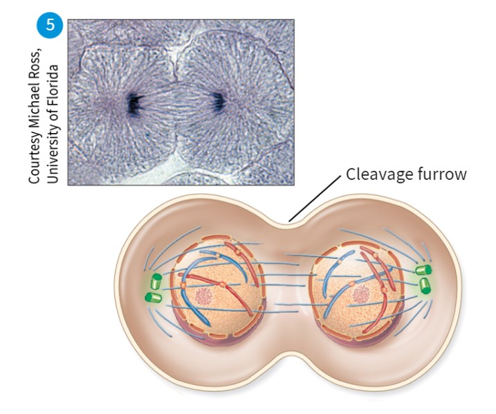
When does cytokinesis appear
Through Anaphase to after Telophase
Miotic Index
number of cells growing divided by the total number of cells
-A high miotic index indicates cancereous tissue
Layers of the epidermis, from deep to superficial
-Stratum Basale
-Stratum Spinosum
-Stratum Granulosum
-Stratum Lucidum (Only in Thick Skin)
-Stratum Corneum
Label all terms

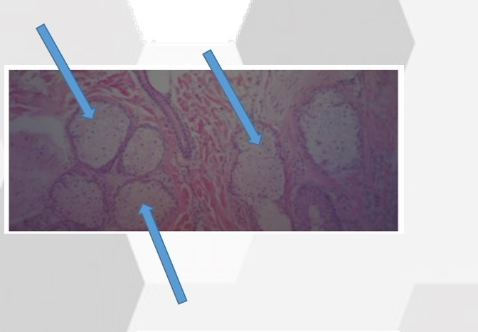
What is this gland
Sebaecous Glands
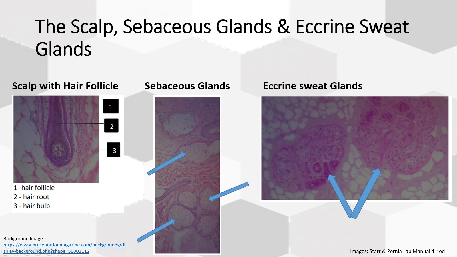
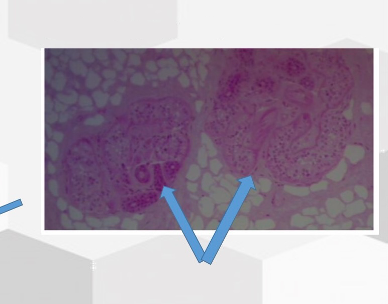
Name this gland
Eccrine (Sweat) Glands

Difference between Thin & Thick Stratum Corneum?
The thick stratum corneum has the layer stratum lucidum present
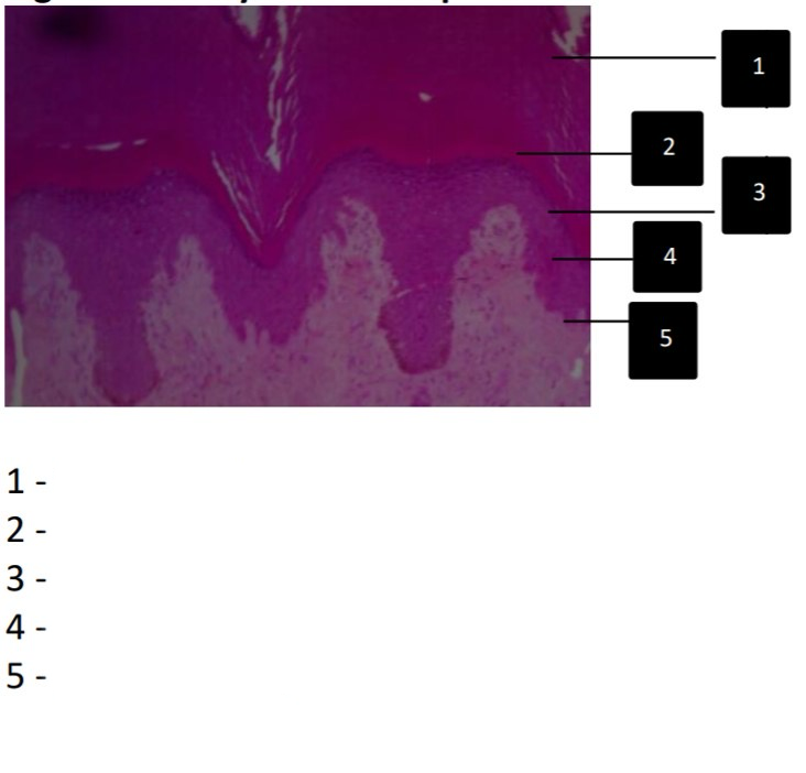
Label all parts
1- stratum corneum
2- stratum lucidum
3- stratum granulosum
4- stratum spinosum
5- stratum basale
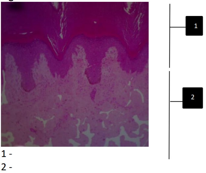
Label all parts
1- epidermis
2- dermis
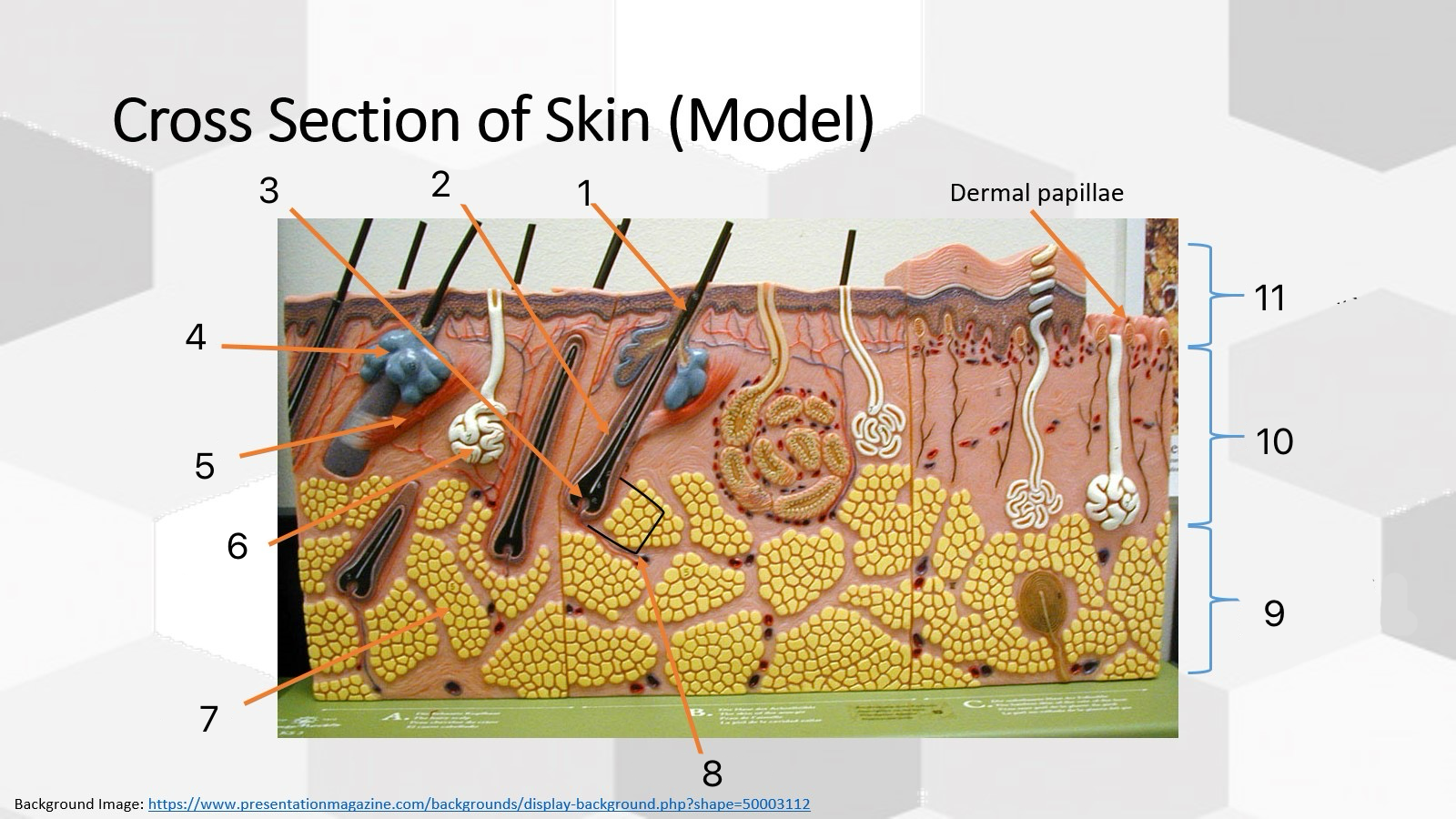
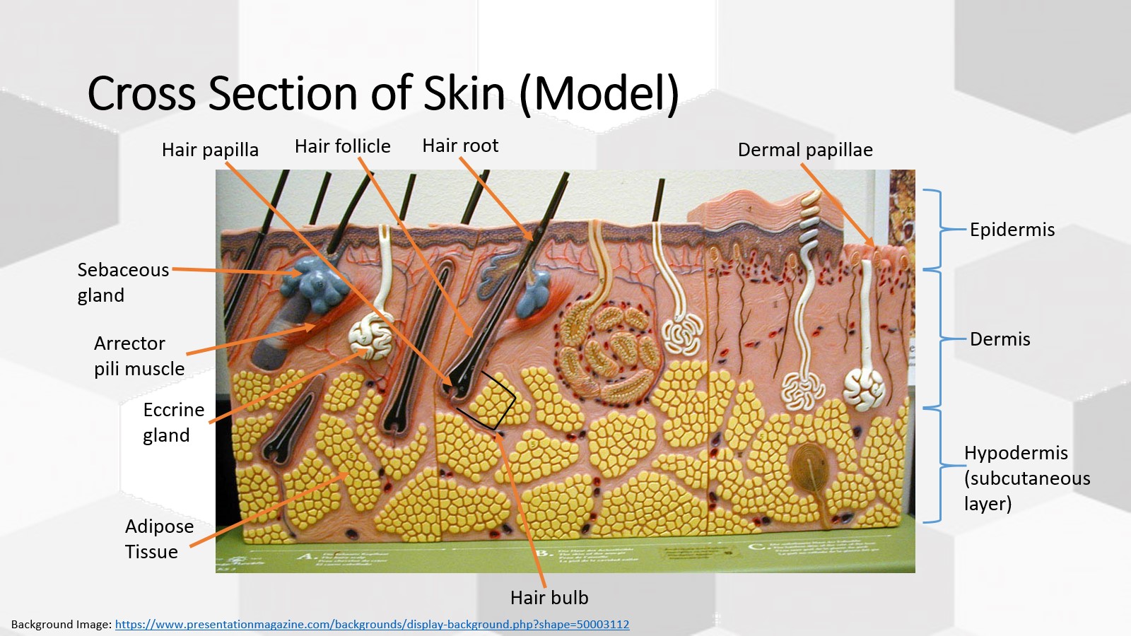
Label all parts
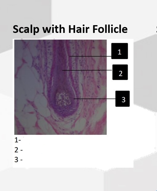
1- Hair root
2- Hair follicle
3- Hair papilla
4- Sebaceous gland
5- Arrector pili muscle
6- Eccrine gland
7- Adipose tissue
8- Hair bulb
9- Hypodermis
10- Dermis
11- Epidermis
Definition of Cancer
Uncontrolled cell division, cells continue to mass produce unregulated daughter cells, which leads to a tumor
maglinant cancer - spreads to other reigons of body