Biology II Exam 3- Stoffer
1/67
There's no tags or description
Looks like no tags are added yet.
Name | Mastery | Learn | Test | Matching | Spaced | Call with Kai |
|---|
No analytics yet
Send a link to your students to track their progress
68 Terms

Distinguish among the approaches used by osmoconformers and osmoregulators, listing examples of each.
Osmoconformers: in osmotic equilibrium with environment
-Marine environments/marine invertebrates
-Salt water
-Hagfish (vertebrates)
Osmoregulators: maintain constant blood + tissue osmolarity different from the environment
-Vertebrates
-Allows for living many places, but requires continuous regulation

Compare and contrast osmotic challenges faced by animals in freshwater, marine, and terrestrial environments, and the adaptations used to address them.
Freshwater: hypertonic (more solute) to surrounding water
-Water wants to enter bodies
-Need to prevent excess water and prevent less of inorganic ions
Marine: hypotonic (less solute) to surrounding seawater
-Water wants to leave
-Need to prevent loss of water
-They drink seawater, and eliminate excess ions with kidneys and gills to help get rid of unnecessary salt, while keeping the water
Terrestrial: higher concentration of water than air; living in air creates constant risk of water loss
-Lose water by evaporation w/ skin and lungs
-Fluctuation of fluid and ions
Describe the different kinds of nitrogenous waste, their relative toxicity and solubility, and list which animals excrete each of them.
1. Ammonia: very toxic, must be removed quickly
-Metabolism of amino acids + nucleic acids ~ deamination (removal of amino, NH2) and combination with H+ to form ammonia (NH3)
-Excretion is easy for bony fish + tadpoles (diffusion through gills and small amount of dilute urine.
Urea and uric acid are less toxic but have different solubilities
2. Urea: organic molecules formed in vertebrate liver
-Mammels
-Water-soluble
-Made in liver, carried in bloodstream to kidneys
3. Uric acid: insoluble; reptiles, birds, insects
-Excreted in a pasty-white material in bird droppings (guano)
-Costs energy to synthesize, but conserves water
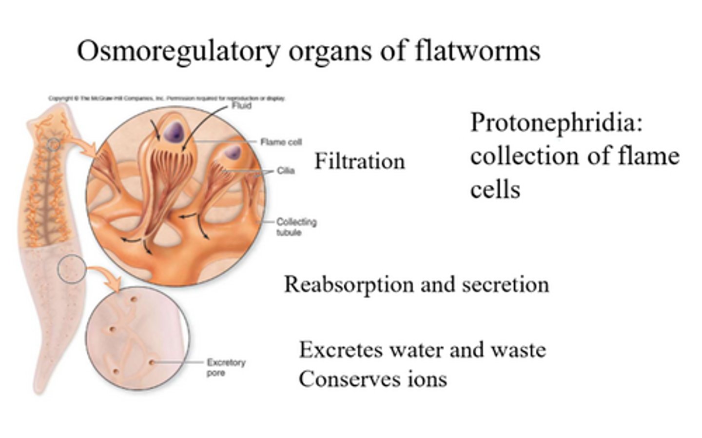
Osmoregulatory organs of flatworms
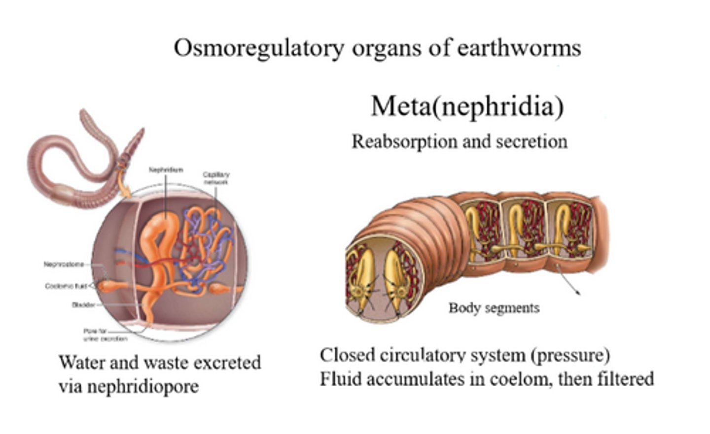
Osmoregulatory organs of earthworms
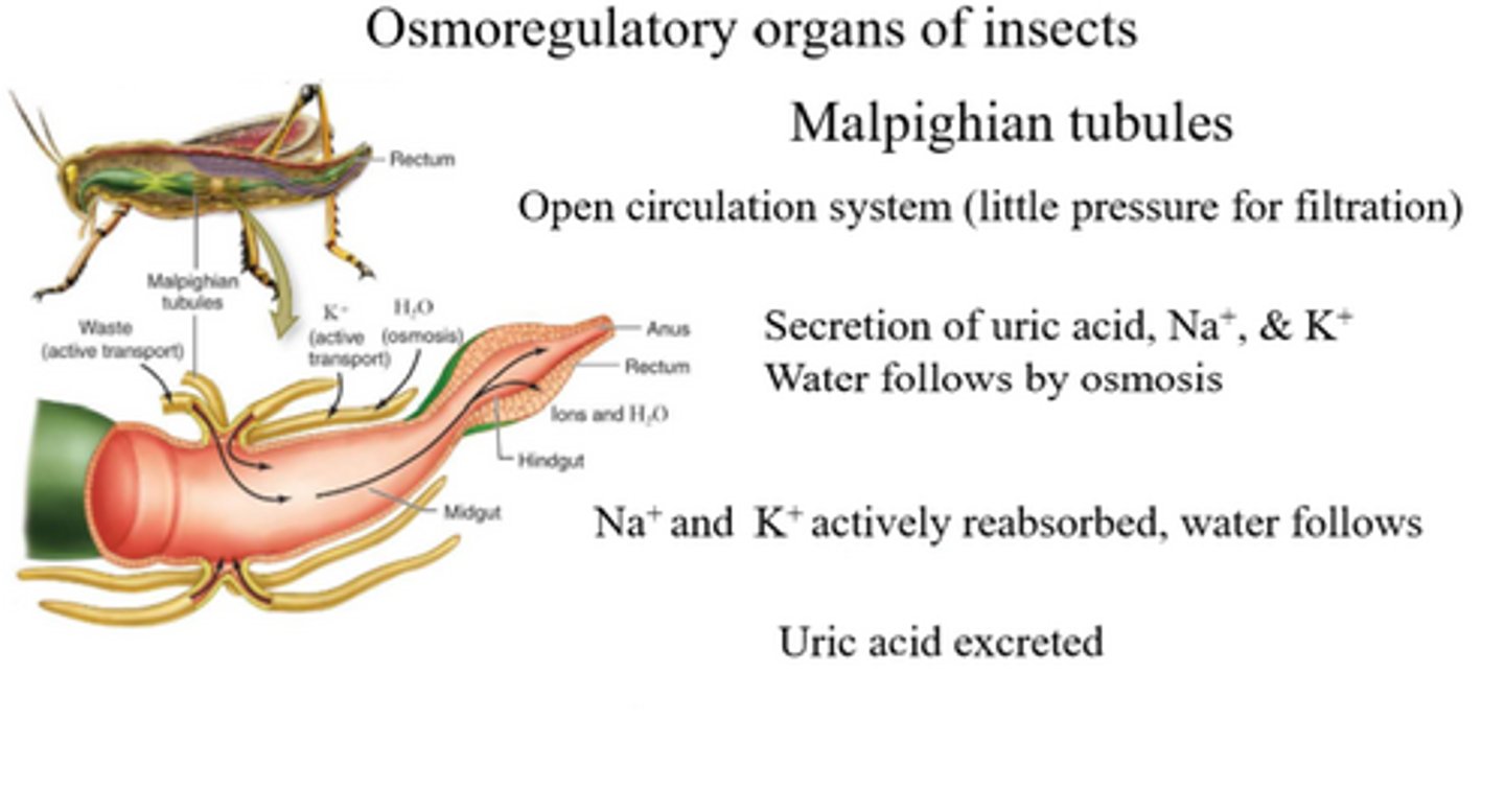
Osmoregulatory organs of insects
How the processes of filtration, reabsorption, secretion, and excretion occur.
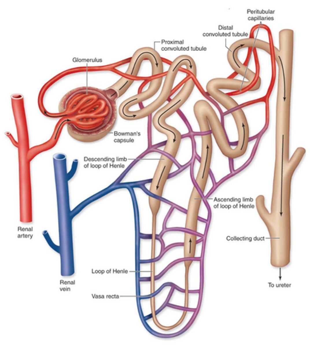
Filtration: driven by hydrostatic pressure (blood pressure); blood pressure in the glomerulus forces water and small solutes through the filtration membrane into Bowman’s capsule, forming the initial filtrate.
Reabsorption occurs through a combination of active Na⁺ transport in the ascending loop of Henle, urea recycling in the collecting duct that increases medullary osmolarity, and ADH‑regulated aquaporin insertion that allows water to follow the osmotic gradient. Together, these mechanisms enable the kidney to reclaim water and produce concentrated urine.
Secretion is when the nephron moves extra wastes from the blood into the filtrate, mainly in the distal tubule but also in the proximal tubule. The cells in these tubules use active transport to push things like hydrogen ions, potassium ions, drugs, and toxins out of the peritubular capillaries and into the tubule. This helps the body get rid of substances that were not filtered earlier and also helps control pH and electrolyte balance.
Excretion is the final step where the fluid leaving the collecting duct is now considered urine. After this point, the urine flows into the renal pelvis, then down the ureter, into the bladder, and finally out through the urethra. This step removes the body’s wastes, extra ions, and extra water.

Explain the significance of the loop of Henle in mammalian and avian kidneys.
The loop of Henle is a key structure in mammalian and avian kidneys that concentrates urine and conserves water. As filtrate moves down the loop, water is reabsorbed into the surrounding tissue because the descending limb is permeable to water. As it moves up, ions like sodium and chloride are actively pumped out, but water cannot follow because the ascending limb is impermeable to water. This creates a concentration gradient in the kidney, allowing the collecting ducts to reabsorb more water and produce hyper‑concentrated urine, which helps mammals and birds survive in environments where water is limited.
Mammalian nephron
Using ADH as an example, explain how homeostatic kidney functions are coordinated.
ADH (antidiuretic hormone): key regulator or maintaining homeostasis by adjusting and water and solute levels in the body.
-When the body is dehydrated, ADH is released from the pituitary gland and acts on the collecting ducts of the kidney, making them more permeable to water. This allows water to be reabsorbed back into the blood, producing concentrated urine, and conserving water.
-When the body has excess water, ADH secretion decreases, the collecting ducts become less permeable, and more water is excreted in dilute urine.
-More ADH = more aquaporins in collecting duct
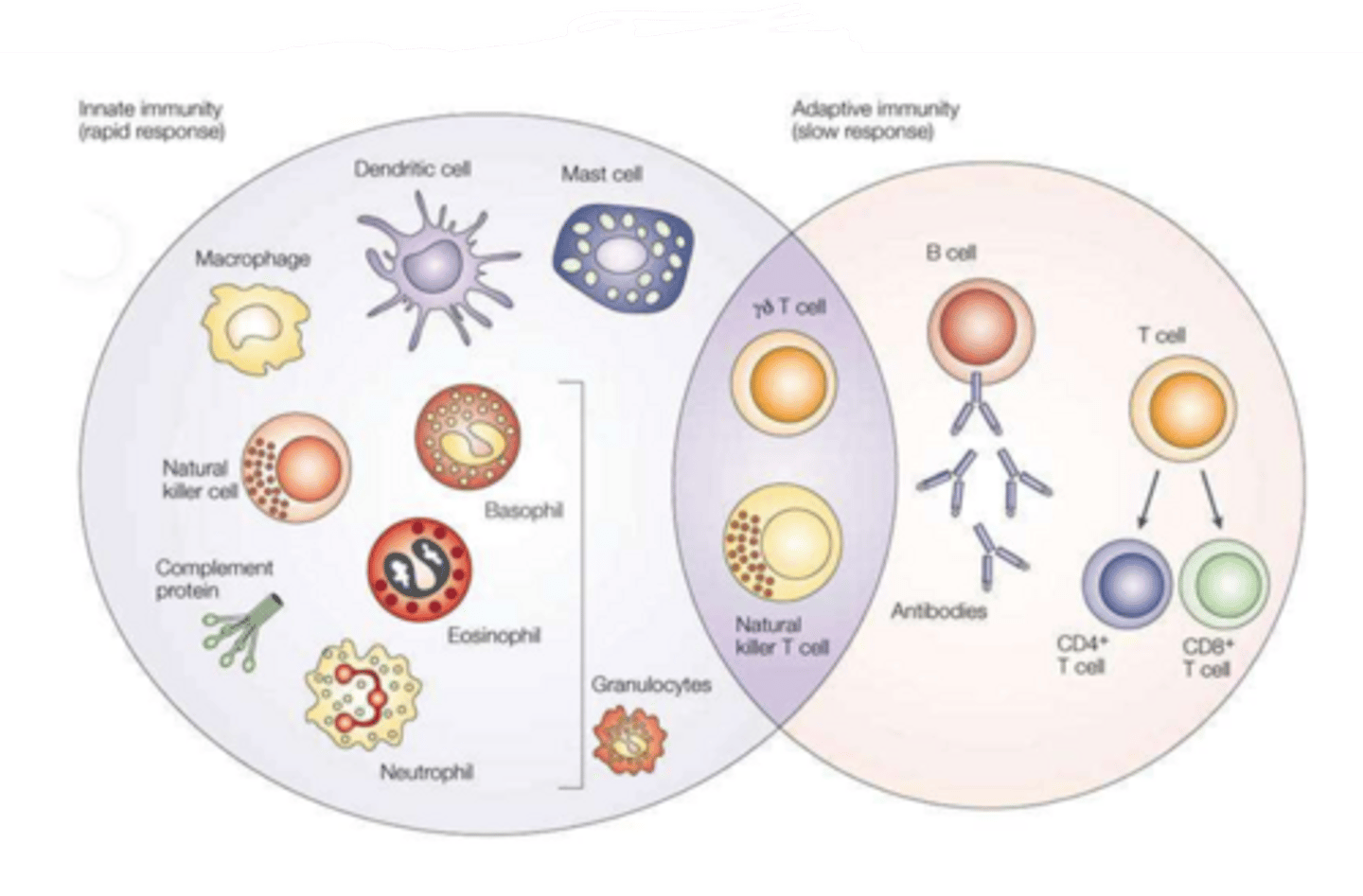
Describe the barrier and internal defenses that functions as the innate immune system of vertebrates. Explain why these are considered non-adaptive defenses.
Innate immunity: recognizes specific features of entire classes of pathogens; can stimulate an adaptive immune response
-skin, mucus, vomiting, diarrhea, coughing, sneezing
-pattern recognition receptors bind to pathogen-associated molecular patterns (PAMP)
Innate because: immediate, nonspecific, consistent
List the distinguishing characteristics of adaptive immunity and how they are brought about.
Specific, diversity, memory, self vs. non-self recognition, recognition of chemical signals
Lymphocytes (WBC, B cells, T cells): carry out adaptive immune responses; they have receptor proteins that recognize epitopes on an antigen
For each of the following molecules describe their composition, where they originate from, and their function in adaptive immunity: Epitope, Antigen, MHC, Antibody, and Cytokines.
Epitope: part of an antigen; target antibodies can bind to trigger an adaptive immune response
Antigen: derived from pathogens; substance capable of eliciting an immune response
MHC (major histocompatibility complex): produced by host cells; membrane glycoproteins
Antibody: y-shaped glycoprotein made of 2 heavy chains and 2 light chains; recognize/bind specific antigens
Cytokines: small signaling proteins secreted by immune cells; regulate proliferation, differentiation, and activation during adaptive immune response
For each of the following cells describe where in the body they originate from and what role each plays in adaptive immunity: Macrophage, Natural killer cells, Mast cells, B-cells, and T-cells.
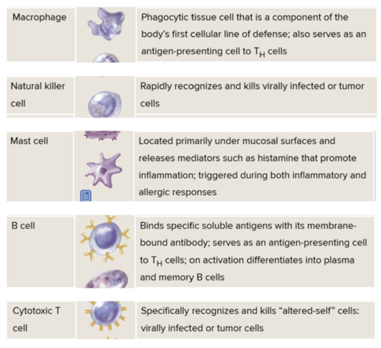
Cells in innate and adaptive immune response- slide from lecture
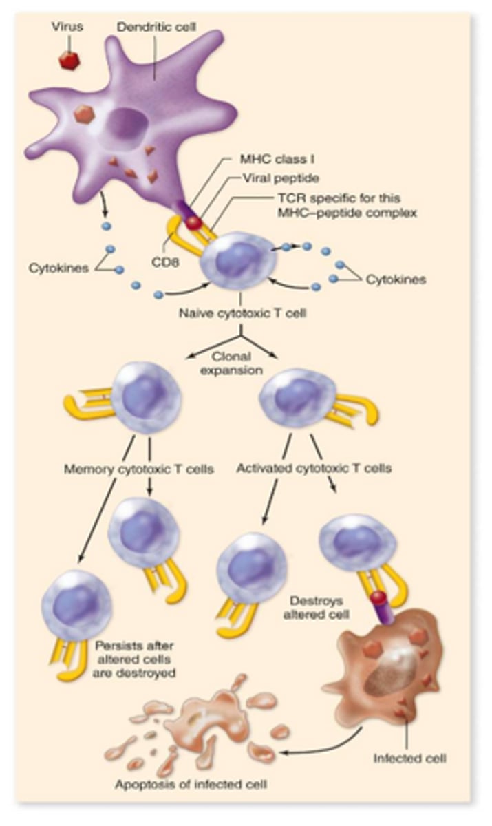
Distinguish between the roles of antigen-presenting cells, B-cells, plasma cells, helper T-cells, and cytotoxic T-cells, and how they carry them out. *image from textbook
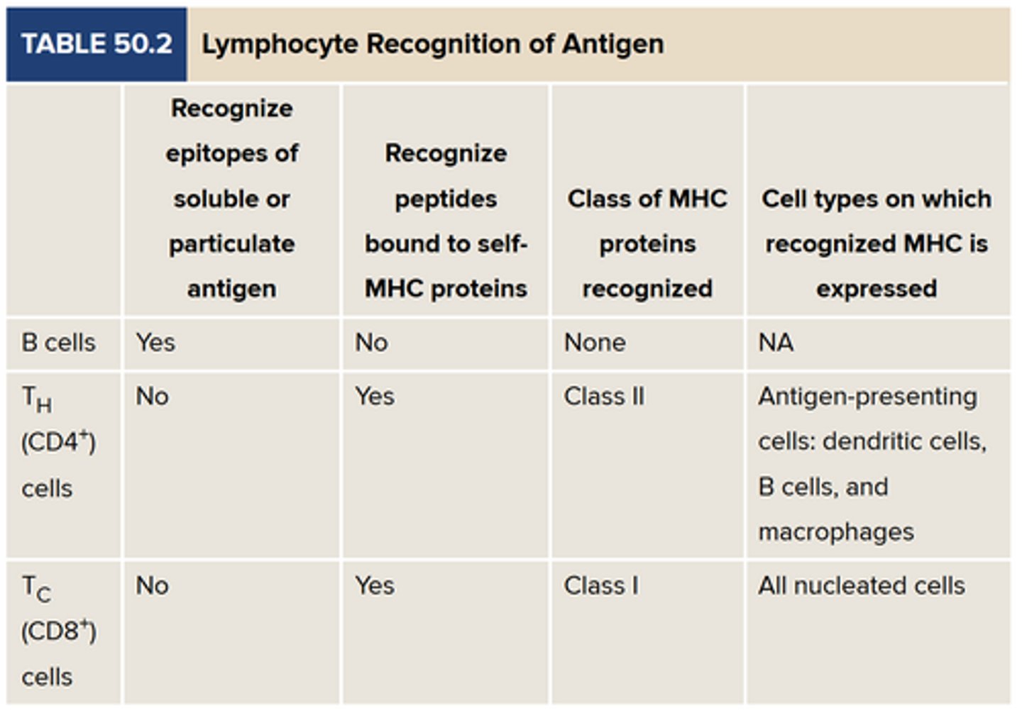
MHC class I proteins
On every nucleated cell of the body
Peptides responded to by cytotoxic T cells
MHC class II proteins
Found on antigen-presenting cells
Macrophages, B cells, dendritic cells
Peptides responded to by helper T cells
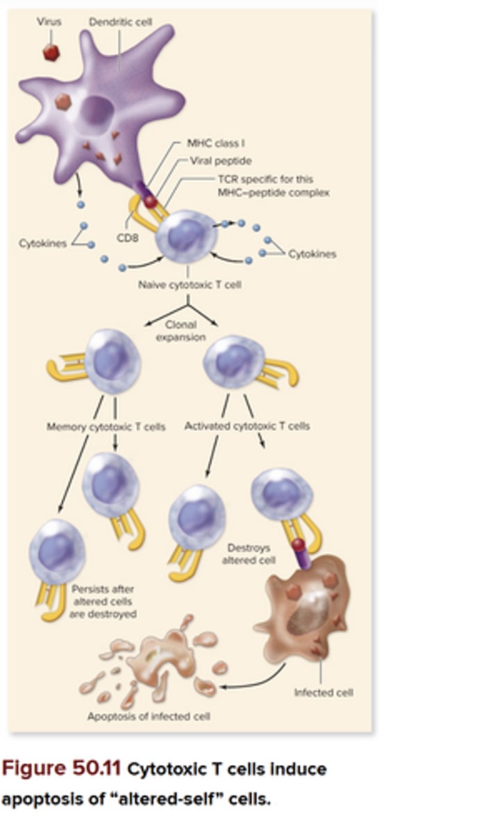
Cytotoxic T cells
-Recognize “altered‑self” cells (infected by virus or tumor)
-TCRs recognize peptides of endogenous antigens bound to MHC class I proteins
-Dendritic cells in particular often present antigens that activate Tc cells
-Dendritic cells ingest viruses or tumors, then cross‑present (place the viral or tumor peptides on MHC class I proteins). Binding of Tc cells through its TCR and CD8 site to the dendritic cell induces clonal expansion of the Tc cells, generating activated Tc cells and memory Tc cells. These will circulate around the body and bind to any host cell expressing the same foreign peptide on self‑MHC class I.
Helper T cells
-Secrete cytokines; bind to specific receptors on membranes of other cells (usually immune system) and initiate signaling cascades to promote activation or differentiation
-Potent; bind to nearby cells + hypothalamus (for fever response)
-Different TH cells secrete cytokines for different cell receptors, so it is mainly the TH cells and the cytokines they secrete that determine whether an immune response will be humoral or cell‑mediated
-Respond to exogenous antigens that were brought into an antigen‑presenting cell: macrophages and dendritic cells (phagocytosis or endocytosis) and B cells (receptor‑mediated endocytosis)
Humoral immunity
Antigen from bacteria engulfed by dendritic cell → carries bacterial peptide (antigen with specific receptor) → binds with TCR → clonal expansion → memory helper T cell → cytokines → recruit B cells + attach to activate T cells → activated B cell → clonal expansion → memory B cell → "activated B cell" = plasma cell
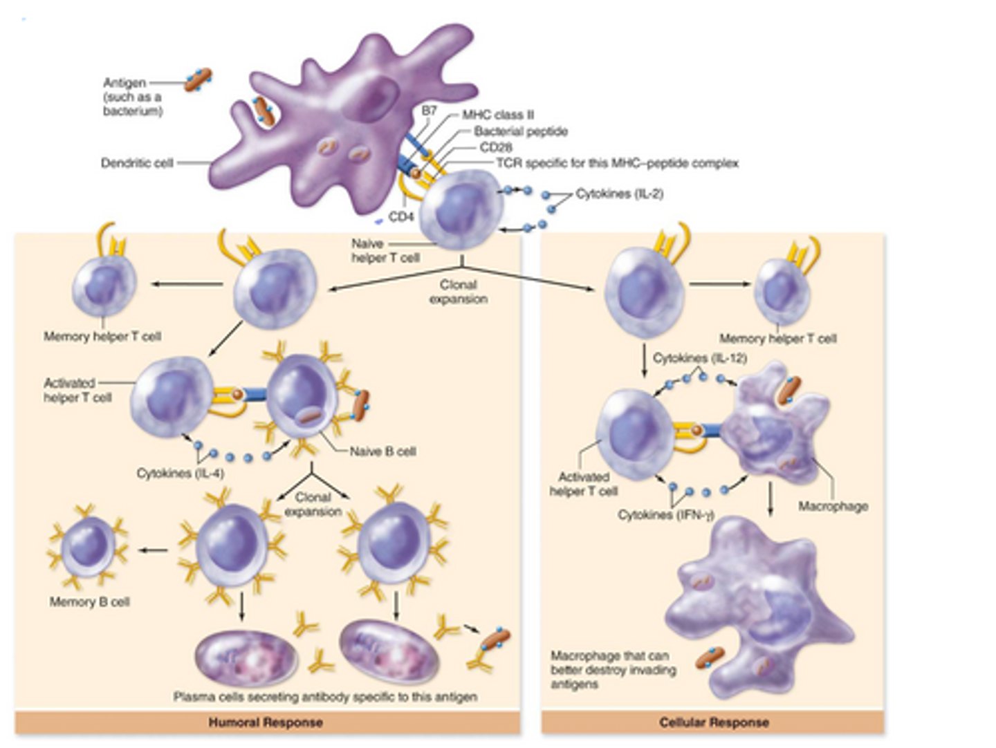
Cell-mediated immunity
-Cytotoxic T cells (directly interact + destroy cells); destroy our own cells that have been infected
Virus → dendritic cell → present antigen (peptide) of virus → naïve Tc cell with TCR receptor → MHC class I and CD8 → cytokines released by dendritic cell → clonal expansion → memory Tc cells stay around, active Tc cells go off and destroy
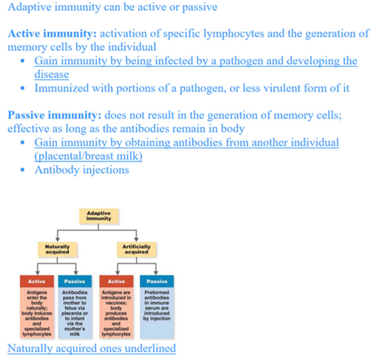
Distinguish between passive and active immunity.
Explain how immunization works.
Immunization:
-Stimulates active immunity, usually through a vaccine
-Don’t cause disease, but they trigger an immune response to produce antibodies and memory cells
Describe what allergies are and what causes them.
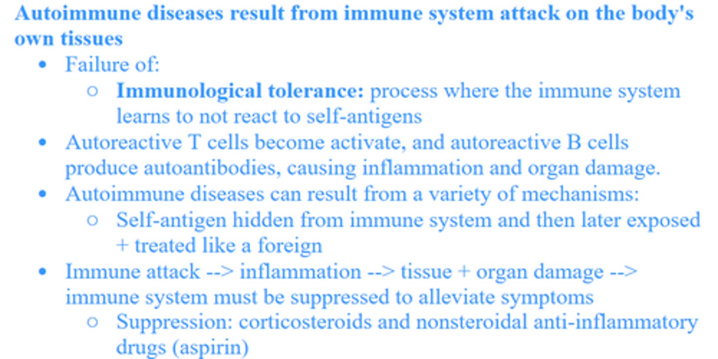
Describe what autoimmune diseases are and what causes them.
Explain the interactions between HIV infection, AIDS, and the immune system, and the difficulty in producing an effective vaccine against HIV.
HIV (human immunodeficiency virus):
-Infects cells of the immune system, especially CD4+ helper T cells, which coordinate immune responses.
-Virus enters these cells, replicates, destroys them, reducing the body's ability to fight infections.
-AIDS: as HIV continues to destroy CD4 T cells, the immune system gets weaker due to less and less CD4 T cells, and the body becomes vulnerable to opportunistic infections and certain cancers (AIDS).
Difficulty in producing an effective vaccine:
-HIV mutates very quickly, constantly changing its surface proteins.
-The virus is attacking the immune systems needed to mount an immune response.
-HIV can hide in host cells, integrate into the host genome, and sit in a latent state where the immune system cannot easily detect it.

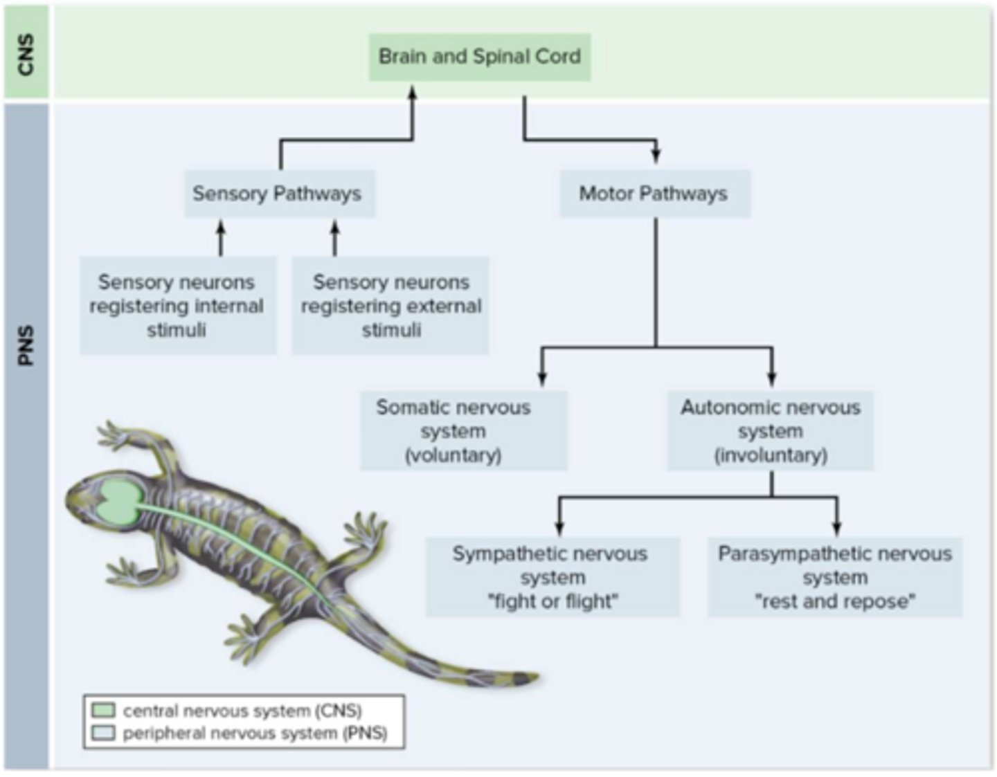
Distinguish between the different types of neurons.
3 types of neurons:
-Sensory neurons (afferent): carry impulses from sensory receptors to CNS
-Motor neurons (efferent): carry impulses from CNS to effectors (muscles and glands)
-Interneurons (association): in brain + spinal cord of vertebrates; help provide complex reflexes / higher associative functions (learning/memory)
Sensory and motor neurons form the PNS
Subdivisions of the nervous system
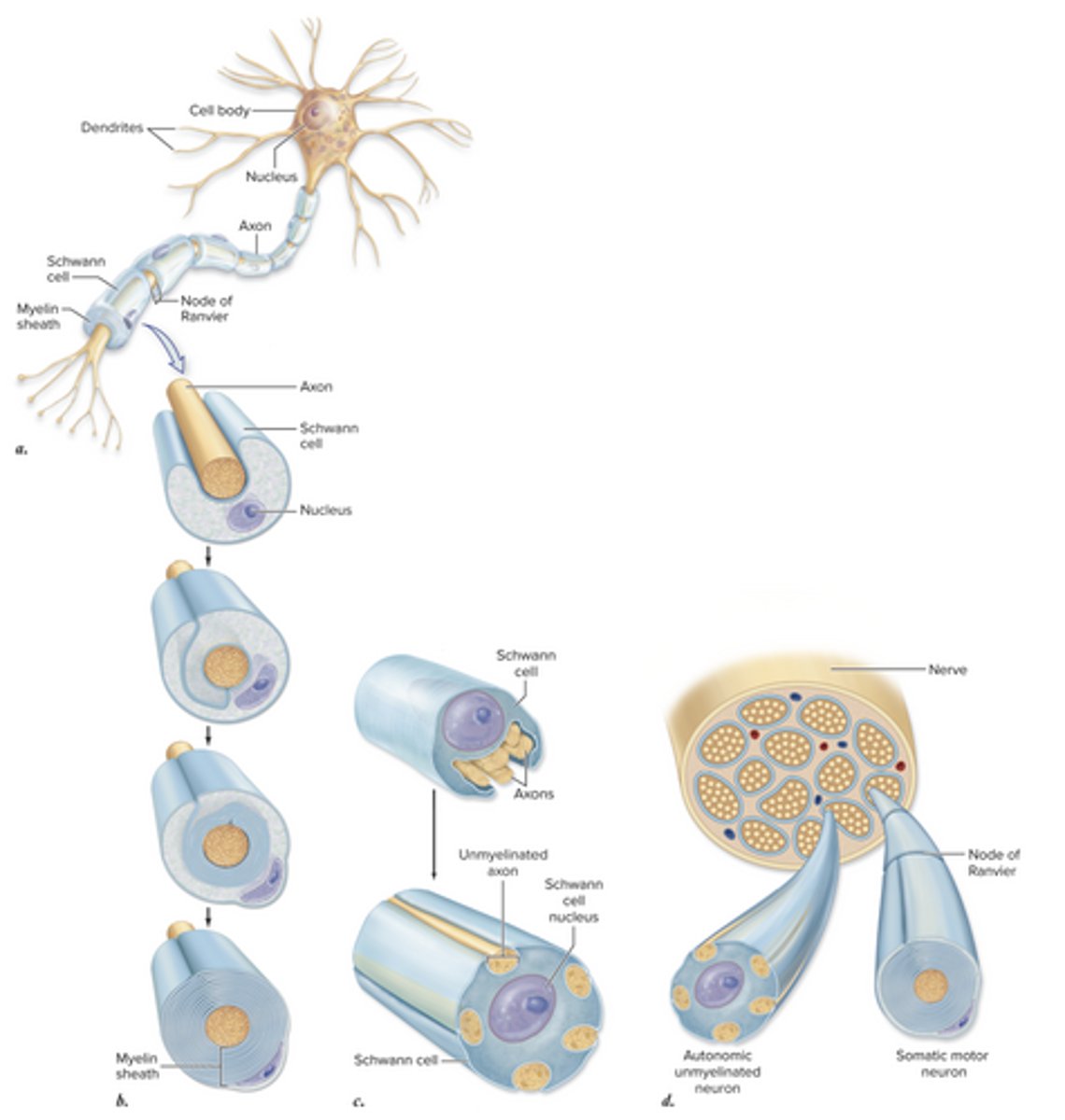
Typical neuron and what happens in specific parts
Cell body: enlarged region w/nucleus
Dendrites: extends from cell body; receives information
Axons: transmits impulses from cell body; many have myelin sheath made by Schwann Cells
Nodes of Ranvier: small gaps that interrupt sheath at regular intervals
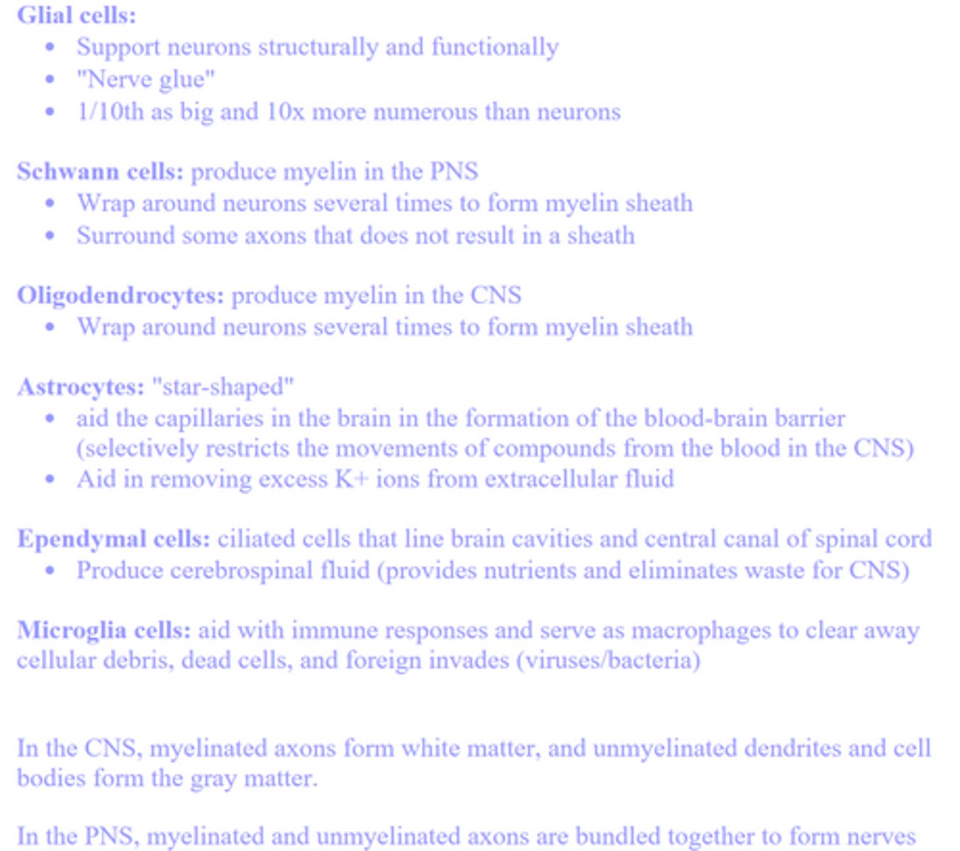
Describe the various functions of neuroglial cells.
Define membrane, resting, and graded potentials, and describe how they are each established, including the relative concentrations of specific ions inside and outside the cell.
Draw a picture of a membrane showing how resting and graded potentials are created and maintained.
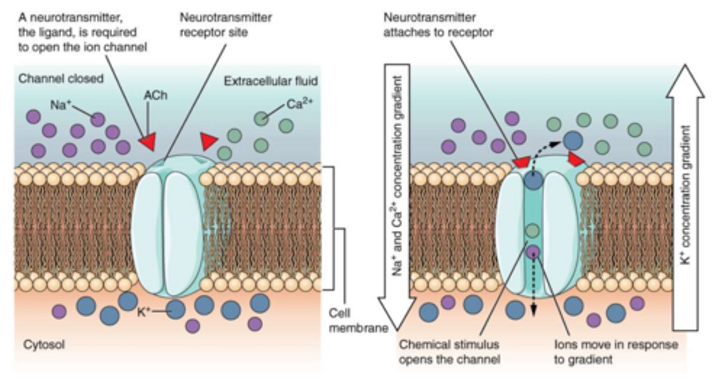
Graded potential hyperpolarization/depolarization
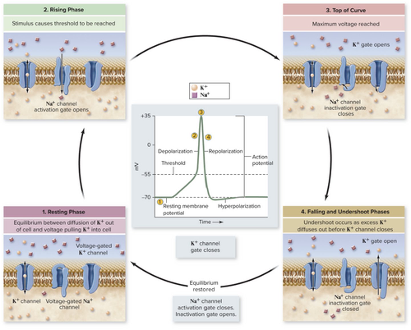
Define action potentials, distinguishing between graded potentials and action potentials.
Graded: small, continuous changes to membrane potentials
Action: transient, all-or-none disruption of the potential triggered by a threshold (minimum) change in potential
Draw an action potential, identifying the phases of the action potential and the causes of the voltage changes at the membrane level.
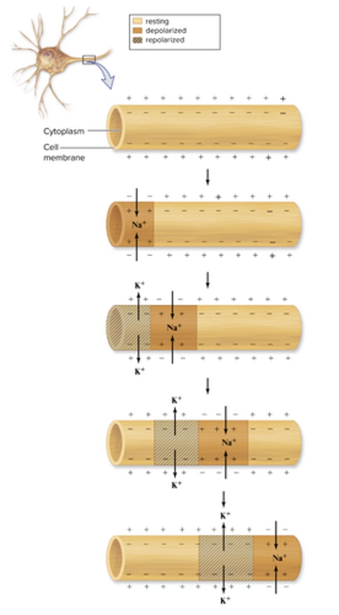
Describe how action potentials are propagated, and sometimes accelerated, along axons.
Propagation: depolarization at one segment triggers the next segment (a wave)
Acceleration methods:
-Saltatory conduction: signal "jumps" between nodes of Ranvier; much faster than continuous conduction
-Axon diameter: larger diameter increases velocity of nerve impulses due to the electrical property of resistance
-Myelination: myelinated axons conduct impulses more rapidly than unmyelinated axons because the axon potentials in myelinated axons are produced only at nodes of Ranvier.
Diagram of propagation
Explain why action potentials are unidirectional and always travel without loss of signal.
They are unidirectional due to the refractory period. After a section of membrane depolarizes, its voltage-gated sodium channels become temporarily inactivated (refractory period), and during this time, the section cannot fire again, which prevents the signal from moving backward. So the depolarization spreads forward to the next section of the axon, triggering a new action potential.
Each segment creates a full action potential rather than passing along a fading signal, so strength remains constant.
Distinguish between electrical and chemical synapses and describe how signals cross synapses.
Electrical: direct connections between cells through gap junctions, allowing ions to flow quickly (often in both directions), making them fast, but less flexible
Chemical: more common; use neurotransmitters to carry the signal across a small gap called the synaptic cleft
-When an action potential reaches the axon terminal, it causes Calcium ions to enter the cell, which triggers vesicles to release neurotransmitters. These cross the synapse and bind to receptors on the next cell, continuing the signal in a controlled, one-way direction.
Using acetylcholine as an example, describe how neurotransmitters transmit the action potential signal.
Action potential arrives at terminal (end of neuron), Calcium enters, vesicles and causes vesicles filled with Acetylcholine to be released into the synaptic cleft. ACh diffuses across the gap and binds to receptors on the postsynaptic membrane, opening ion channels (usually letting sodium enter). This depolarizes the postsynaptic cell, and if it is strong enough to reach threshold, a new action potential is generated. The signal is stopped when ACh is broken down by the enzyme acetylcholinesterase.
Distinguish between excitatory and inhibitory postsynaptic potentials, and between spatial and temporal summation.
Excitatory postsynaptic potentials: make the inside of a cell more positive, bringing it closer to threshold for firing an action potential.
Inhibitory postsynaptic potentials: make the inside more negative, moving it further from threshold.
A neuron receives many of these signals at once and must integrate them through summation.
Spatial summation: occurs when multiple neurons send signals at the same time from different locations
Temporal summation: occurs when one neuron sends signals rapidly in succession
Combined effect of EPSPs and IPSPs determine if a neuron reaches threshold and fires.
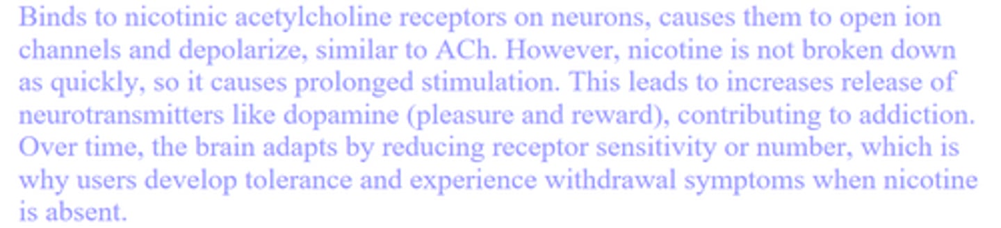
Explain the effects of nicotine on the nervous system.
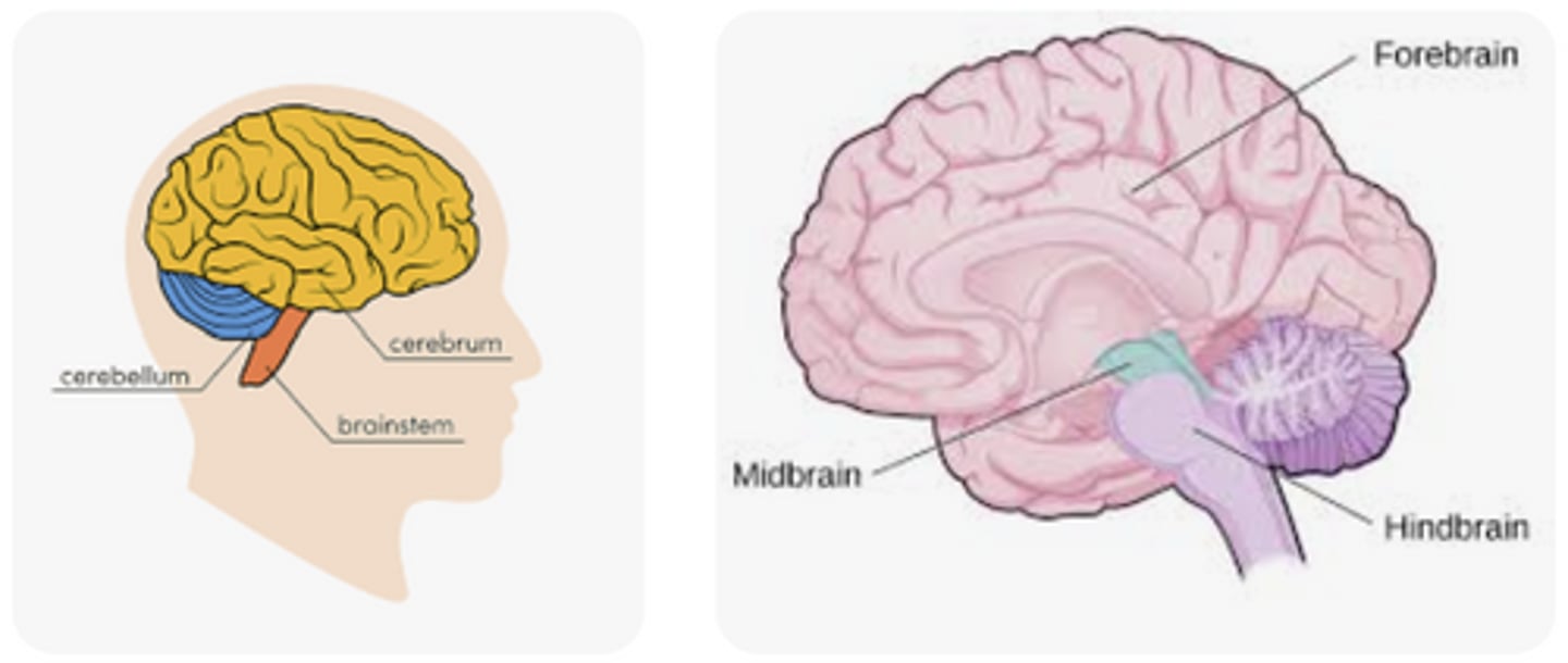
Describe the organization of the animal brain, and how this has evolved over time in vertebrates.
Hindbrain, midbrain, forebrain
Fish rely on hindbrain for motor coordination/basic functions
Forebrain has become increasingly more dominant (cerebrum)
Describe the location and function of the major portions of the human cerebrum.
Cerebrum:
-2 hemispheres connected by Corpus callosum
4 lobes:
Frontal: voluntary movement, decisions, personality; primary motor cortex
Parietal: touch, temperature, pain; primary somatosensory cortex
Occipital: visual
Temporal: hearing, language comprehension (Wernicke’s area)
Outer layer = cerebral cortex
Distinguish between short-term and long-term learning and memory. Describe a possible mechanism of long-term memory.
Short‑term memory:
-lasts seconds to minutes
-limited capacity
-does not require new proteins
-based on temporary changes in synaptic activity (like brief increases in neurotransmitter release)
Long‑term memory:
-synaptic plasticity
-long-term potentiation (repeated stimulation of neurons strengthens synaptic connections; activation of NMDA receptors, Ca²⁺ influx, and addition of AMPA receptors to postsynaptic membrane)
-hippocampus
Explain how a simple reflex works.
-Rapid motor response to a stimulus because the sensory neuron passes info. to a motor neuron in the spinal cord
-No higher level processing
-Sensory neuron detects stimulus and sends signal to the spinal cord
Monosynaptic reflex arc:
-sensory nerve cell makes synaptic contact directly w/ a motor neuron in the spinal cord whose axon travels directly back to the muscle
-knee-jerk
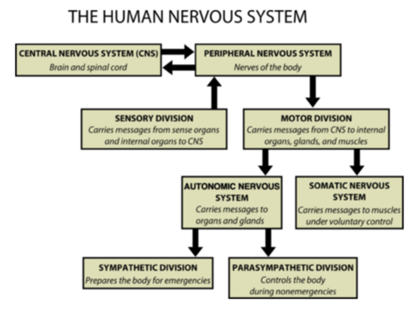
Describe the organization of the peripheral nervous system, distinguishing between the functions of the motor and autonomic nervous systems.
The motor division has two parts. The somatic nervous system controls voluntary movements by sending signals to skeletal muscles. The autonomic nervous system controls involuntary functions by sending signals to smooth muscle, cardiac muscle, and glands. The autonomic system is further divided into the sympathetic (“fight or flight”) and parasympathetic (“rest and digest”) branches.
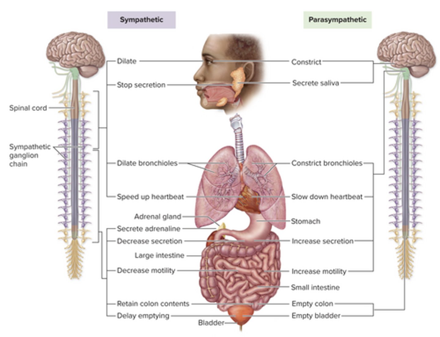
Differentiate between the sympathetic and parasympathetic divisions of the autonomic nervous system
Sympathetic:
-Fight-or-flight
-Prepares for stress
-↑ HR, dilate pupils, inhibit digestion
Parasympathetic:
-Rest and digest
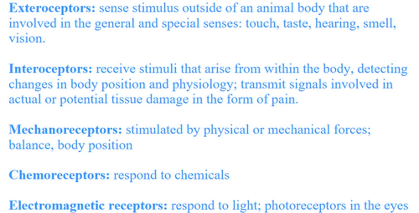
Distinguish between exteroceptors, interoceptors, mechanoreceptors, chemoreceptors, and electromagnetic receptors.
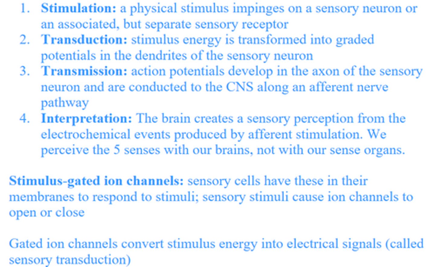
Explain how sensory information is conveyed from sensory neurons to the CNS, including the role of gated ion channels, and how a sensory stimulus is translated into an action potential.
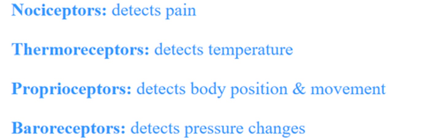
Distinguish among nociceptors, thermoreceptors, proprioceptors, and baroreceptors.
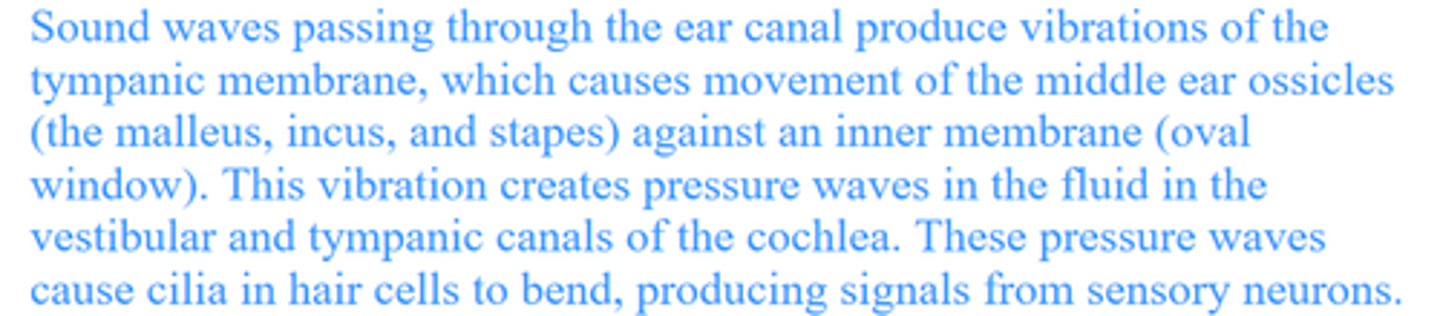
Explain how sound waves in the environment lead to production of action potentials in the inner ear and how different sound frequencies are detected and distinguished in terrestrial vertebrates.
Describe how hearing differs between aquatic and terrestrial animals.
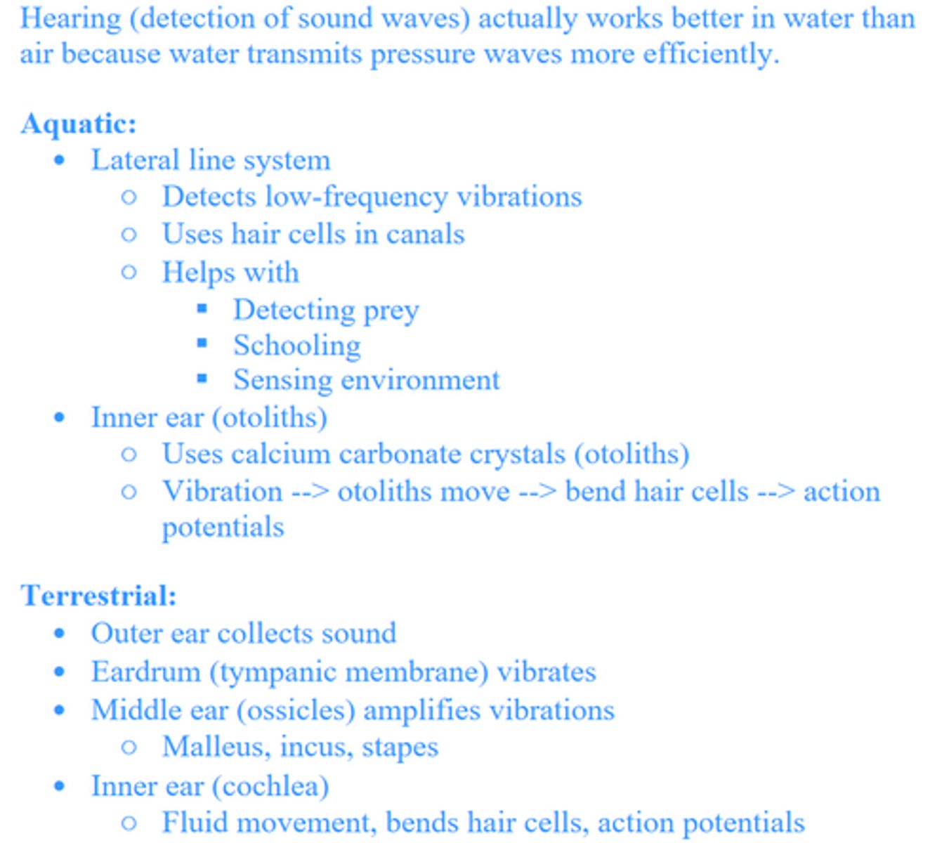
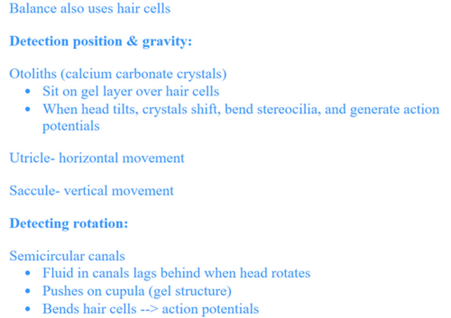
Compare and contrast how body position and movement are detected in terrestrial and aquatic animals.
Describe how taste buds sense and tramsit information
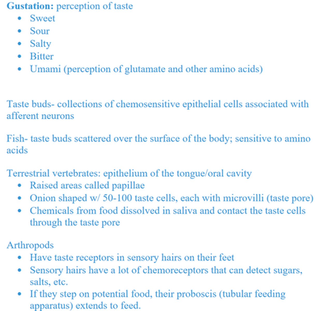
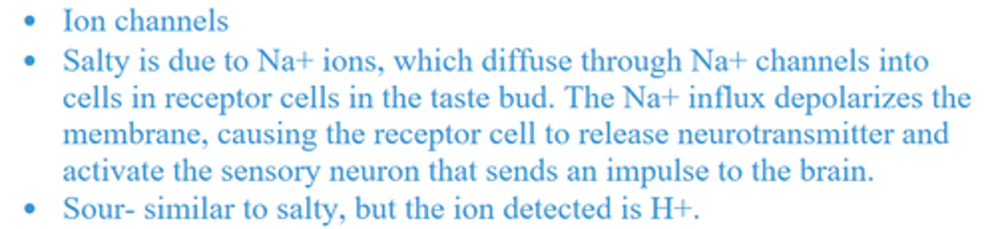
Salty and sour
Sweet, bitter, and umami
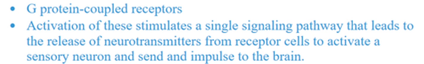
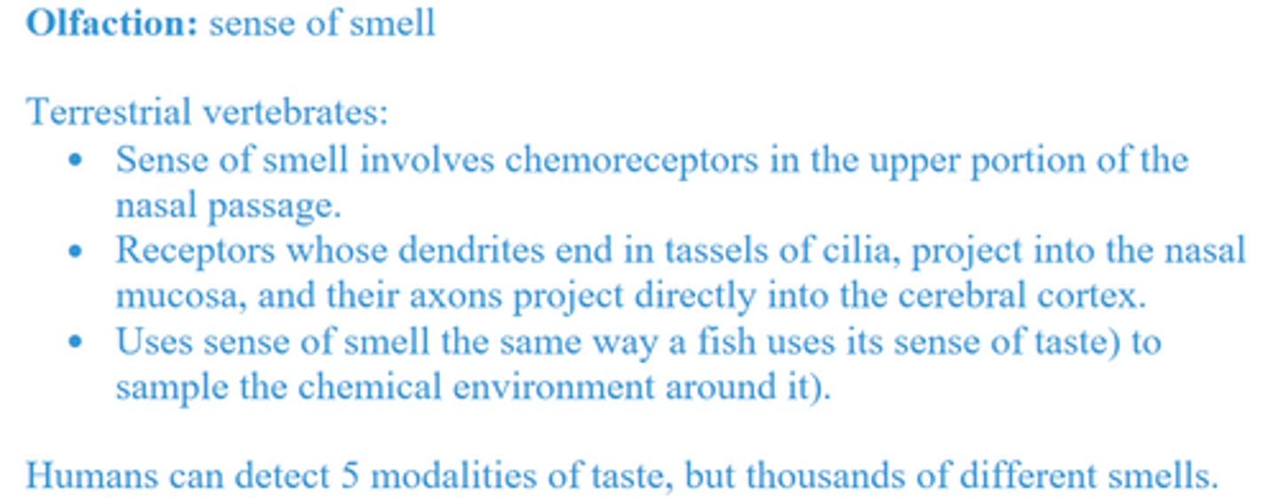
Describe how olfactory neurons sense and transmit information
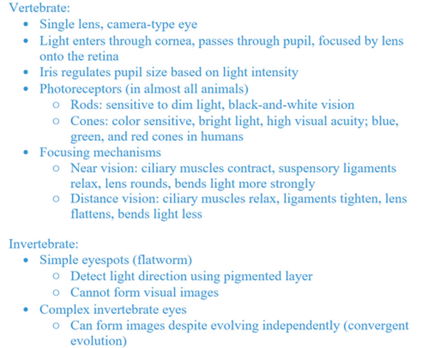
Compare and contrast vertebrate and invertebrate eyes, explaining how vertebrate eyes focus.
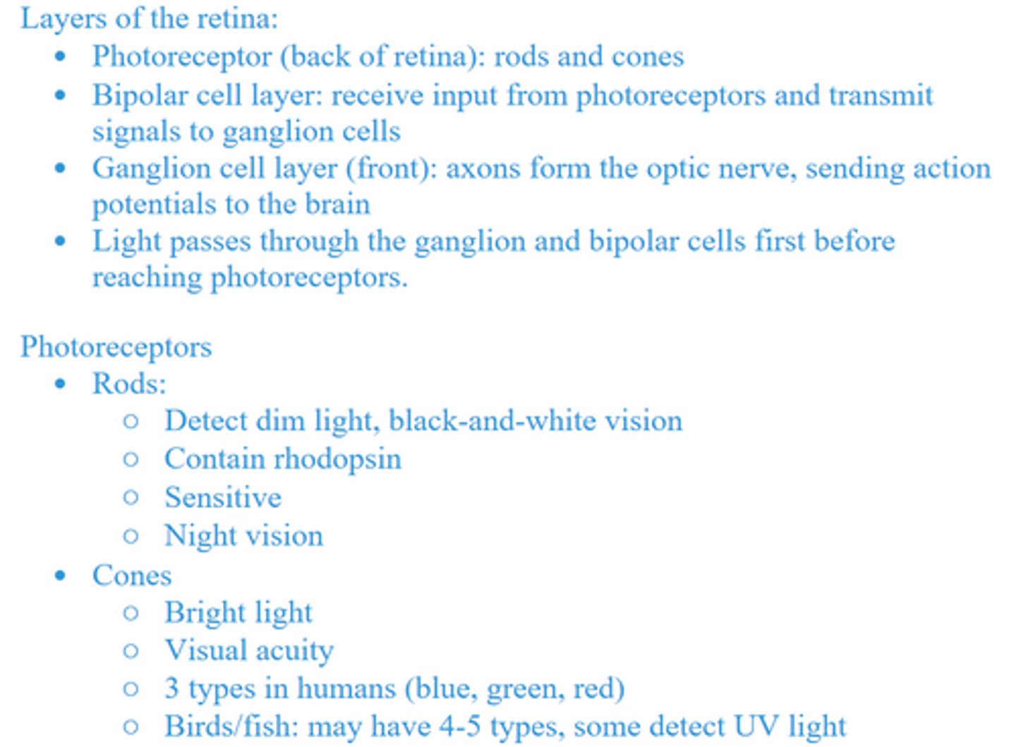
Describe the types of cells, and their functions, in the vertebrate retina
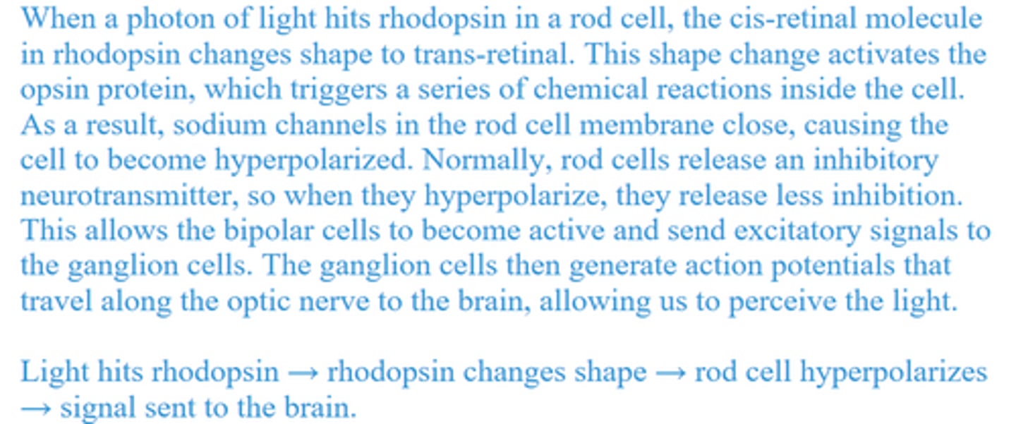
Explain how photons affect rhodopsin.
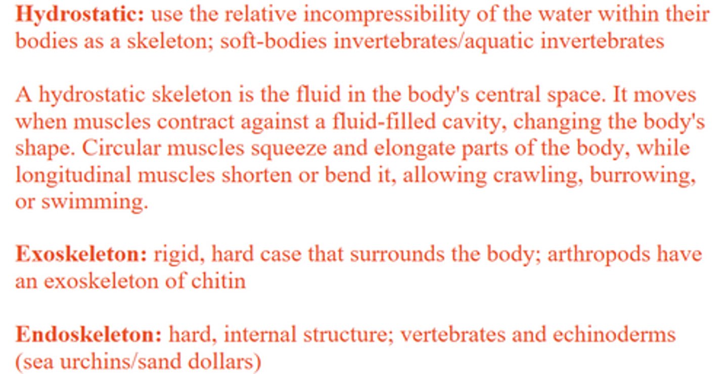
Compare and contrast hydrostatic skeletons, exoskeletons, and endoskeletons. Explain how animals with hydrostatic skeletons move.
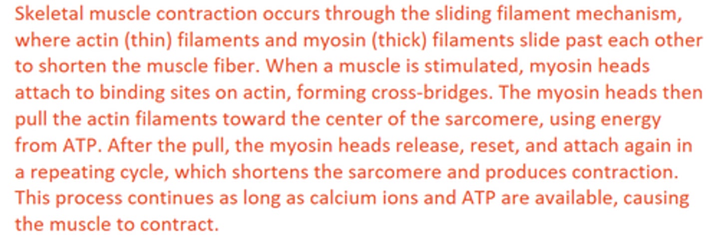
Explain the sliding filament mechanism of skeletal muscle contraction.
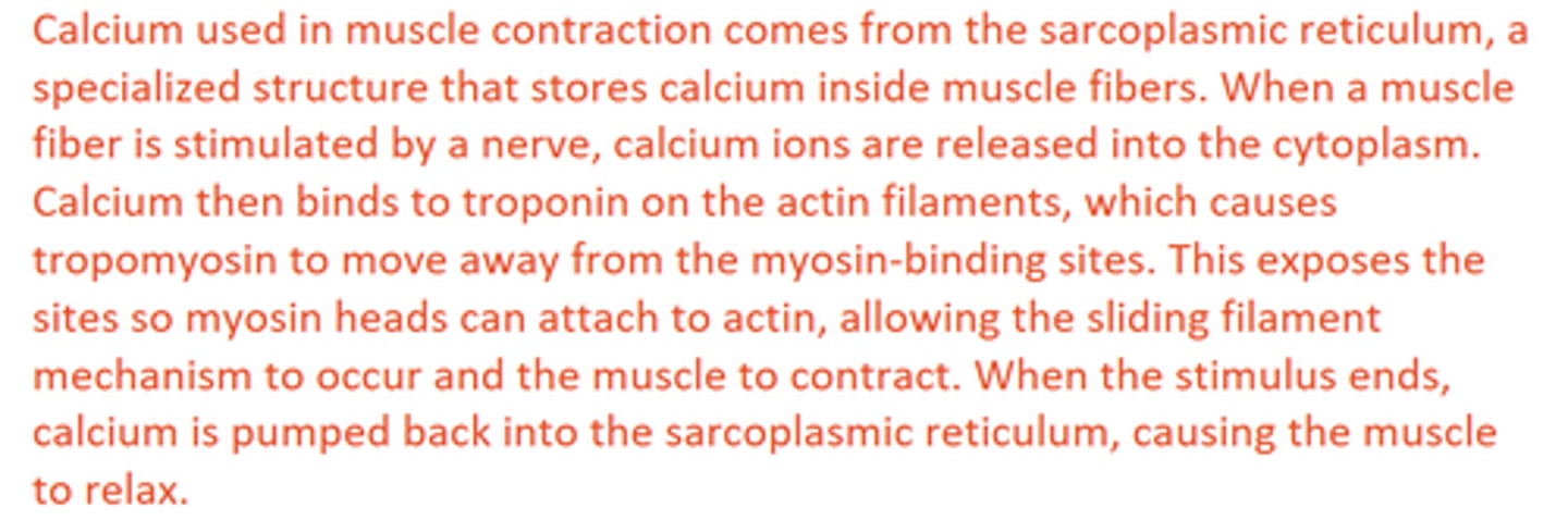
Describe the origin and role of calcium in muscle contraction.
Explain how motor units produce precise coordinated movement.
Differentiate between tetanus and twitches, and between slow-twitch and fast-twitch muscle fibers.
Still learning (1)
You've started learning these terms. Keep it up!