Biology IB - Muscle and motility
1/104
There's no tags or description
Looks like no tags are added yet.
Name | Mastery | Learn | Test | Matching | Spaced | Call with Kai |
|---|
No analytics yet
Send a link to your students to track their progress
105 Terms
Do all organisms have movement within their body?
Yes
Are all organisms capable of locomotion (moving from one place to another)?
No
What is a motile organism?
An organism that uses its own energy to move from one place to another
What are characteristics of motile organisms?
- They are active feeders (in search for food)
- Require higher amounts of nutrients
- Have higher metabolic rates
- Must search for mating partners
What are examples of some motile organisms?
- Some prokaryotes (e.g. E. coli)
- Single-celled eukaryotes (e.g. Amoeba)
- Most animals
What is a sessile organism?
An organism that can't direct its movement from one place to another
What are characteristics of sessile organisms?
- They are autotrophs or passive feeders
- Require fewer nutrients
- Have slower metabolic rates
- May be easily attacked by predators
What are examples of sessile organisms?
- Some prokaryotes
- Some animals such as sponges and cnidaria (jellyfish and polyps)
- Most fungi and plants
What are the types of muscle tissue?
Smooth muscle (involuntary control), cardiac muscle (involuntary control) and skeletal muscle (voluntary control)
What are muscles made of?
Fascicles
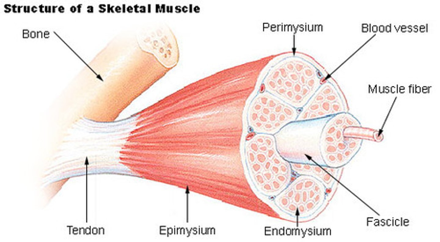
What are fascicles made of?
Muscle fibres (cells)
What are muscle fibres made of?
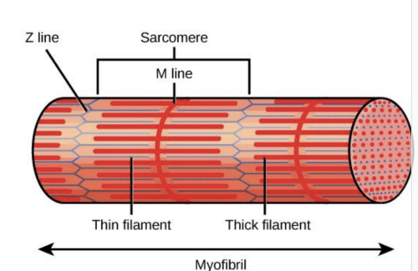
Myofibrils
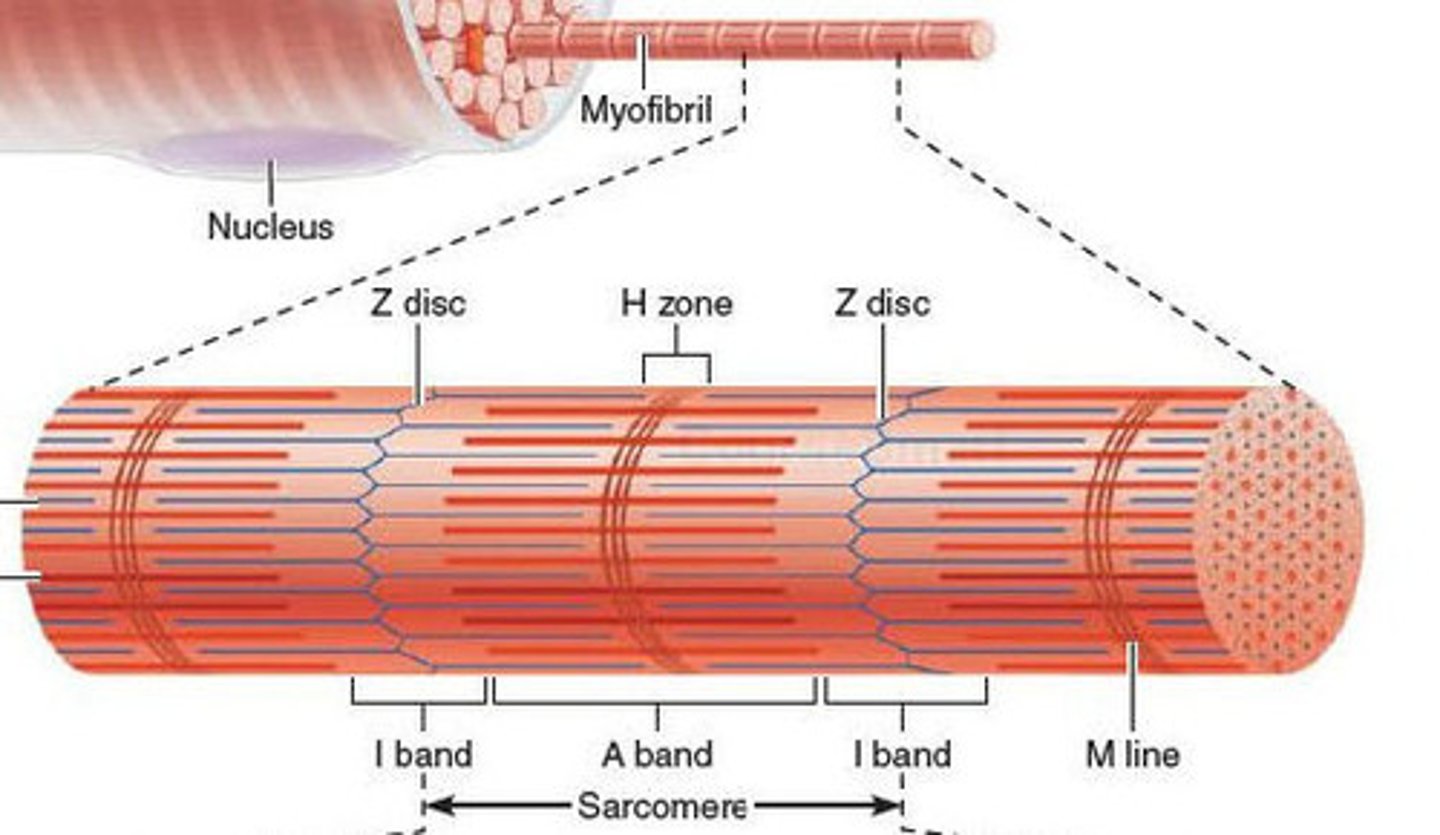
What are myofibrils made of?
Sarcomeres
How many nuclei do skeletal muscle fibres have?
Many
What is the cell membrane of muscle fibres called?
Sarcolemma
What is the endoplasmic reticulum of muscle fibres called?
Sarcoplasmic reticulum
What is the cytoplasm of muscle fibres called?
Sarcoplasm (which contains mitochondria and myofibrils)
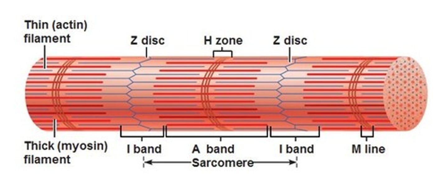
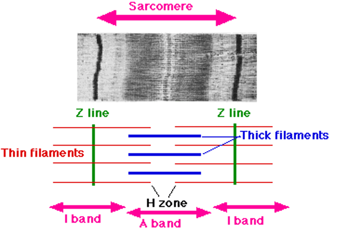
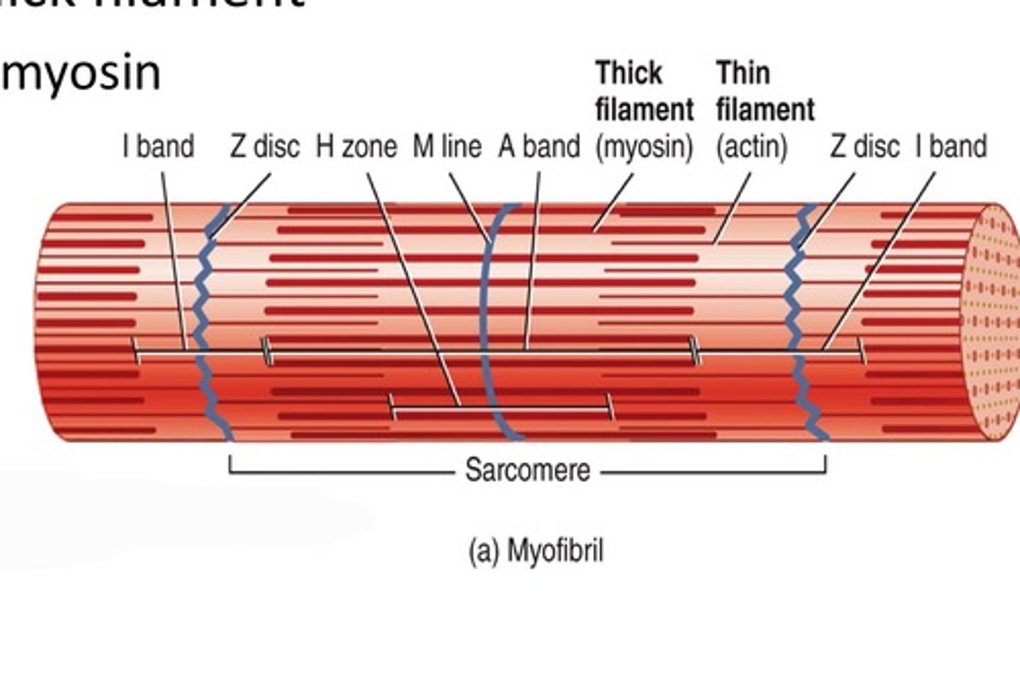
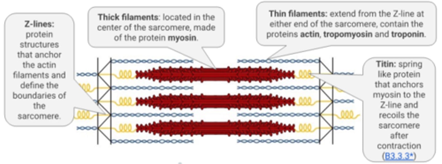
What is the Z line in sarcomeres?
Fixed protein structures that anchor the actin filaments and defines the boundaries of the sarcomere
What is the H band in sarcomeres?
The band where only thick myosin filaments present
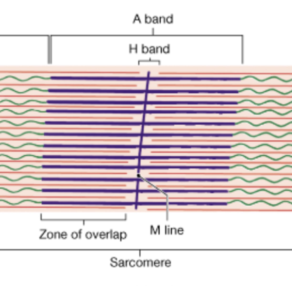
What is the A band in sarcomeres?
The length of the myosin filament (it contains areas where only myosin filaments are present and areas where myosin and actin filaments overlap)
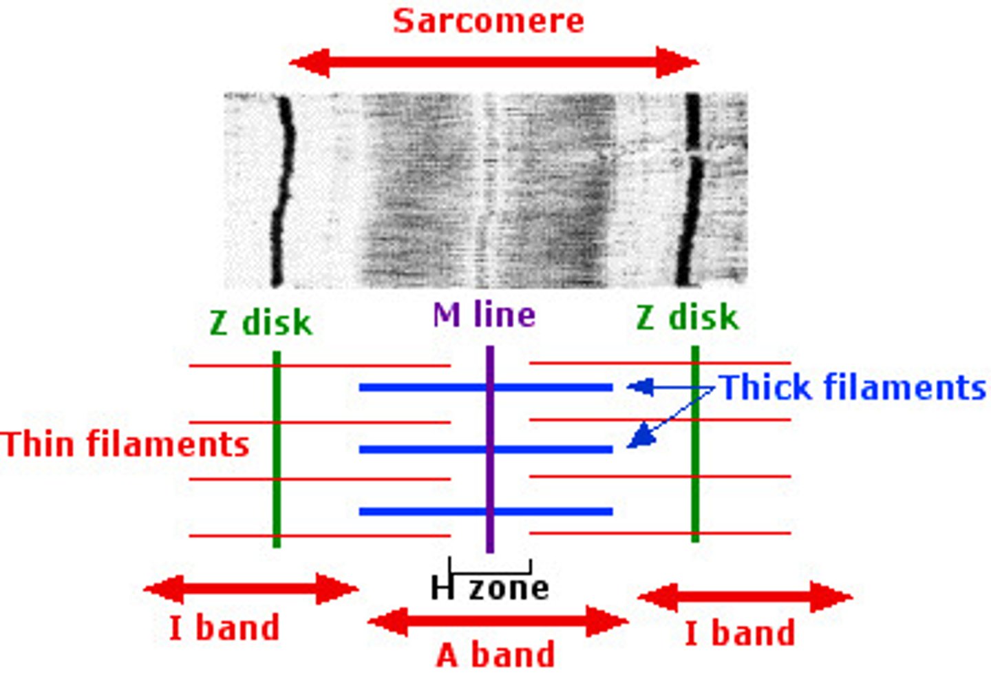
What is the I band in sarcomeres?
The band where only thin actin filaments are present
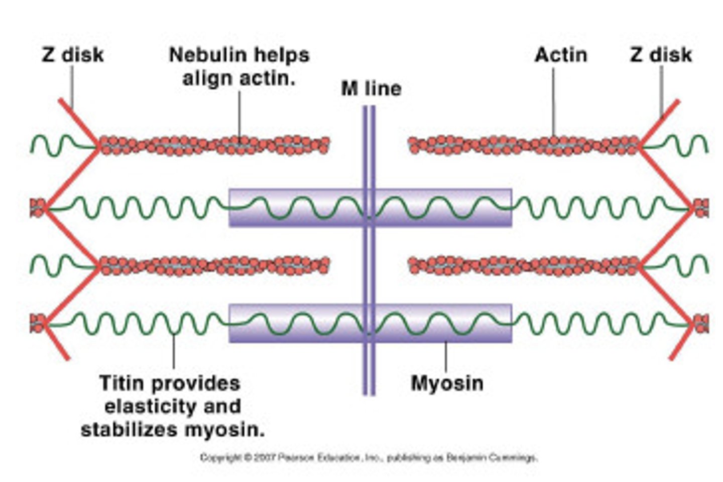
What is the M line in sarcomeres?
The central line and anchor point for myosin filaments
What is titin in sarcomeres?
Spring-like proteins that anchor myosin to the Z-line and recoils the sarcomere after contraction
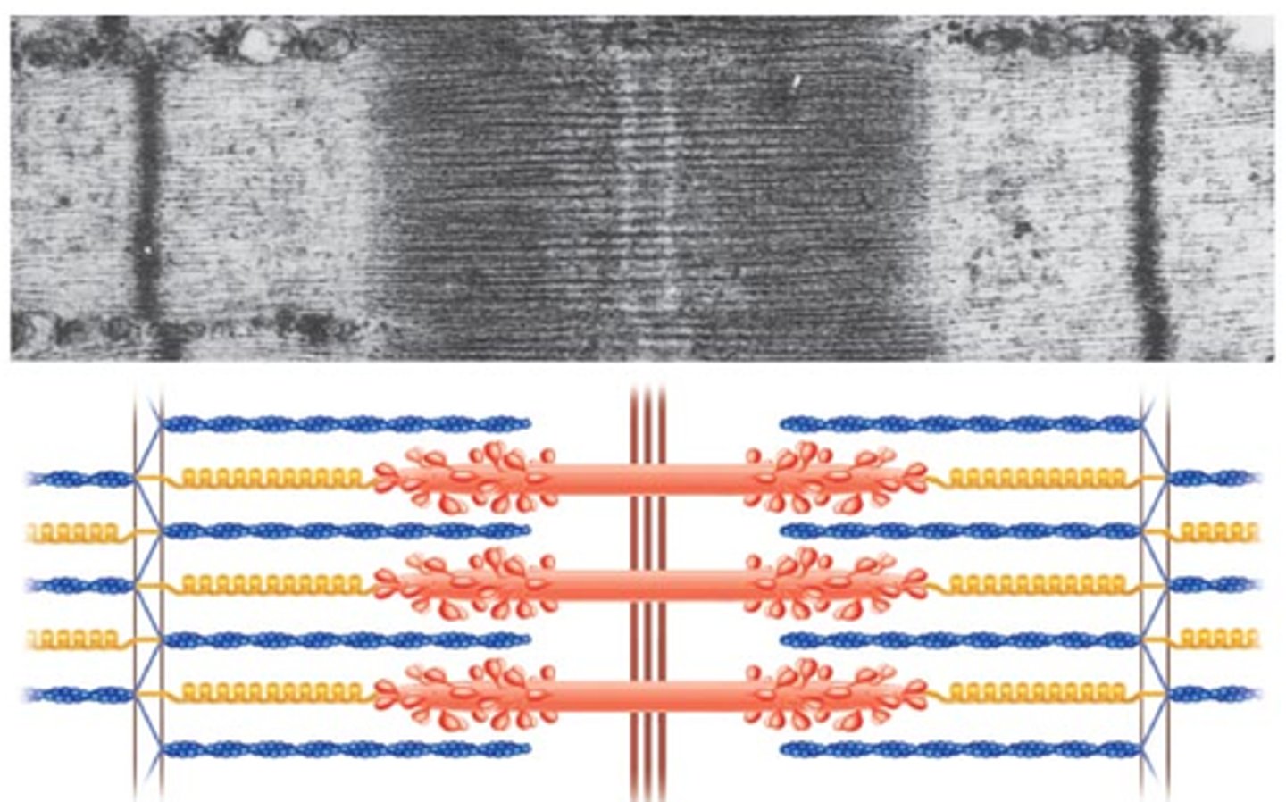
What does the structure of the sarcomere result in?
A pattern of light and dark bands (A band appears dark and I bind appears light)
What is a cross bridge?
When the myosin head attaches to actin
What happens when a muscle contracts?
The sarcomere gets shorter (thin filaments are pulled inwards to slide over the thick filaments and the Z-lines are pulled closer together)
During muscle contraction, does the I band of the sarcomere get smaller or larger?
Smaller
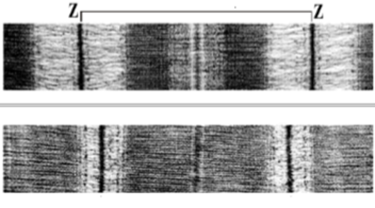
During muscle contraction, does the H band of the sarcomere get smaller or larger?
Smaller
During muscle contraction, does the A band of the sarcomere get smaller or larger?
It does not change (the length of the myosin filament doesn't change during contraction)
What is the sliding filament theory?
A theory that describes the cycle of molecular events that cause thin actin filaments to slide past thick myosin filaments within sarcomeres
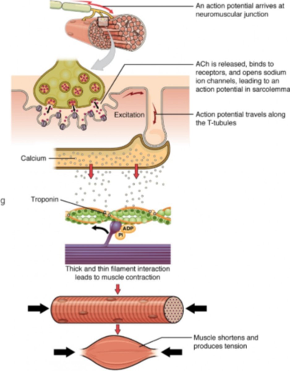
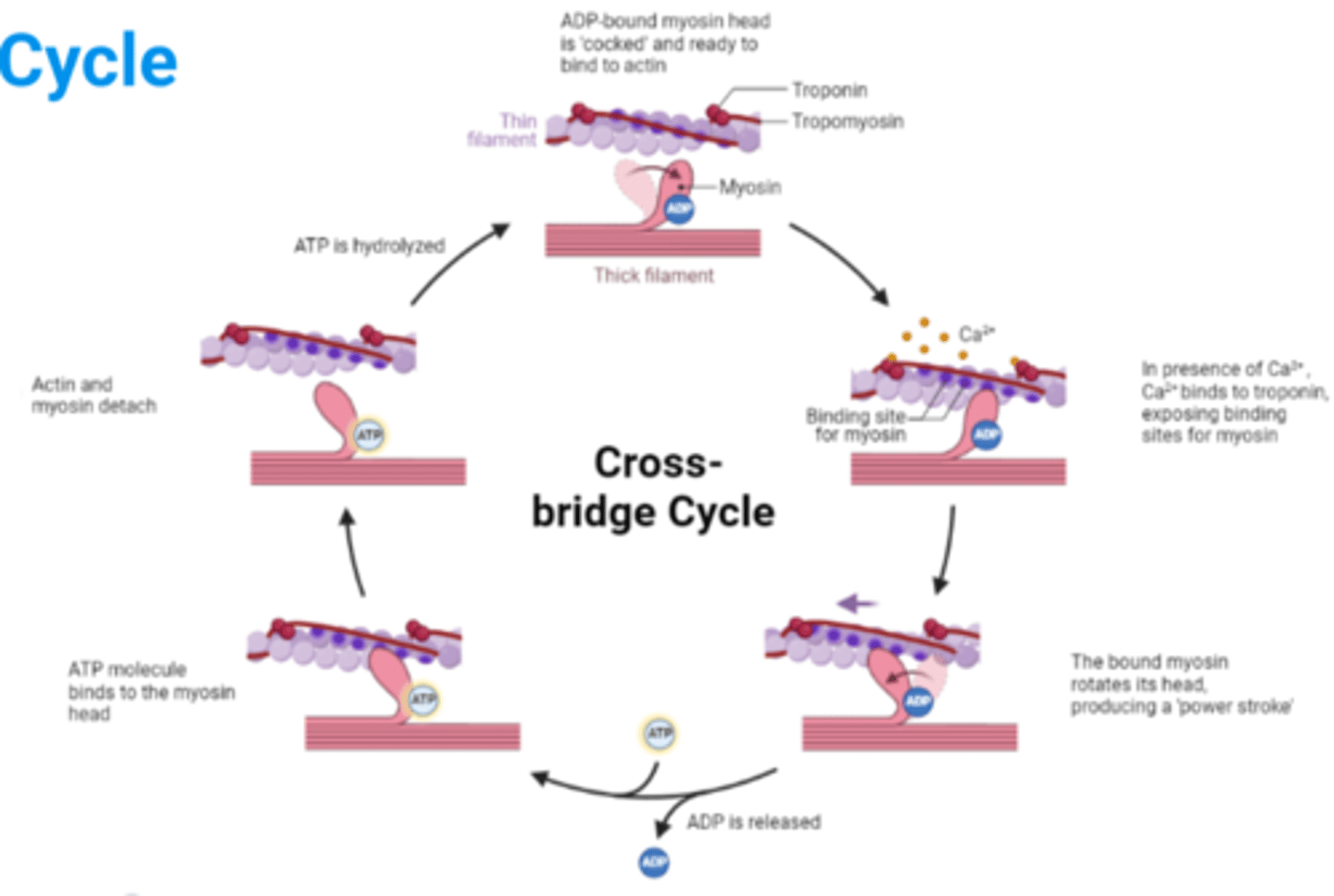
What are the steps of the sliding filament theory?
1. Muscle at rest
2. Arrival of action potential triggers release of acetylcholine at the neuromuscular junction
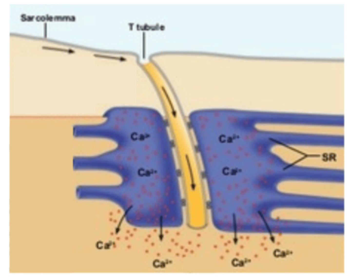
3. Action potential travels along the sarcolemma membrane and down T-tubules
4. Release of Ca²⁺ from the sarcoplasmic reticulum
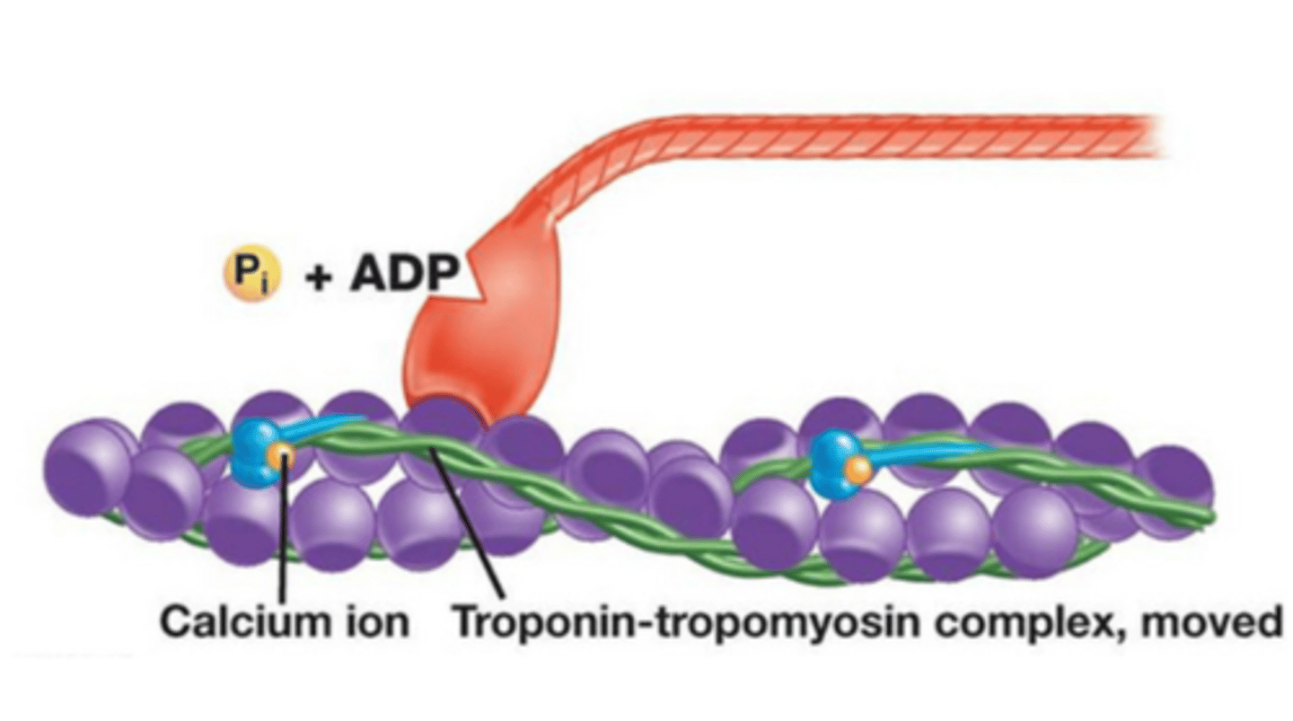
5. Ca²⁺ binds to troponin, causing tropomyosin to move away from myosin binding sites on actin
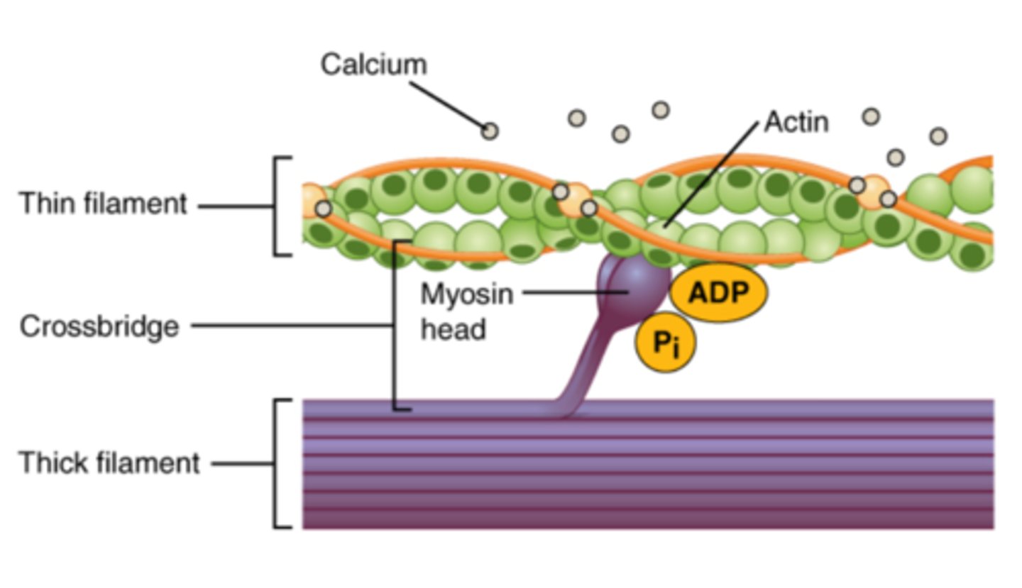
6. Myosin heads bind to actin, forming a crossbridge
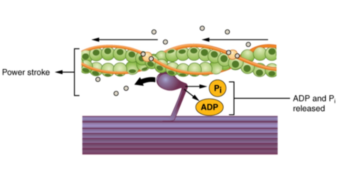
7. Myosin head flexes, moving the actin filament inwards and shortening the sarcomere
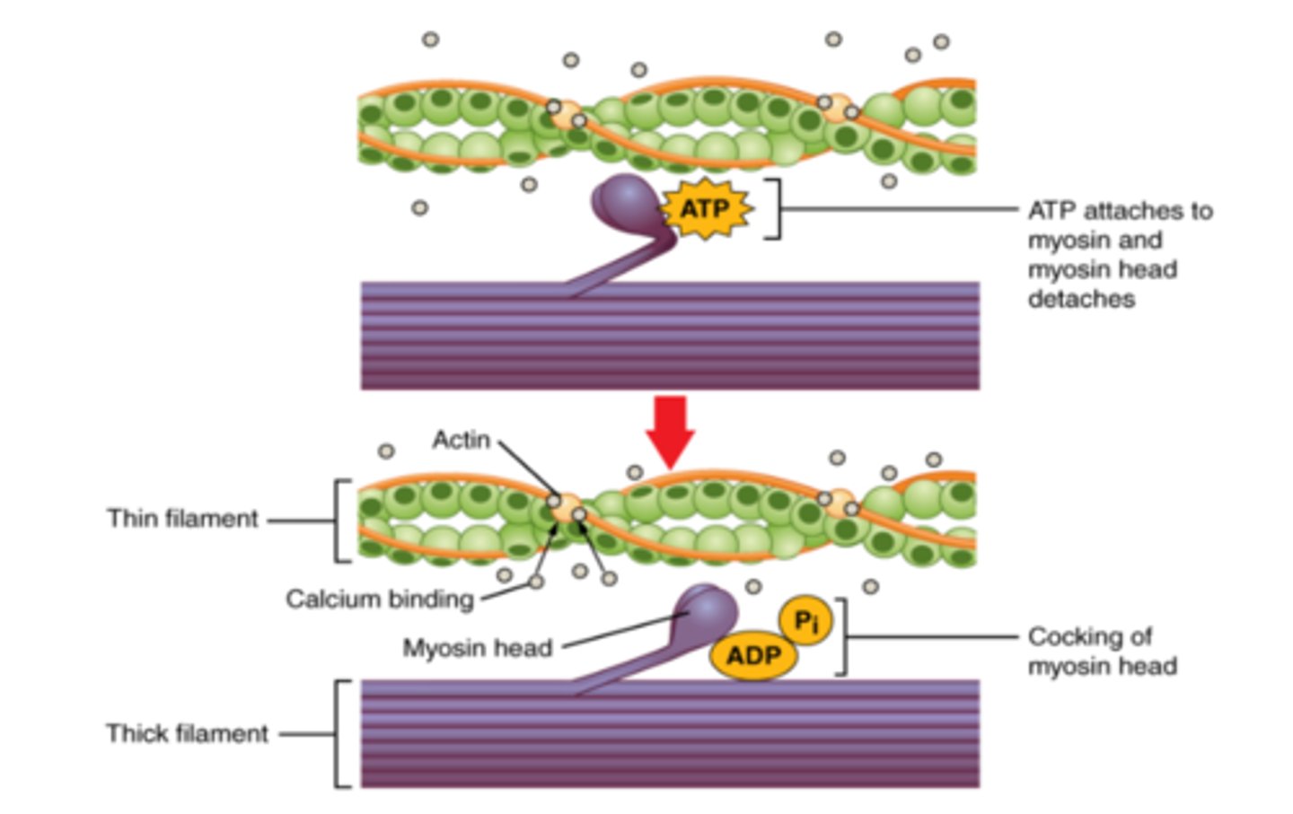
8. ATP attaches to the myosin heads, breaking the crossbridge
9. Steps 6-8 repeat in a cross-bridge cycle
10. Contractions end when Ca²⁺ is pumped back into the sarcoplasmic reticulum
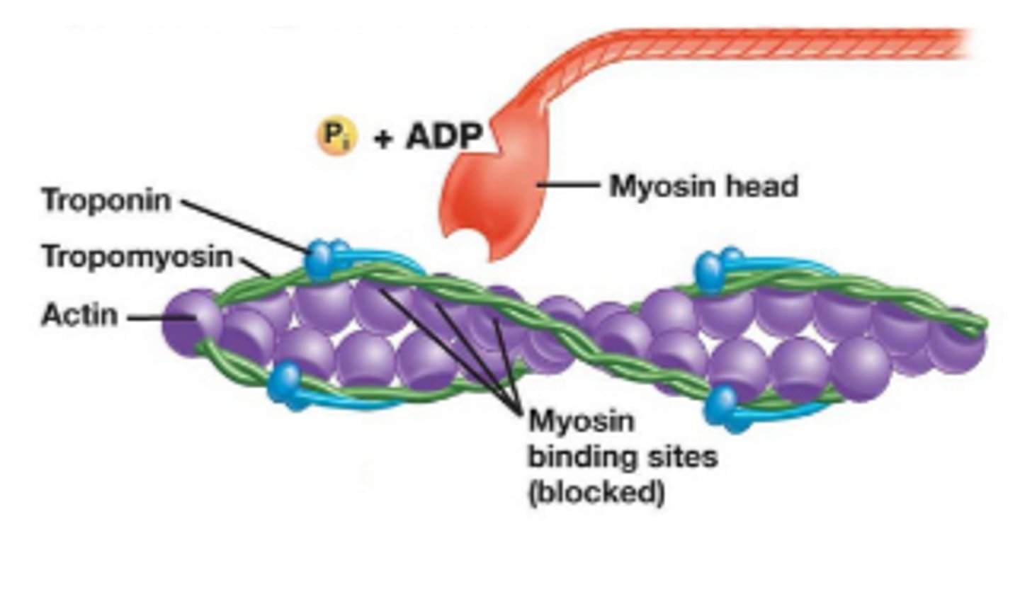
What is a description of step 1 (muscle at rest)?
- The motor neuron is not signalling the muscle to contract so sarcomere is relaxed
- Myosin heads are "cocked" with bound ADP and an inorganic phosphate (Pi) so they are not bound to actin
- Myosin binding sites on actin are blocked by tropomyosin
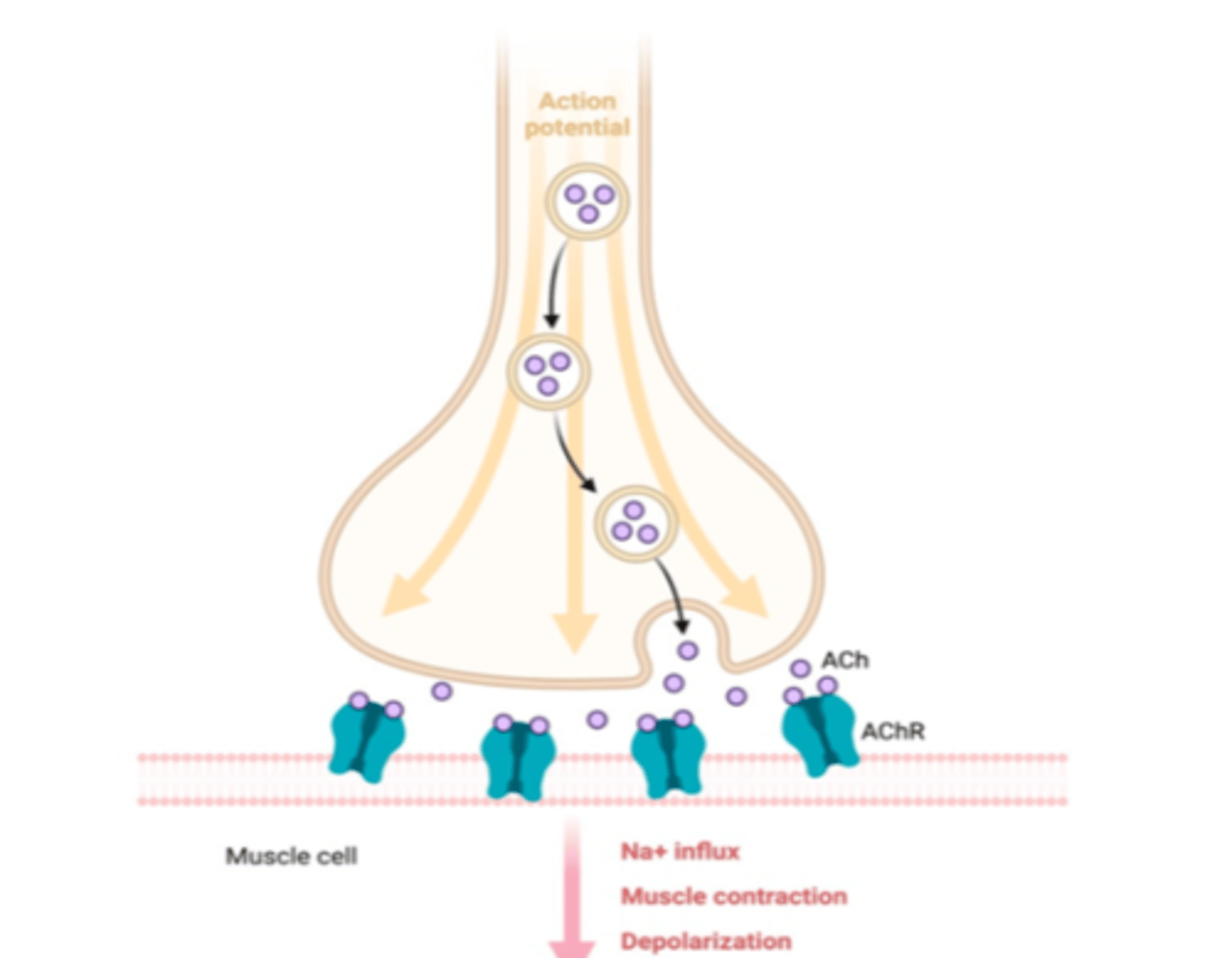
What is a description of step 2 (arrival of action potential triggers release of acetylcholine at the neuromuscular junction)?
- An action potential reaches the neuromuscular junction and ACh is released and binds to receptors in sarcolemma, triggering the opening of ligand-gated Na⁺ channels
- Na⁺ depolarise the the membrane and trigger a wave of action potentials along the sarcolemma
What is a description of step 3 (action potential travels along the sarcolemma membrane and down T-tubules)?
- The electrical impulse travelling across the sarcolemma continues down the T-tubules (invaginations in the sarcolemma that carry the electrical impulse into the interior of the muscle fibre)
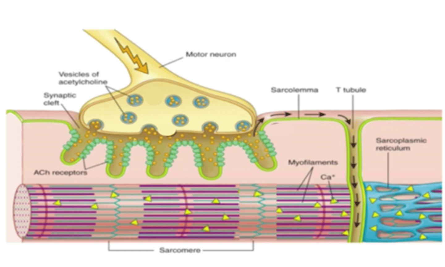
What is a description of step 4 (release of Ca²⁺ from the sarcoplasmic reticulum)?
- The electrical signal in T-tubules trigger the release of Ca²⁺ form the sarcoplasmic reticulum
- Ca²⁺ move by facilitated diffusion out of the sarcoplasmic reticulum and onto the myofibril (it acts as an intracellular signalling molecule to trigger contraction)
What is a description of step 5 (Ca²⁺ binds to troponin, causing tropomyosin to move away from myosin binding sites on actin)?
- Ca²⁺ bind to troponin and triggers a conformational change that shifts tropomyosin, exposing the myosin binding sites on actin
What is a description of step 6 (myosin heads bind to actin, forming a crossbridge)?
- Myosin binding sites are now exposed so the myosin heads can bind to actin, creating a cross-bridge
What is a description of step 7 (myosin head flexes, moving the actin filament inwards and shortening the sarcomere)?
- When the myosin head binds to actin, it flexes and pulls the actin slightly towards the centre of the sarcomere (this is called the power stroke)
- ADP and Pi are released in this process
What is a description of step 8 (ATP attaches to the myosin heads, breaking the crossbridge)?
- Crossbridge is broken when ATP binds to the myosin head (it causes the myosin head to detach from actin)
- The hydrolysis of ATP gives the energy needed for the myosin head to extend itself (to be "cocked") ready to bind to a new binding site
What is a description of step 9 (steps 6-8 repeat in a cross-bridge cycle)?
- As long as Ca²⁺ are present and bound to troponin, myosin will repeatedly bind and pull on the actin in a crossbridge cycle
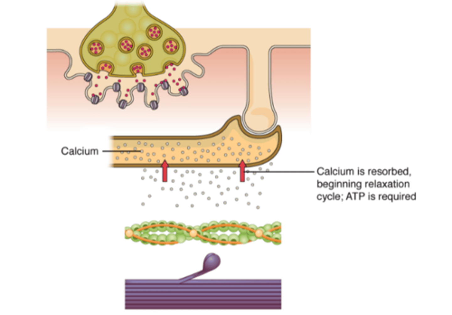
What is a description of step 10 (contractions end when Ca²⁺ is pumped back into the sarcoplasmic reticulum)?
- Muscle contraction stops when the motor neuron stops sending ACh which causes:
--> Repolarisation
--> Ca²⁺ channels close
--> Ca²⁺ are pumped back into the sarcoplasmic reticulum
--> Myosin binding sites are blocked
What would be a full explanation to explain muscle contraction?
1. Muscle fibres are made of myofibrils, which are made of sarcomeres.
2. Sarcomeres are arranged end to end and shorten during muscle contraction.
3. Sarcomeres are made by actin and myosin filaments which overlap (the thick filament which has a darker band is myosin and the thin filament which has a lighter band is actin)
4. When nerve impulses arrive, it causes the depolarisation of the sarcolemma, triggering the release of Ca²⁺ from the sarcoplasmic reticulum.
5. The Ca²⁺ bind to troponin, causing tropomyosin to move and exposing the binding sites on actin. This allows the myosin heads to form cross bridges and bind to actin.
6. The myosin head moves and pulls actin towards the centre of the sarcomere (causing more overlap between actin and myosin).
7. ATP is used to provide energy to cause the movement of the myosin heads
What would be a diagram of a sarcomere?
.
When can a muscle only exert force?
When it contracts
When does muscle relaxation happen?
When the sarcomere returns to a relaxed state after a contraction
What is muscle relaxation influenced by?
Titin and antagonistic muscles
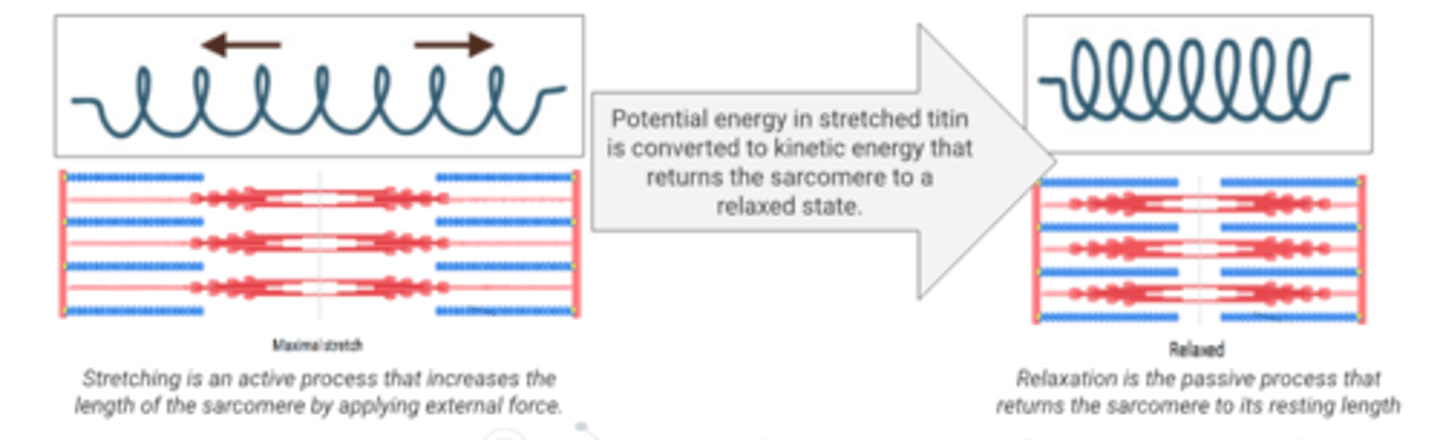
What does titin provide?
Passive elasticity that helps muscles return to their resting length during relaxation
What are the primary functions of titin?
1. Provides sarcomere stability by anchoring the myosin filament to the Z-line.
2. Helps sarcomeres recoil after contraction so that the sarcomere returns to its relaxed state.
3. When stretched, titin generates power for rapid motion when it recoils
4. It prevents overstretching
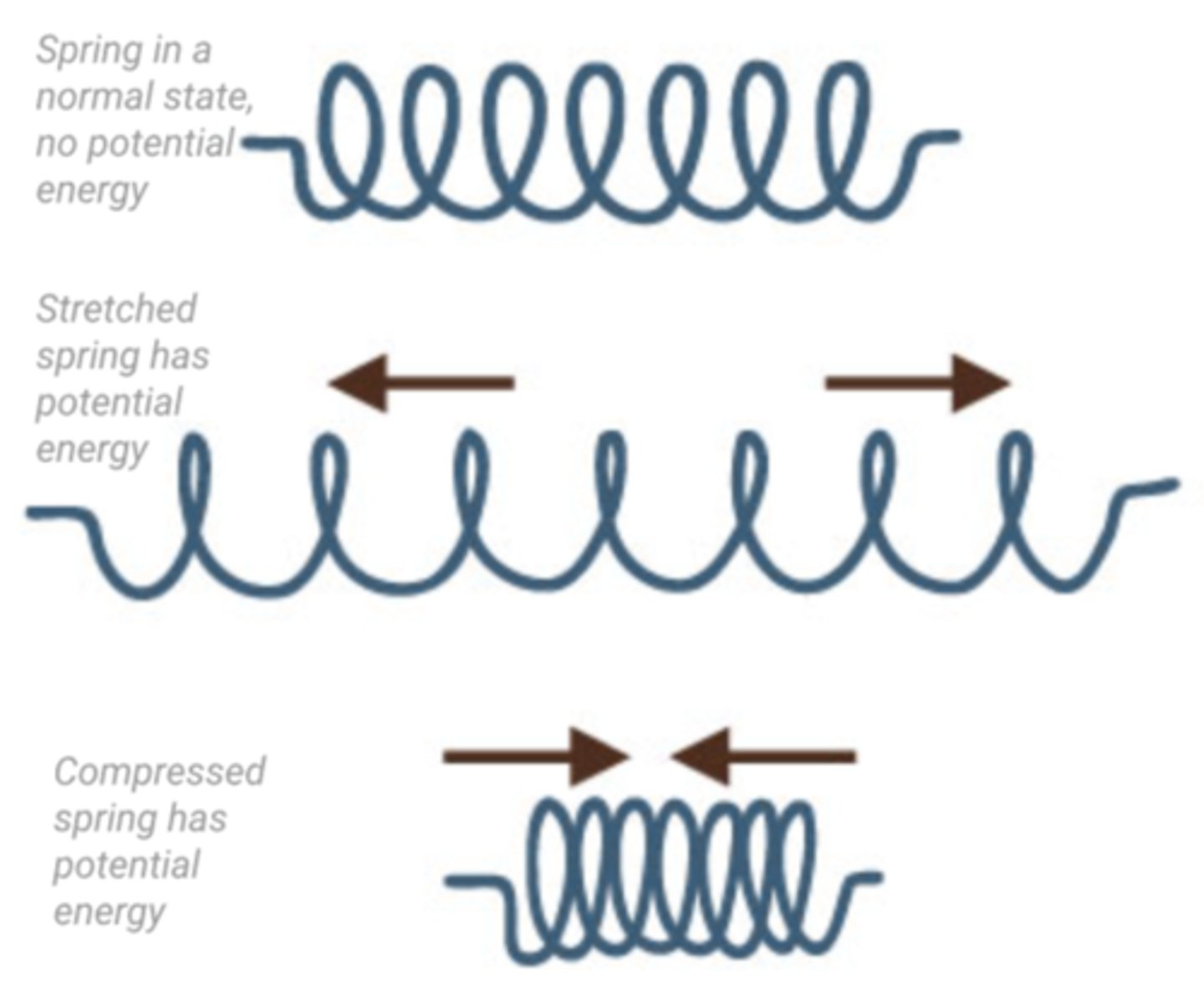
By which mechanism does titin store potential energy?
When it is stretched or compressed, it stores elastic potential energy which can be converted into kinetic energy when it is released and returns to its original shape (causing movement)
What are antagonistic muscles?
A pair of muscles that cause opposite movements as when one muscle contracts the other one relaxes
What are examples of antagonistic muscles?
- Biceps and triceps work together to move the forearm
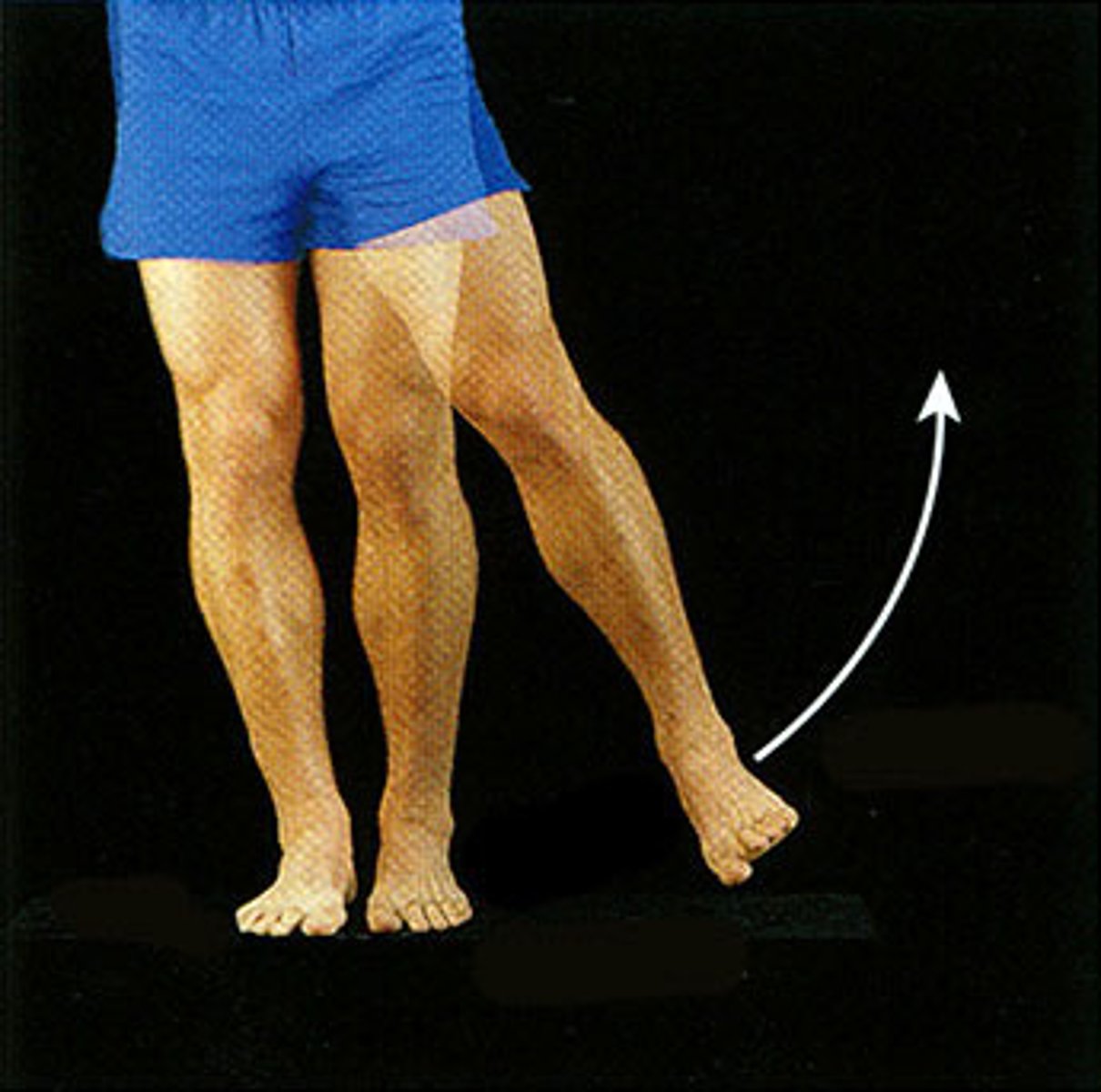
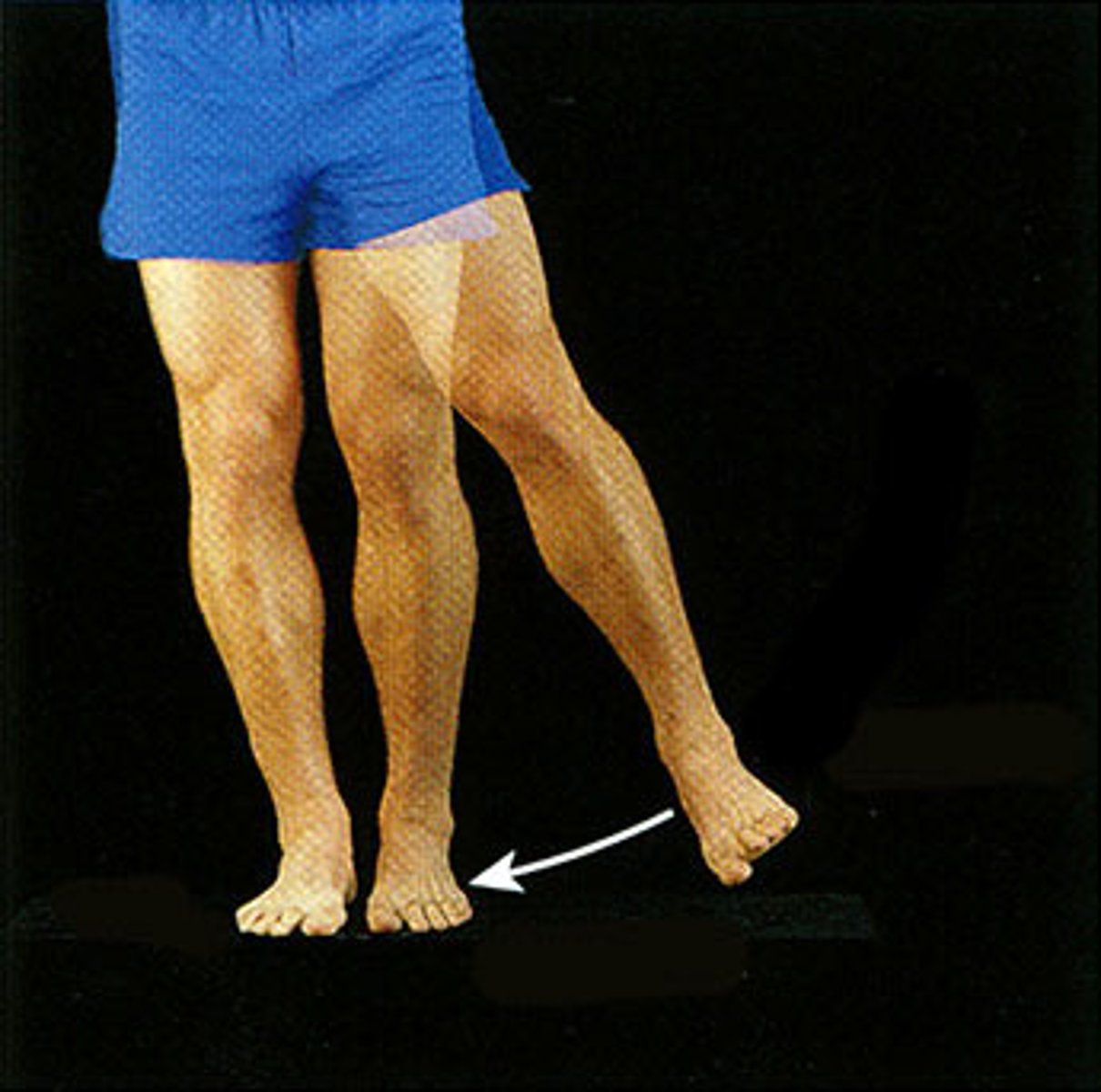
- Quadriceps and hamstrings work together to move the lower leg
- Internal and external intercostals work together to move the ribs
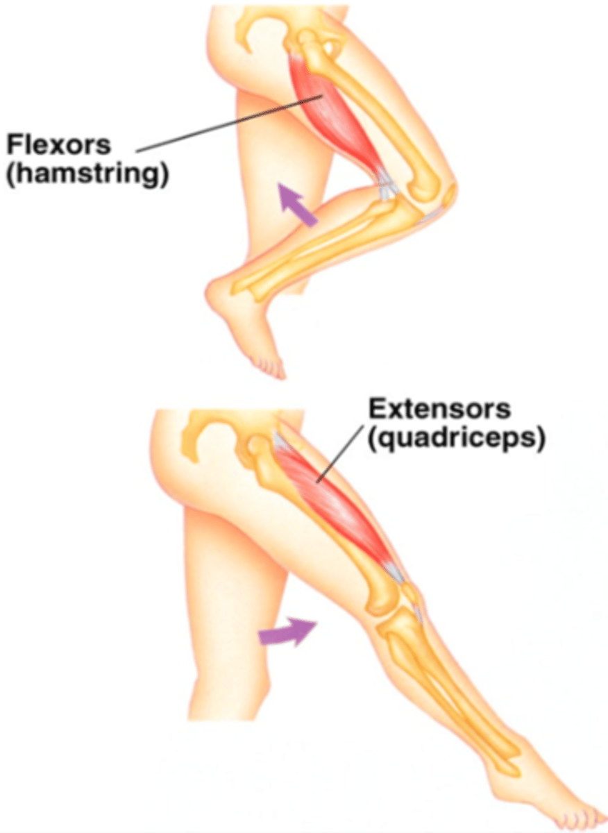
In the case of quadriceps and hamstrings, which one would be the flexor and which one the extensor?
The quadriceps would be the extensor and the hamstrings the flexor (as when the leg extends, it's the quadriceps that contracts and when it bends, it's the hamstrings that contract)
Why are antagonistic muscle pairs necessary?
Because they can only cause movement by contracting (it can't supply the energy it needs to lengthen itself so this is provided by the contraction of an antagonistic muscle)
What is a motor unit?
The single motor neuron together with all of the muscle fibres it stimulates (it includes the axon terminal of the motor neuron, neuromuscular junction, muscle fibres and myofibrils)
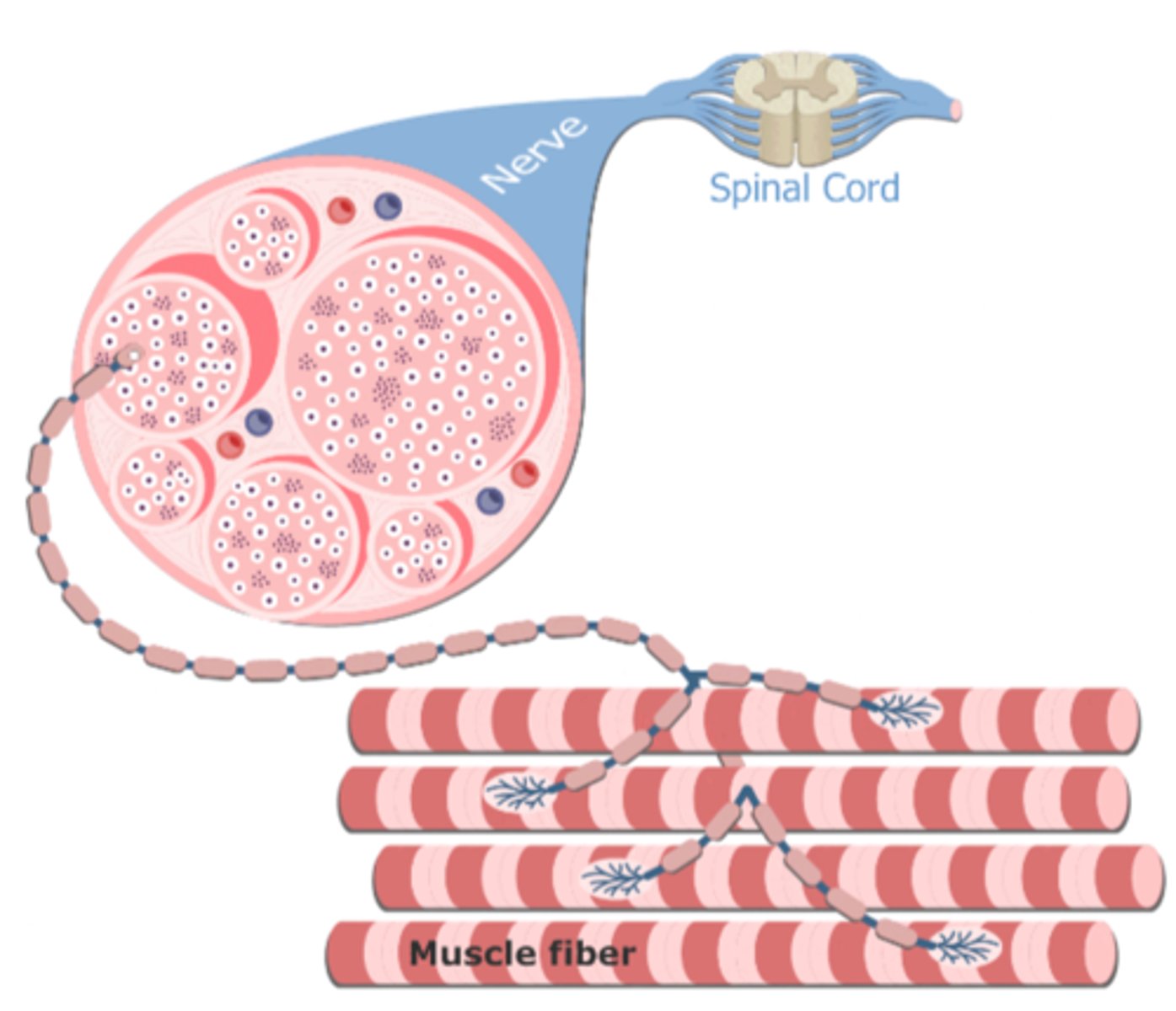
What are neuromuscular junctions?
The synapse between a motor neuron and a muscle cell
What happens when an action potential reaches the synaptic terminal of the motor neuron?
It causes the release of the neurotransmitter acetylcholine into the synaptic cleft which binds to receptors in the sarcolemma and opens ligand-gated Na⁺ channels leading to muscle contraction
What is a skeleton?
A supportive framework that supports and protects an animal's body (it also facilitates locomotion by providing anchorage for muscles and acting as levers)
What are exoskeletons?
Hard, protective skeleton made of chitin on the outside of the body (e.g. arthropods)
What are endoskeletons?
Skeleton made of bone and cartilage on the inside of the body (e.g. vertebrates)
In the musculoskeletal system, what would be the lever (la palanca)?
The skeleton
In the musculoskeletal system, what would be the fulcrum (el punto en medio justo debajo de la palanca)?
A joint
In the musculoskeletal system, what would be the effort?
The muscle that pull the bone
In the musculoskeletal system, what would be the load?
The mass being moved (usually body mass)
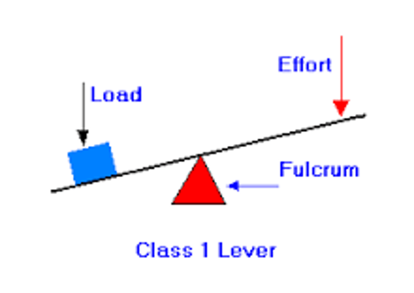
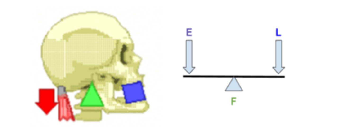
What is the first type of lever in the body?
First-class levers (it has the fulcrum placed between the load and the effort) (e.g. contractions of the muscle in the neck pull on the skull so that the face rises)
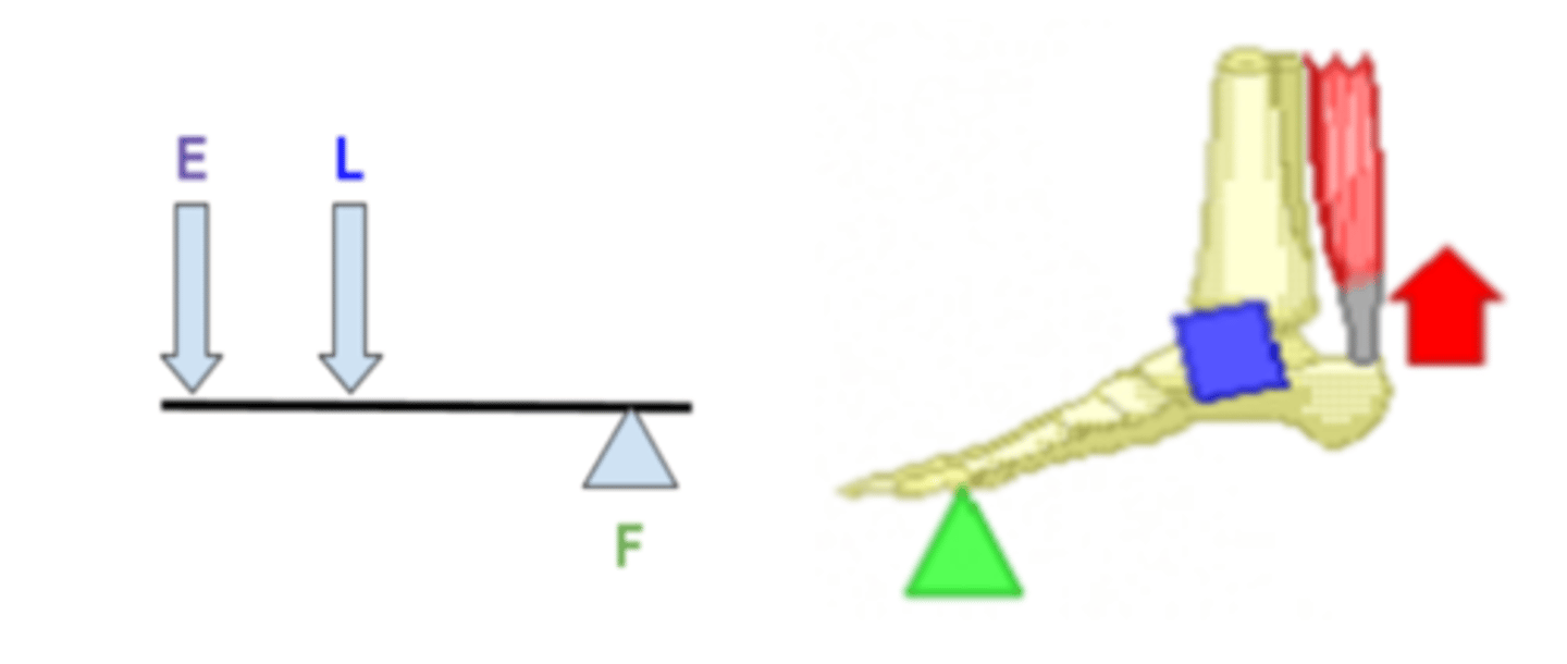
What is the second type of lever in the body?
Second-class levers (it has the load between the effort and the fulcrum) (e.g. contractions in the calf muscle pull the on the heel making the foot rise)
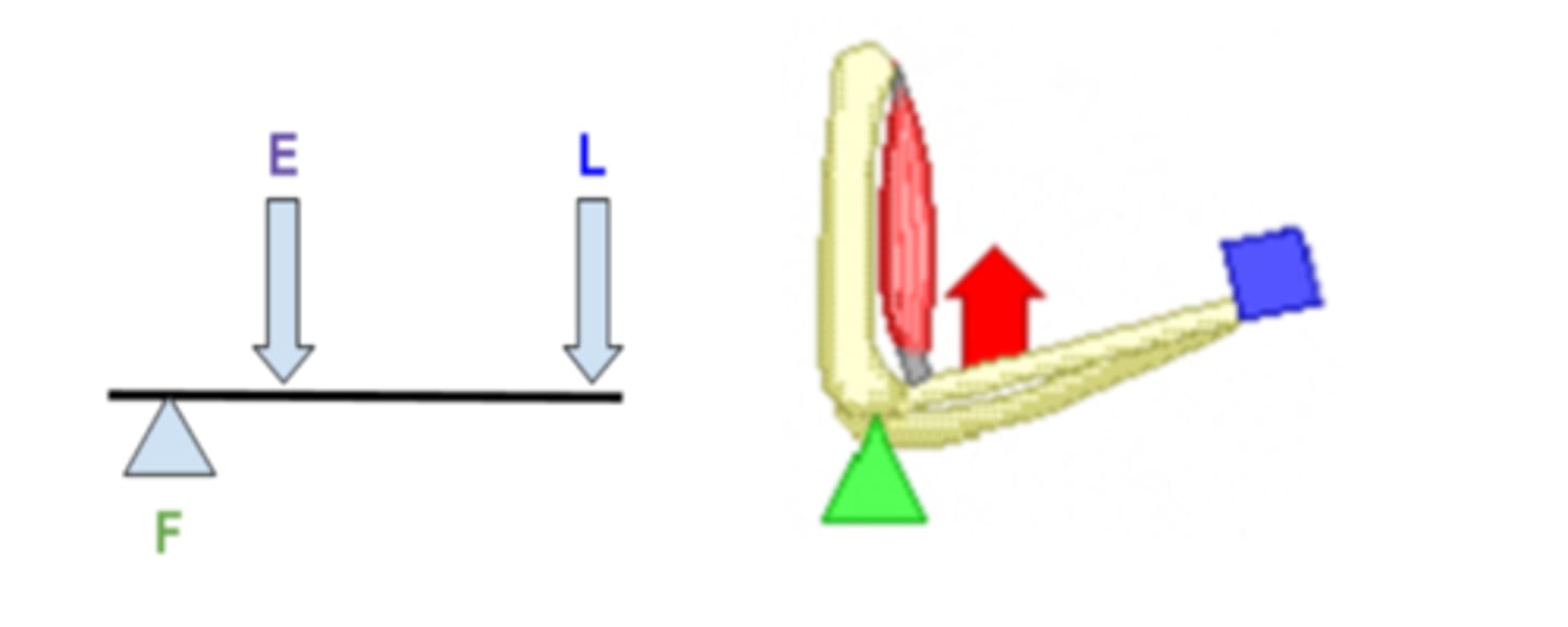
What is the third type of lever in the body?
Third-class levers (it has the effort placed between the load and the fulcrum) (e.g. contractions in the biceps muscle pull the radius making the hand rise)
What is a joint?
The site of the junction of two or more bones of the body (they are classified based on the degree of movement they permit)
What are the three types of joints?
- Immovable, fibrous (e.g. in the skull)
- Slightly movable, cartilaginous (e.g. spine)
- Freely movable, synovial (e.g. elbow)
What does the synovial joint feature?
A fluid-filled space between smooth cartilage pads at the end of articulating bones that contains synovial fluid
What is the synovial joint held together by?
Ligaments which also allows for flexibility
What does the synovial joint include?
Joint capsule, bones, cartilage, synovial fluid, ligaments, muscles and tendons
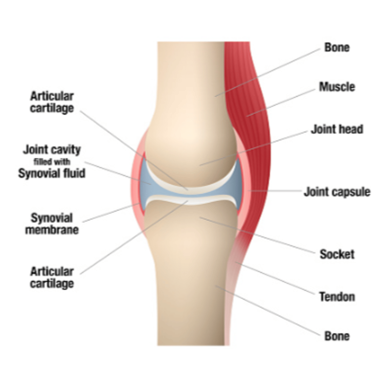
What is the function of the joint capsule?
A flexible, fibrous tissue that surrounds a joint to retain the synovial fluid and provides protection and stability
What is the function of the bones?
Serves as a lever and anchors muscles
What is the function of the cartilage?
Tough tissue that covers the bone at the joint, prevents friction and absorbs shock
What is the function of the synovial fluid?
Fills the cavity in the joint, lubricates the joint and reduces friction
What is the function of the ligaments?
Tough cords that connect bone to bone at the joint
What is the function of the muscles?
Provides effort force to move the bone at the joint
What is the function of the tendons?
Attach the muscle to the bone
What are the two types of synovial joints?
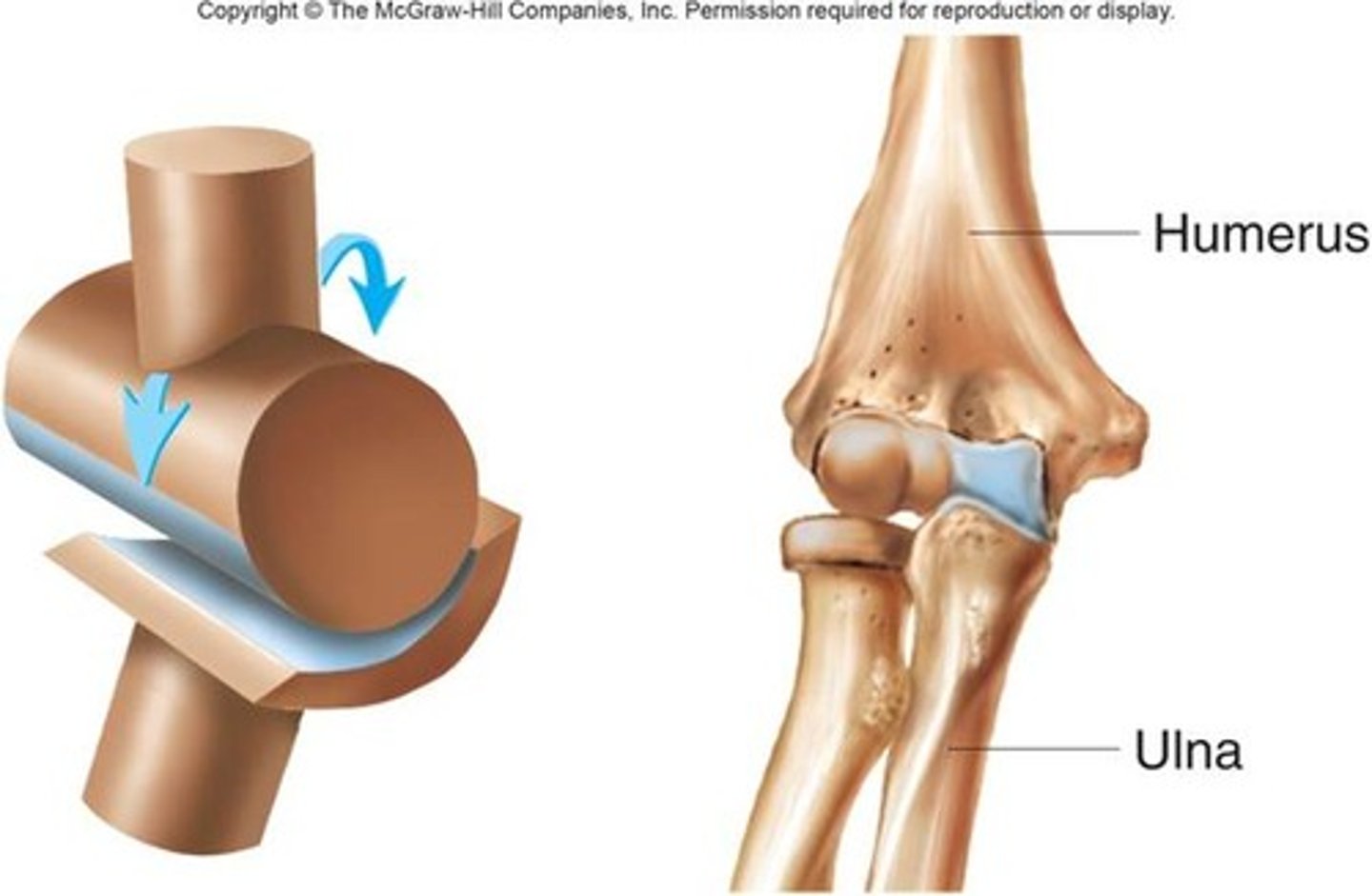
Hinge and ball and socket joints
What is a hinge joint?
A joint that only allows flexion and extension in one plane (e.g. elbow, knee)
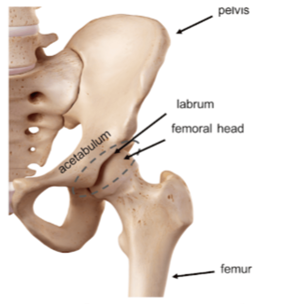
What is the ball-and-socket joint?
A type of synovial joint in which the ball-shaped surface of one rounded bone fits into the cup-like surface of another bone and allows for multiple directions of movement
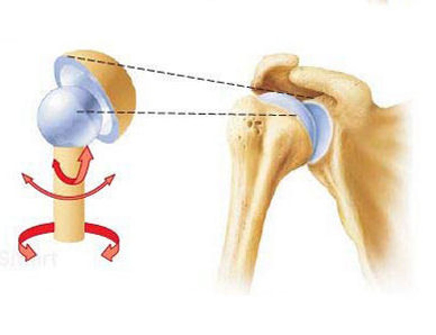
What is an example of a ball-and-socket joint?
The hip joint
What is the range of movement?
The extent to which a joint can move, measured in degrees and in a specific direction (it's a measure of flexibility that involves muscles, tendons, ligaments and bones of the joint)
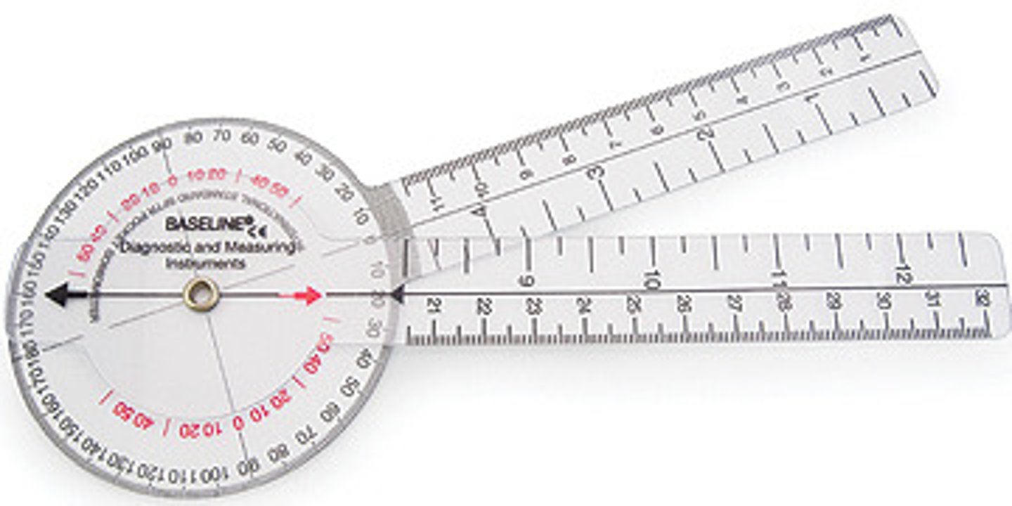
What can be used to measure the range of movement of a joint?
- Goniometer: a tool with two arms that are hinged together and positioned at a joint to measure the angle
- Analysis of images: using computer programs or phone applications that measure angles
What are the possible movements in a joint?
Flexion, extension, rotation, abduction and adduction
What is flexion?
Bending a joint, decreasing the angle of bones at these joints
What is extension
Straightening a joint, increasing the angle of bones at these joints
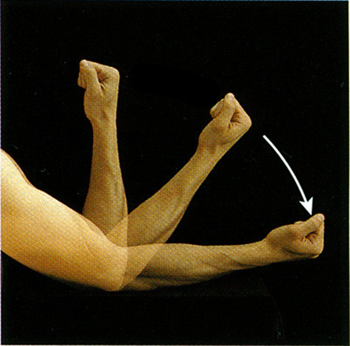
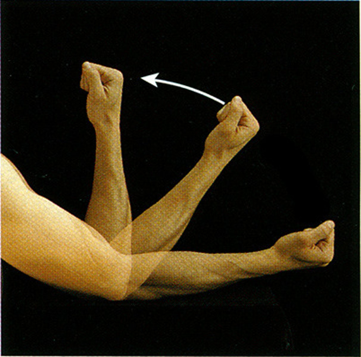
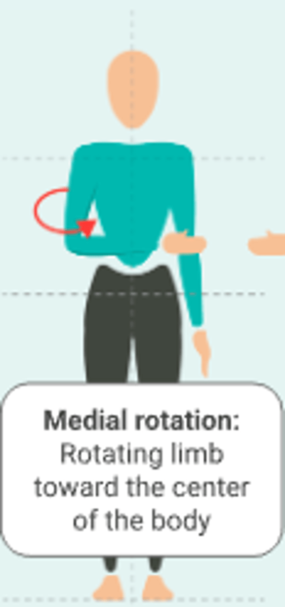
What is medial rotation?
Rotating limb towards the centre of the body
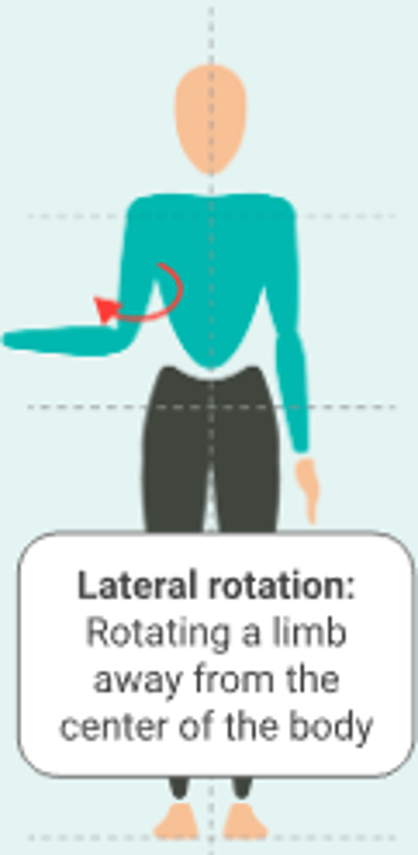
What is lateral rotation?
Rotating a limb away from the centre of the body
What is abduction?
Movement of a limb away from the centre of the body
What is adduction?
Movement of a limb towards the centre of the body
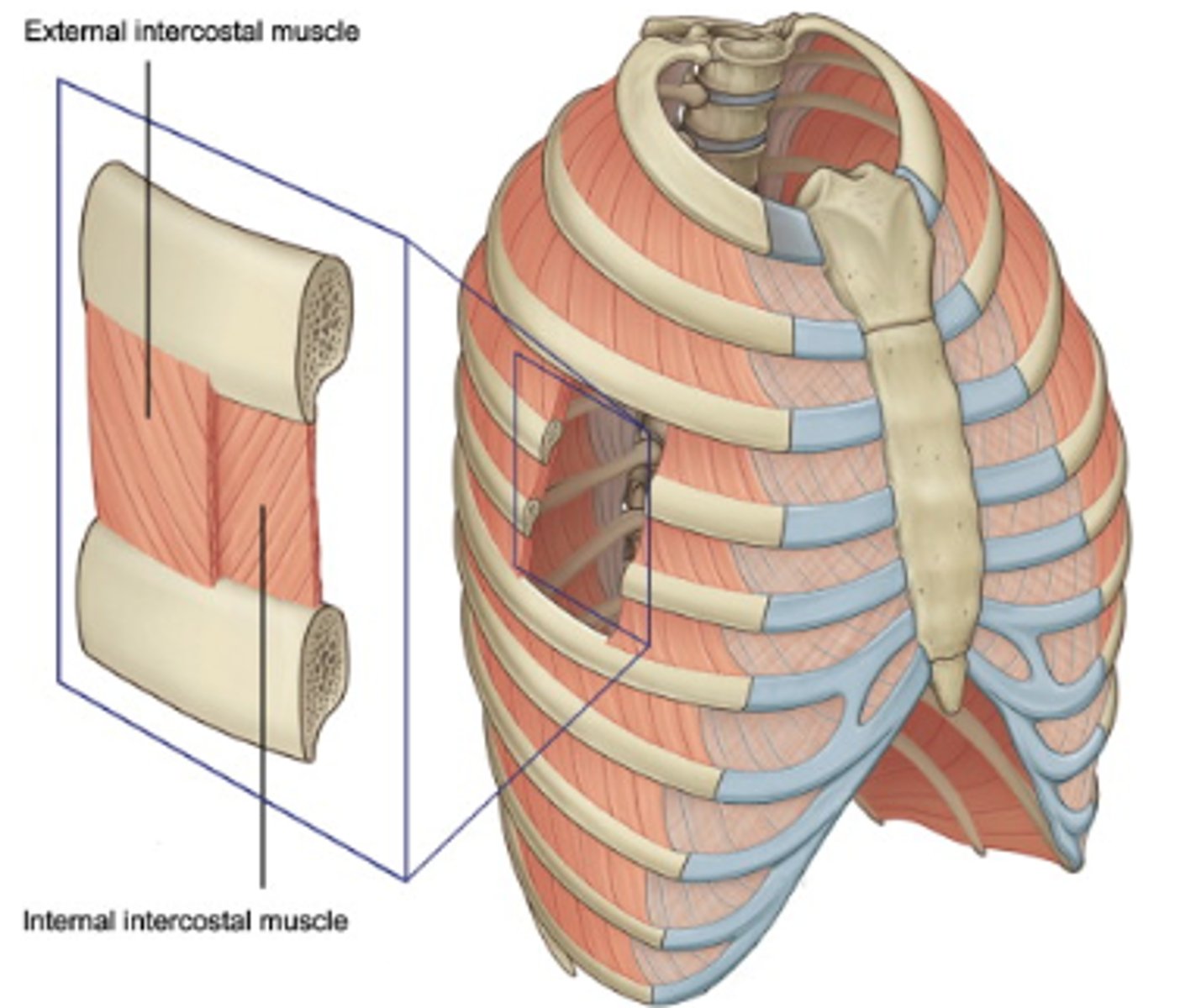
Are the external and internal intercostal muscles examples of antagonistic muscles?
Yes
What is the mnemonic for the direction of external and intercostal muscles?
Hands on pockEts (external = diagonally down)
Hands on tIts (internal = diagonally up)
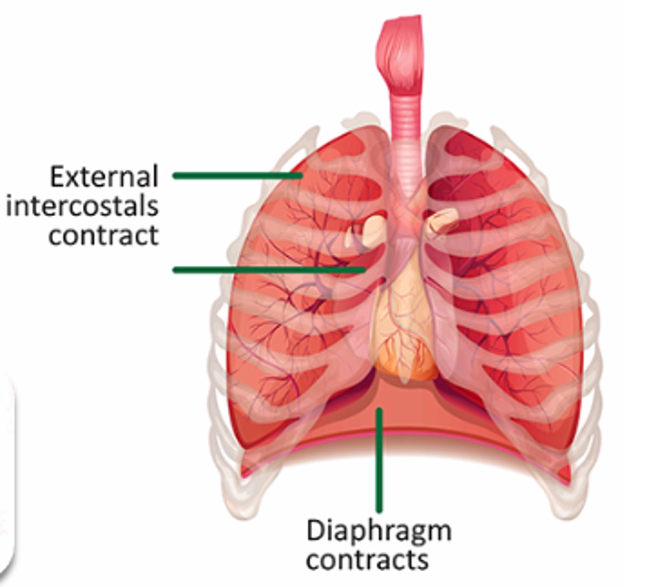
What happens to external intercostal muscles in inspiration?
They contract to pull the ribcage up and out, expanding the chest cavity and allowing air to flow into the lungs
What happens to internal intercostal muscles in expiration?
They contract to pull the rib cage down and in, reducing the chest cavity volume and pushing the air out of the lungs
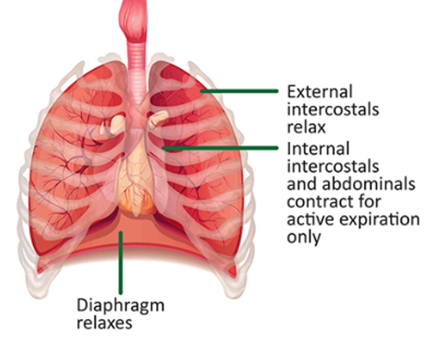
Why do the intercostal muscles move the rib cage in opposite directions?
Because of the different orientations of muscle fibers in the internal and external layers
What happens when one of the intercostal muscles contracts?
The other stretches, storing potential energy in the sarcomere protein titin
What are some reasons why animals need locomotion?
Finding food (e.g. pollinators or grazing animals), escaping danger (e.g. antelope escaping from a lion), searching for a mate (e.g. a roaming lion) and migration (e.g. salmon or geese)
Do marine mammals have special adaptations for swimming?
Yes
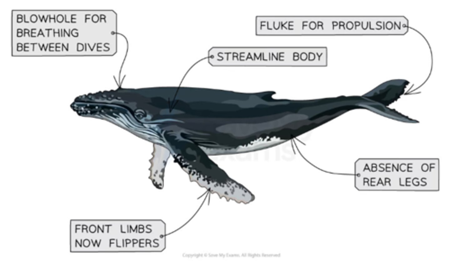
What are some adaptations of marine mammals?
- Streamlining (larger at front, thinner at back, smooth and hairless): facilitates movement through relatively viscous water with ease and great speeds
- Adaptations of front limbs to form flippers (used mainly for steering) and rear legs have been lost
- Adaptation of tail to form a fluke (capable of up and down movement and used for propulsion)
- Changes to airways by the evolution of a blowhole allows periodic breathing between dives (they can be sealed between dives so that water does not enter the airways)
- Blubber tissue for insulation