a+p lab test winter
1/351
There's no tags or description
Looks like no tags are added yet.
Name | Mastery | Learn | Test | Matching | Spaced | Call with Kai |
|---|
No analytics yet
Send a link to your students to track their progress
352 Terms
adult skeleton
206 bones
axial skeleton
80 bones - skull, vertebral column, and thoracic cage
appendicular skeleton
126 bones - upper and lower limbs
long bones
longer then they are wide - humerus, femur
diaphysis
shaft region of bone - compact bone occurs there
epiphysis
ends of long bones - spongy bone occurs there
short bones
similar length and width - carpal bones
flat bones
thin flattened and slightly curved bone - scapula
irregular bone
bones w/ complex shapes - vertebrae
sesamoid bone
small round sesame-like - patella
process
projection of bone - raised bump
tuberosity
large rounded projection - attachment point
tubercle
small rounded projection - attachment point
condyle
rounded articular projection - attachment point
foramen
round oval opening through bone
fontanels
fiborous connective tissue on baby skull - allows skull to compress through birth canal and permit growth during infancy.
fontanels become
sutures by 2 years of age
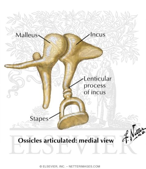
auditory ossicles
malleus, incus, stapes
foramen magnum
spinal cord passes through
occipital condyles rest upon
vertebral column
number of bones forming face
14:
2 maxillae, 2 zygomatic, 2 nasal, 1 mandible, 2 lacrimal, 2 palatine, 2 inferior nasal conchae, 1 vomer
number of bones forming eye orbit
7:
frontal, sphenoid, zygomatic, maxilla, palatine, lacrimal, ethmoid
hyoid bone
attachment point for muscles of the tongue and larynx
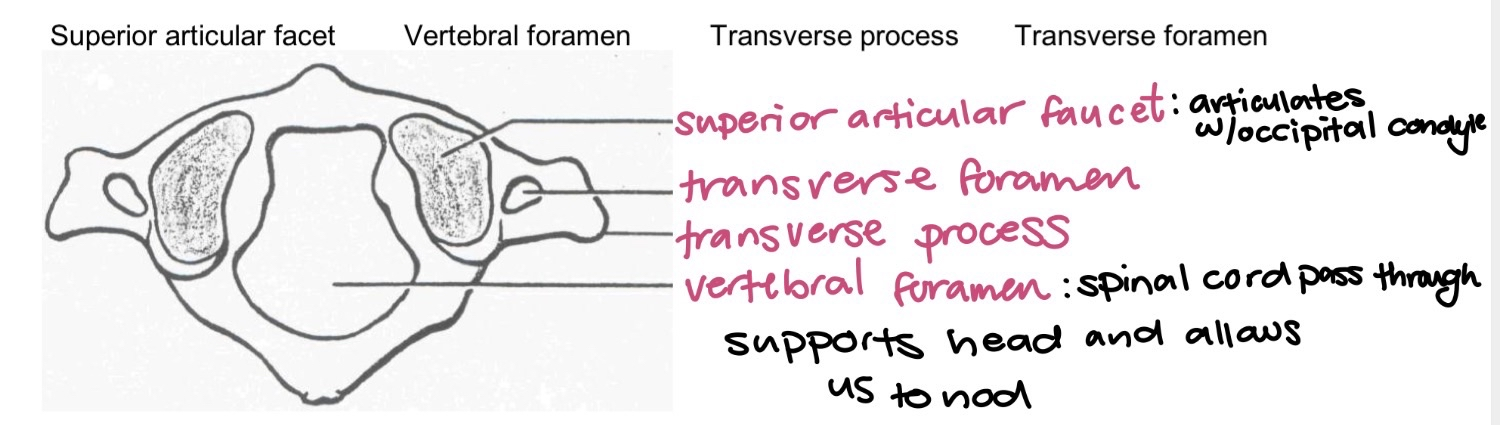
C1 atlas
receive occipital condyles of skull that allows for nodding using superior articular faucet
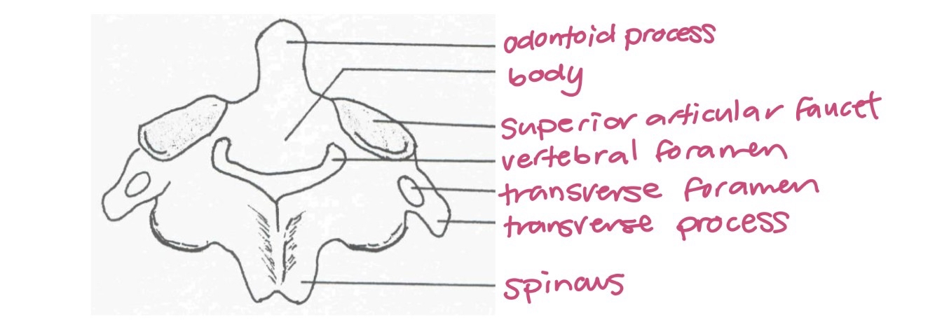
C2 axis
articulates with atlas and allows head to shake no
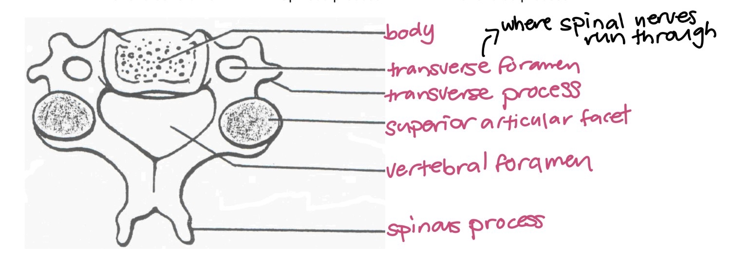
C3-C7 cervical vertebrae
mantis looking
transverse foramen - where spinal nerves run through
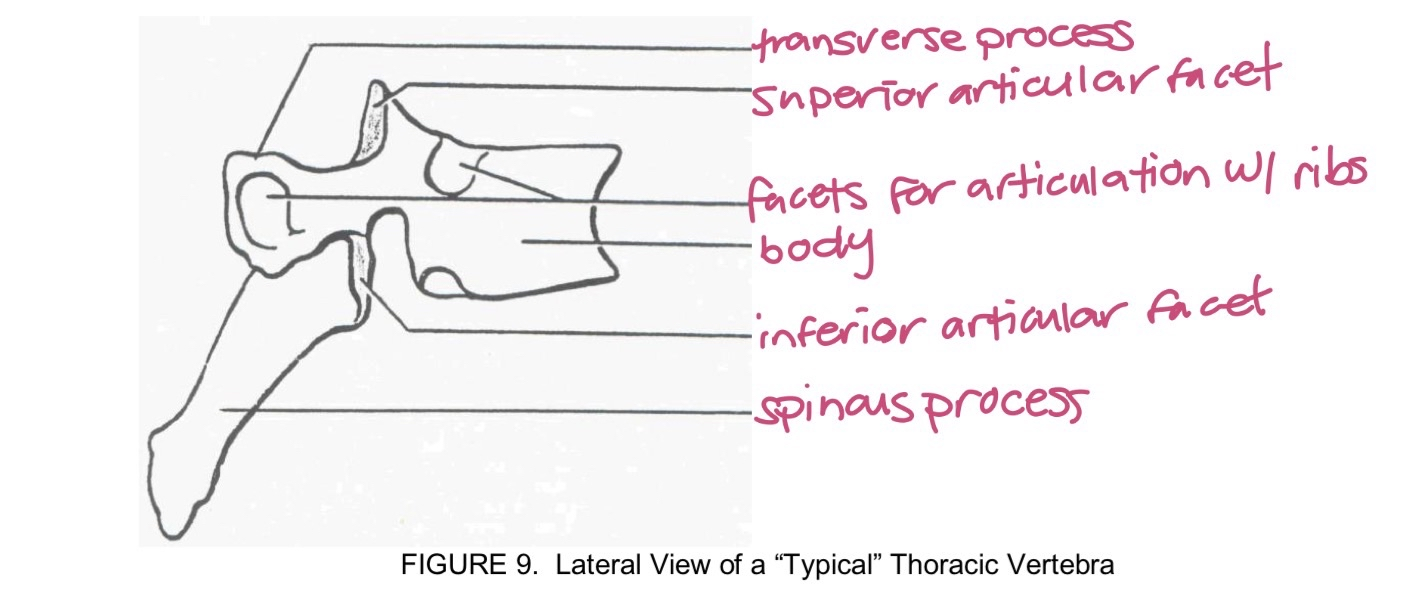
T1-T12 thoracic vertebrae
giraffe looking
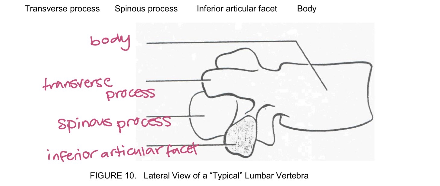
L1-L5 lumbar vertebrae
moose looking
largest and heaviest: weight bearing
sacroiliac joint
sacrum articulating with ilium of each hipbone
coccyx position
inferior coccyx: female - allows passage of baby
anteriorly coccyx: male
true rib
direct cartilage connection to sternum - 7 pairs
false rib
does not directly attach to sternum - 5 pairs
floating rib
only attaches to spine not sternum - 2 pairs
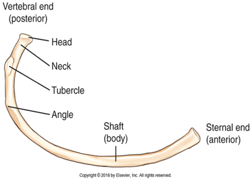
individual rib
vertebral end is bumpy
sternal end is smooth
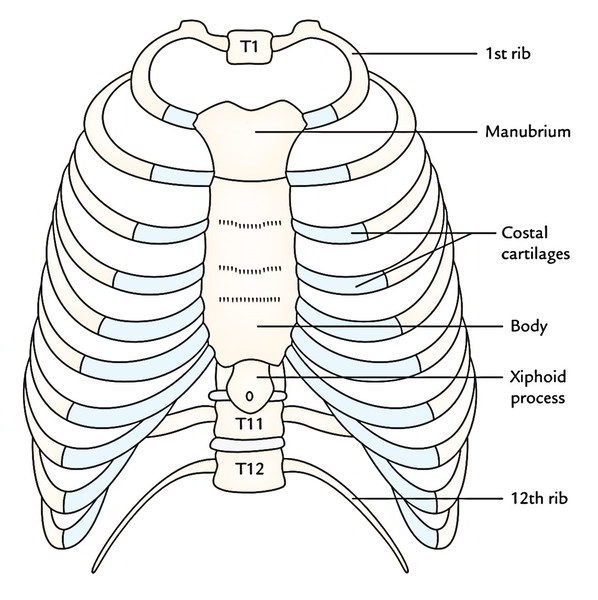
thoracic cage
manubrium
sternum
xiphoid process
2 bones composing zygomatic arch
zygomatic process of temporal bone and temporal process of cheekbone
bones composing hard palate
palatine and maxilla
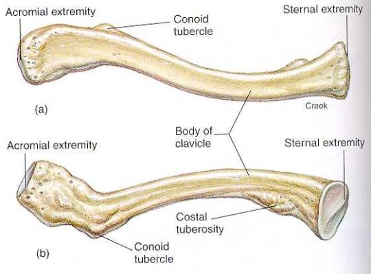
clavicle
rounded end lateral - shoulder scapula side
flat end medial - towards manubrium
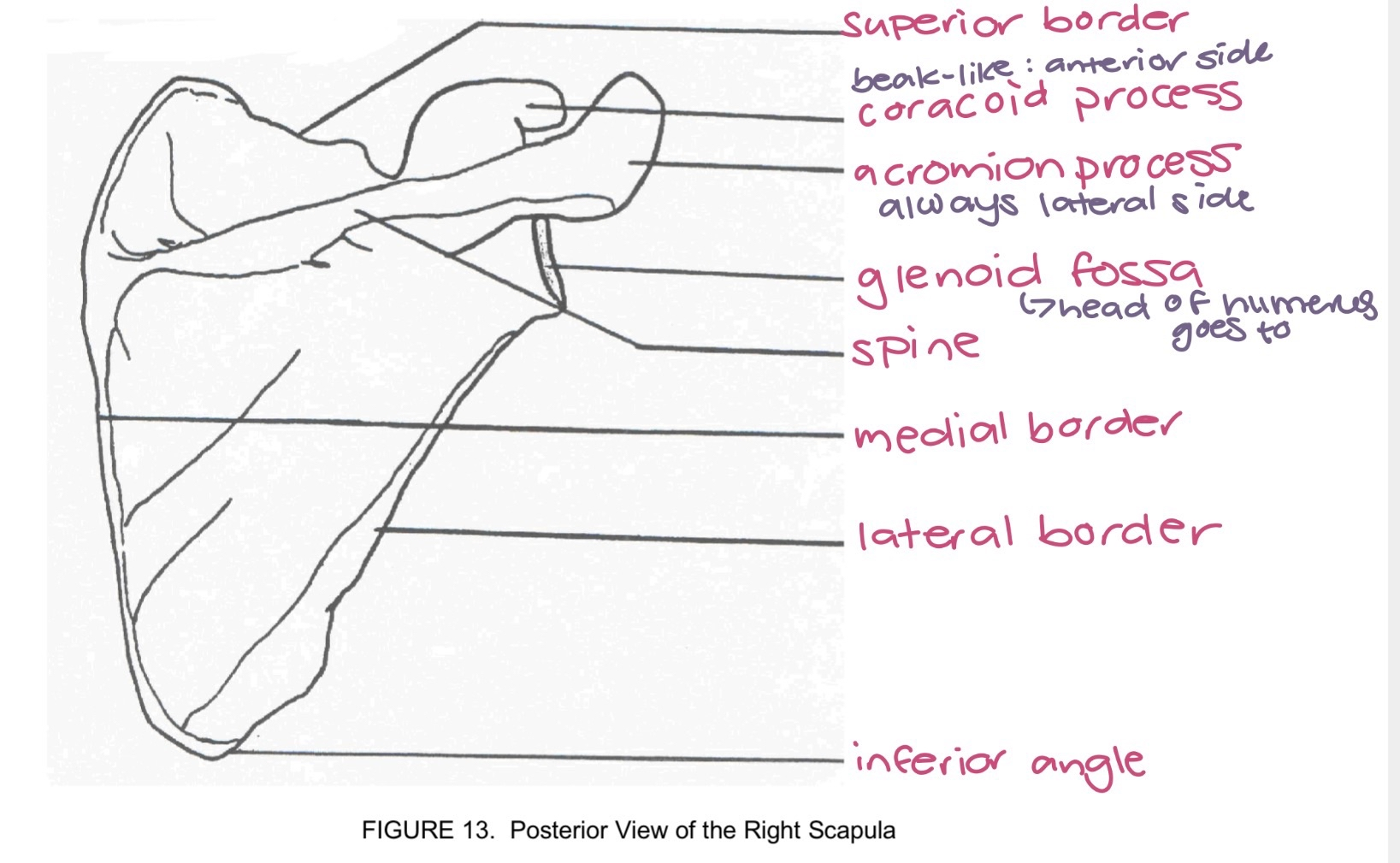
scapula
acromion process always anterior and lateral
glenoid fossa - where humerus goes to
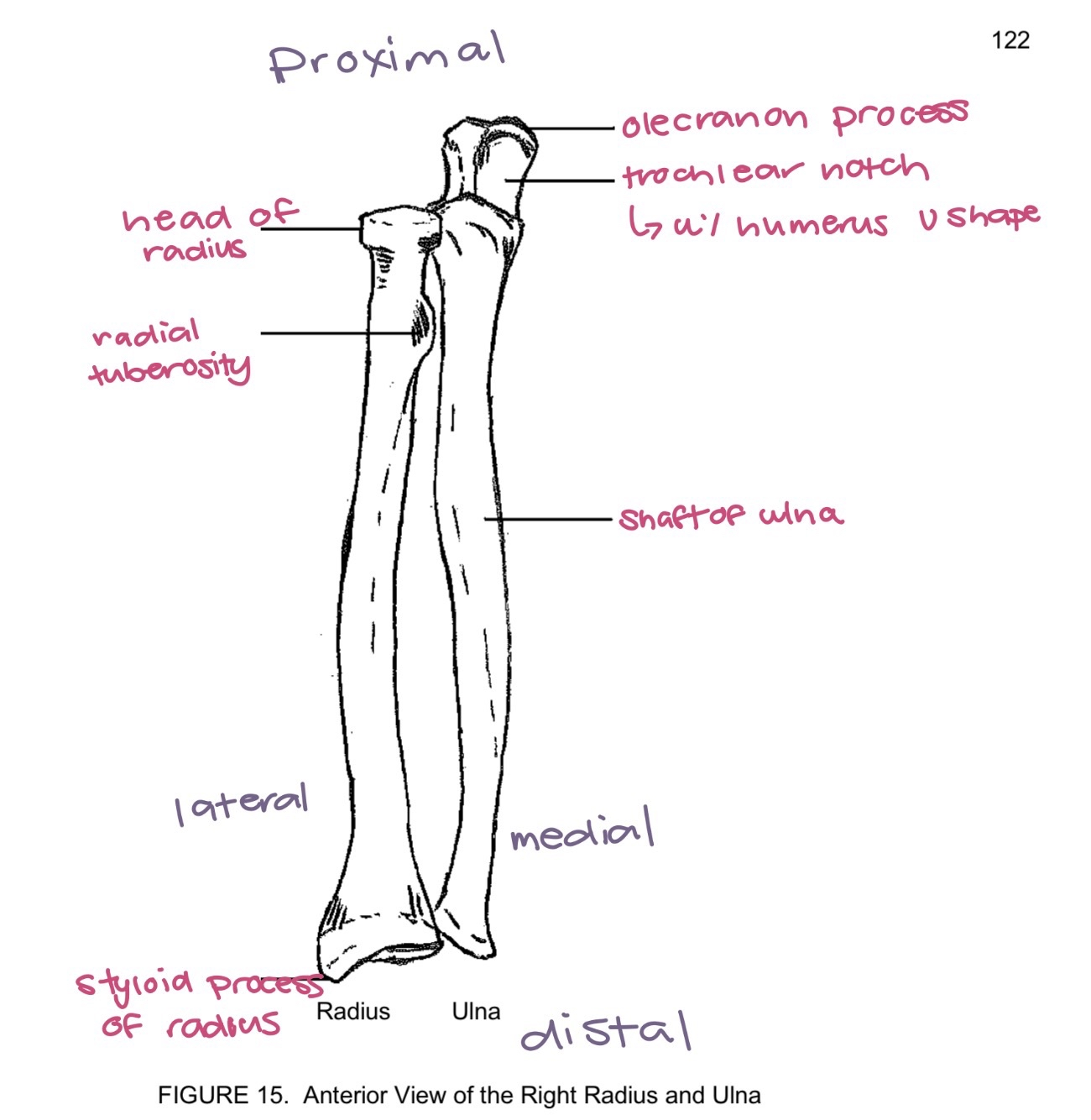
ulna and radius anatomical position
ulna medial radius lateral
proximal end radius head articulates with
humerus and ulna - specifically using the trochlear notch
distal end of radius articulates with
carpal bones of wrists
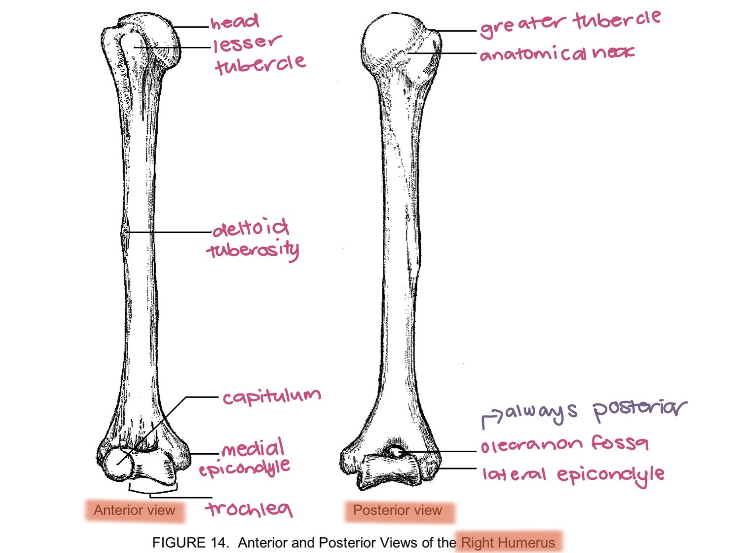
humerus
olecranon fossa always anterior
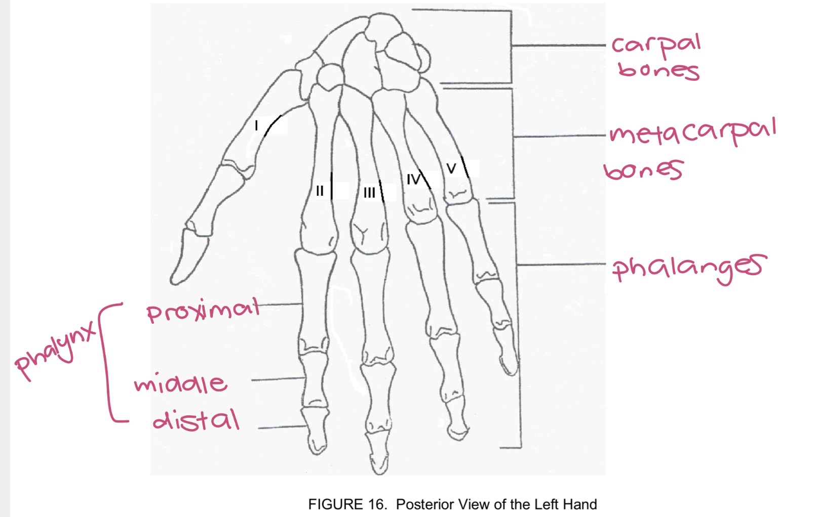
phalanges
proximal phalanx middle phalanx and distal phalanx
bony pelvis consists of
coxae, sacrum, and coccyx
true pelvis
below pelvic brim
false pelvis
above pelvic brim
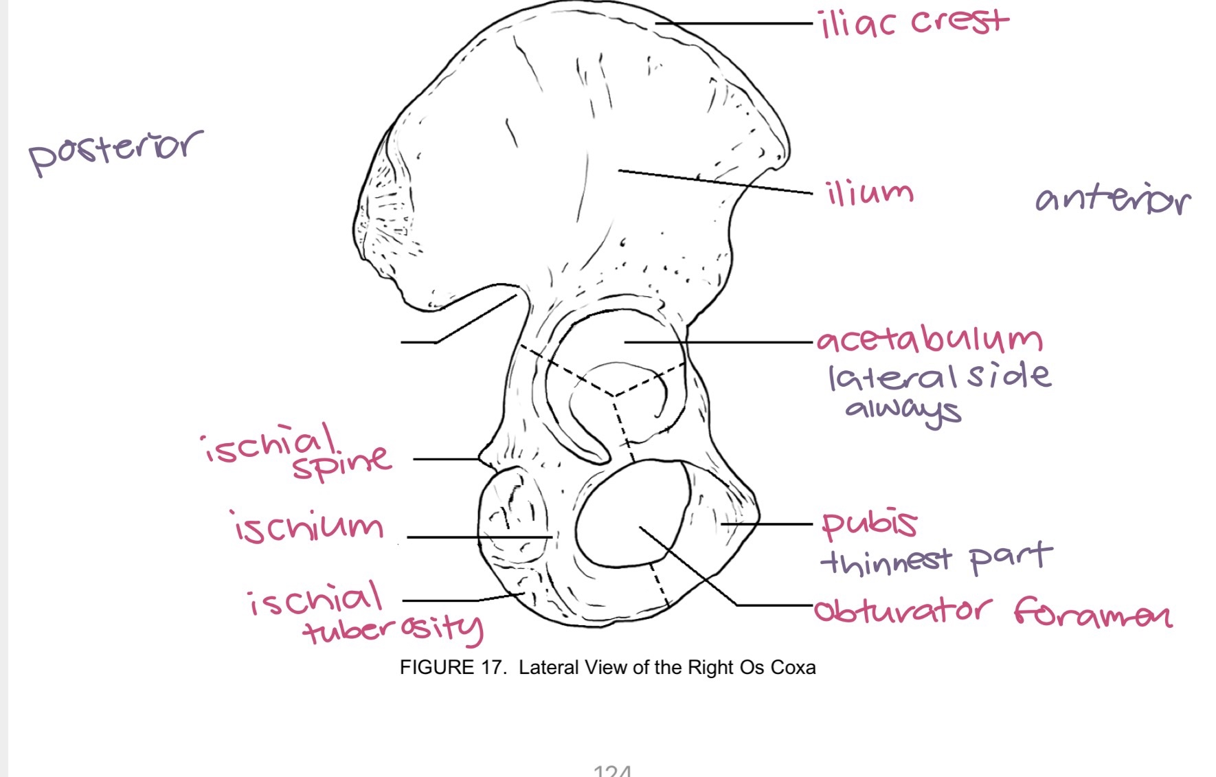
os coxae
made of: ilium ischium and pubis top
acetabulum socket for femur is always lateral
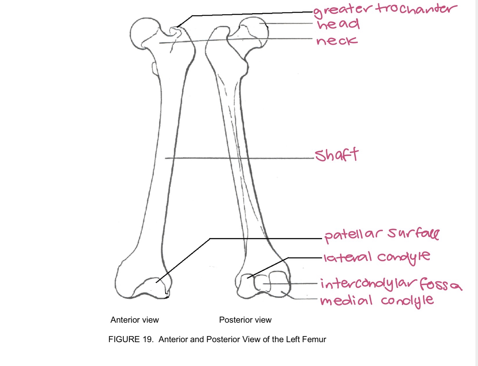
femur
has intercondylar fossa for medial condyle that articulates with acetabulum and tibia
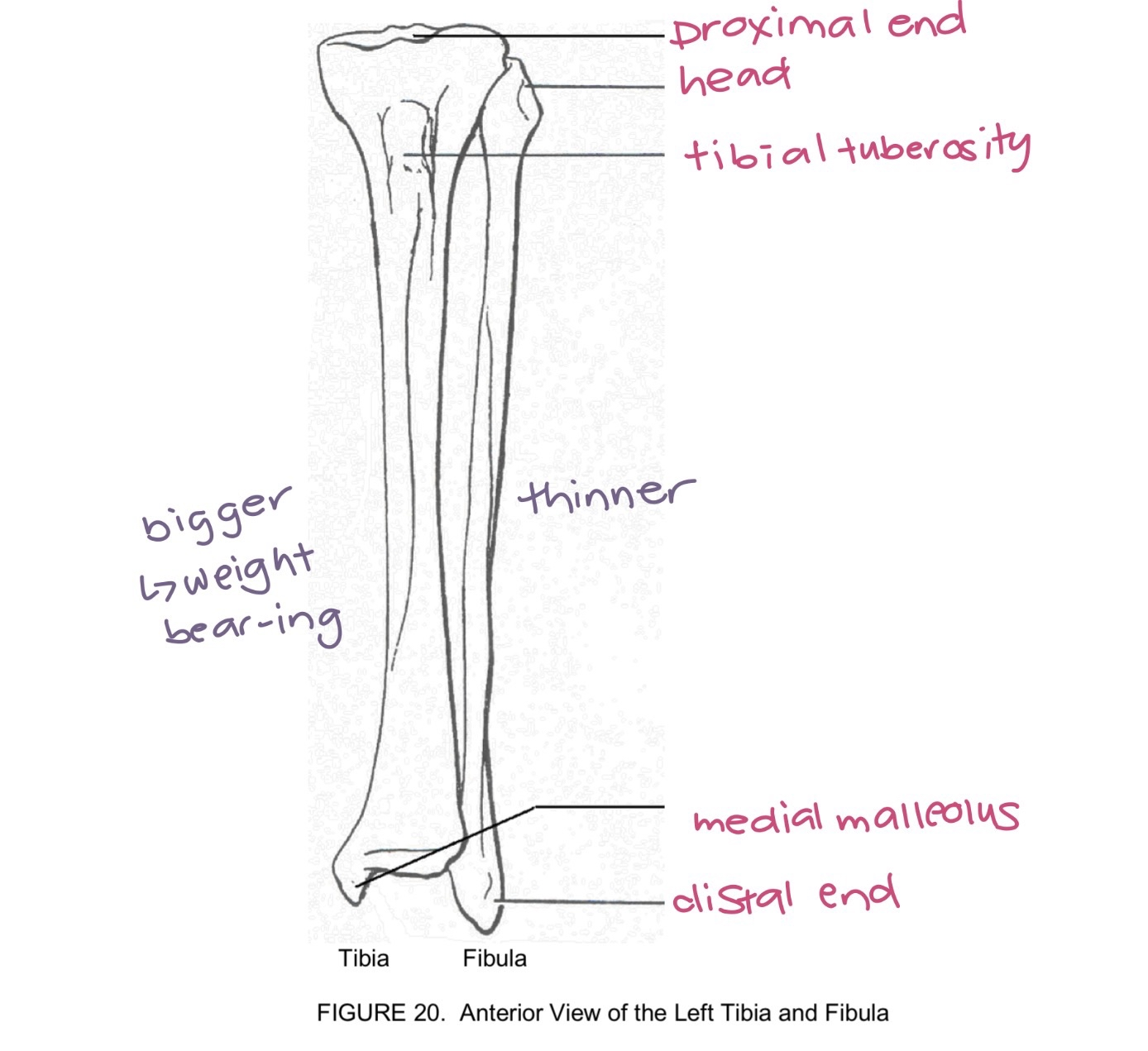
tibia and fibula
tibia is bigger weight bearing
talus
articulates with tibia and fibula
calcaneus
heel bone
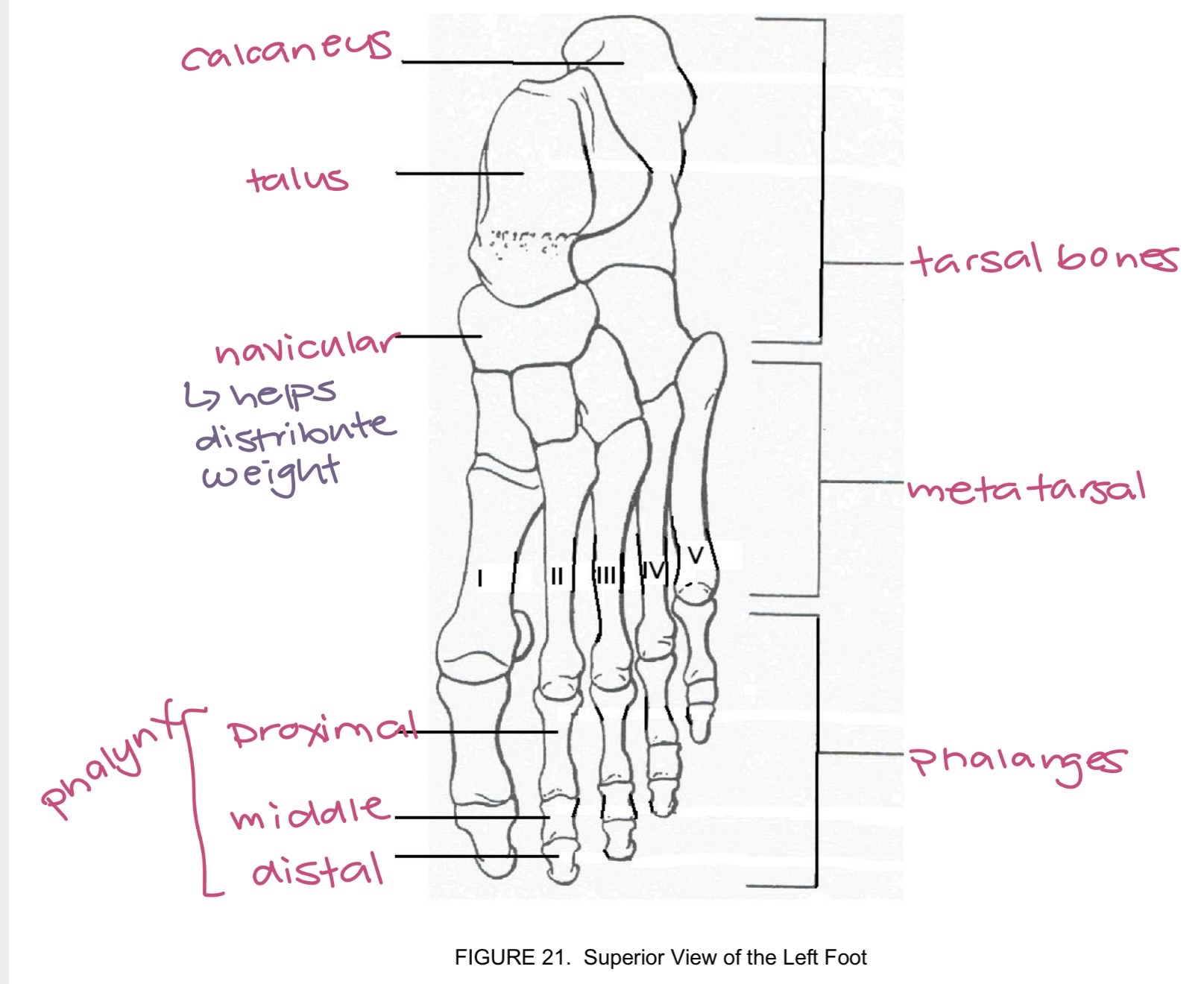
foot
has navicular that helps distribute weight
bone we sit down on
ischium
pectoral girdle attaches to
sternum
functional classification of joints
based on amnt of movement that can occur:
immovable joints
slightly movable joints
freely movable joints
structural classification of joints
type of connective tissue that binds articulating surfaces together at a joint cavity
fibrous joint
cartilaginous joint
synovial joint
fibrous joint
bound by dense fibrous connective tissue - little to no movement
no joint cavity
ie sutures
cartilaginous joint
articulating bones bound tgt by cartilage - little to no movement
no joint cavity
ie epiphyseal plates in long bones of children
synovial joint
hyaline cartilage covers the surface of articulating bones and has synovial membrane producing fluid that lubricates the joint - reinforced by ligaments of dense regular connective tissue
has a joint cavity
freely movable
gliding synovial joint
non axial slide - side to side in one plane
little movement
ie joints b/w carpal bones of the wrist
hinge synovial joint
1 axis: flex and extend
movement occurs in a single axis: permit flexion and extension
ie elbow joint
pivot synovial joints
1 axis, rotation
ie joint b/w atlas and axis
condyloid synovial joint
2 axes 1 better than other
permits: flexion extension adduction and circumduction
ie joint b/w radius and carpal bones
saddle synovial joint
2 axes, both equally good
flexion extension abduction adduction circumduction and rotation
ue joint b/w carpal and metacarpal bone of thumb
ball and socket synovial joint
multiaxial movement
flexion and extension abduction and adduction circumduction and rotation
ie shoulder joint
abduction
movement of limb away from medial line of body - going away remove
lifting arm up
adduction
movement of limb towards medial line of body - towards midline add
arm going back to side
flexion
decreases angle between articulating bones
contraction of flexor muscle
bending arm
dorsiflexion
toes point upward
plantar flexion
toes point downward
extension
bending movement that increases angle b/w articulating bones
straightening of limb
hyperextension
extension beyond anatomical position
rotation
movement of bone around longitudinal axis
ie shaking head no
circumduction
movement when proximal limb stays stationary and distal limb moves in a circule
pronation
hand from palm up to palm down
pro=pull up position
supination
palm down to palm up
sup= soup holding bowl
eversion
movement of soles turning outward laterally
inversion
movement of soles turning inward medially
glenohumeral joint
ball and socket synovial joint
glenoid fossa humerus
greatest range of motion
coracohumeral ligament
coracoid process of scapula and humeral head
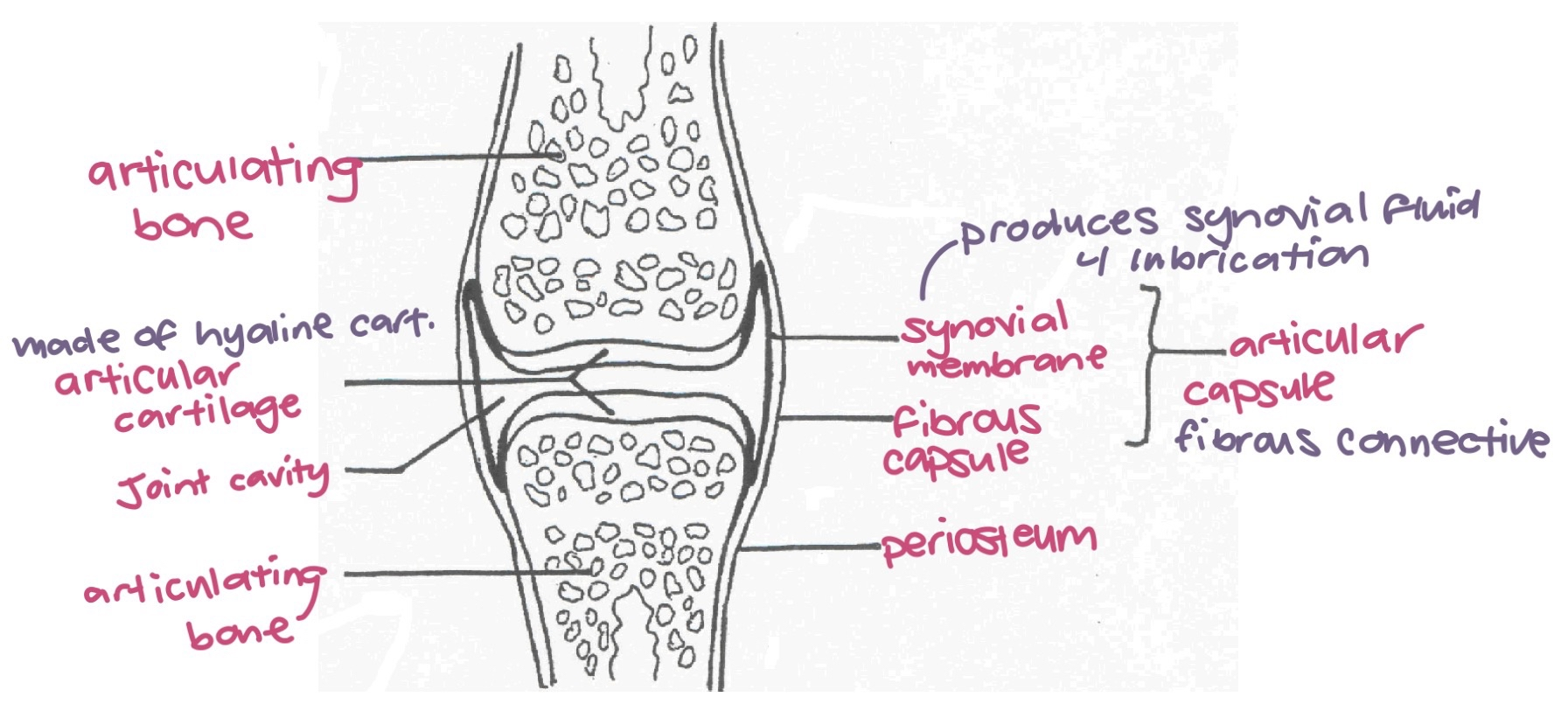
synovial joint diagram
synovial membrane produces synovial fluid for lubrication
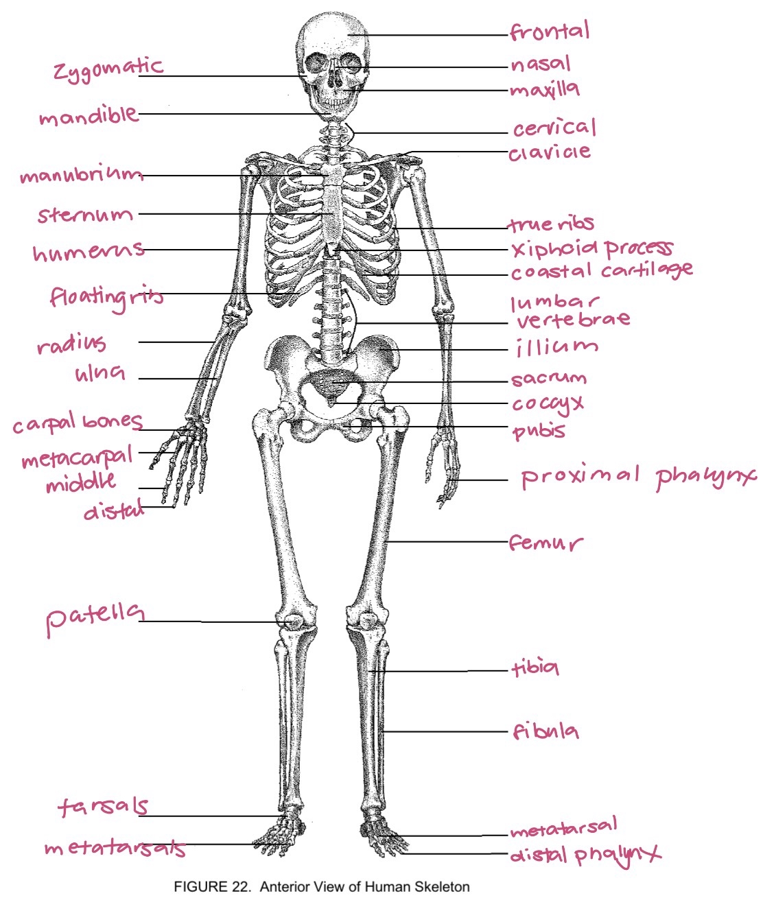
full body skeleton
anterior view
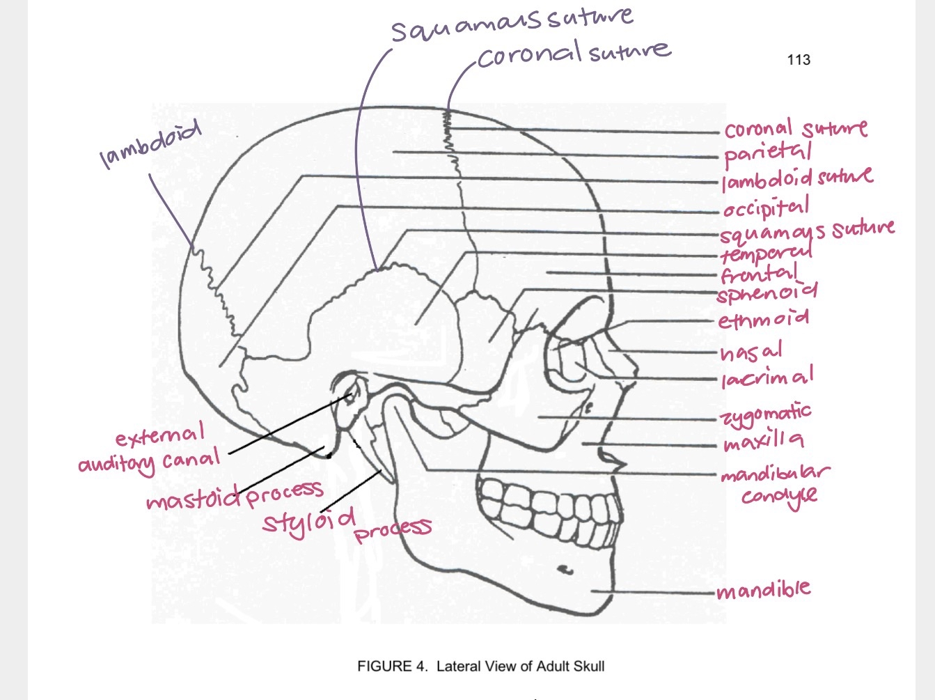
skull lateral
lateral view
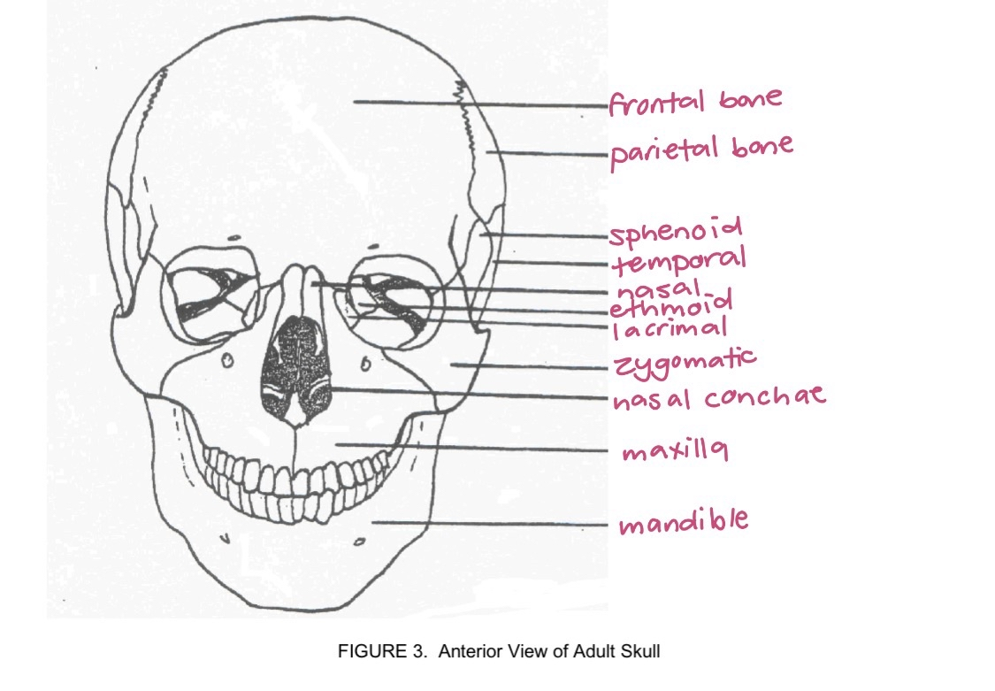
skull frontal
frontal view
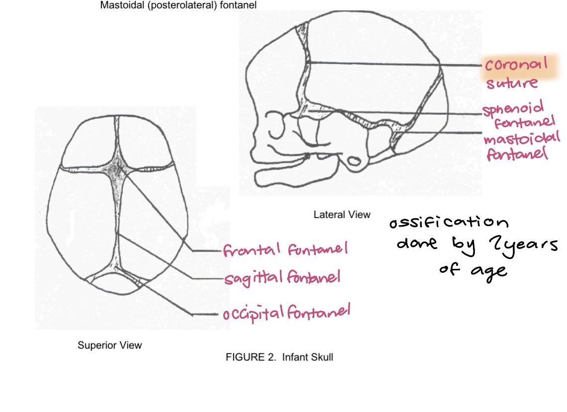
infant skull
fontanels: sphenoid fontanel, mastoid fontanel, frontal fontanel, sagittal fontanel, occipital fontanel
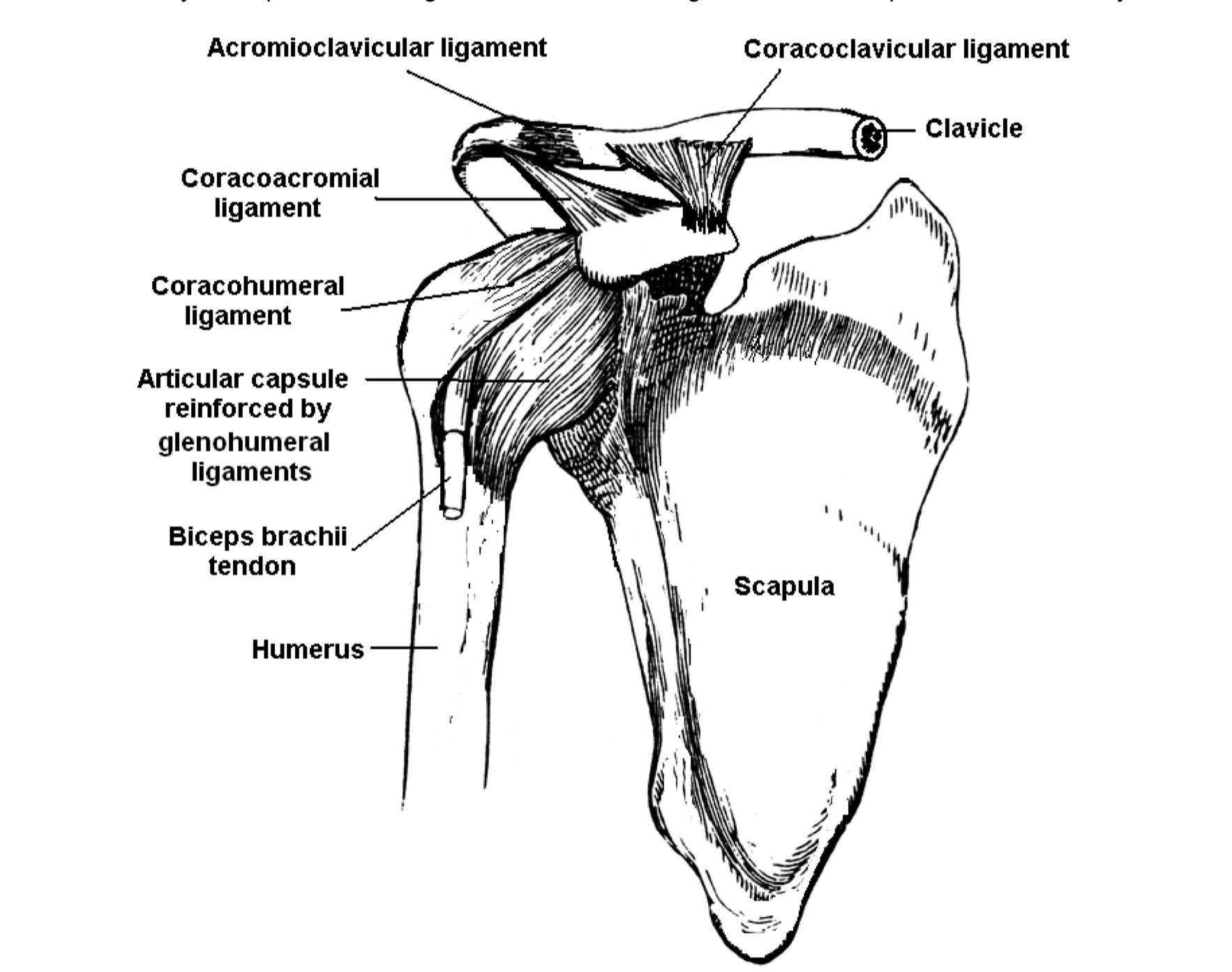
shoulder joint
coracoclavicular ligament binds clavicle to coracoid process of scapula
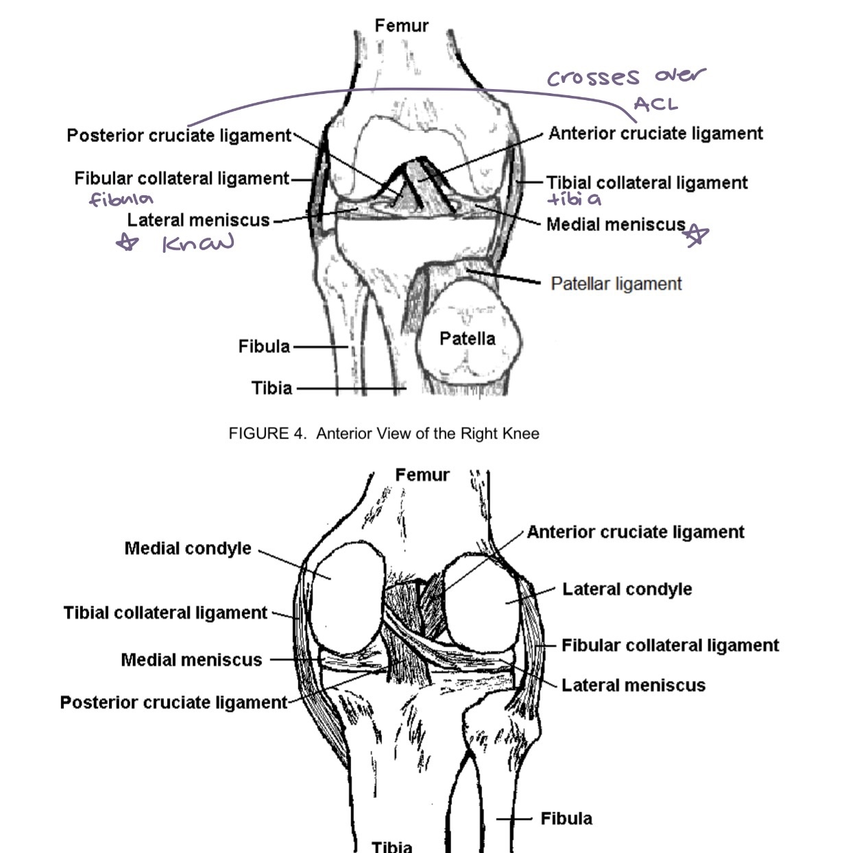
knee diagram
lateral and medial meniscus - frequently damaged and torn due to injury
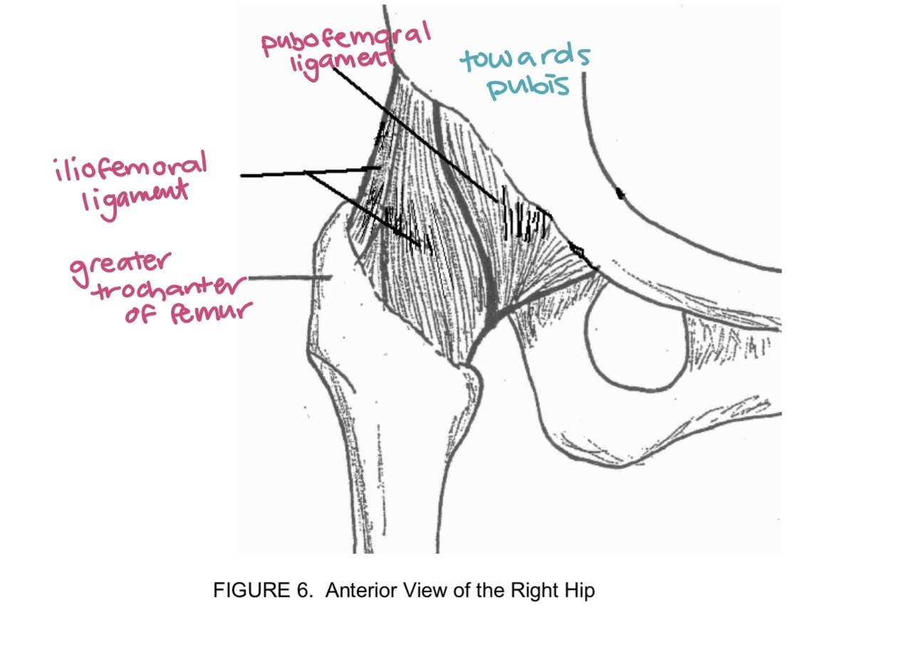
hip joint
pubo near pubis
ilio ilium
ischio ischium
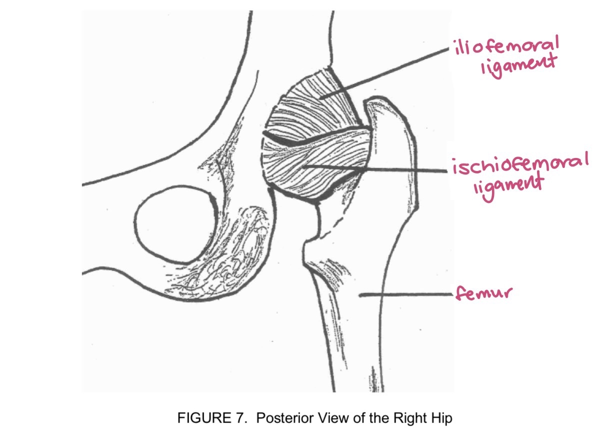
ligaments around joint help
stabilize and limit movement and prevent dislocation
bursitis
inflammation of fluid sacs that cushions: bones tendons and muscles
arthritis
swelling and tenderness of one or more joints - leads to pain and stiffness
origin
stationary end of a bone
insertion
attachment site on the move that moves
masseter
origin zygomatic arch
insertion lateral surface of mandible
closing jaw
sternocleidomastoid
origin manubrium of sternum
insertion mastoid process of temporal bone
flex neck
pectoralis major
origin clavicle
insertion humerus
arm medially rotates at shoulder joint
deltoid
origin acromion process of scapula
insertion deltoid tuberosity of humerus
laterally rotate arm
rectus abdominus
origin pubic symphysis
insertion xiphoid process of sternum
compresses abdominal contents
gluteus maximus
origin sacrum
insertion posterior surface of femur
extends thigh at hip