Looks like no one added any tags here yet for you.
The cornea is ____ to the sclera
anterior
What is the thin membrane that lines the sclera anterior?
conjunctiva
What is the inside layer of the sclera called?
uvea
What does the conjunctiva NOT cover?
cornea
What does the anterior chamber contain?
lens, iris, ciliary body
How does the blood flow from the ophthalmic artery?
deep to peripheral
How does the blood flow from the choroid plexus?
peripheral to deep
What is the site where all vessels terminate?
macula
What supplies blood to the macula and fovea?
choroid plexus
What CN provides sensory to cornea and conjunctiva?
CN V
Which CN is for vision?
CN II
What CN controls opening of the eye, eye movement, sympathetic pupil dilation, and parasympathetic pupil constriction?
CN III
What is OD?
oculus dexter; right eye
What is OS?
oculus sinistral; left eye
What is OU?
oculus uterus; both eyes
What is emmetropic?
no vision correction needed
What is myopic?
nearsighted
What is hyperopic?
farsighted
What is presbyopia?
age related farsightedness
What is amblyopia?
poor vision of eye that is other wise physically normal; “lazy eye”
What is a scotoma?
blind spot
What is entropion?
eyelid folds inward
What is ectropion?
eyelid folds outward
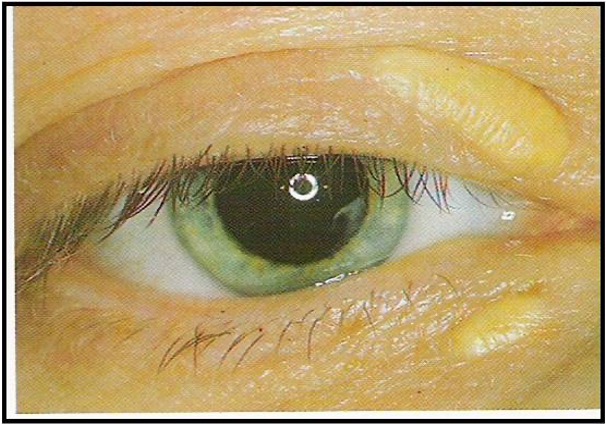
what is hypopyon?
pus in anterior chamber
What is epiphora?
overflow of tears
What is synechiae?
iris adheres to cornea or lens
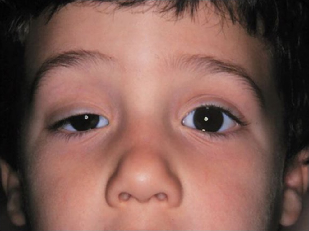
What is ptosis?
drooping of upper eyelid margin
What is hyperemia?
congestion of blood in any part of eye
What is stroma?
connective tissue framework
What is heterochromia?
different colored eyes
What is aphakia?
no lens in the eye
What is dyschromatopsia?
acquired color blindness which occurs due to CN disease or degenerative disease of macula
What is a slit lamp used for?
measure depth of an abrasion or of an infection
What should you use if slit lamp is unavailable?
ophthalmoscope
what would you use to differentiate between ulcer and abrasion?
fluorescein stain
What is a tono-pen / tonometer?
measures the intraocular pressure; anesthetize w/ tetracaine and compare 3 measurements
What is normal intraocular pressure?
10-21 mmHG
What is the intraocular pressure for chronic open angle glaucoma?
20-30 mmHg
What is the intraocular pressure for acute angle closure glaucoma?
> 40 mmHg
How do you measure setting in the orbit?
place a flat object over eyebrow and maxilla and note the distance between the closed eye and flat object
When the patient is looking forward, the upper lid should cover …
the upper portion of the cornea
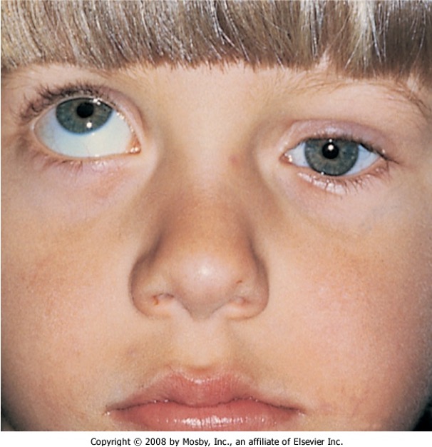
What is exophthalmos?
bulging of eye anterior out of orbit
What is bilateral exophthalmos due to?
abnormal connective tissue deposition in the orbit and extraocular muscle that can be visualized on CT or MRI
What is bilateral exophthalmos associated with?
graves disease
What is unilateral exophthalmos associated with?
orbital tumor
What are findings you might see with exophthalmos?
stare on frontal gaze, lid lag up and down, anxiety, heat intolerance, palpitations
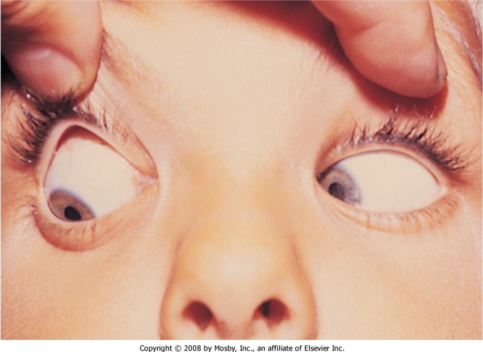
What is this?
enophthalmos
What type of enopthalmos is congenital?
primary
Which enopthalmos is acquired change in volumetric relationship b/t rigid bone cavity, the orbit, and it’s contents?
secondary
What is Horner’s syndrome?
damage to pathway in SNS that regulates HR, pupil size, perspiration, BP, etc; results in ptosis, miosis, anhidrosis
what would CN III palsy result in?
ptosis
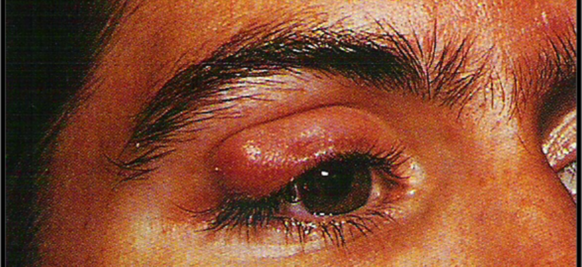
What is an internal hordeolum?
acute infection of meibomian gland deep to the eyelid surface
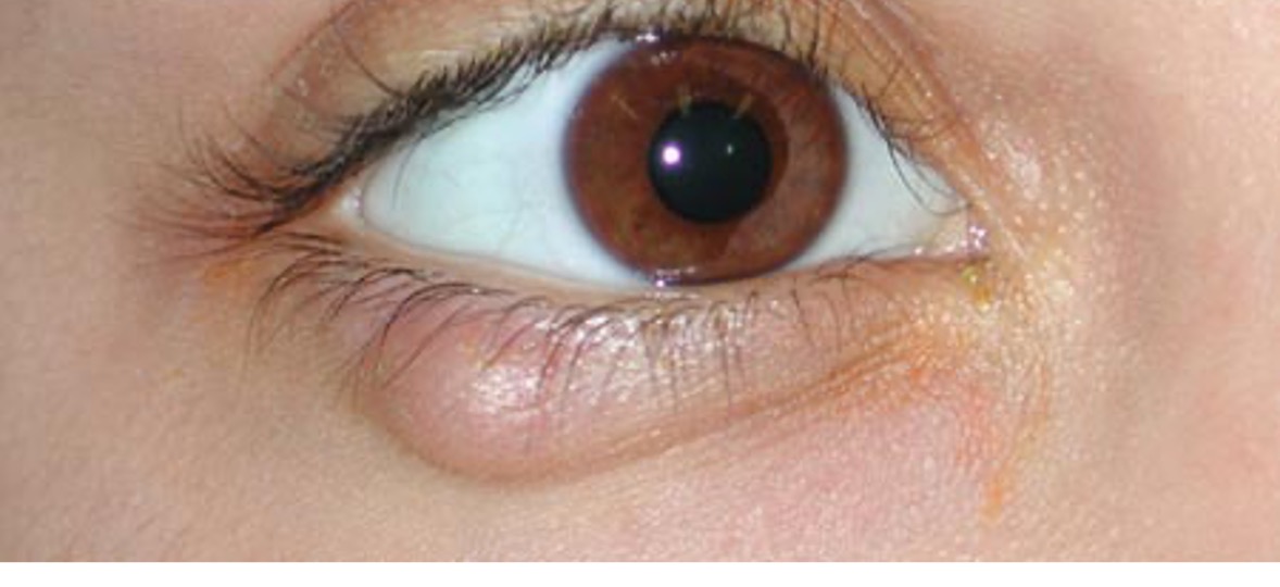
What is an external hordeolum?
pustule in an eyelash gland (moll or zeis)
What is a chalazion?
chronic enlargement of meibomian gland due to obstruction
What is xanthelasma?
yellow plaque or patch on eyelid or periorbital skin
What are causes of xanthelasma?
familial hyperlipidemia, DM, hypothyroidism
What might patients with xanthelasma also present with?
abdominal pain from pancreatitis or eruptive xanthomas
What is dacryostenosis?
blocked tear duct
What is special about newborns with dacryostenosis?
usually gets better without treatment
What is a marker for elevated lipids?
xanthelasma
How often should patients aged 40-65 years have a visual acuity examination?
every 2-4 years
How often should patients over 65 have a visual acuity examination?
every 1-2 years
What can you use to exam a child’s visual acuity after the age of 3 instead of snellen?
tumbling E chart or picture chart
What visual acuity is considered legally blind?
20/200 or worse
What do you do when testing for visual acuity if patient is legally blind?
test again at 15 and 10 feet, then assess with fingers, hand movement, and a light source
Which conditions improve with lenses- refractory or nonrefractory?
refractory
When no light is perceived (NLP), that is considered ____
total blindness
How do you differentiate between nonrefractory and refractory?
have patient read snellen chart through a pinhole to see if there is improvement
What chart is used to test color vision?
ishihara
What is the only part of the retina with cones?
fovea centralis
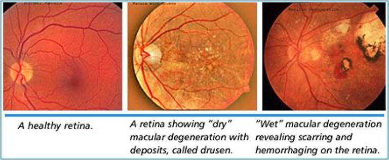
What is the earliest detectable clinical feature of macular degeneration?
drusen
What is used to asses macular degeneration and gross scotoma?
amsler visual grid
What is macular degeneration?
acute or chronic deterioration of central vision characterized by drusen and no pain or redness
What CN keeps eyes open?
CN III
What CN closes eyelid?
CN VII
How would visual acuity present in a patient with an optic chiasm tumor?
loss of entire half of temporal field bilaterally
How would a normal patient see the amsler visual grid?
not wavy and central point through visual fields; absolute scotoma 10 degrees on temporal midline
How would a patient with macular degeneration see the amsler visual grid?
wavy and decreased ability to see central dot
How would a patient with chronic glaucoma see the amsler visual grid?
multiple scotomas on periphery and scotoma on an arc extending from blind spot to superior nasal area
At what visual acuity, with visual sx, would you refer to ophthalmologist?
less than 20/50 in one or both eyes
At what visual acuity, without visual sx, would you refer to ophthalmologist?
less than 20/40 in one or both eyes
When do you evaluate visual fields?
after visual acuity has been done
When testing visual fields by confrontation, what do you do if there is a temporal defect on one eye?
have patient cover opposite eye and slowly move object from defective area to the better vision in order to define the border
Where is the lesion location in horizontal (altitudinal) visual field defects and how does the VF loss present?
branch of central retinal after or ischemia of optic nerve
unable to see below a horizontal point on the same eye
Where is the lesion location in a blind eye? How does VF loss present?
optic nerve; unilateral blindness
Where is the lesion location in bitemporal hemianopsia? How does VF present?
optic chiasm; temporal half of each field
Where is the lesion location for left homonymous hemianopsia? How does VF present?
right optic tract;
affects temporal visual field on the left and nasal visual field on the left
Where is the lesion location in homonymous left superior quadratic defect? How does VF loss present?
partial right optic radiation;
homonymous quadratic defect “pie in the sky”
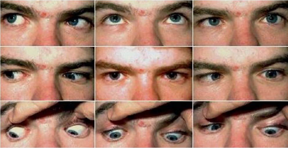
What is nystagmus?
repetitive, oscillatory, jerky movements of eyes
What 3 disorders are associated with Horner’s syndrome?
partial ptosis, anhidrosis, miosis
What muscles does CN III innervate?
superior rectus, inferior rectus, inferior oblique, medial rectus
what is seen with CN III?
ptosis and inability to elevate and adduct eye
What is normal motion of superior oblique?
look down and medially
What would defects with the superior oblique be caused by?
trauma to orbit or uncle herniation
What does CN IV innervate?
superior oblique
What are deficits seen with the superior oblique muscle?
moderate exotropia or slight deviation upward
What does CN VI innervate?
lateral rectus muscle
What is the normal movement of the lateral rectus muscle?
move eyes laterally
What is a deficit with the lateral rectus muscle?
esotropia
What causes deficits with the lateral rectus?
congenital weakness, trauma