occipital lobe
1/38
There's no tags or description
Looks like no tags are added yet.
Name | Mastery | Learn | Test | Matching | Spaced | Call with Kai |
|---|
No analytics yet
Send a link to your students to track their progress
39 Terms
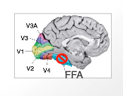
V1 (4)
functionally heterogeneous
vision begins here
outputs to V2 and specialized zone (V3-5)
if damaged patients think they are blind
V2 (2)
functionally heterogenous
inputs from V1 + outputs to specialized zones
V3 (2)
specialized zone
form perception
ie. people, basketball
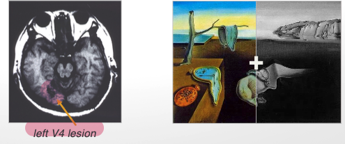
V4 (4)
in lingual gyrus
primarily colour perception → also form perception
also involved in thinking in colour → colour blind painter
ie. blue, yellow, beige in an image
colour vision is important for (3)
motion
depth
location perception
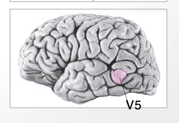
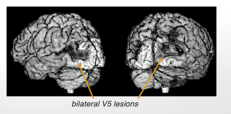
V5
motion perception
more posterior
ie. players moving around
geniculostriate pathway
visual info → eye → optic tract → lateral geniculate nucleus (thalamus) → striate cortex (V1) → other visual cortical areas
defacto pathway
tectopulvinar pathway
visual info → eye → optic tract → superior collicus (midbrain) → pulvinar (thalamus) → other visual cortical areas
usually used to locate objects
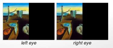
homonymous hemianopia
blindness to one visual field (left/right) of both eyes
complete cut of optic tract/lateral geniculate nucleus
lesion to V1
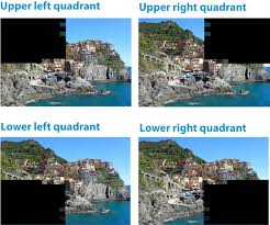
quadrantanopia
loss of vision in ¼ of visual field
small lesion to occipital lobe
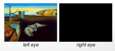
monocular blindness
loss of sight in one eye
destruction of retina/optic tract
bitemporal hemianopia
loss of vision from both temporal (outer) fields
lesion to medial part of optic chiasm
right nasal hemianopia
loss of vision from one nasal (inner) field
lesion to right lateral chiasm
same applies to left
scotoma
loss of vision in a small area of visual field
even smaller lesion to occipital lobe
dorsal stream (where/how)
visual guidance of movements
outputs to parietal lobe
ventral stream (what)
object perception
outputs to temporal lobe
STS stream
visuospatial function → biological motion
outputs to superior temporal sulcus
ie. watching someone walk → species specific
depends on proximity
activated by shape perception
______ activates parietal region
location processing
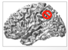
________ activates temporal region (FFA)
facial processing
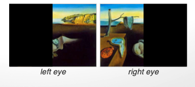
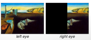
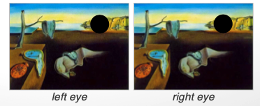
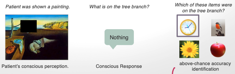
cortical blindness (blindsight)
damage to V1
cannot consciously perceive visual information BUT can identify above chance lvls nature of visual stimuli w/o conscious perception
achromatopsia
loss of vision for colour
due to lesion in V4
akinetopasia
loss of vision for movement
unable to intercept moving objects
due to lesion in V5
Visuospatial Agnosia (aka Topographical Disorientation)
inability to find one’s way in relation of self to environment
damage to right medial occipitotemporal region (including fusiform and lingual gyri)
(e.g.) patient can accurately report number of rooms in the house,
disruption of dorsal stream results in
optic ataxia
disruption of ventral stream results in (2)
object agnosia
prosopagnosia
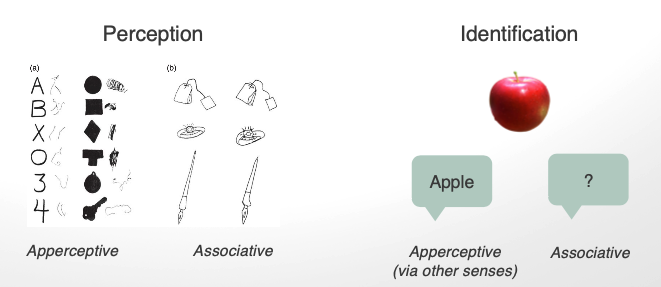
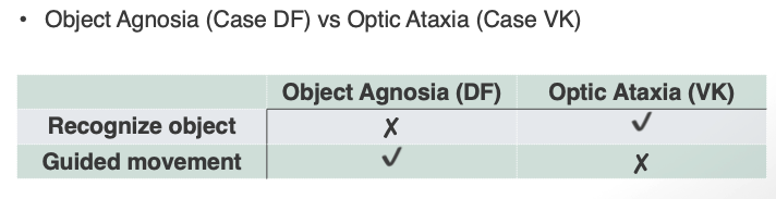
object agnosia (apperceptive agnosia)
deficit to perceive object structure/recognize object
basic sensory functions intact (color/motion perception)
bilateral damage to lateral parts of occipital lobes
patients with object agnosia ARE able to
recognize/draw objects from memory
guide hand movements towards objects
Lesson from DF Case → Bilateral Damage in Lateral Occipital Cortex (LO), Junction of Temporal/Parietal & Occipital Cortex
use of visual info to guide movements to objects (dorsal stream) involves different brain areas than those used to recognize those objects (ventral stream)
simultagnosia
type of apperceptive agnosia
inability to perceive more than one object simultaneously
associative agnosia
can perceive form
cannot identify objects
ie. recall name of object from memory
lesion to anterior temporal lobes (ventral stream)
apperceptive vs associative agnosia (3)
double dissociation btw perception + memory identification for objects
ability to perceive the structure of an object + ability to identify/label the object requires different neural structures
evidence for different cognitive systems
prosopagnosia
unable to recognize familiar faces (even one’s own)
able to recognize ppl using other features (voice, hair)
bilateral damage to fusiform face area (FFA)
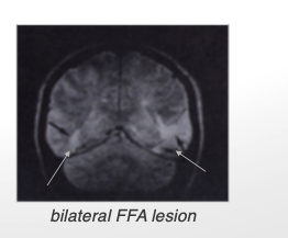
fusiform face area (FFA) (3)
strongly associated with face perception rather than general object recognition.
Found in the inferior temporal cortex, within the ventral visual stream ("what" pathway).
Typically more active in the right hemisphere, but present bilaterally.
Damage to the FFA → Prosopagnosia ("Face Blindness")
FFA stimulation patient video: what happens after sham + stimulation condition
sham: no ∆ in perception
stimulation condition: major ∆s in face only
Why is the sham condition important?
It ensures that the observed effects (such as face distortion or improved recognition) are truly due to stimulation of the FFA and not other confounding factors like placebo effects, expectation, or general brain stimulation.
optic ataxia (3)
deficit in visually guided movements
other functions intact (motor, somatosensory, visual acuity)
damage to posterior parietal lobe
object agnosia vs object ataxia (3)
double dissociation between dorsal and ventral streams
ability to perceive an object and to use visual information to guide hand movements towards objects requires different neural structures
visual experience is not unified, despite our phenomenological experience of it
calcarine sulcus
contains much of 1º cortex
divides upper/lower halves of the visual world
parietal-occipital sulcus
separates parietal lobe from occipital