A&P EXAM 1 (CH 1, 4, & 5)
1/87
Earn XP
Description and Tags
CT - connective tissue | NT - nervous tissue | MT - muscle tissue | ET - epithelial tissue
Name | Mastery | Learn | Test | Matching | Spaced | Call with Kai |
|---|
No analytics yet
Send a link to your students to track their progress
88 Terms
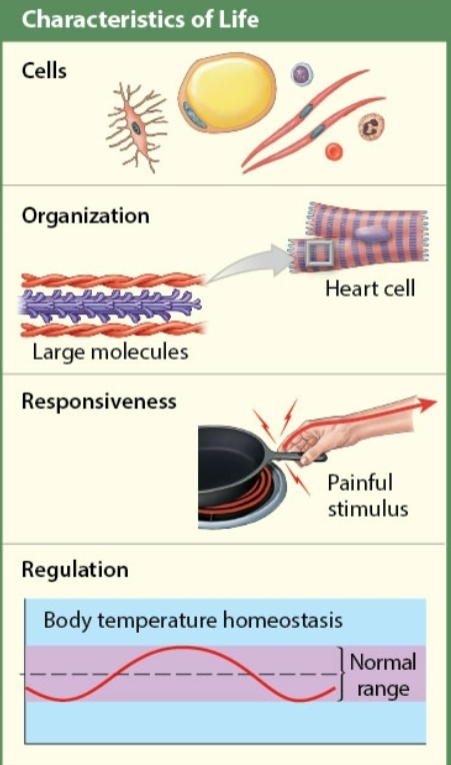
List the common characteristics shared by all living things
Composed of cells
Complex structural arrangement
Detect & respond to stimuli
Maintain a relatively stable internal environment
Organisms grow (increase in size) & develop (natural progression in physical maturation)
Offspring are produced by reproduction
Have metabolism - all essential processes occurring in cells
Anabolic - building up
Catabolic - breaking down
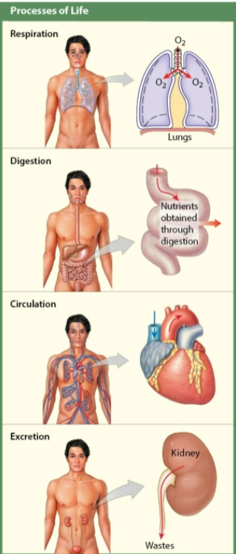
Describe the basic processes in humans & other animals
Respiration - O2 required for chemical processes obtained from atmosphere & delivered to cardiovascular system, CO2 removed by cardiovascular system
Digestion - mechanical & chemical process to convert ingested food into simple absorbable substances
Circulation: internal movement & distribution of O2 wastes, & digestion products
Excretion: undigested food & wastes of metabolism eliminated from body
What are the differences between gross anatomy & macroscopic anatomy?
Gross anatomy: you can see large structures & features usually visible w/ unaided eye
Microscopic anatomy: it is the study of structures that cannot be seen w/ out magnification (need microscope)
Explain the link between anatomy & physiology
Physiology: study of function & how organisms perform vital functions. complex & more difficult to examine than anatomical structures. Focuses on functional properties
Name the simplest lvl of organization that includes the smallest living units in the body
Cellular lvl = the simplest lvl of organization
Cells = the smallest living units in the body
Functions depend on organelles composed of molecules
Each organelle has a specific function
Ex: a mitochondrion provides energy for ❤ muscle cell contraction
Define organ
Composed of 2 or more tissues working together to perform specific functions
Ex: layers of cardiac muscle tissue along w/ connective tissue from the heart
Name & define the unit used to measure cell size
Cells in the human body
contains trillions of cells
Only an estimated 200 different types of cells
measured in micrometers
vary greatly in size
List the 3 basic principles of the cell theory
Cells are the structural building blocks of all plants & animals
Cells are produced by the divisions of pre-existing cells
Cells are the smallest structural units that perform all vital functions
Describe the general roles of the different types of 💪 tissue
Skeletal 💪 tissue
usually attached to the skeleton
moves or stabilizes position of skeleton or internal organs
Cardiac 💪 tissue
only in

propels 🩸 thru 🩸 vessels
Smooth 💪 tissue
In 🩸 vessel walls, within glands, along respiratory, circulatory, digestive, & reproductive tracts
moves fluids & solids along digestive tract
regulates diameter of small arteries, among other functions
Identify the 4 primary tissue types & explain the functions of each
Epithelial tissue
most common
forms a barrier w/ specific properties
covers every exposed body surface
lines digestive, respiratory, reproductive, & urinary tracts
surrounds internal cavities (ex: chest cavity)
lines inner surfaces of 🩸 vessels & 🫀
produces glandular secretions
Connective tissue
diverse in appearance but all forms contain cells surrounded by ECM
matrix has ➡ protein fibers & ground substance
amount & consistency of matrix varies by the particular connective tissue type
🩸 - watery matrix
🦴 - crystallized matrix w/ little ground substance
fills internal spaces
provides structural support
stores energy
💪 tissue
has the ability to contract forcefully (to produce movement)
includes skeletal, cardiac, & smooth muscle
major functions
skeletal movement
soft tissue support
maintenance of 🩸 flow
movement of materials internally
stabilization of body temp
Nervous tissue
conducts electrical impulses
carries info
Which 2 organ systems are involved w/ circulation within the body?
Cardiovascular & Lymphatic
Why is homeostatis regulation important to an organism?
The organism is able to physiologically adjust itself to preserve homeostasis in variable environments
Describe the 3 parts necessary for homeostatic regulation
Receptor (sensor)
sensitive to environmental change
Control center (integration center)
processes info from the receptor & sends out commands
Effector
responds to commands opposing stimulus
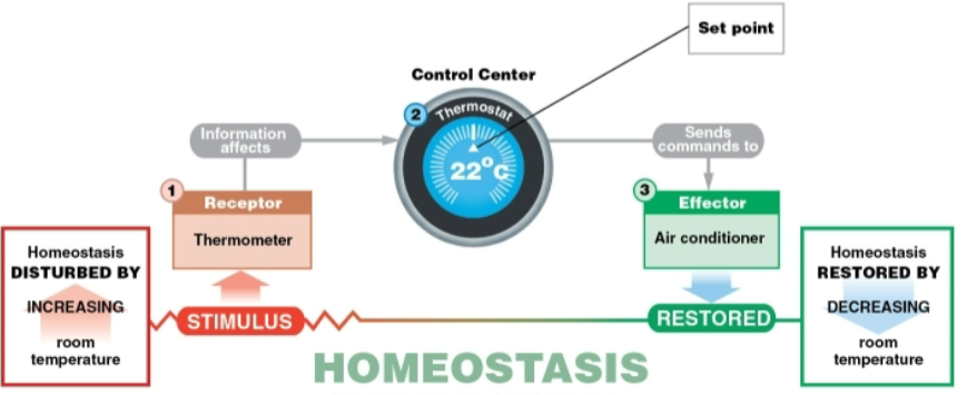
Explain the function of negative feedback systems, give an example of homeostatic regulation by negative feedback in the body
Effector opposes or negates the original stimulus
Minimizes change
Primary mechanism of homeostatic regulation in the body
Dynamic process
set point varies w/ varying environments & activity lvls
Ex: body temp
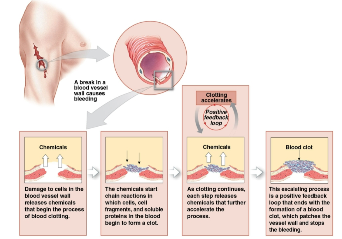
Why is postiive feedback helpful in 🩸 clotting but unsuitable for regulating body temp?
It usually occurs in dangerous responses & produces a response that enchances the original change
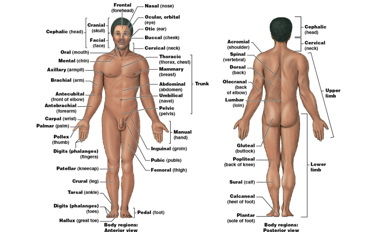
Describe a person in the anatomical position
🧍 up
👐 at the side
🤲 facing forward
🦶🦶 together & facing forward
👀 facing forward
Lying down in anatomical position
Supine when face up
Prone when face down
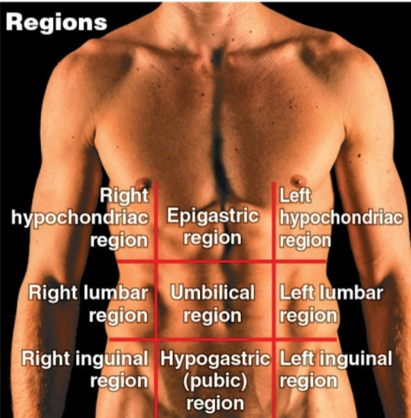
Contrast the descriptions used by clinicians & anatomists when referring to the positions of injuries of internal organs of the abdomen & pelvis
The abdominopelvic quadrants is used by clinicians to describe locations of patient pains, aches, or injuries, whereas the abdominopelvic regions is used by anatomists to describe precise location & orientation of internal organs
What is the purpose of anatomical terms?
To find exactly which body part is in crucial pain
In the anatomical position, describe an anterior view & a posterior view
Anterior - facing the front
Posterior - facing the back
What type of section would separate - the 2 👁👁, the 👃 & the 2👂, the neck & navel?
2 👁👁 - sagittal
the 👃 & the 2 👂 - frontal
the neck & navel - transverse
What is the purpose of directional & sectional terms?
it is sometimes the only way to show the relationship between parts of a 3-dimensional body
medical imaging techniques utilize sectional views
used for visualization purposes
important to consider when looking at 🔬 slides & CT or MRI scans
views change throughout structure
Describe 2 distinctive features & 2 essential functions of true body cavities
2 distinctive features
from common embryological origin
covered by serous membrane
2 essential functions
protect organs from shocks & impacts
permit changes in sizes & shape of organs
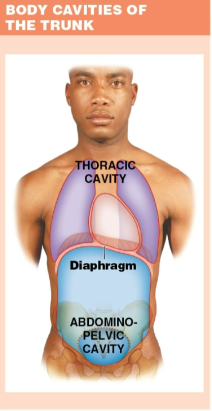
Identify the body cavities of the trunk
Thoracic cavity
everything deep to the chest
Abdominopelvic cavity
all structures deep to abdominal & pelvic walls
Give the term for “the study of tissues”
Histology
What is a tissue?
Cells working together
How do early microscopes compare w/ modern microscopes?
Early magnification lvls of an early 🔬 lvls 10-20 times the actual size
Differentiate among LM (light), TEM (transmission electron), and SEM (scanning electron).
LM
detects visible light through thin section of tissue
2 lenses magnify specimen
objective lens located on revolving nosepiece
ocular lens located in the eye piece
total magnification (calculated by multiplying the 2 lens power (obj x ocu)
Resolution
ability to distinguish between 2 separate points
wavelength of light limtis resolution on light 🔬 to about 200 nm
TEM
transmits through specimen
uses 🧲s to direct beam of electron’s through the surface of a very thin object onto a photographic plate
wavelength of electron beam 0.00001 of white light
maximum resolution 0.2 nm
SEM
uses electrons but not by sending them through a specimen
specimen coated w/ electron dense material
electron beams are focused on the specimen
reflections of electrons bouncing off object produce 3 3-dimensional image of the surface
can view surface features only
maximum resolution of about 10 nm
List 4 essential functions of epithelial tissue
Provide physical protection
protect surfaces from abrasion, dehydration, or destruction by chemical or biological agents
Control permeability
most epithelia are capable of selective absorption or secretion
epithelial barrier can be modified in response to stimuli (ex: calluses)
Provide sensation
specialized epithelial cells, detect changes in environment (ex: touch receptors)
Neuroepithelium
sensory epithelium found in special sense organs
produce specialized secretions
glandular epithelial cells produce secretions
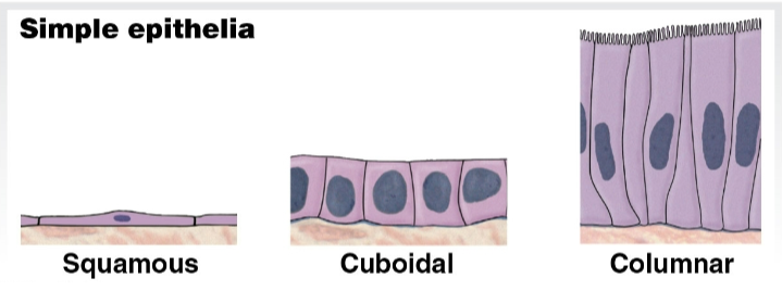
Summarize the classification of an epithelium based on cell shape & # of cell layers
3 basic shapes
Squamous
thin and flat
Cuboidal
cube-shaped
like little boxes 📦
Columnar
taller than they are wide
slender rectangles
Epithelial cell layers
single layer
simple epithelium
several layers of cells
stratified epithelium
found in areas that need protection from abrasion or chemical stress
ex: surface of skin, lining of 👄
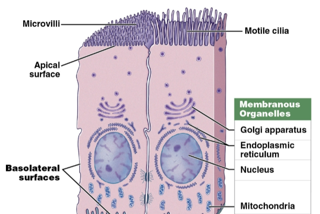
Identify the various types of epithelial intercellular connections
Hemidesmosomes
attach deepest epithelial cells to basement membrane
Tight junctions
interlocking membrane proteins bind adjacent plasma membranes together
prevent passage of 💧 & solutes between cells
isolate basolateral surfaces & deeper tissues from contents in lumen
found in intestinal tract
Adhesion belts
continuous band of membrane proteins
strengthens apical region of cells
reinforces tight junctions
dense proteins attached to microfilaments of the terminal web (part of cytoskeleton)
belts encircle cells & bind to adjacent cells
Gap junctions
held together by interlocking transmembrane proteins (connexons)
assist chemical communication to help coordinate functions such as secretion or beating cilia
also found in cardiac 💪 & smooth 💪 muscle tissue to coordinate contraction
Desmosomes
provide firm attachments by interlocking adjacent cells’ cytoskeletons
opposing plasma membranes locked together by cell adhesion molecules
thin layer of proteoglycans may also bond
contain polysaccharide, notably hyaluronic acid
very strong; resist stretching & twisting
found in superficial layers of skin
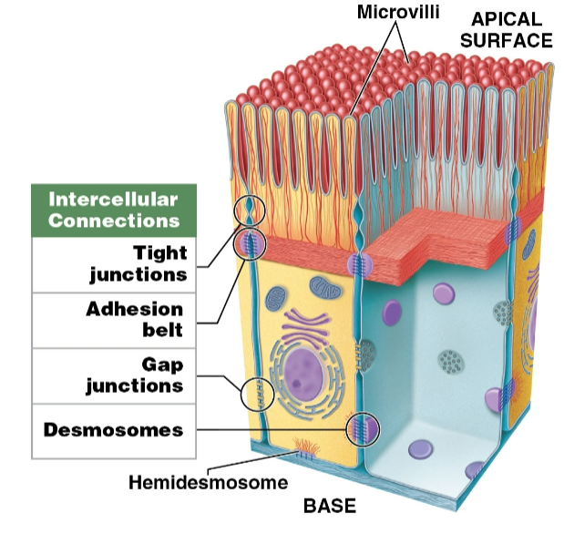
What is the functional significance of gap junctions?
They assist chemical communication to help coordinate functions such as secretion or beating cilia
How do epithelial tissue obtain needed nutrients?
They require attachment to underlying connective tissue for nourishment from 🩸 vessels there
What 2 types of tissue contribute to the formation & maintenance of the basement membrane?
Epithelial & connective
What do mesothelium & an endothelium have in common?
Both are simple squamous epithelium
Why do the pharynx, esophagus, anus, & vagina have a similar epithelial organization?
They have many layers of cells, superficial layers are flattened, & are all stratified squamous epithelium
What properties are common to keratinized epithelia?
superficial layers packed w’ keratin
tough & 💧 resistant
resists both mechanical stress & dehydration
found on surface of skin, hair, & in nails
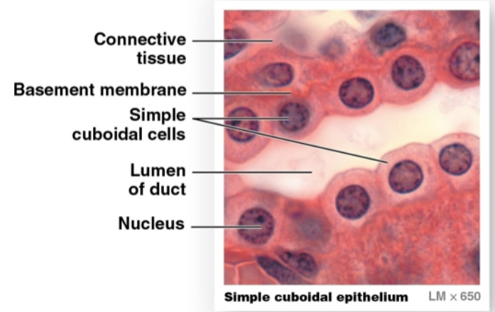
Describe the appearance of simple cuboidal epithelial cells in sectional view
Cells appear cube shaped
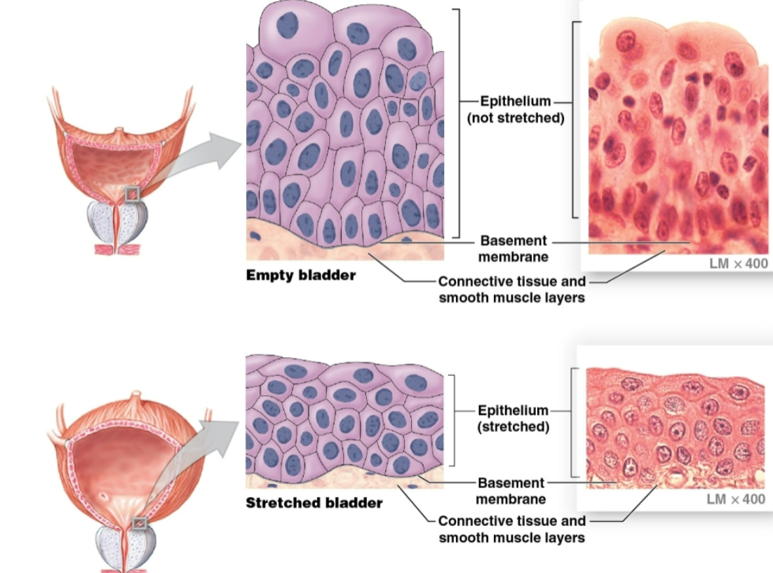
Identify the epithelium that lines the urinary bladder, & describe its unusual functional characteristics
Transitional epithelium = unusual stratified epithelium that can stretch & recoil w/ out damage
Describe the changes in appearance of the transitional epithelium lining the urinary bladder as stretching occurs
Relaxed (empty bladder) - superficial cells cuboidal
Stretched (full bladder) - superficial cells flattened
What functions are associated w/ a simple cuboidal epithelium & a transitional epithelium?
SCE:
functions in secretion & absorption
lines exocrine glands & ducts
lines part of kidney tubules & thyroid gland
TE:
it changes appearance (can stretch & recoil w/ out damage)
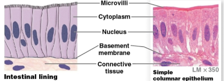
Describe the appearance of simple columnar epithelial cells in a sectional view
Cells appear rectangular
Explain why a pseudostratified columnar epithelium is not truly stratified
It has varying shapes & functions
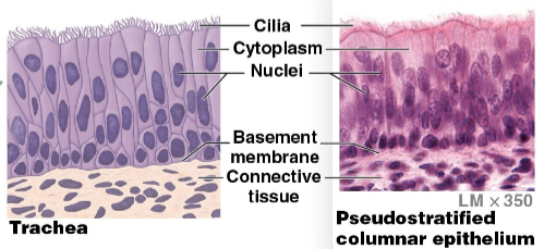
Describe the structures found on the surfaces of simple columnar & pseudostratified columnar epithelia
SC: microvilli (absorption) or cilia (movement) on apical surface
PC: usually has cilia
Describe the 2 primary types of glands
Endocrine glands
release secretions into interstitial fluid
Exocrine glands
release secretions into ducts onto epithelial surface
By what 3 methods do various secretory cells of exocrine glands release their secretions?
Merocrine
product released from secretory vesicles by exocytosis
most common mode of secretion
ex: salivary gland secretion
Mucin
merocrine secretion that mixes w/ 💧 to form mucus
Apocrine
apical cytoplasm packed w/ secretory vesicles
cell releases cytoplasm as well as secretory product
ex: mammary gland secretion (combo of merocrine & apocrine)
Holocrine
destroys gland cell
entire cell bursts, releasing secretions & killing cell
destroyed cells replaced by stem cell division
ex: sebaceous glands
What characteristics are used to describe multicellular glands?
Based on duct structure
simple (single duct that does not ➗)
compound (duct ➗’s 1 or more times)
Based on the shape of the secretory area
Tubular (glandular cells form tubes)
Alveolar or acinar (glandular cells form sacs)
Tubuloalveolar (glandular cells forms tubes & sacs)
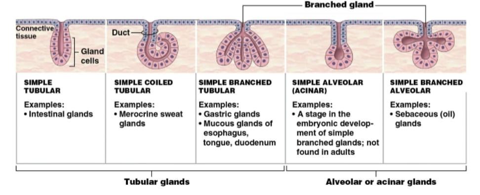
Describe the simplest type of multicellular exocrine gland
Simple tubular
Identify the 3 basic components of connective tissue
Specialized cells
Extracellular protein fibers
Fluid called ground substance
Summarize the functions of connective tissue
establish a structural framework for the body
transport fluids & dissolved materials
protect delicate organs
support, surround, & interconnect other types of tissue
store energy, especially in the form of triglyceride
defend the body from invading microorganisms
Distinguish among connective tissue proper, fluid connective tissues, & supporting connective tissues
Connective tissue proper
contains many types of cells
extracellular fibers in syrupy ground substance
loose (fibers create loose, open framework)
dense (fibers densely packed)
Fluid connective tissue
distinctive group of cells
💧y matrix
🩸 (within cardio)
lymph (within lymphatic)
Supporting connective tissue
less diverse cell population
more densely packed matrix
cartilage (solid, rubbery matrix)
🦴 (solid, crystalline matrix)
Identify the types of cells found in connective tissue proper
Fixed cells
Fibroblasts (synthesize extracellular fibers)
Adipocytes (store lipid reserves)
Fibrocytes (differentiate from fibroblasts & maintain extracellular fibers)
Wandering cells (move through out tissue; function in defense and repair)
plasma cells (immune cells producing antibodies)
free macrophages (engulf debris & pathogens)
mesenchymal cells (stem cells that aid tissue repair)
neutrophils & eosinophils (phagocytic 🩸 cells)
lymphocytes (immune system cells)
Describe the roles of fibroblasts in connective tissue
It synthesizes extracellular fibers
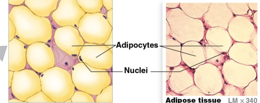
Which type of loose connective tissue contains primarily lipids?
Adipose
What term means the fibrous supporting network formed of reticular fibers?
Stroma
What types of phagocytic cells are present in connective tissue proper?
Neutrophils & eosinophils
What makes a connective tissue “loose” or “dense”?
The amount of volume or fibers
Which connective tissue fiber is characteristic of the cartilage supporting the ear?
Elastic cartilage
Describe the 2 layers making up the perichondrium
outer layer of dense irregular connective tissue
functions —> mechanical support, protection, attachment
inner cellular layer
where cartilage growth & maintenance occurs
Contrast appositional & interstitial growth of cartilage
Appositional growth (at cartilage surface)
chondroblasts ➗ in cellular layer of perichondrium
chondroblasts secrete new matrix
once surrounded by matrix, chondroblasts mature into chondrocytes
Interstitial growth (within cartilage)
chondrocytes ➗ within a lacuna
daughter cells secrete additional matrix & move apart
both types of cartilage growth occur during development
normally no growth & repair in adults
w/ slight damage or w/ hormonal stimulation some appositional growth possible
Describe bone matrix
2/3 of matrix is Ca (calcium) salts
mostly Ca phosphate
some Ca carbonate
What are mature bone cells in lacunae called?
Osteocytes
What is the functional unit of compact 🦴?
Osteon
Distinguish between the 2 types of supporting connective tissues w/ respect to their characteristic fibers
Cartilage characteristics
Cells: chondrocytes in lacunae
Ground substance: chondroitin sulfate (in proteoglycan) & 💧
Fibers: collagen, elastic, & reticular fibers in varying proportions
Vascularity: no internal 🩸 vessels
Covering: perichondrium (2 layers)
Strength: limited, bends easily, but hard to break
🦴 characteristics
Cells: osteocytes in lacuna
Ground substance: a small volume of liquid surrounding insoluble crystals of Ca salts
Fibers: collagen fibers predominate
Vascularity: extensive 🩸 vessels
Covering: periosteum (2 layers)
Strength: resists distortion until breaking point
Which cavities in the body are lined by serous membranes?
Pleural cavity, pericardium, & peritoneum
Name the 4 types of membranes found in the body
Mucous
Serous
Cutaneous
Synovial
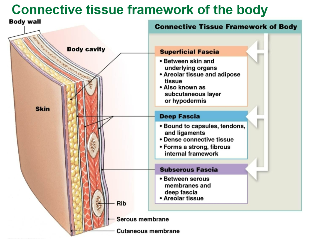
Name the 3 layers of fascia & their types of connective tissue
Superficial fascia
under skin
consists of areolar & adipose tissue
Deep fascia
continuous w/ capsules, ligaments, & other connective tissue structures
consists of dense irregular connective tissue
forms strong, fibrous internal framework
Subserous fascia
between serous membranes & deep fascia
consists entirely of areolar tissue
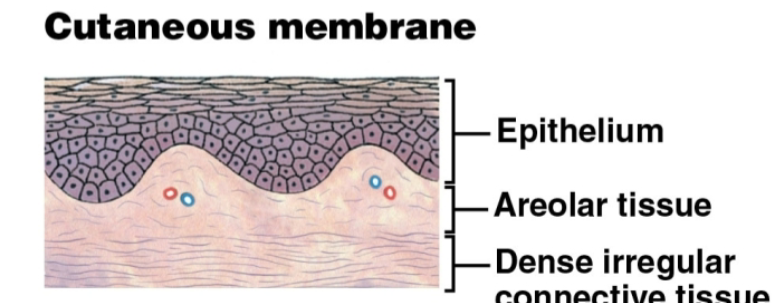
Which of the 4 tissue membranes is relatively waterproof & usually dry?
Cutaneous membrane
Which of the 4 tissue types has the highest body percentage weight and the lowest?
Highest = CT
Lowest = NT
Which type of 💪 tissue regulates 🩸 vessel diameter?
Smooth 💪
Distinguish between neurons & neuroglia
Neurons
transfer info around body & perform info processing
vary in size and shape
longest cells in body r neurons (up to 1 m)
Neuroglia
several different structural types w/ associated functions
maintain physical structure of NT
repair NT framework after injury
perform phagocytosis
provide nutrients to neurons
regulate the composition of the interstitial fluid surrounding neurons
Organs are made up of different tissues. What tissues are found in skeletal muscles?
Skeletal muscle tissue
Identify the 2 processes in the response to tissue injury
Inflammation
Regeneration
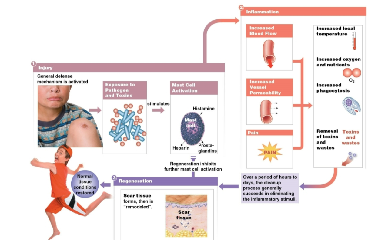
What are the 4 indications of inflammation that occur following an injury?
Swelling
Redness
Warmth
Pain
Why can inflammation occur in any organ in the body?
It occurs in connective tissue (all organs have connective tissue)
Compare thin skin w/ thick skin
Thin skin
covers most of body surface
contains 4 strata (layers)
Thick skin
found on palms of hands & soles of feet
contains 5 strata (layers)
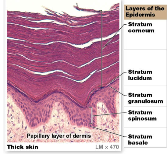
Identify the 5 layers of the epidermis (from superficial to deep)
Stratum basal
attached to basement membrane by hemidesmosomes
most cells here r basal cells, stem cells that ➗ to replace more superficial keratinocytes
merkel cells that respond to touch r also found here
Stratum spinosum (“spiny layer”)
composed of 8-10 layers of keratinocytes bound together by desmosomes
only looks spiny when on a prepared slide
contains dendritic (langerhans) cells
part of immune response defending against microorganisms & superficial skin cancers
Stratum granulosum (“grainy layer”)
composed of 3-5 layers of keratinocytes
most cells have stopped dividing & started producing keratin & keratohyalin
cells grow thinner & flatter
cell membranes thicken & become less permeable
Stratum lucidum (“clear layer”)
found only in thick skin
separates stratum corneum from underlying layers
flattened, densely packed dead cells filled w/ keratin & keratohyalin
Stratum corneum (cornu, horn)
outermost, protective region w/ 15-30 layers of keratinized cells (filled w/ keratin)
dead cells still tightly connected by desmosomes
💧 resistant, not waterproof
lose 💧 through insensible perspiration (unable to see or feel) & sensible perspiration (sweat)
Name the 2 pigments contained in the epidermis
Carotene
Melanin
Why is basal cell carcinoma considered less dangerous than malignant melanoma?
It does not metastasize (spread) & most people live
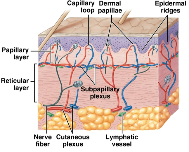
Describe the layers of the dermis
Papillary layer
named for dermal papillae in this region
composed of areolar tissue
contains capillaries, lymphatic vessels, & sensory neurons
Reticular layer
interwoven meshwork of dense, irregular connective tissue w/ collagen & elastic fibers
contains 🩸 vessels, lymphatic vessels, nerve fibers, & accessory organs (hair follicles, sweat glands)
Subcutaneous layer (not part of skin)
separates skin from deeper structures
dominated by adipose tissue
important energy storage site
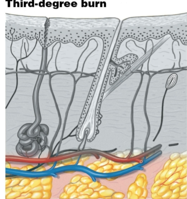
Distinguish among a 1st degree, 2nd degree, & a 3rd degree burn
Partial-thickness burns
First-degree burn
only the surface of the epidermis affected
ex: most sunburns
skin redness (erythema) results from inflammation
Second-degree burn
entire epidermis & maybe some of dermis damaged
accessory structures not affected
blistering, pain, & swelling occur
infection can develop from ruptured blisters
healing takes 1-2 weeks
Full-thickness burns
Third-degree burns
destroys epidermis, dermis, & damage extends into subcutaneous layer
less painful than 2nd-degree burns
extensive burns of this type cannot repair themselves
skin grafting usually necessary
Which type of burn usually requires skin grafting? Why?
Third degree burn
Why? - because these type of burns cannot fix themselves, so a skin graft is needed
Describe the 3 types of skin grafts. Which one is best? Why?
- Autograft - patient’s own undamaged skin
best choice if possible; no rejection by immune system
Allograft - frozen skin from a cadaver (dead body)
Xenograft - animal skin
Autograft is the best skin graft
Why? - because it does not reject the immune system
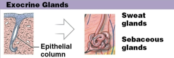
What are epidermal derivatives?
Accessory structures of the integumentary system
Which exocrine glands are in the integument?
Sweat glands & sebaceous glands
Describe a typical strand of hair
found almost everywhere on the body
except palms of 🙌, sides & soles of 🦶🦶, sides of 👆’s & toes, 💋, & parts of external genitalia
each hair produced by a hair follicle
complex structure composed of epithelial & connective tissue that forms a single hair
What happens when an arrector pili muscle contracts?
It pulls the hair erect
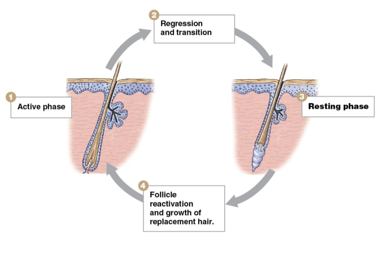
Describe the 4 phases of the hair growth cycle
Active phase
lasts 2-5 years
hair grows at rate of 0.33 mm/day
Resting phase
hair loses attachment to follicle
becomes club hair
club hair is shed when follicle is reactivated
new hair formation begins
Regression & transition
Follicle reactivation & growth of replacement hair
Steps in order ——→ Active phase, regression & transition, resting phase, & follicle reactivation & growth of replacement hair
Why does hair turn white or gray with age?
Because of decreased melanocyte activity