nervous tissue
1/114
There's no tags or description
Looks like no tags are added yet.
Name | Mastery | Learn | Test | Matching | Spaced | Call with Kai |
|---|
No analytics yet
Send a link to your students to track their progress
115 Terms
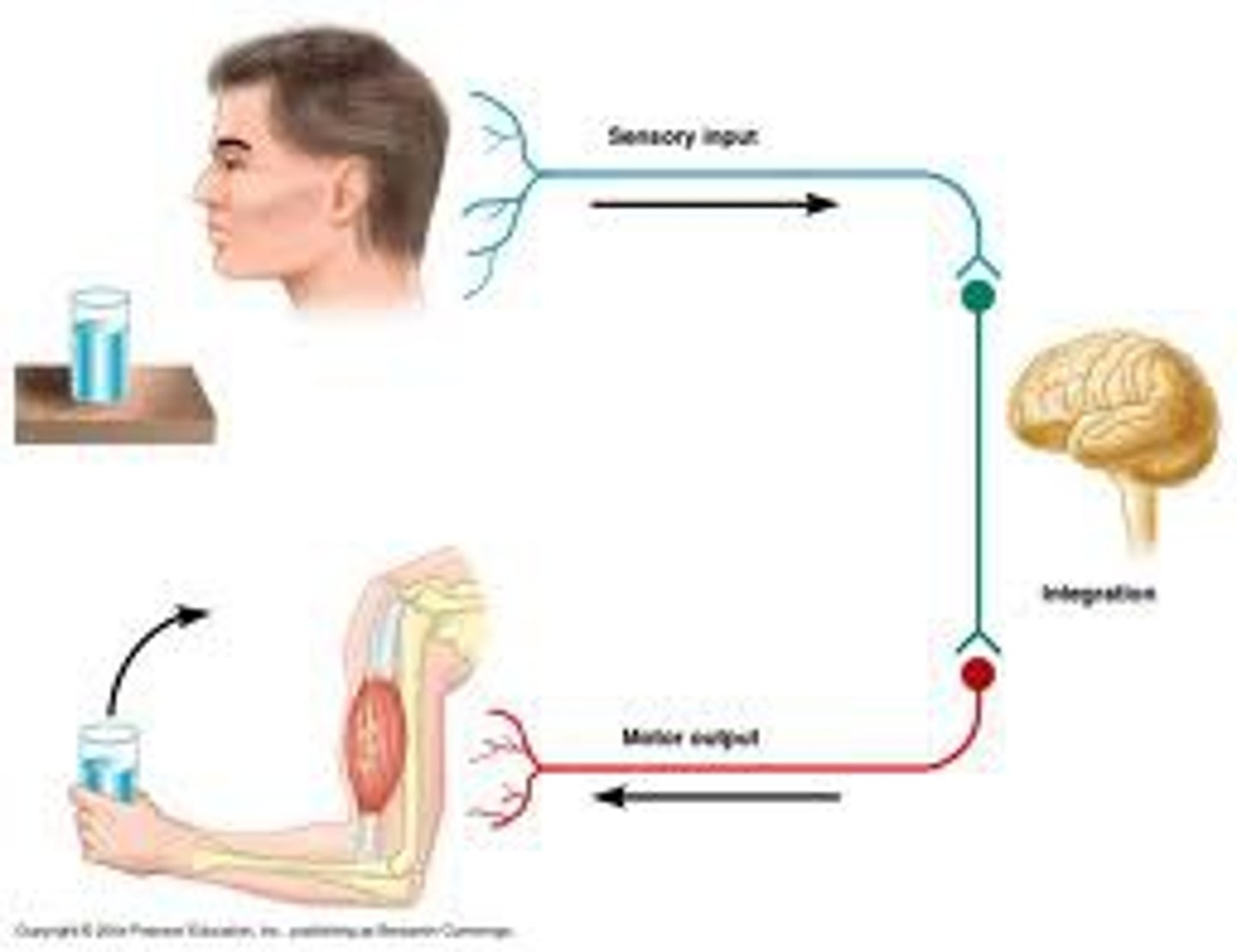
Nerve tissue is involved in the transmission of
information throughout the body
electrochemical signals cause muscles to
contract or relax when needed to move body parts
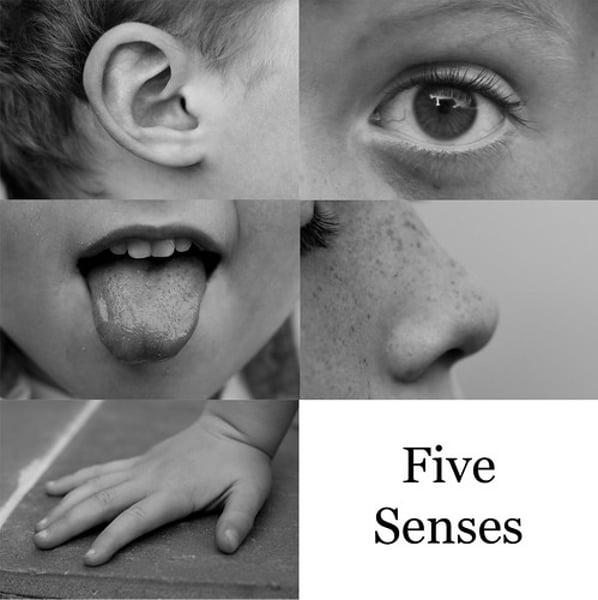
nervous system allows us to
sense the world around us; 5 senses
Divisions of the Nervous System
CNS, PNS, ANS
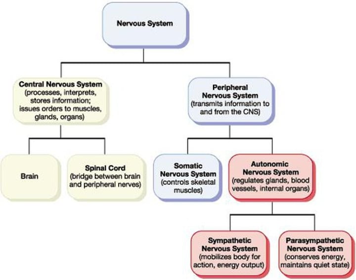
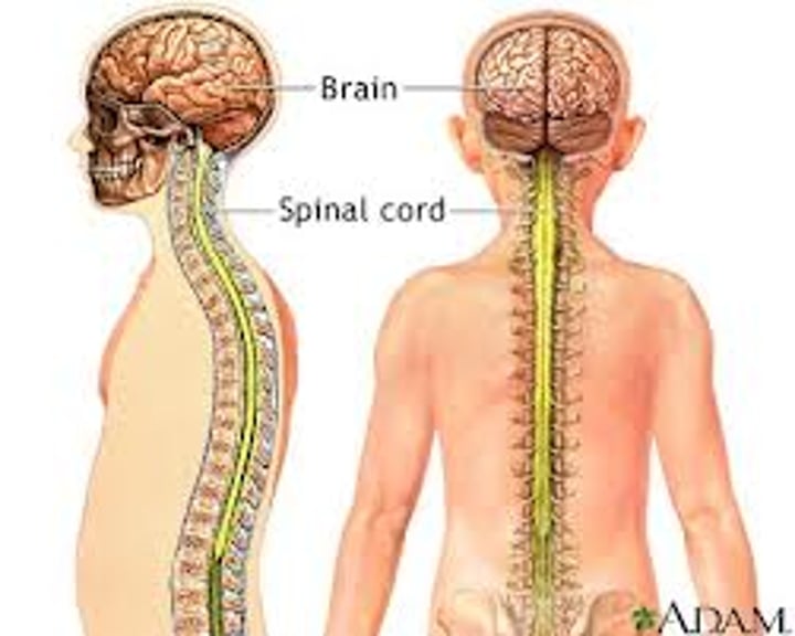
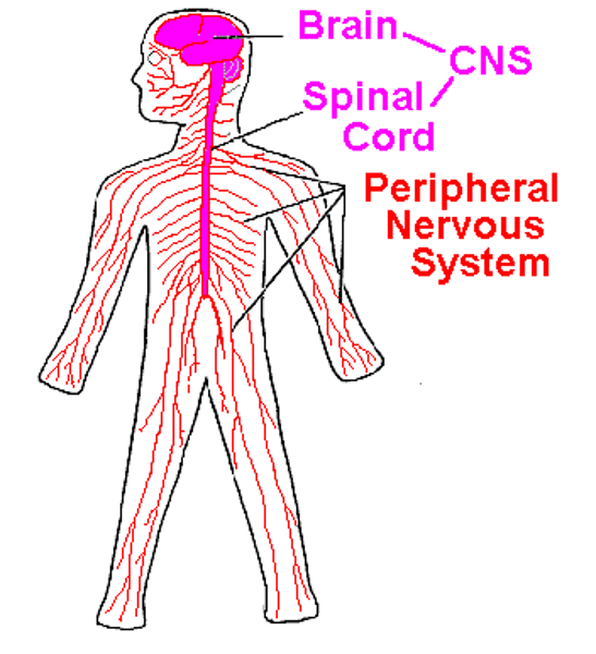
CNS
brain and spinal cord
PNS
motor neurons and sensory neurons
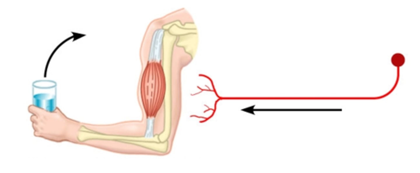
motor neurons
efferent neurons; neurons that carry messages from spinal cord/brain to muscles and glands
sensory neurons
afferent neurons; neurons that carry messages from sensory organs to the brain and spinal cords
ANS
innervates organs and glands;
in both CNS and PNS;
parasympathetic and sympathetic
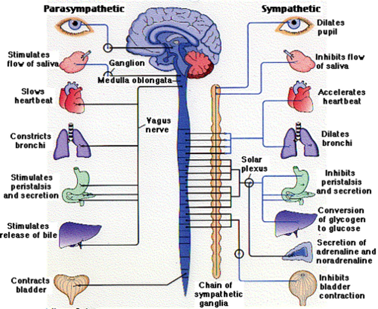
parasympathetic nervous system
rest and digest
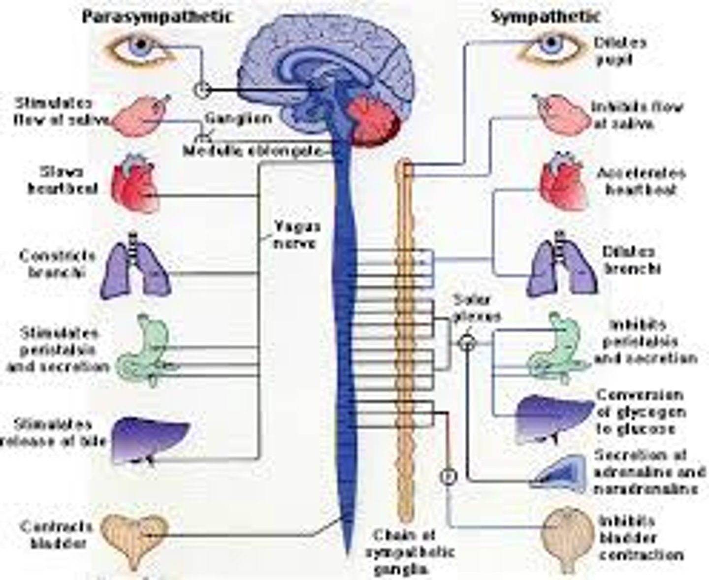
sympathetic nervous system
fight or flight

enteric nervous system
the nervous system of the digestive tract;
operates independently and in conjunction with the ANS
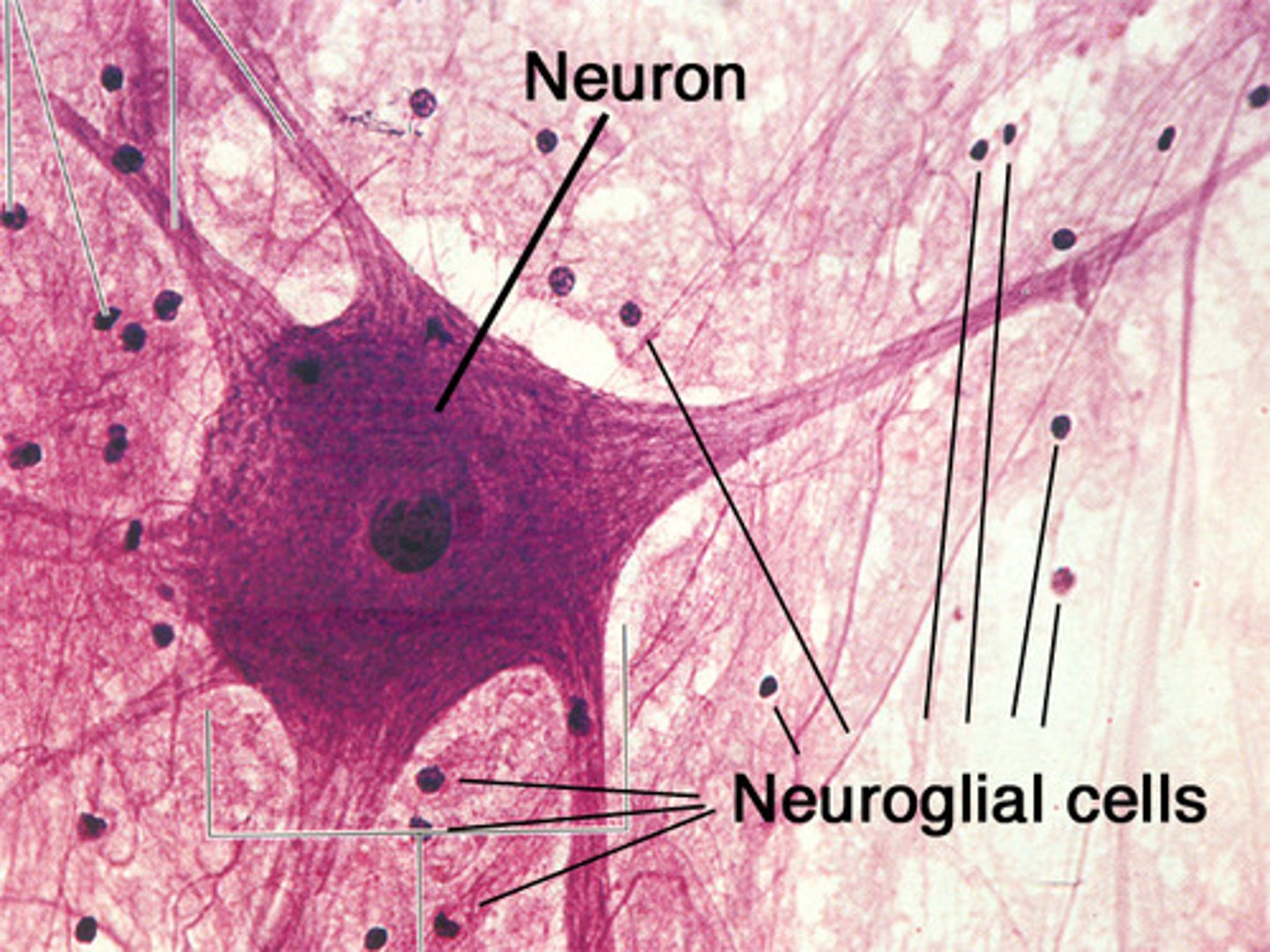
2 general classes of nerve cells
neurons, glial cells
neurons
nerve cells, with highly excitable membranes;
10% of cells
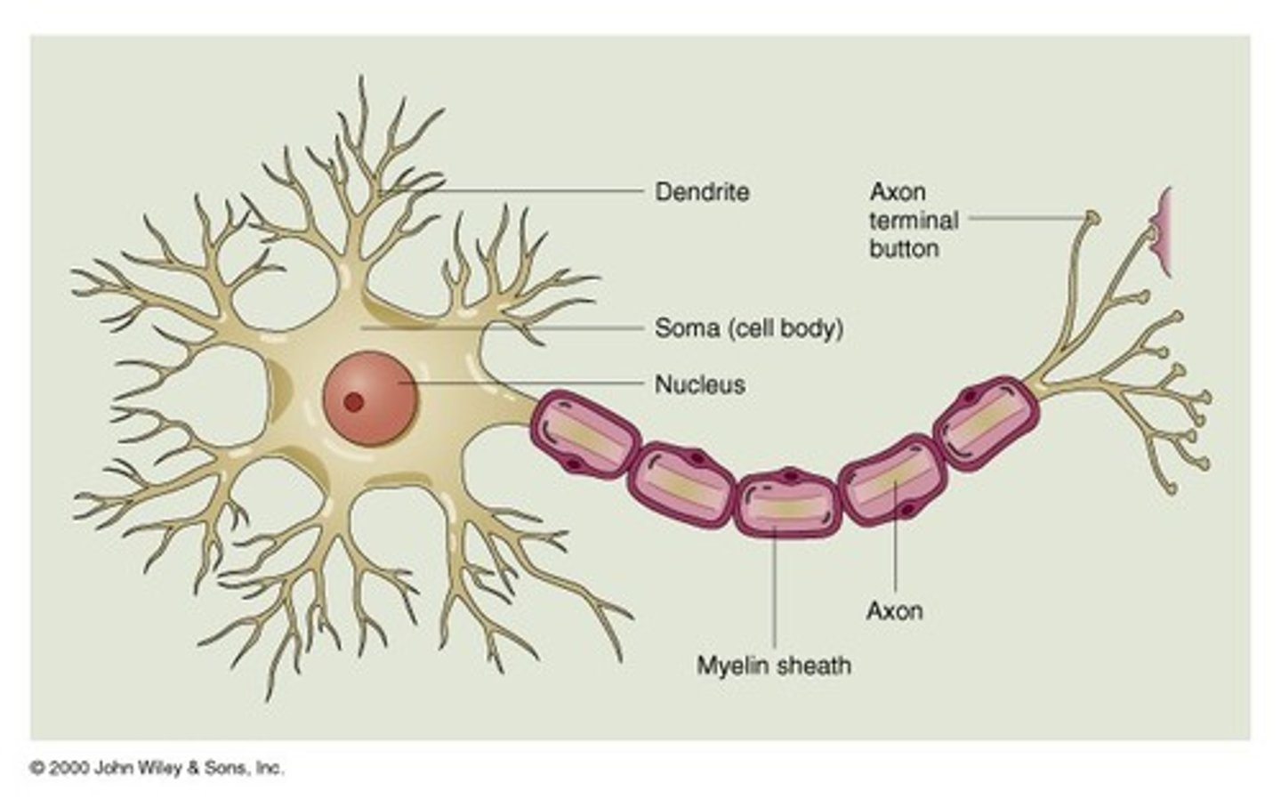
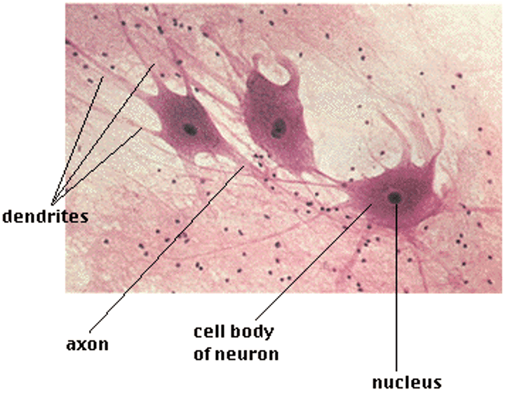
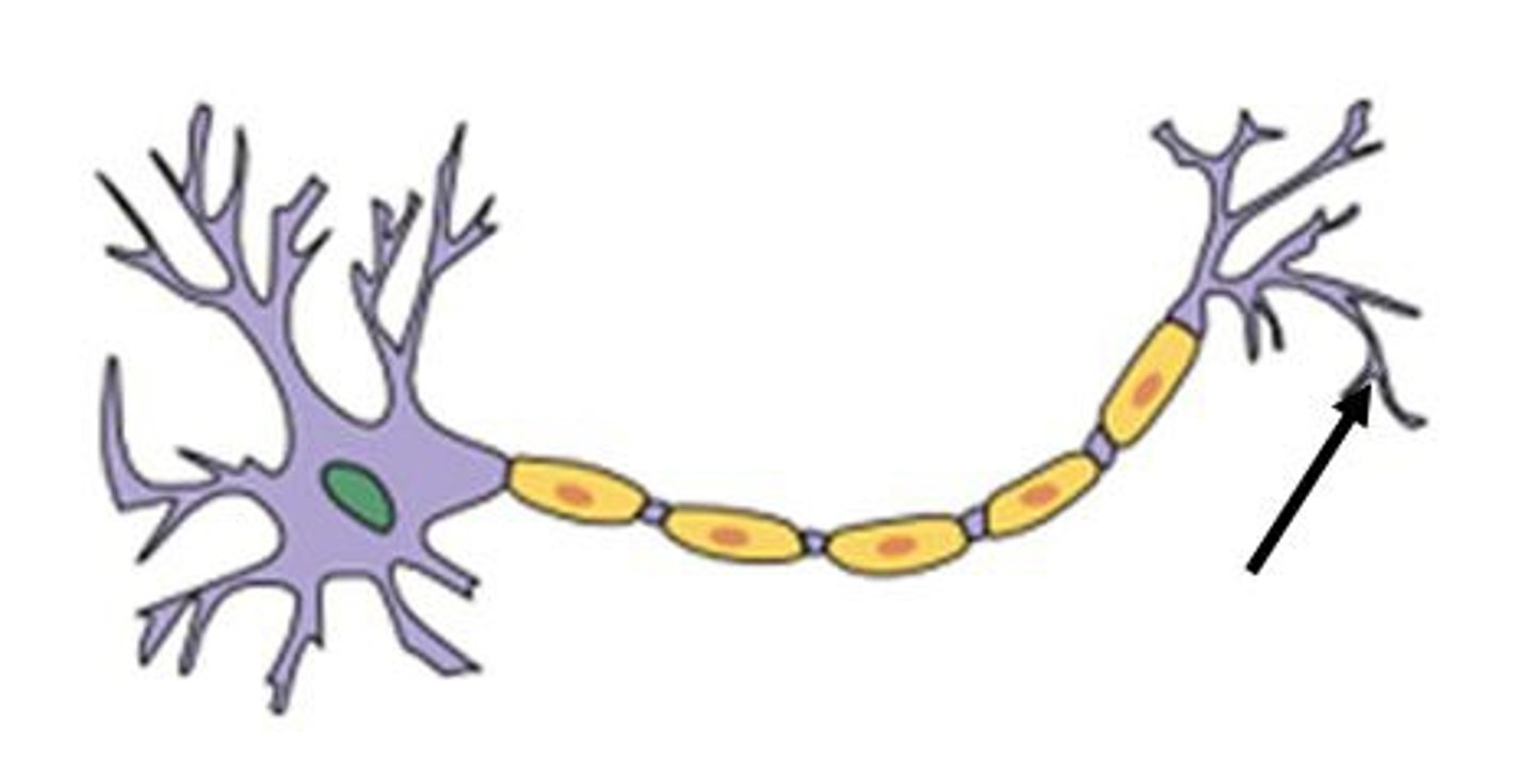
Part of a neuron
cell body, dendrites, axon
cell body AKA soma
surrounded by cytoplasm, contains organelles
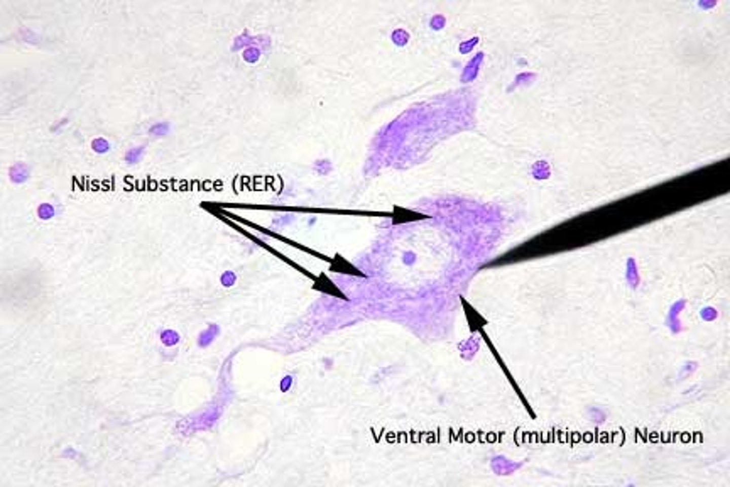
Nissl substance
RER found in cell body and dendrites, but not axon of neurons.
Dendrites
Branchlike parts of a neuron that are specialized to receive information from other neurons
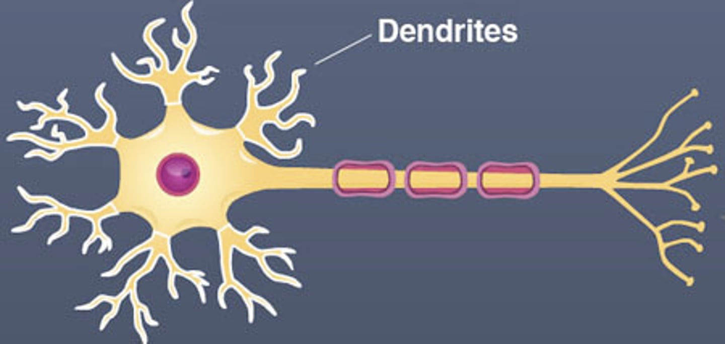
axon
the extension of a neuron, ending in branching terminal fibers, through which messages pass to other neurons or to muscles or glands
neurons connect to each other by
electrochemical synapses
nerves
collections of hundreds of axons, 10% of cells
glial cells
support cells found in the nervous system, 90% of cells
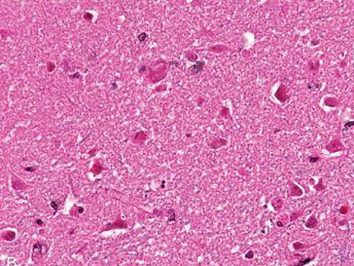
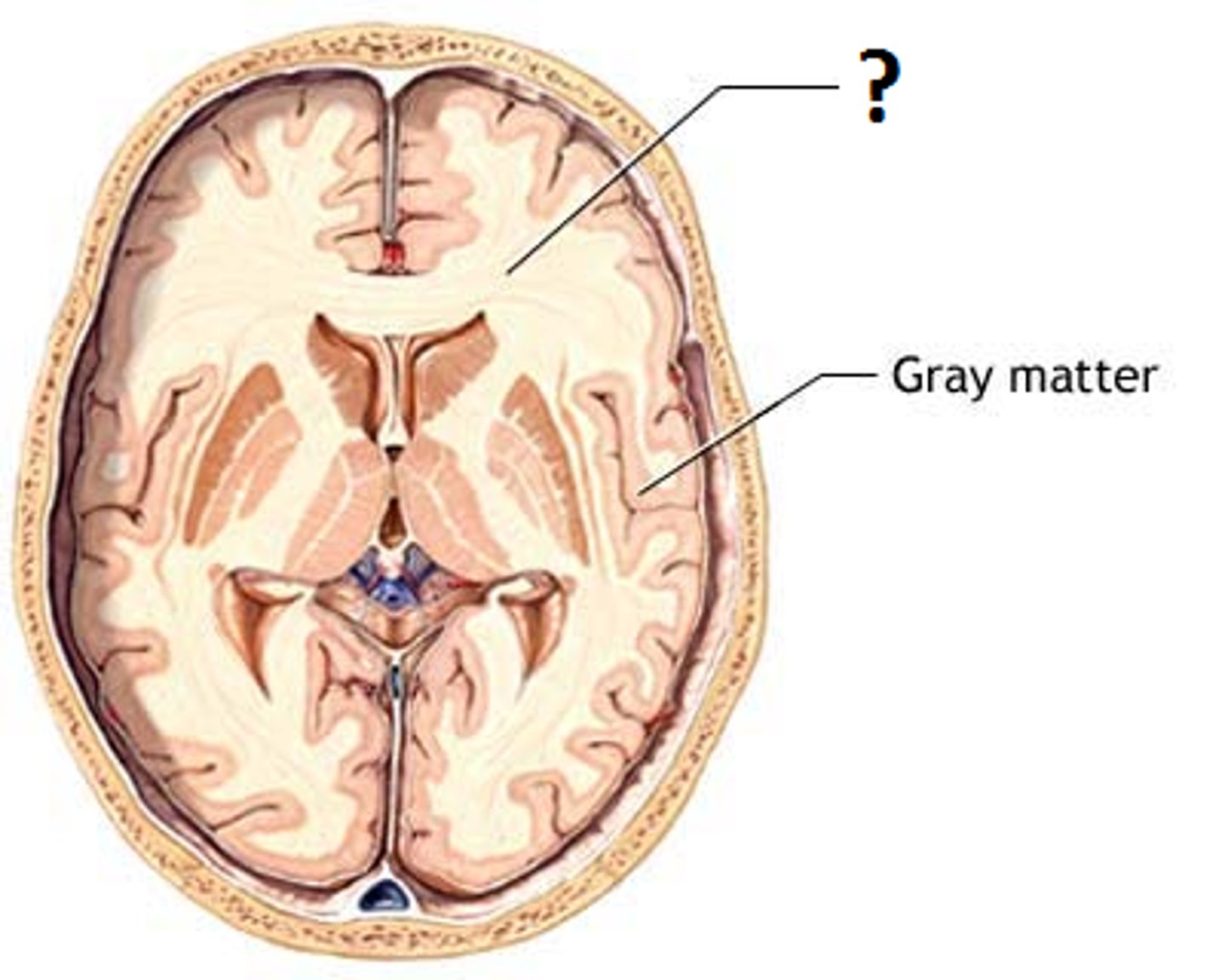
cerebral cortex has
grey and white matter
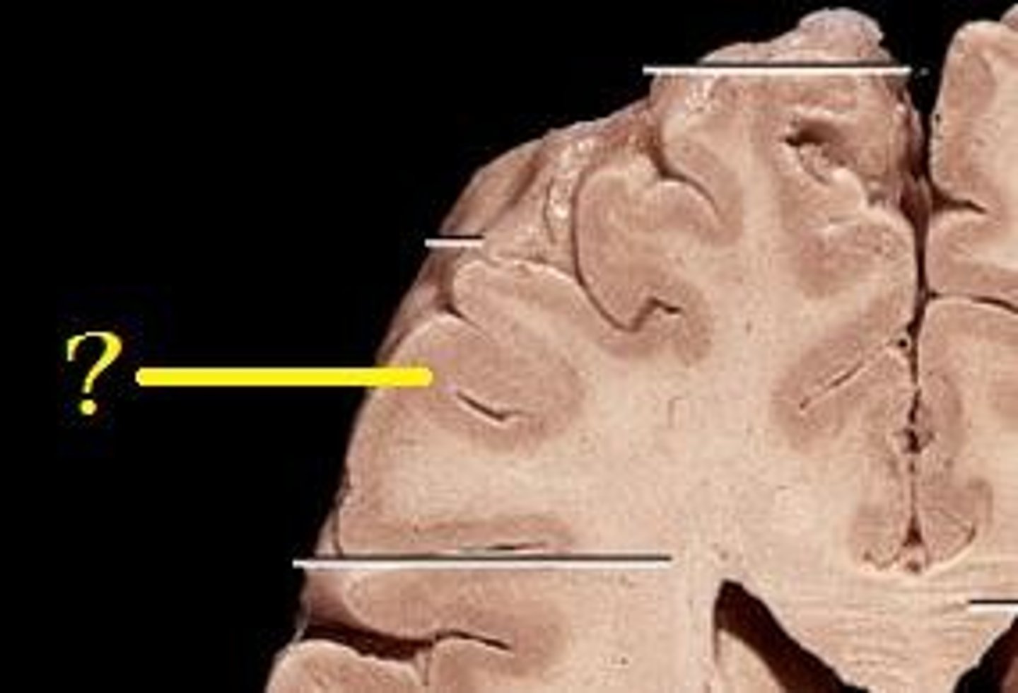
gray matter
outer layer; composed of neurons and glia; 6 layers can be seen with light microscopy;
contains pyramidal cells

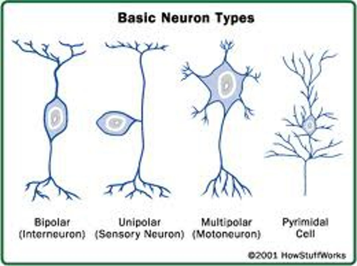
pyramidal cells
large neurons that allow conscious control of precise, skilled, skeletal muscle movements;
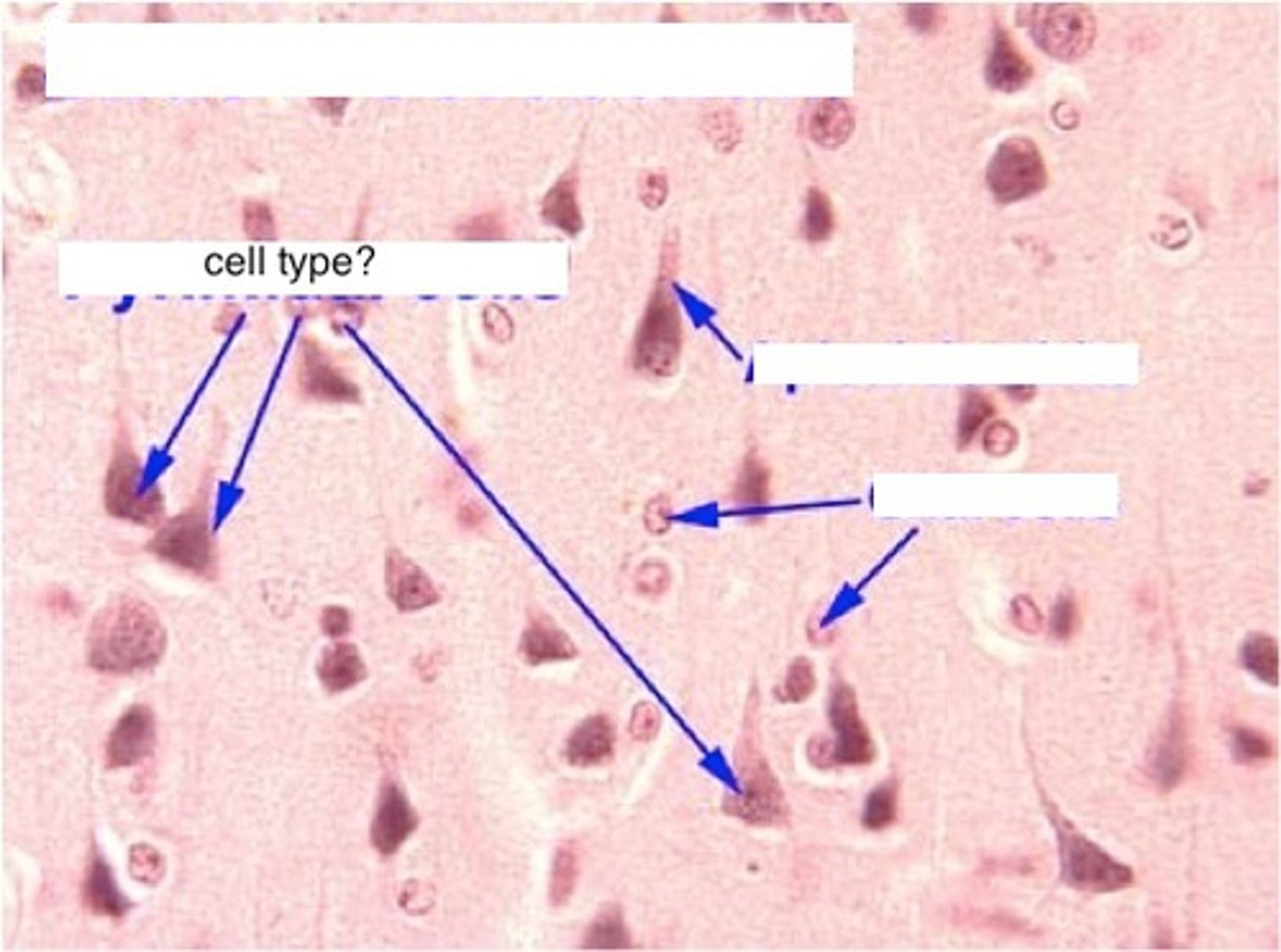
pyramidal cells shape
triangular shape;
Dendrites seen at base and apex of the pyramid;
axon is in middle of base
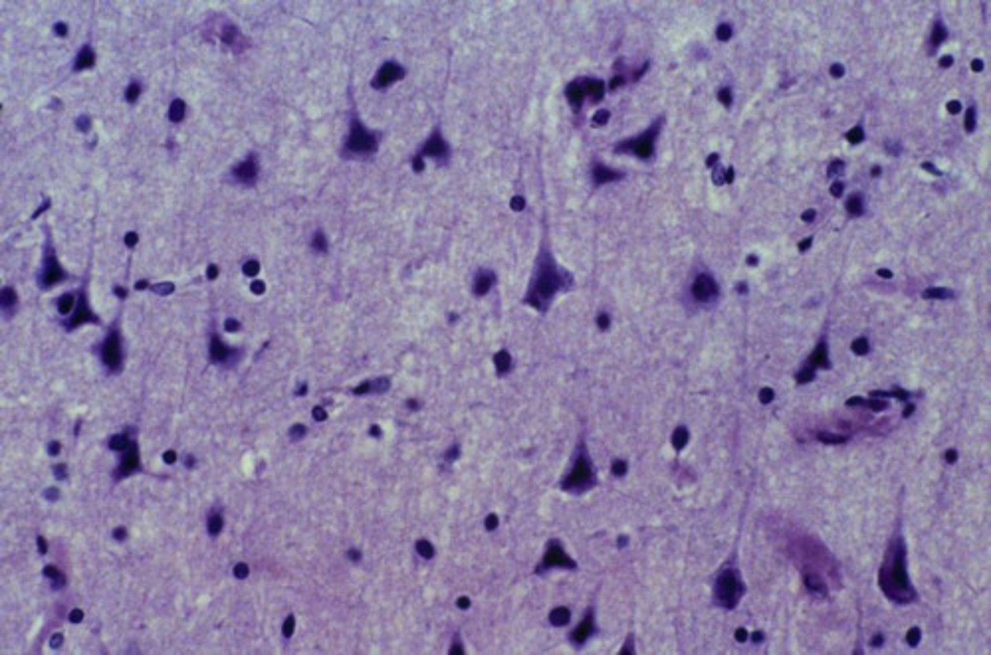
pyramidal cells H&E
see soma and proximal dendrites

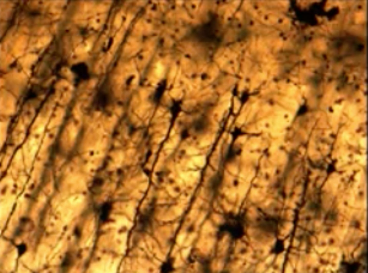
pyramidal cells golgi stain
labels all dendrites and axons
white matter
inner layer; composed of axons (going to and from cortex
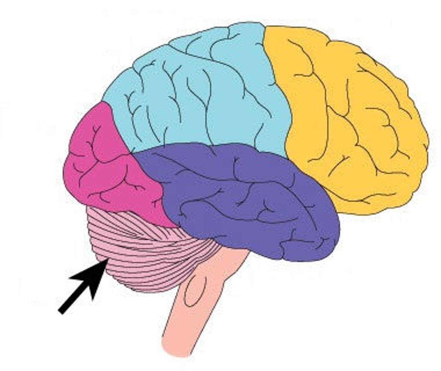
Cerebellum
A large structure of the hindbrain that controls fine motor skills;
has a 3 layered cortex
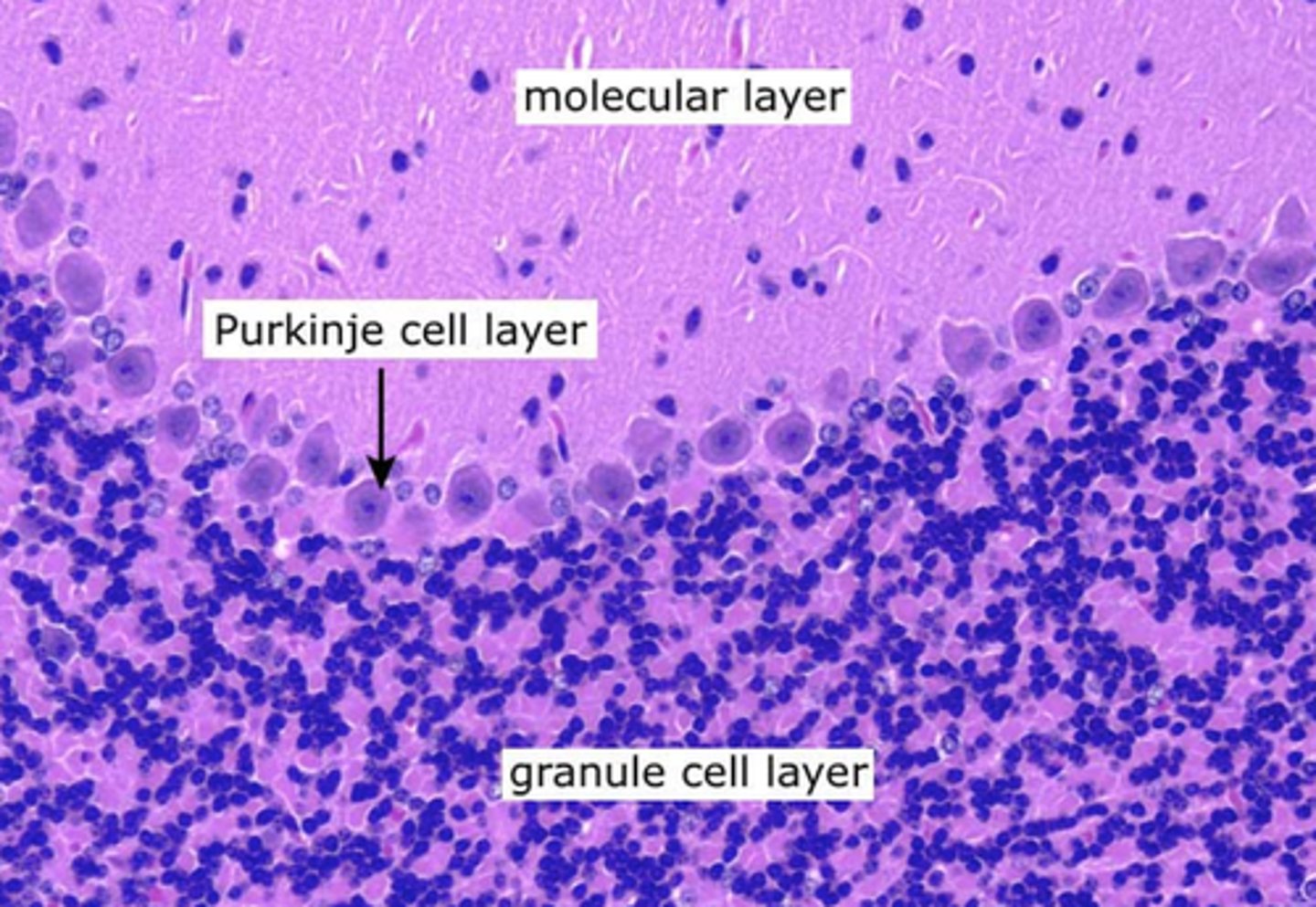
3 layered cortex of cerebellum
molecular;
purkinje;
granular
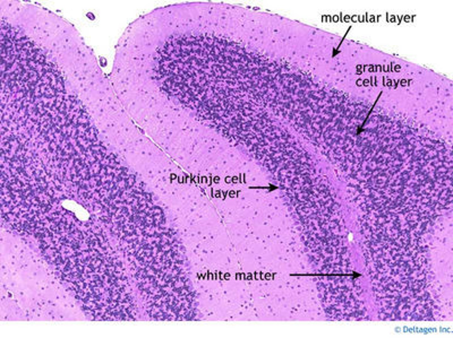
Molecular layer of cerebellum
outer most layer

Purkinje layer of cerebellum
visible as thin layer of globular shaped cells

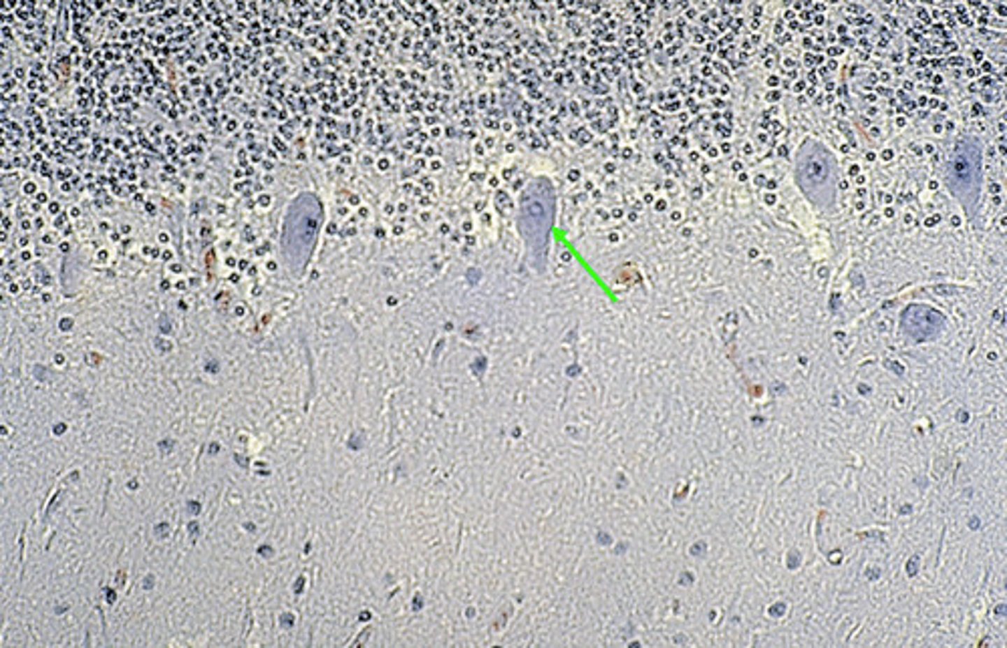
Purkinje cells
flat cells in sequential planes, parallel to one another
purkinje cell nissl stain
only soma is visible
purkinje cell immunofluorescence stain
see massive dendritic tree extending towards molecular layer
granular cerebellum layer
innermost layer
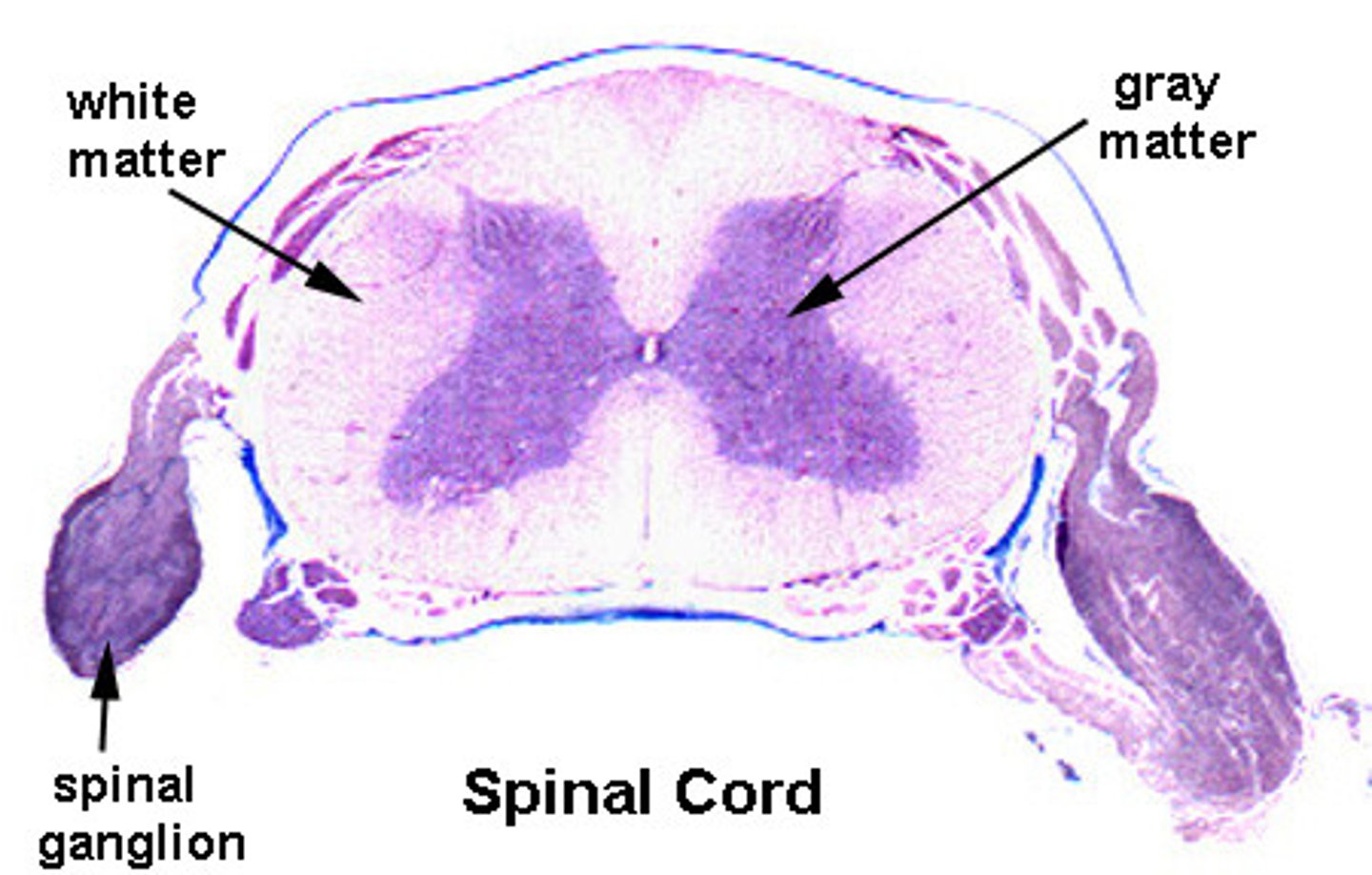
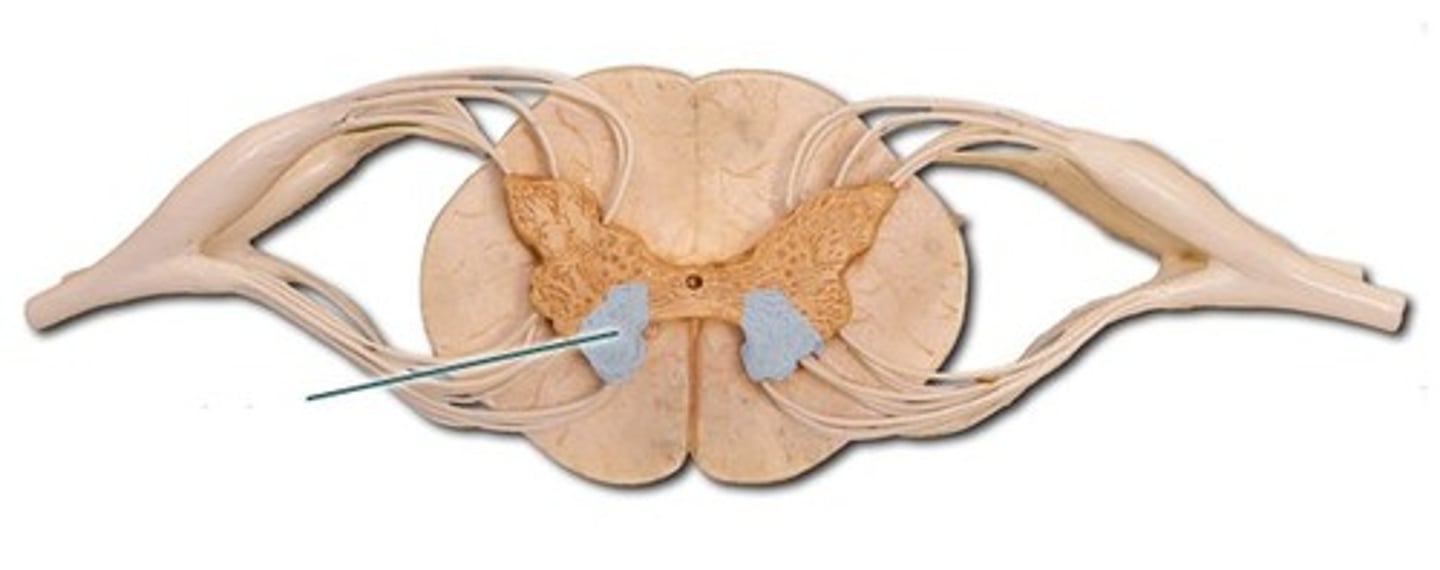
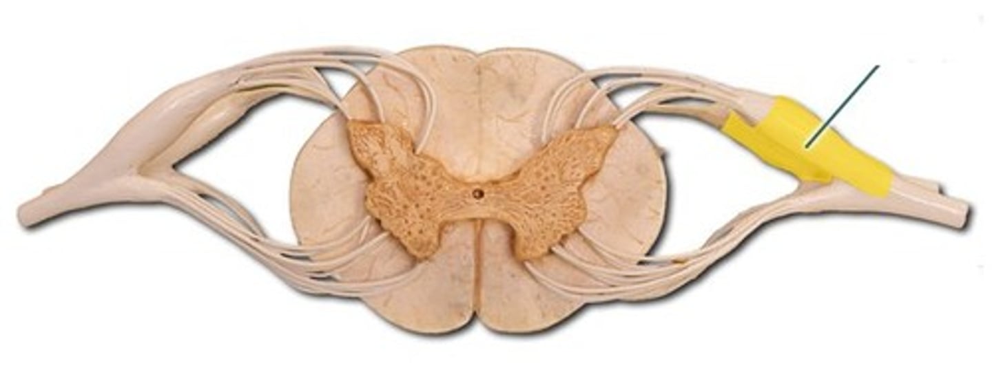
Spinal cord gray and white matter
Central cavity surrounded by gray matter, with outer white matter

gray matter contains
cell bodies and dendrites
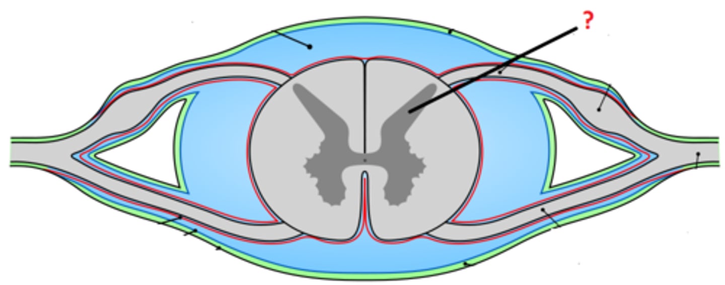
white matter contains
myelinated axons
Horns of the spinal cord
The extensions of the gray matter that form the letter "H"
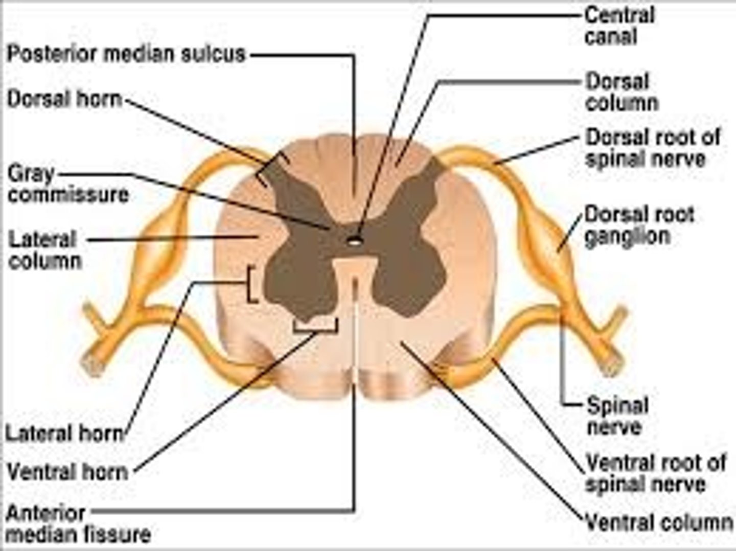
dorsal horns
interneurons that receive somatic and visceral sensory input
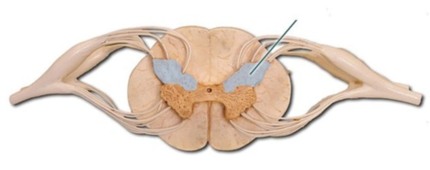
ventral horns contain
motor neurons
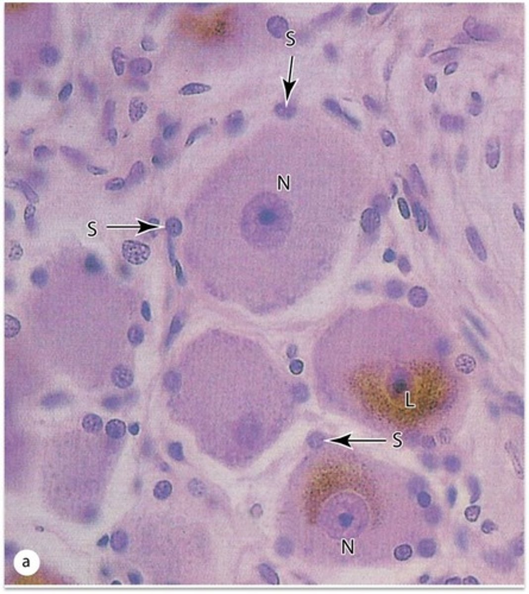
motor neurons nissl stain
all one can see are the cell body (soma) and proximal dendrites;
can sometimes see beginning of the axon, the axon hillock and initial segment, can be seen
Motor neurons have cell bodies in the
CNS and send their axons into the PNS
In the PNS hundreds of
of axons travel together in bundles that we call nerves
White matter
axons from neurons at other levels of the brain and spinal cord
white matter has a high
lipid content
PNS begins
outside the spinal cord;
but within the vertebral column in the spinal canal, close to where sensory axons enter & motor axons leave the spinal cord
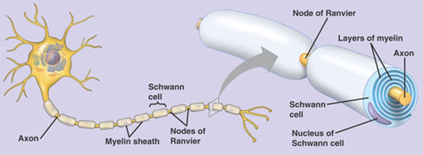
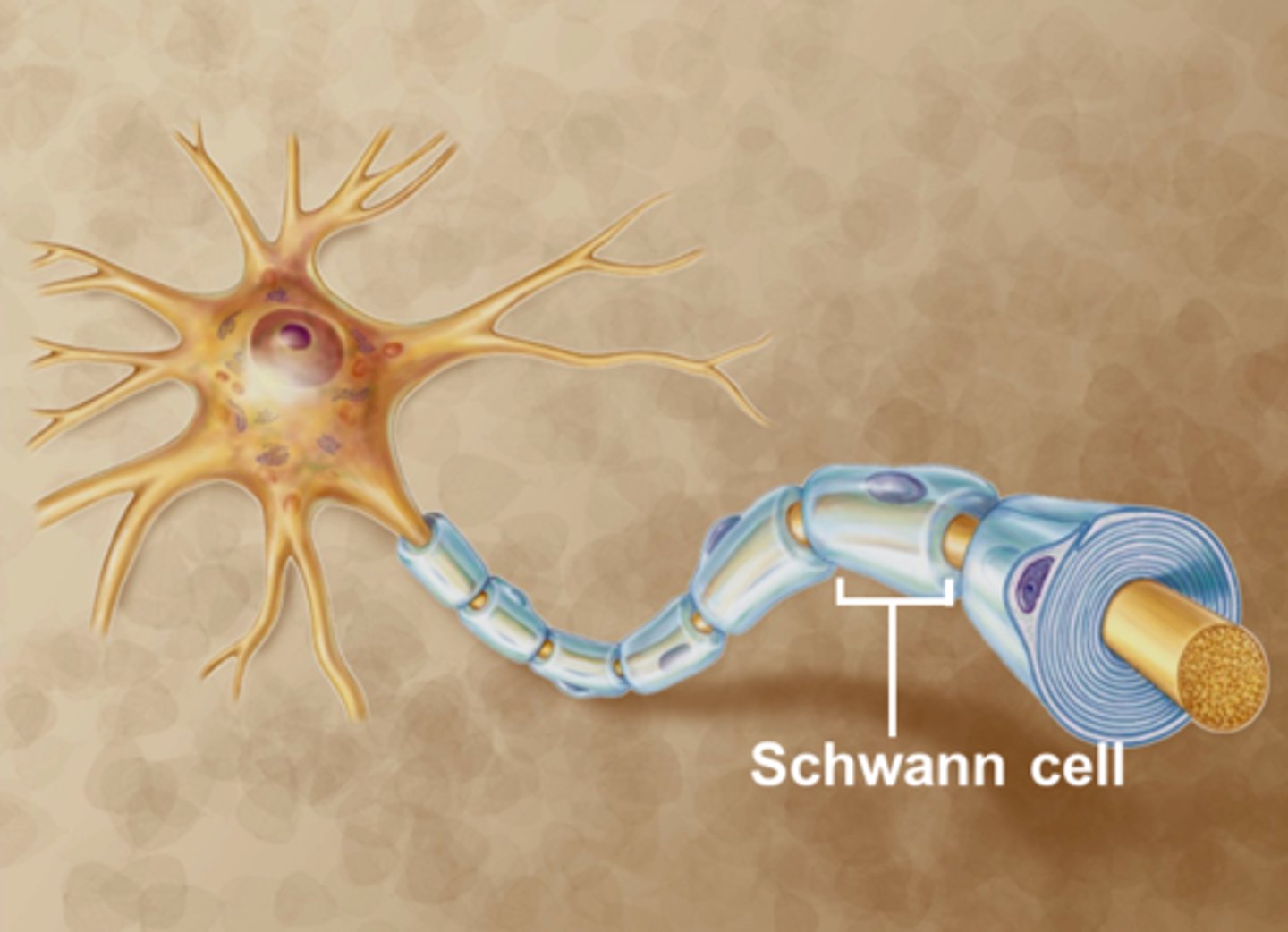
schwann cells
produce myelin in PNS;
only wrap around 1 axon
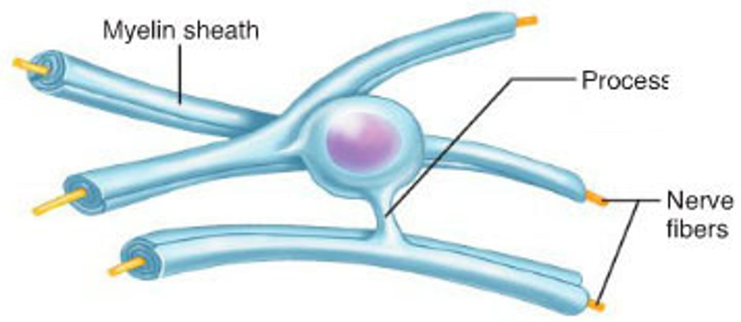
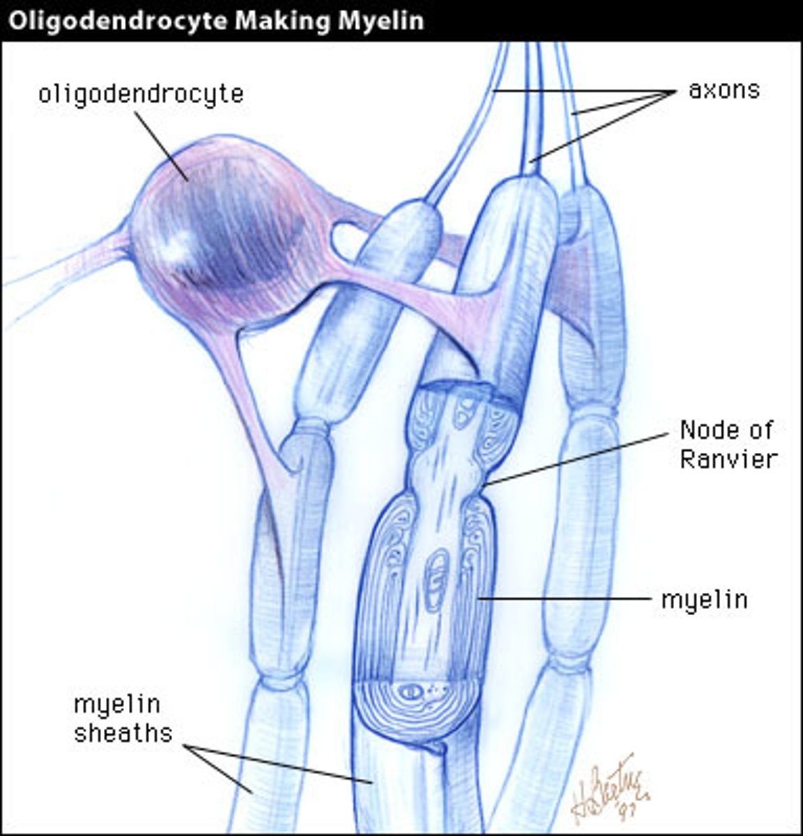
oligodendroglia
glial cells in the CNS that myelinate multiple axons
some neurons are in both the
CNS and PNS
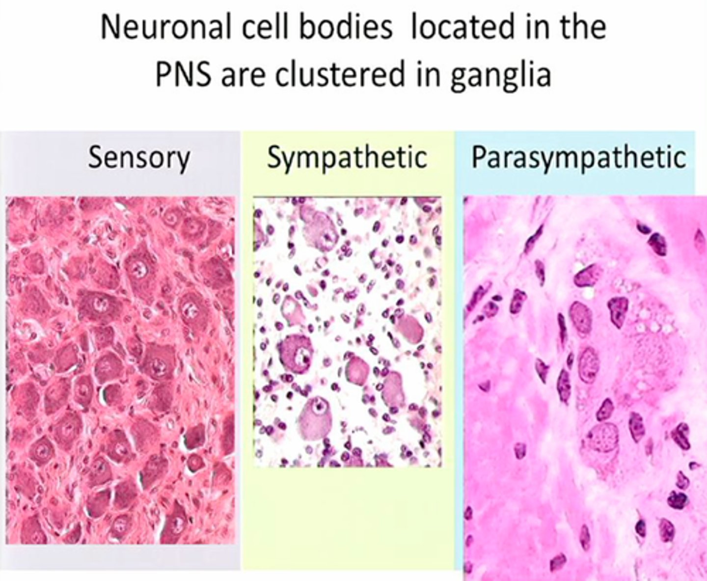
neurons with cell bodies in PNS
sensory ganglia;
autonomic ganglia
sensory ganglia
no dendrites;
T shaped axon;
long peripheral branch of sensory ganglia
go to body
long central branch of sensory ganglia
enters spinal cord;
short stem axon of sensory ganglia
connect to soma
sensory neurons have some of the
longest axons in the body
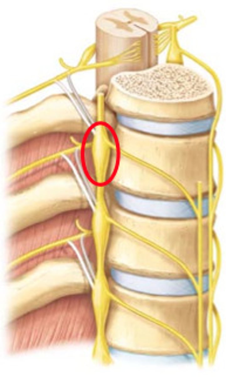
sensory neuron cell bodies are located in the
dorsal root ganglia, which sits in the intervertebral foramina of the vertebral column
sensory ganglia structure
bipolar during early development;
becomes pseudounipolar
autonomic ganglia
Collections of nerve cell bodies, belonging to the autonomic division of the peripheral nervous system, that are found in various locations and innervate the major organs.
both divisions of the ANS have the same basic structure
have a two neuron chain between the CNS and their target organs.
A preganglionic neuron is located within the
spinal cord or brainstem;
The axon of this neuron passes out through the ventral roots and synapses on another neuron in a ganglion out in the body
ganglion
a small collection of neurons in the peripheral nervous system
autonomic ganglia are located in various places in the body. One well-known location is the
sympathetic chain
sympathetic chain ganglia
lies bilaterally adjacent to the vertebral column;
a series of tiny ganglia that contain ganglionic neurons AKA postganglionic neurons
postganglionic neurons have
short dendrites and unmyelinated axons
postganglionic neuron innervate
smooth muscle and glands
autonomic ganglion looks very much like the
dorsal root ganglion;
distinguish by looking for evidence of the dendrites on the autonomic neurons;
satellite cells of DRG more prominent
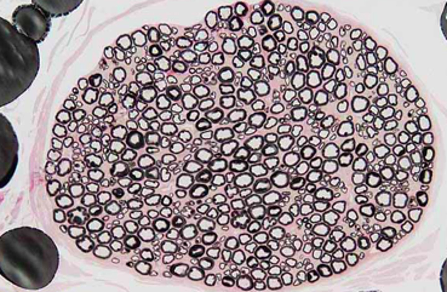
myelinated axons
axons covered with myelin sheaths
myelinated axons in light microscopy
tiny dark rings stacked on one another
myelinated axons in electron microscopy cross section
myelin layers aka lamellae;
Microtubules, neurofilaments, mitochondria
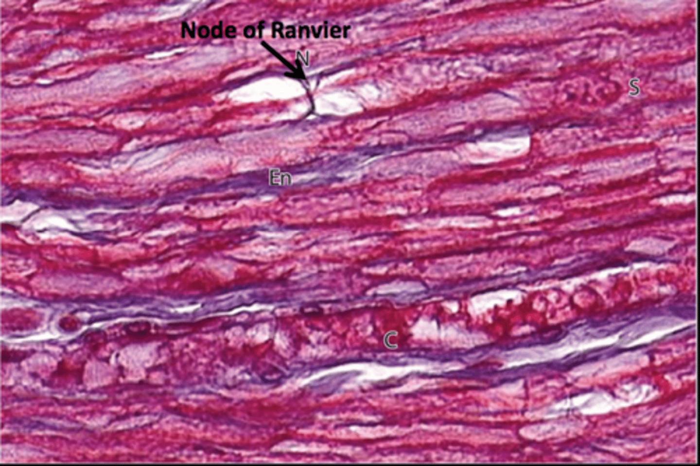
myelinated axons in electron microscopy longitudinal section
nodes of ranvier;
Myelin on either
Nodes of Ranvier
Gaps in the myelin sheath to which voltage-gated sodium channels are confined.
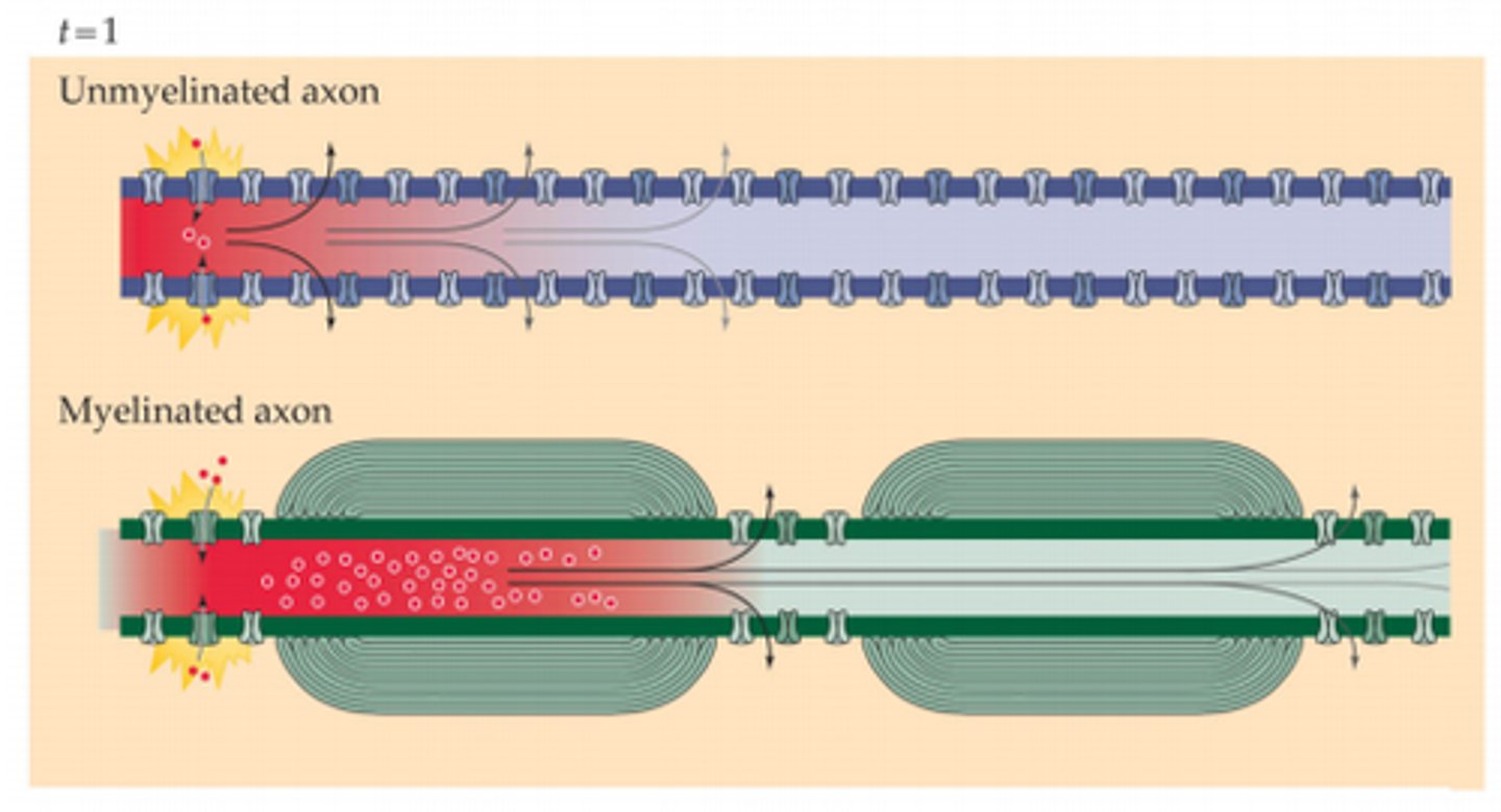
unmyelinated axons
no nodes of ranvier;
slow conduction;
Smaller diameter, but covered by cell membrane from Schwann cells
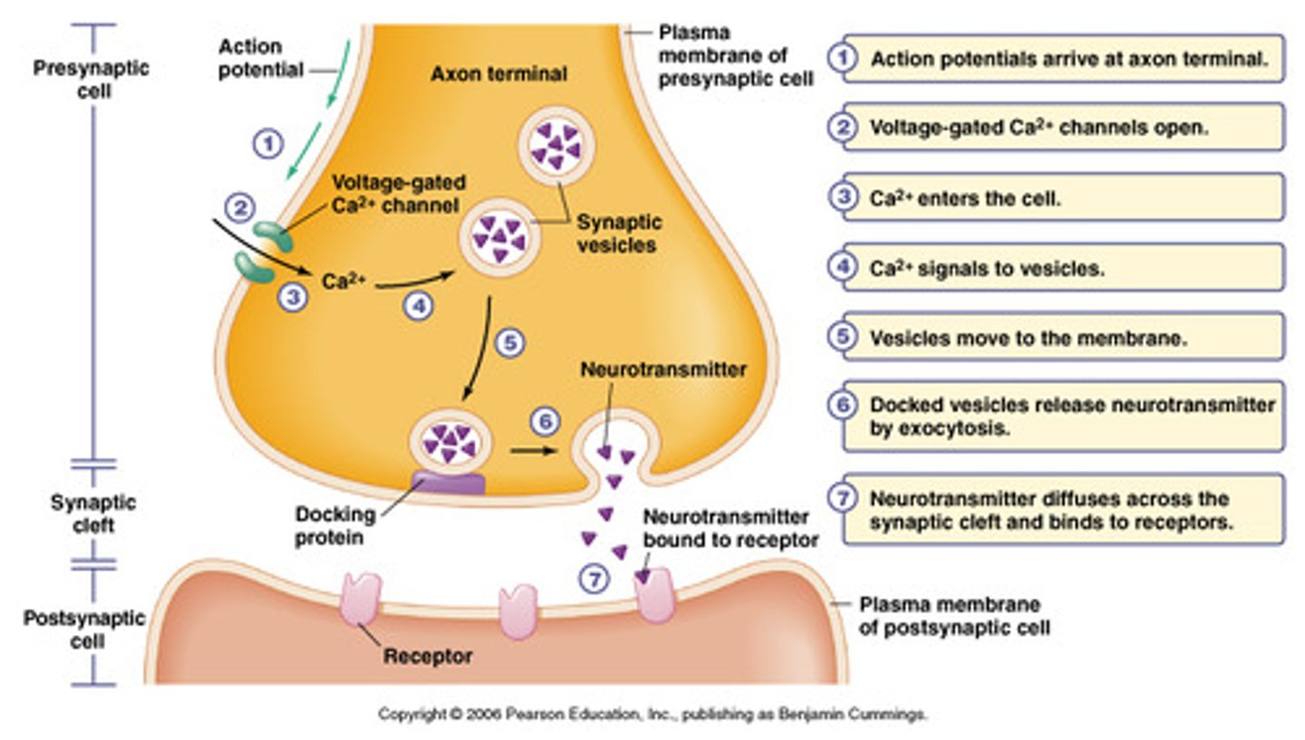
When a neuron is excited it generates an
electrical action potential that is conducted down its axon to its terminals synaptic boutons or a neuromuscular junction (NMJ)
Most axons have multiple
terminals;
These terminals connect to the dendrites and cell bodies of other neurons through synapses
Evidence of the synapse that can be seen with LM at
200-400x magnification is a tiny varicosity called a synaptic bouton.
a single axon may make only a few
few synapses upon one cell (5 or 10);
but receive hundreds of synapses from many different axons
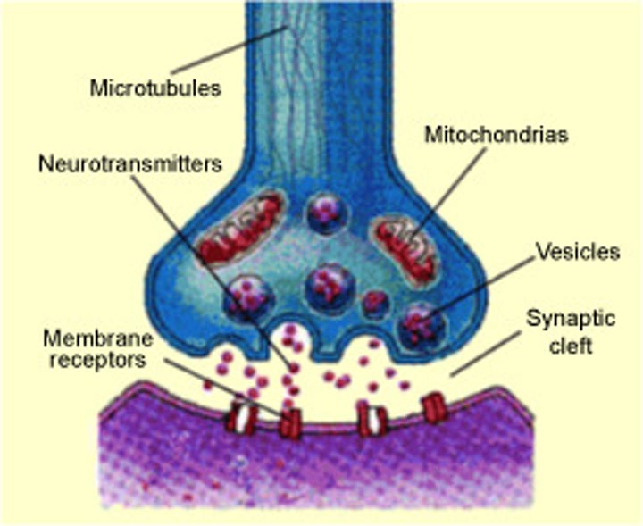
At high magnification of a bouton, one can see
vesicles and synaptic specializations in both the bouton and the dendrite that it is contacting
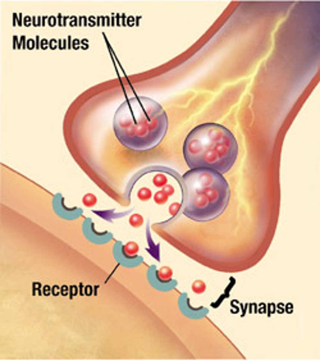
synaptic cleft
The narrow gap that separates the presynaptic neuron from the postsynaptic cell.
Neurotrasmitters
chemical found in the synaptic vesicles that, when released, has an effect on the next cell;
released into synaptic cleft;
excites/inhibits
higher magnification of EM (50,000x magnification) reveals
bouton and synaptic vesicles with neurotransmitter
glial cells come from
neuroectoderm except microglia
neuroglial cells in cns
oligodendrocytes;
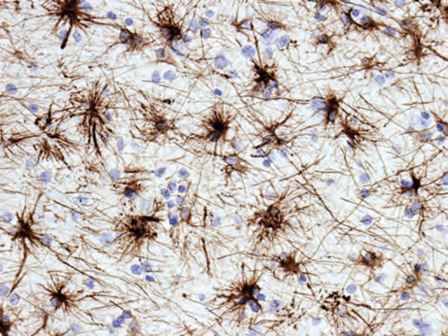
astrocytes;
ependymal;
microglia
Oligodendrocytes
Type of glial cell in the CNS that wrap axons in a myelin sheath.
fibrous astrocytes
in white matter;
long slender processes w/numerous filaments;
induce formation of blood brain barrier
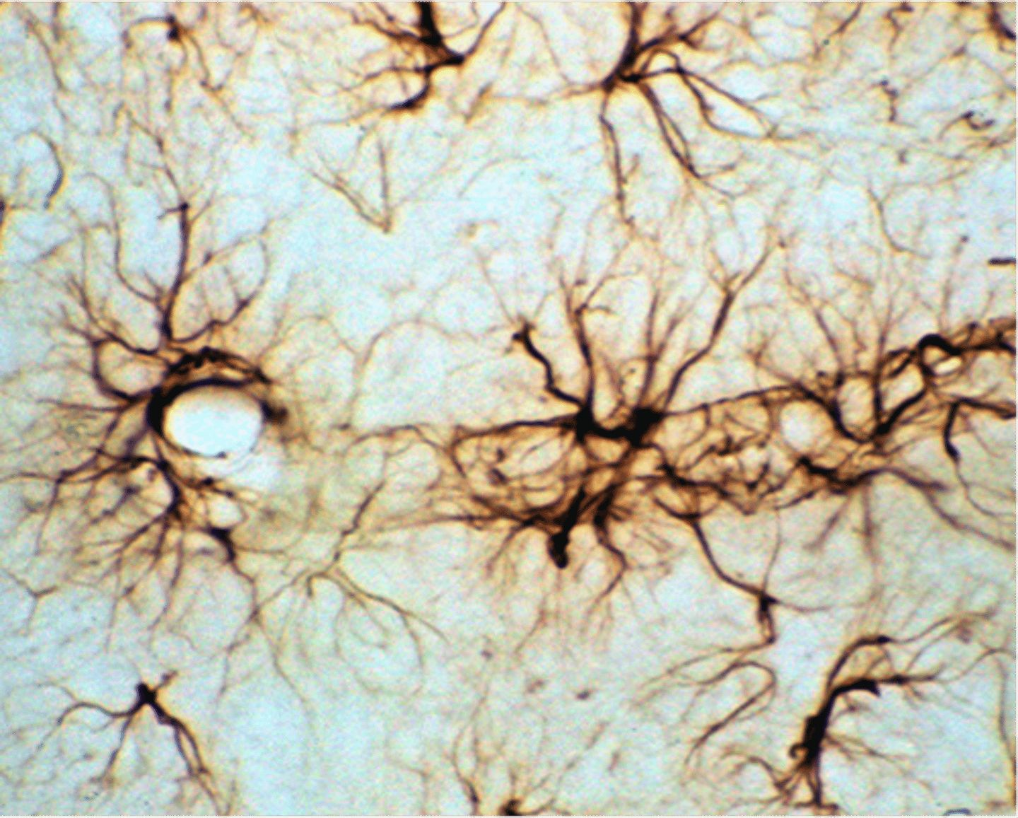
protoplasmic astrocytes
in gray matter;
thick bushy processes;
induce formation of blood brain barrier
Blood Brain Barrier (BBB)
a selective mechanism that protects the brain from toxins and infections;
formed by endothelial tight junctions that divides blood and neuron extracellular space
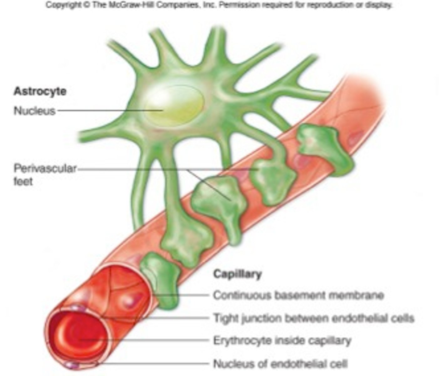
Both types of astrocytes require
special stains to see more than their nuclei
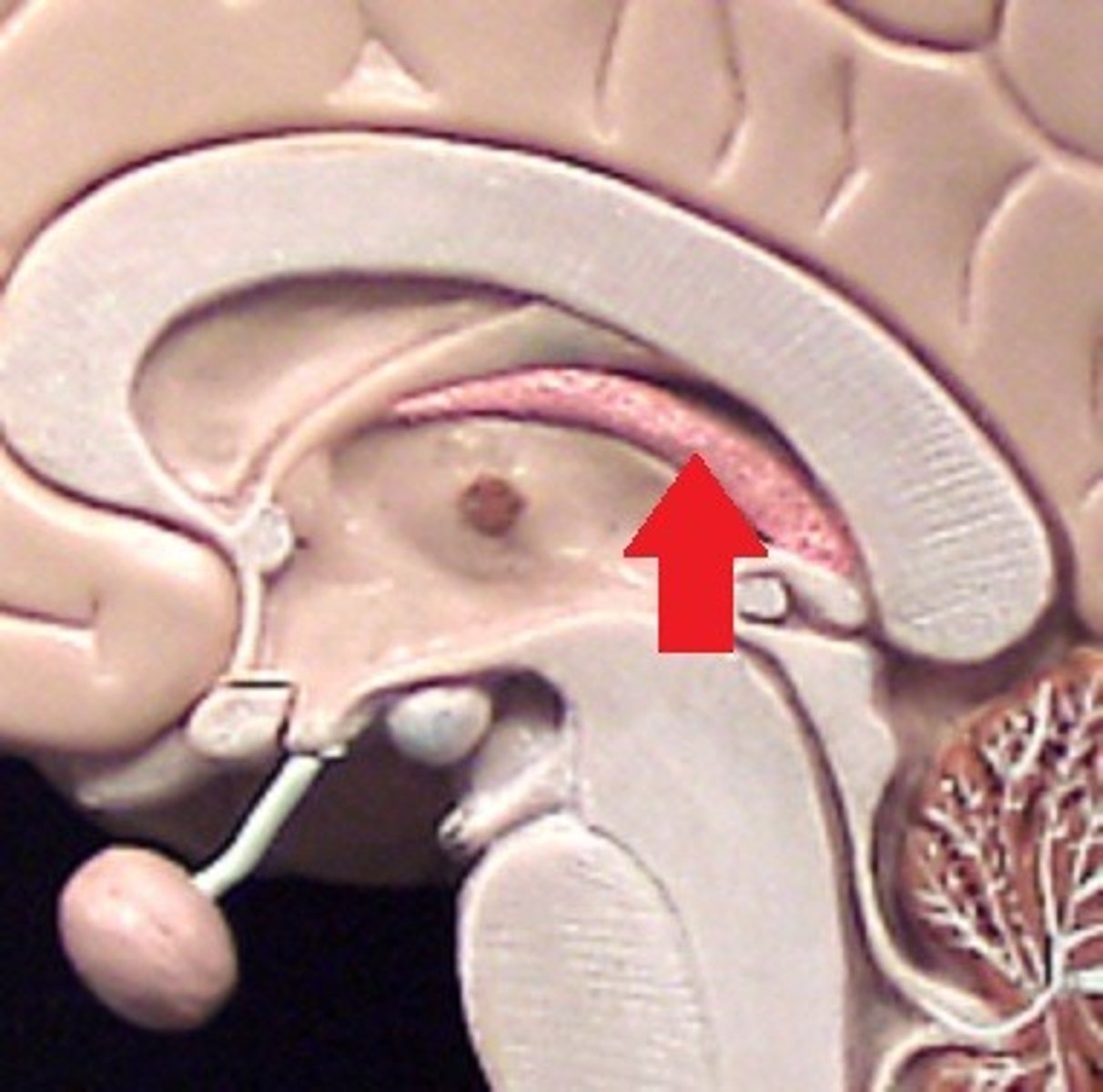
ependymal cells
line ventricles in brain, central canal in spinal cord;
form choroid plexus
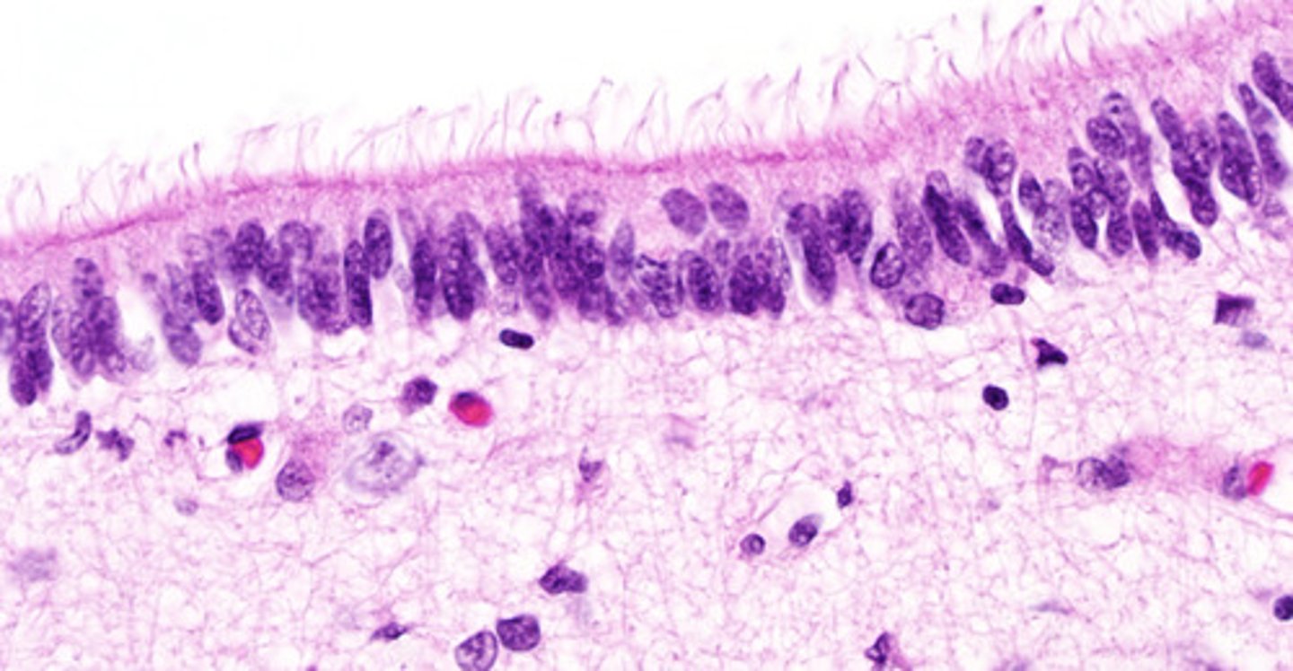
choroid plexus
produces CSF
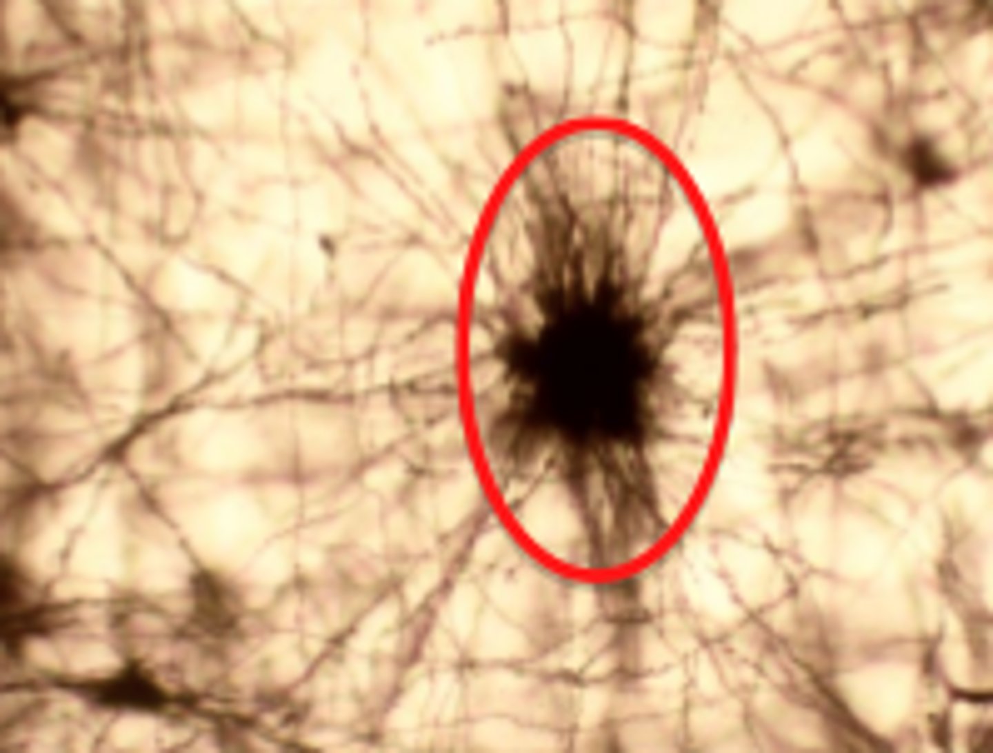
microglia
macrophages of the CNS;
derived from bone marrow;
phagocytose injured tissue
Myelination of a single peripheral nerve axon occurs via
a series of Schwann cells
Cells in PNS
Schwann cells and satellite cells
schwann cells wraps a
wraps a single short-length portion of the axon
in the CNS oligodendroglia can
wrap short-length portions of several axons.
Schwann cells & oligodendroglia both wrap multiple
unmyelinated axons with single layer
satellite cells
found around ganglia neurons (e.g. DRG & autonomic ganglia)
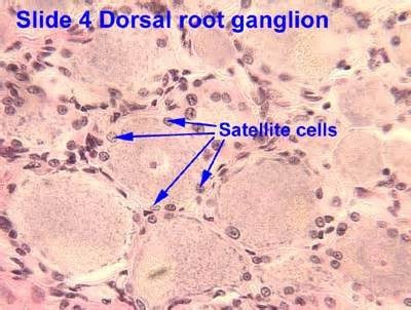
satellite cells assist in
regeneration following peripheral nerve injury