A2.2 Cell Structure
1/23
There's no tags or description
Looks like no tags are added yet.
Name | Mastery | Learn | Test | Matching | Spaced | Call with Kai |
|---|
No study sessions yet.
24 Terms
Prepare an onion for a microscope slide.
Peel thin layer
Place on slide
Add 1-2 drops of stain (iodine) (this stains cell structures)
Cover with cover slip
Gently tap any bubbles
View on lowest objective lens 1st
Make an onion cell slide.
View it.
Draw 6 – 12 cells.
Label & annotate.
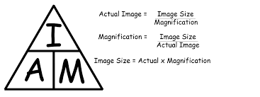
Magnification equation
Disadvantages of electron microscope
Electron microscopes do have some disadvantages.
They can only give black & white images - so, any colour in electron micrographs has to be added artificially.
Methods used to prepare slides may damage the specimen.
Only dead specimens can be viewed
However, light microscopes can be used to examine living material and produce colour images.
Fluorescent stains and immunofluorescence
Fluorescence is when a substance absorbs light then re-emits it at a longer wavelength.
Some absorb UV and re-emit it as blue light.
Antibodies that bind to specific antigens in the cell are produced.
Fluorescent markers of different colours are then linked to them.
A multicoloured fluorescent image can then be produced showing the location of the antigens.
It can be used to find out if one specific type of protein is being produced in a cell, or if specific antigens are present.
Freeze fracture electron microscopy
Used to produce images of surfaces within cells or the internal structure of membranes.
Sample is plunged into liquefied propane (-190oC) and rapidly freezes.
It is about 2nm thick on average but the thickness varies due to the angle at which the coating is applied – this gives the impression of a 3D image through shadowing.
Cryogenic electron microscopy
Principally used to research protein molecular structure.
A thin layer of a pure protein solution is applied to a grid.
The solution is flash-frozen, to create smooth vitreous ice and prevent water crystal formation – usual coolant is liquid ethane (-182oC).
Due to the random orientation of the protein molecules, many different patterns are produced.
Using computers these are combined to produce a 3D image of the molecules using computational algorithms.
What structures are common to all cells?
Cells vary in size, shape & structure but share some common structures.
Plasma membrane:
Outer boundary – encloses all contents
Controls entry & exit of substances
Can pump substances in even if external concn is very low
Cytoplasm (cytosol):
Main component is water – polar substances dissolved to allow reaction to occur
DNA:
Genes hold instructions for Protein Synthesis
Eukaryotic cells have a nucleus that contains all their DNA
(our cells contain DNA from other organisms)
Bacteria do not have a nucleus – DNA is in cytoplasm including plasmid DNA.
Prokaryotic cell structure
First organisms to evolve
Simplest cell structure (+ small!)
No nucleus
Cell wall made of peptidoglycan (murein)(maintains shape & prevents lysis & toxins)
Interior entirely filled with cytoplasm (which is not divided into compartments)
No cytoplasmic organelles apart from …
Ribosomes (size 70S - smaller than eukaryotic ribosomes with are 80s)
Has one circular DNA chromosome (found in a lighter area of cell called the nucleoid – lighter due to lack of proteins & ribosomes.)
DNA is ‘naked’, ie. not associated with proteins.
Eukaryotic cell structure
Much more complicated internal structure.
Compartmentalised (divided up by partitions into compartments – by internal membranes).
Compartments in cytoplasm are known as organelles – each is specialised (has a distinct structure & function).
3 other key features distinguish them from prokaryotes:
(1) Nucleus containing DNA molecules (chromosomes) are linear
associated with histone proteins.
(2) 80s ribosomes: Structurally different and larger.
(3) Mitochondria: Site of aerobic respiration.
Nucleus role
Double membraned with pores. Where the DNA is stored
RER function
Formation of proteins including assembly of Quaternary Structures, packaging into vesicles for transport to the Golgi Apparatus - usually for excretion (exocytosis).
SER function
Formation of lipids and packaging into vesicles
Lysosomes function
Vesicles that contain digestive enzymes
Mitochondria function
Double Membraned. Own DNA, 70s Ribosomes. Contains Enzymes and structures for respiration.
Free ribosomes
Sites of translations for proteins that will typically remain in the cell. (translation)
Chloroplasts function
Double Membraned. Own DNA, 70s Ribosomes. Contains Enzymes and structures for photosynthesis such as chlorophyll.
Vacuoles and Vesicles function
Membrane bound structures for storage and transport
Microtubules function
Protein Tubes that have many functions including the movement of organelles within the cell. E.g. spindle fibres and protein motors for cilia and flagella.
Centrioles function
Anchor points for spindle fibres in Animal Cells only.
Cytoskeleton function
A system of structures made of Tubulin including microtubules that maintain the shape of the cell
Golgi apparatus function
Processes proteins from RER, takes them to vesicles, to cell membrane for secretion
Evidence for endosymbiosis
Mitochondria & Chloroplasts are double membrane bound. They contain their own DNA (carry out protein synthesis), their own cytoplasm and enzymes and have 70s ribosomes.
They divide by a process similar to binary fission to form more of themselves.
What is the endosymbiosis theory?
That mitochondria & chloroplasts were once free living prokaryotic organisms - that were taken in by larger prokaryotes & kept alive - both benefiting from them being there - resulting in eukaryotic cells.
Evolution of multicellularity
All plants and animals are multicellular. (algae are protists!)
Multicellularity has evolved independently more than once in the origins of plants and at least once in animals.
Many fungi and eukaryotic algae are multicellular.
Most cells in a multicellular organism have lost the ability to live independently or to divide.
There are several advantages to multicellularity organisms are generally larger, so they can exploit niches that unicellular organisms cannot it allows for complexity as there can be differentiation of cell types.