lab particle
1/303
There's no tags or description
Looks like no tags are added yet.
Name | Mastery | Learn | Test | Matching | Spaced |
|---|
No study sessions yet.
304 Terms
where are the eye muscles attached to?
the sclera
function of the superior rectus?
elevates and adducts
function of the lateral rectus?
lateral movement
function of the medial rectus?
medial movement
function of the inferior oblique?
lateral rotates
function of the inferior rectus?
depresses
function of the superior oblique?
medially rotates
what duct and gland are located diagonally from each other?
lacrimal gland and nasolacrimal duct
the nasolacrimal duct if lower than?
lacrimal gland
what does the position of the nasolacrimal and lacrimal create?
flow of fluid across the eye
what do tears consist of?
water, salts, mucus, and protective enzymes
what do tears do for the eye?
lubricate the eye and keep pathogens from infecting it
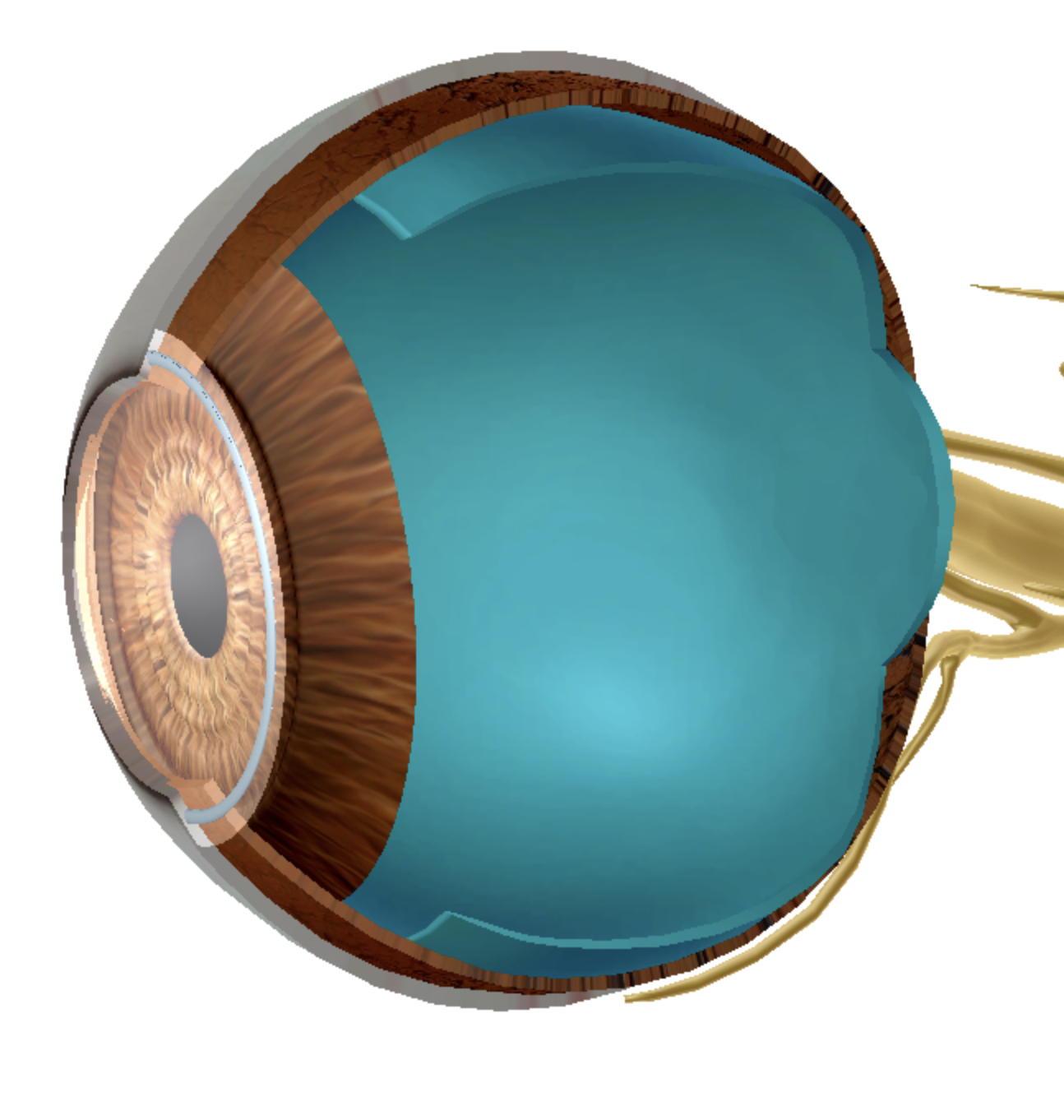
what is the cornea?
transparent layer covering the iris
what’s the function of the cornea?
focuses light through the pupil to the retina and allows light to pass easily through it
what make it hard for the cornea to heal?
no blood vessels
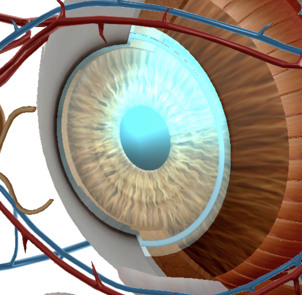
what is the iris?
the colored part of the eye
what gives the iris its color?
melanin
what two parts does the iris separate into?
anterior and posterior chambers
what’s the function of the iris?
adjusts to the size of the pupil to allow different amount of light though the eye , constricting to allow in less light in bright environment and dilating in more light when light is scarce
what does the pupil allow?
light to enter the eye
what does the lens change?
shape to focus light on the retina
what does the ciliary body adapts to?
the lens shape for focusing light to adjust for objects at different distances
when does the pupillary dilator dilate?
when it receives parasympathetic inputs
when does the pupillary sphincter constrict?
when it receives sympathetic inputs
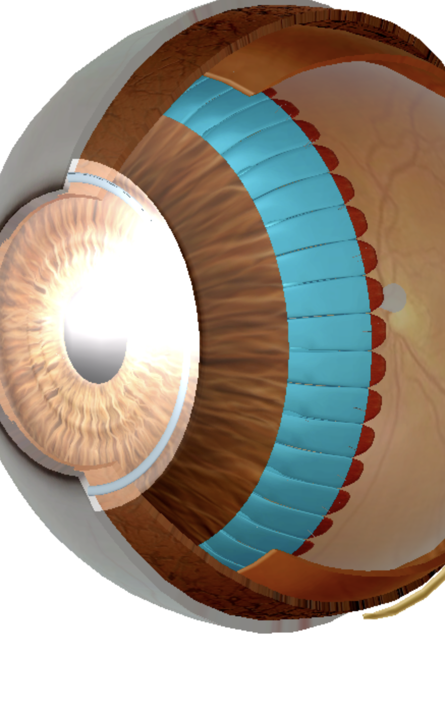
what does the sclera do?
maintain shape and ridigity of the eyeball and provides an attachment surface for the eye muscles
what does the choroid do?
provides nutrients to the retina and absorbs stray beams of light, contains a high abundance of melanin
what does the retina do?
houses the photoreceptors cells and some cells that process visual information
the retina contains photoreceptors called?
rods and cones
what does the photoreceptors do?
they change light signals into neural signals
rods can sense what?
dim light
cones can detect what?
color
how many rods?
120 million
how many cones?
6 million
what is the central part of the retinal with a high abundance of cones?
fovea centralis
what’s the function of the fovea centrals?
higher visual acuity
what is the blind spot?
the parts of the retina with no rods or cones
what the site where the optic nerve exits the eye/ axons of the retinal cells exits in the eye
the blind spot
function of the blind spot
cannot transducer any light from this region
what happens to the lens when the ciliary muscles contract?
the lens becomes more round and thicker
what is the fluid matrix in the eye>
vitreous humor
what is the function of the cilliary muscle?
helps the eye focus

what is A?
spinal cord
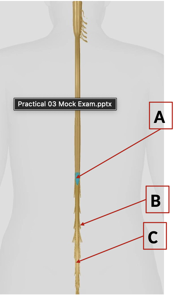
what is B?
conus medullaris

what is C?
cauda equina

what is F?
cochlear duct

what is top E?
scala vestibuli
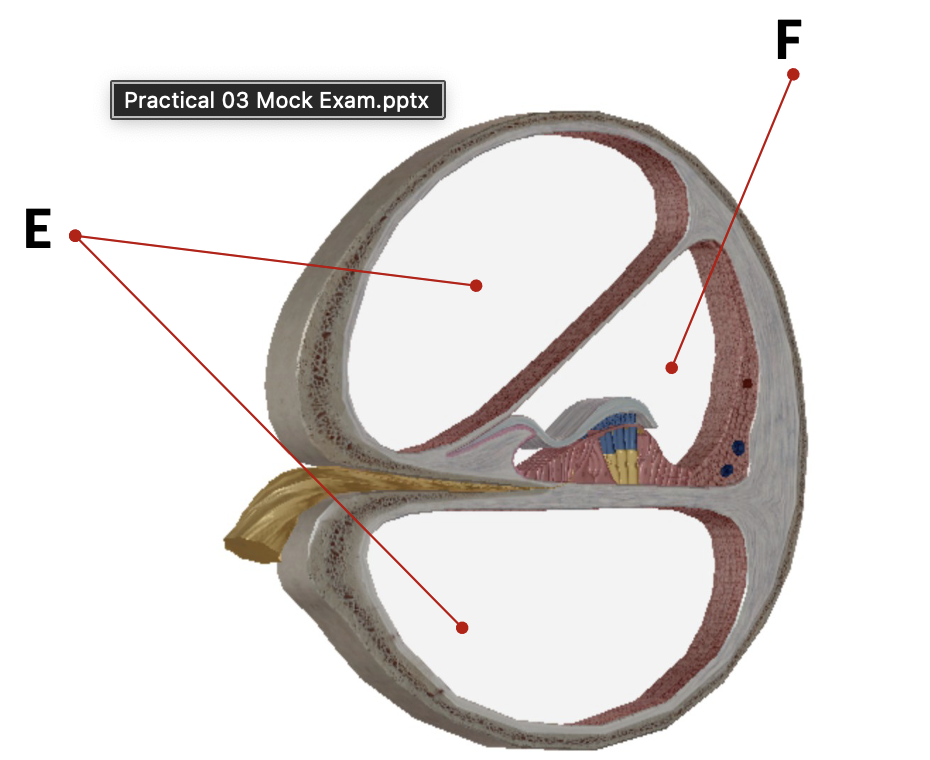
what is bottom E?
scala tympani
what is highlighted
cerebrum
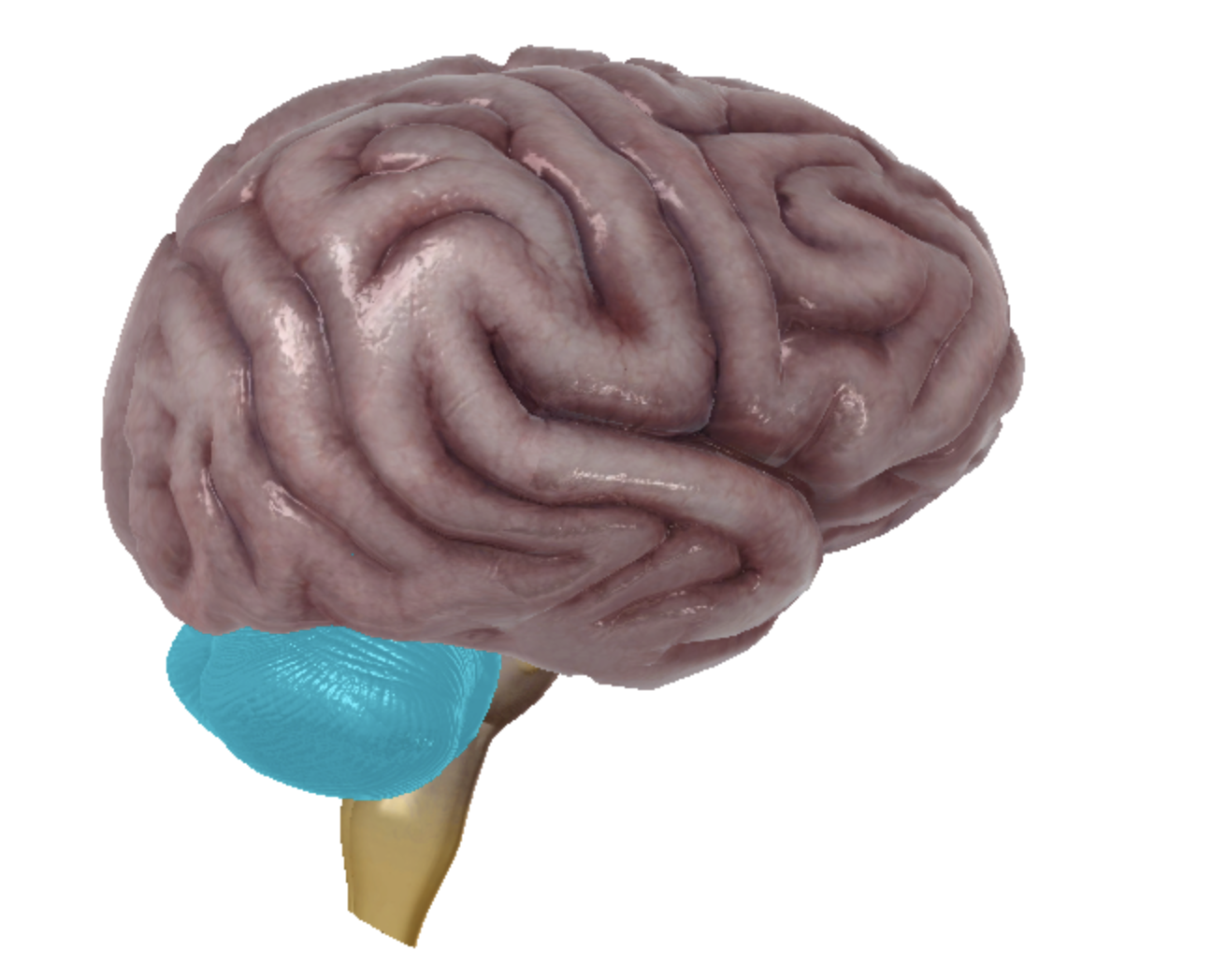
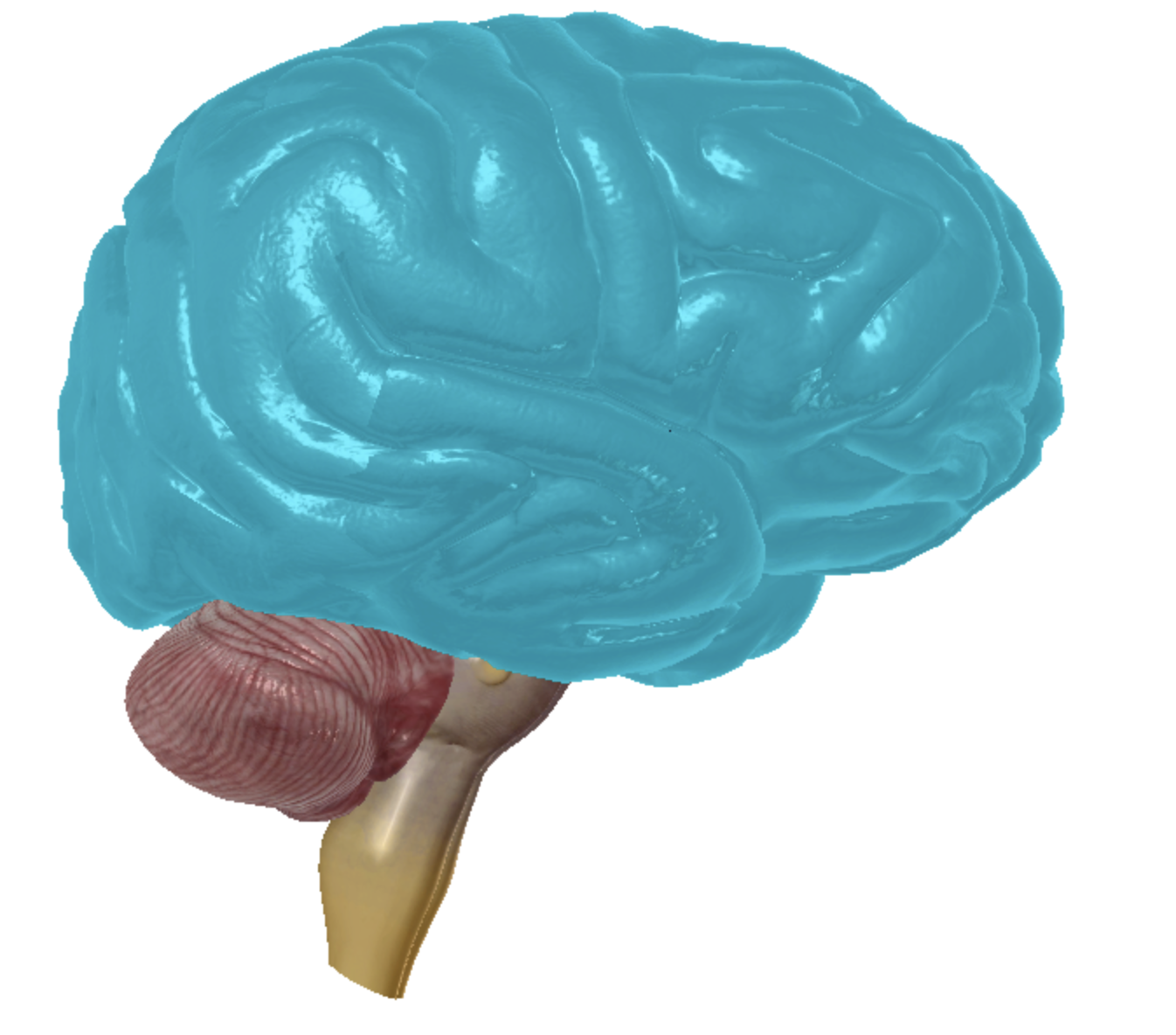
what is highlighted
cerebellum
what is highlighted
diencephalon
what is highlighted
midbrain
what is highlighted
pons
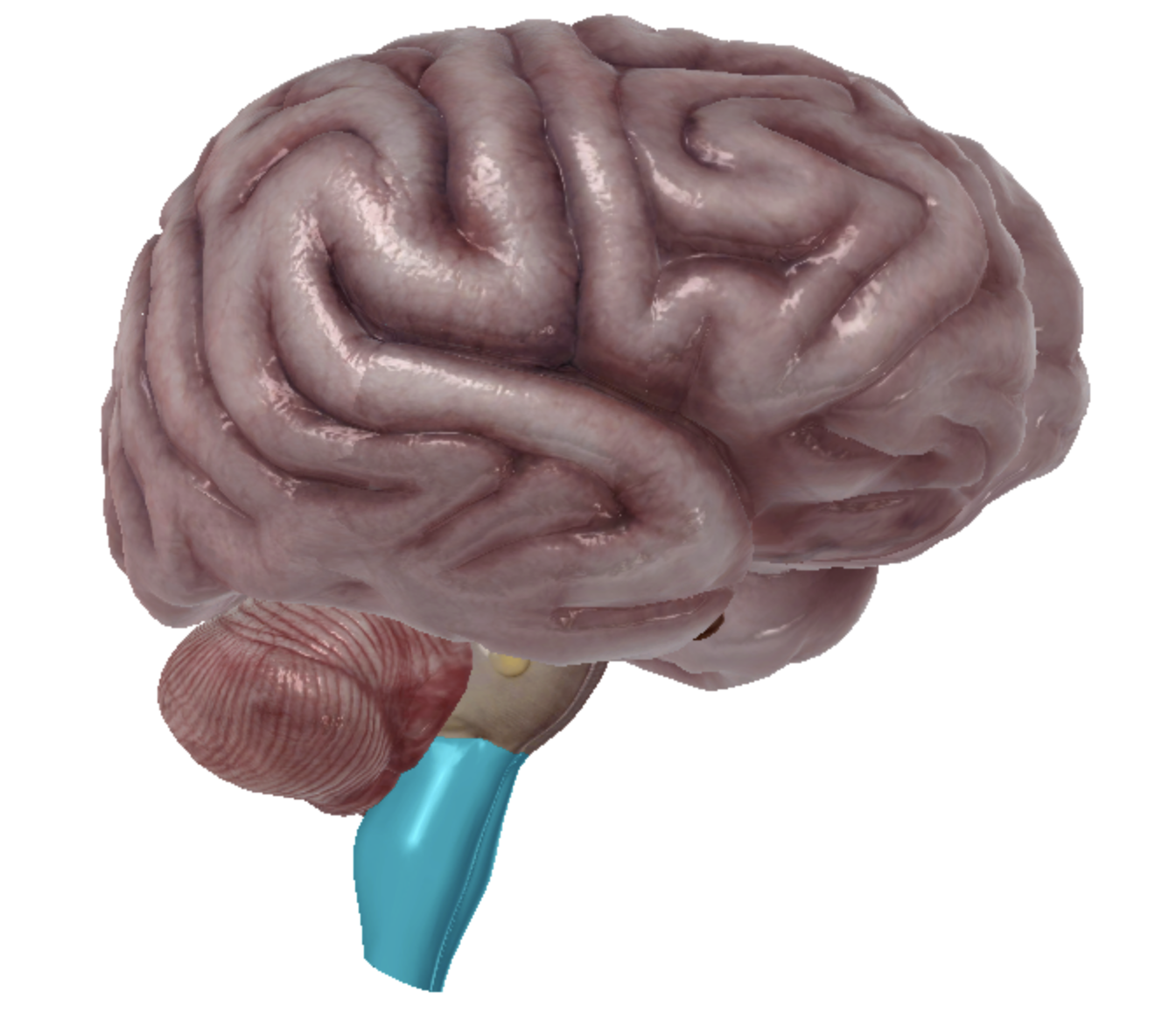
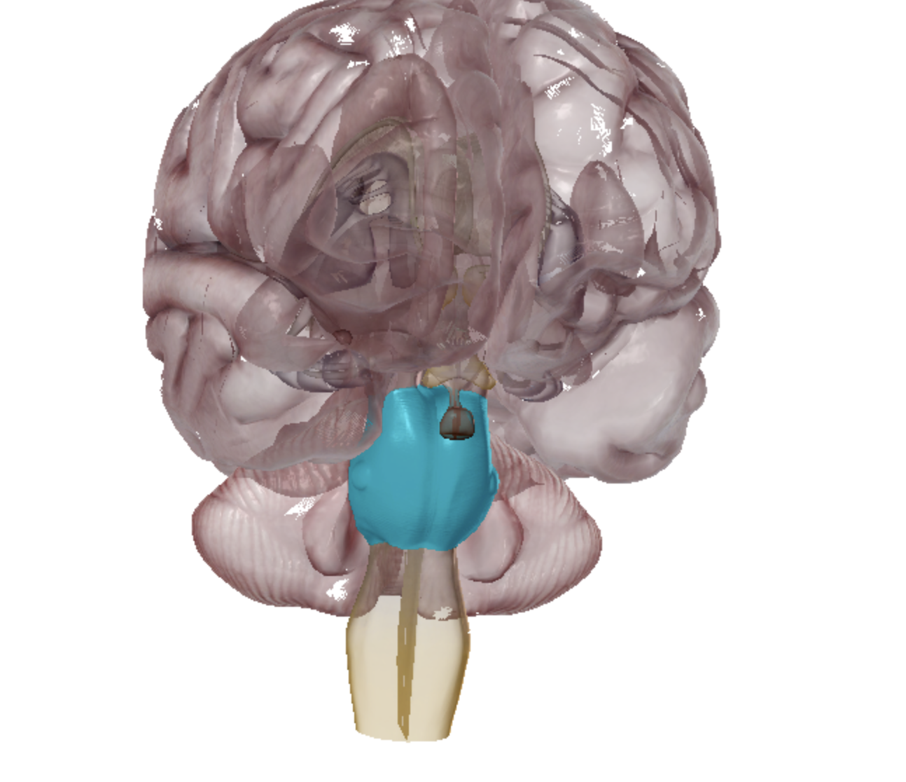
what is highlighted
medulla oblongata
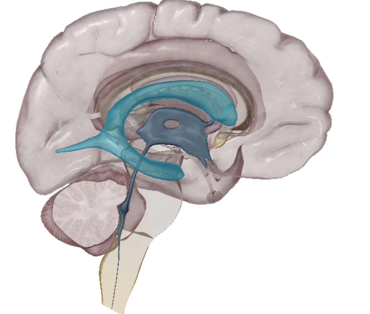
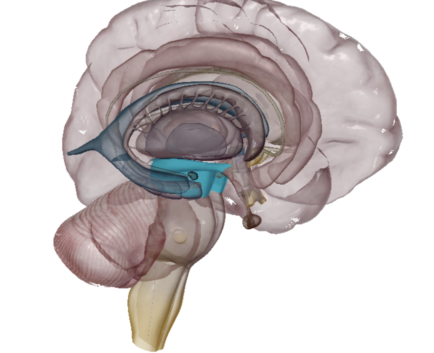
what is highlighted
lateral ventricle
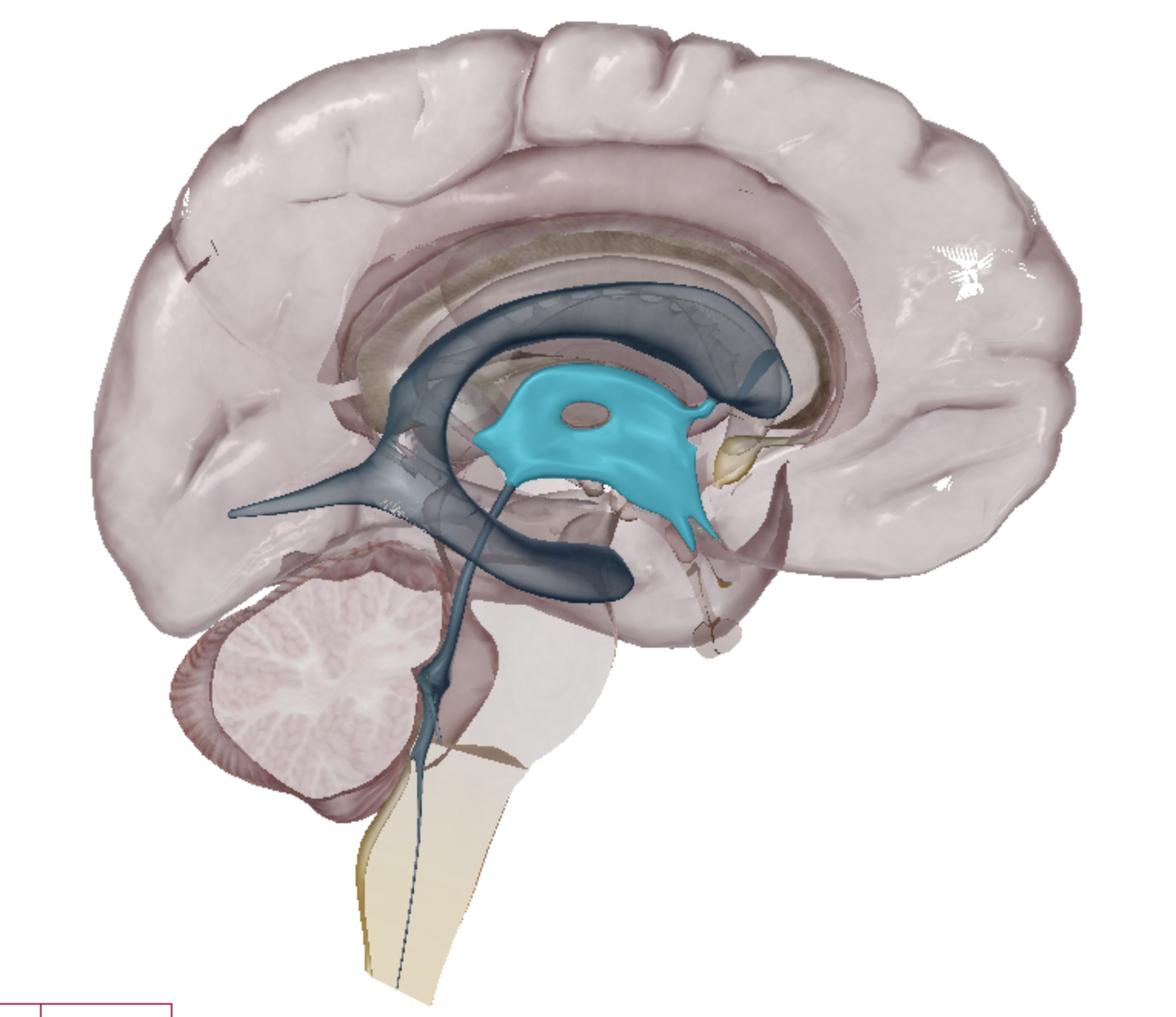
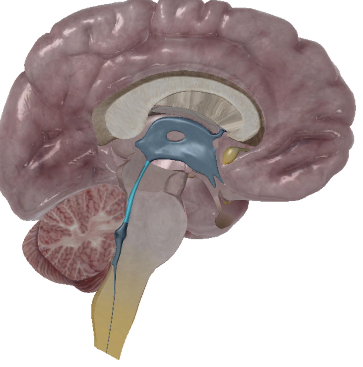
what is highlighted
third ventricle
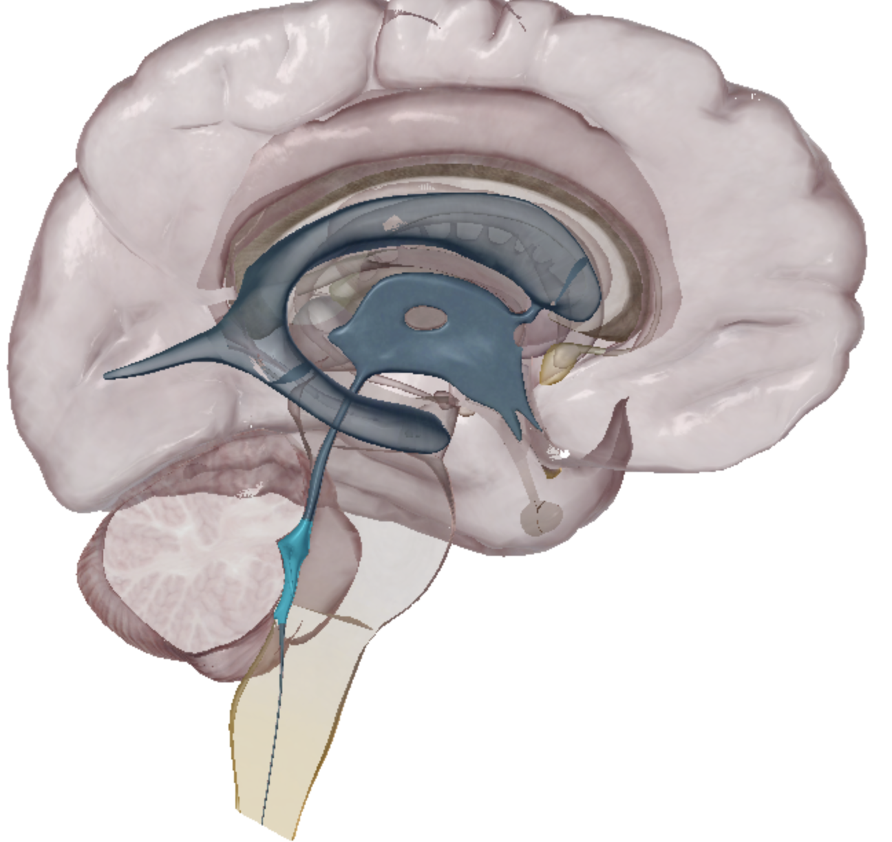
what is highlighted
fourth ventricle
what is highlighted
cerebral aqueduct
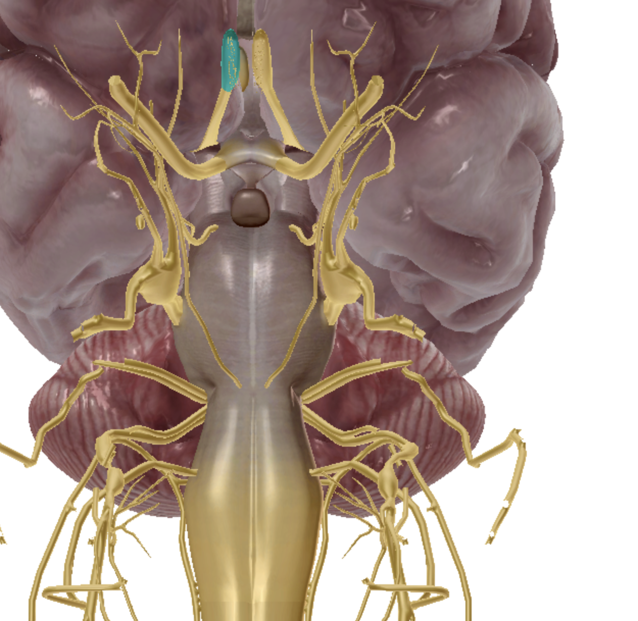
what is highlighted
olfactory CN I
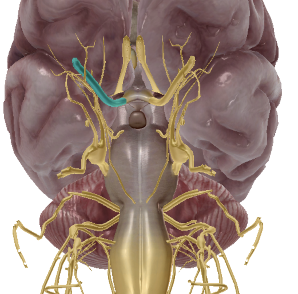
what is highlighted
optic CN II
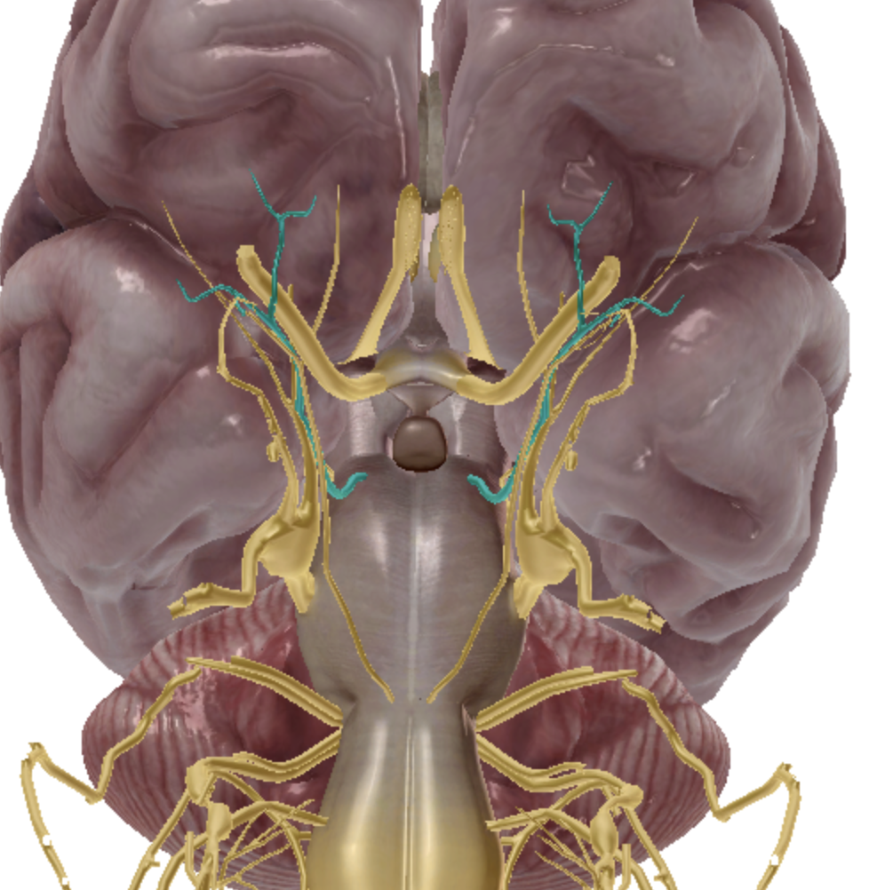
what is highlighted
oculomotor CN III
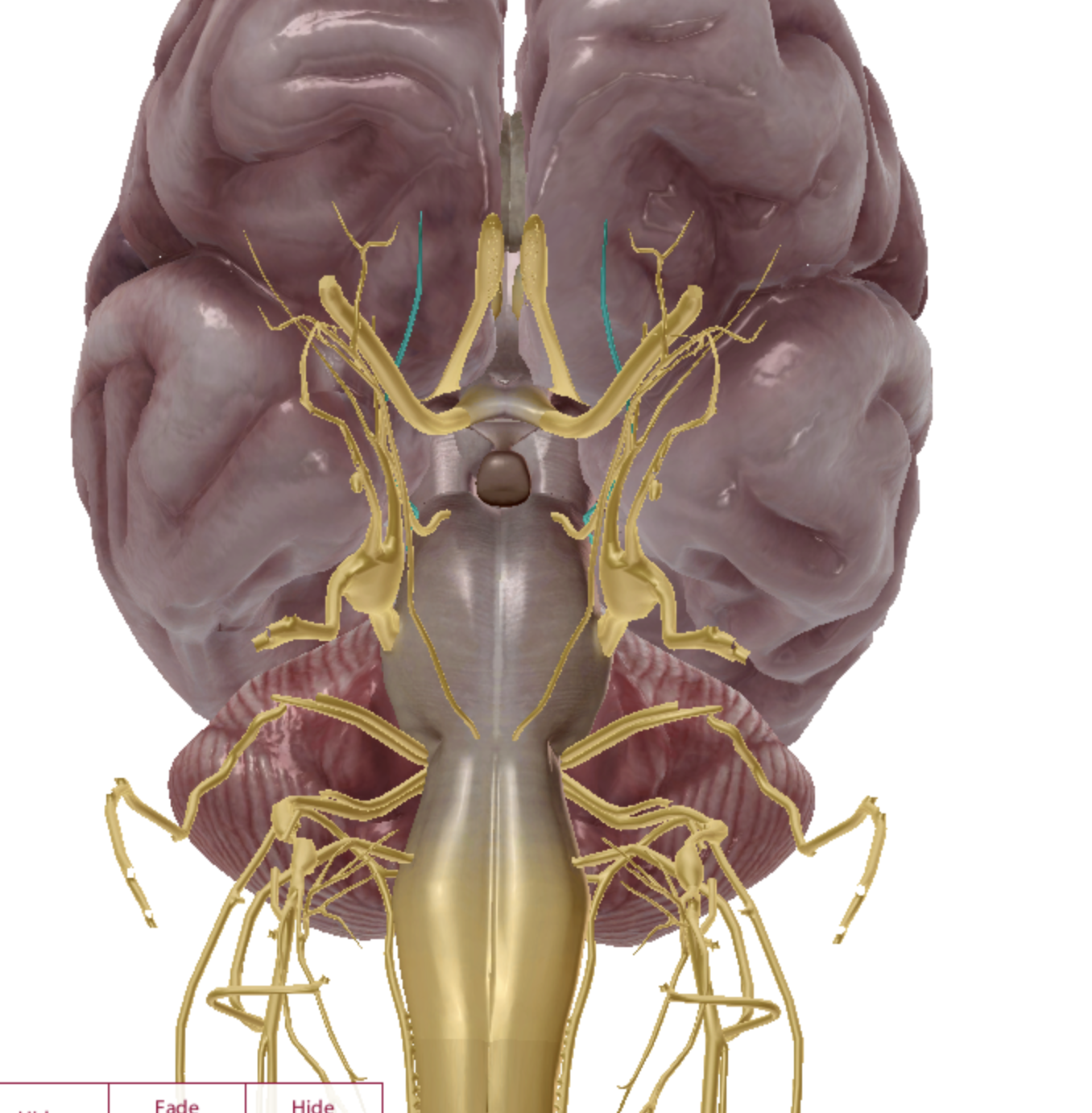
what is highlighted
trochlear CN IV

what is highlighted
Trigeminal CN V
what is highlighted
abducens CN VI
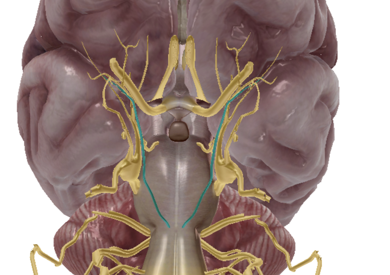
what is highlighted
facial CN VII
what is highlighted
vestibulocochlear CN VIII
what is highlighted
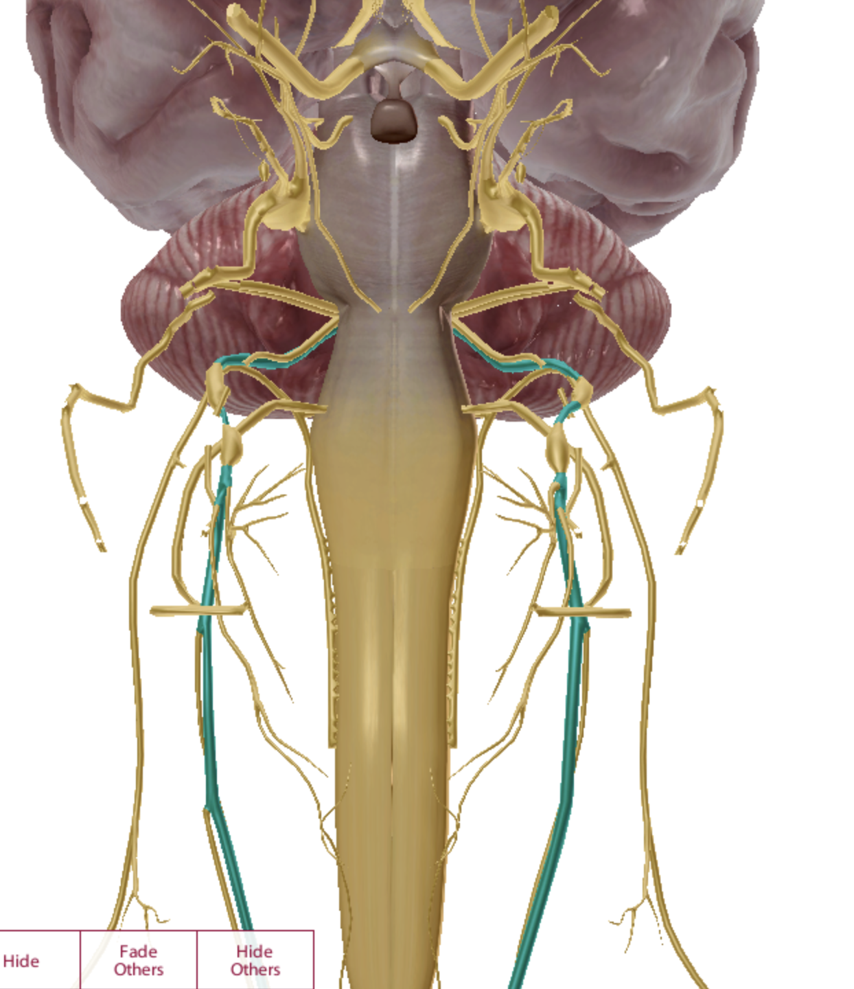
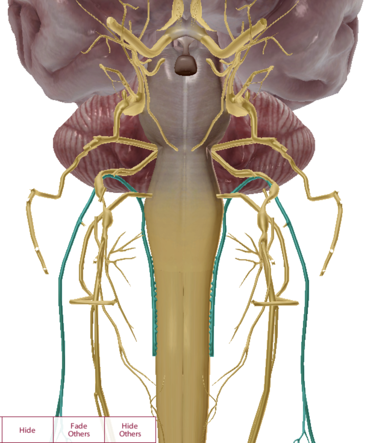
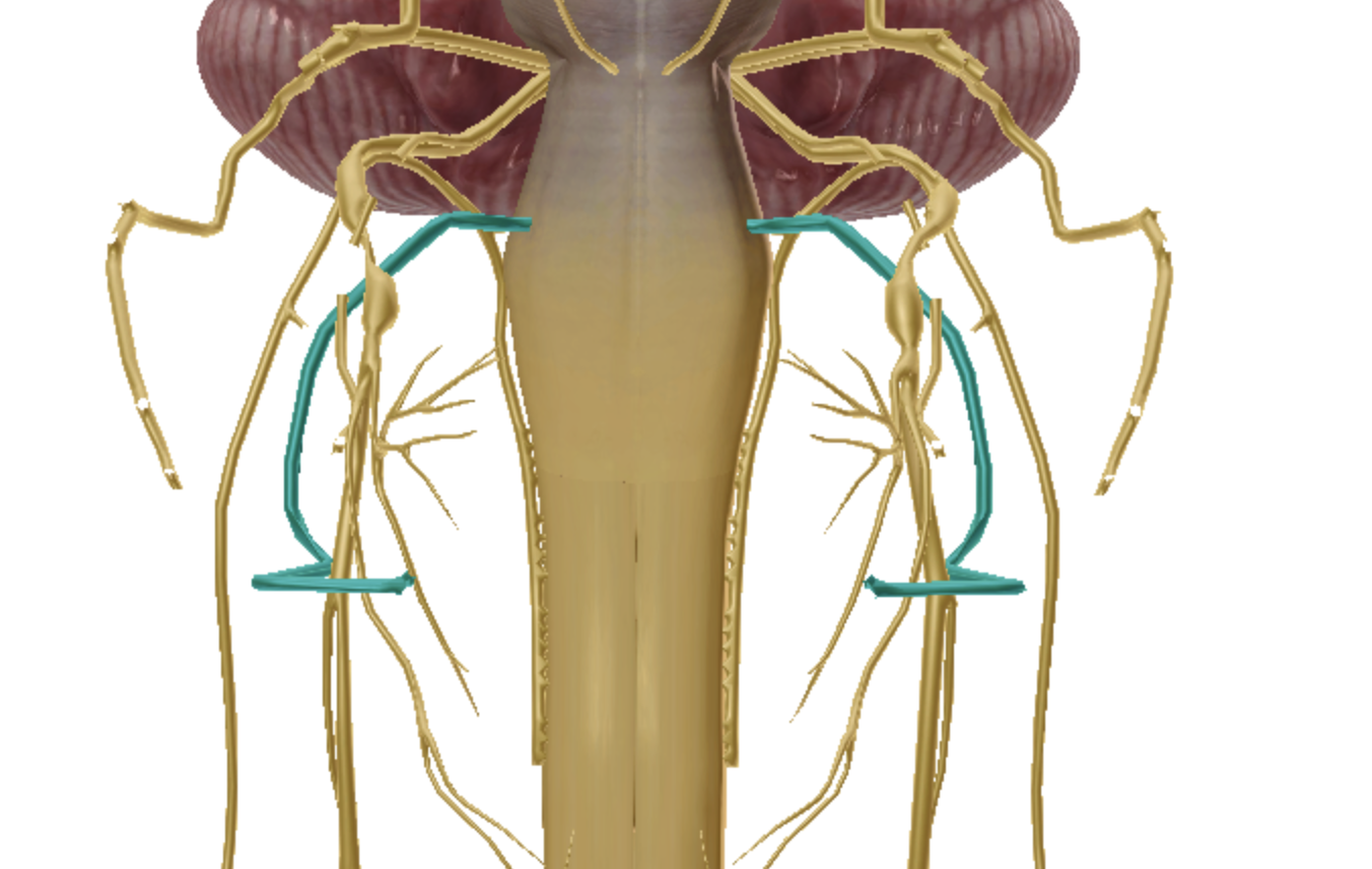
glossopharyngeal CN IX
what is highlighted
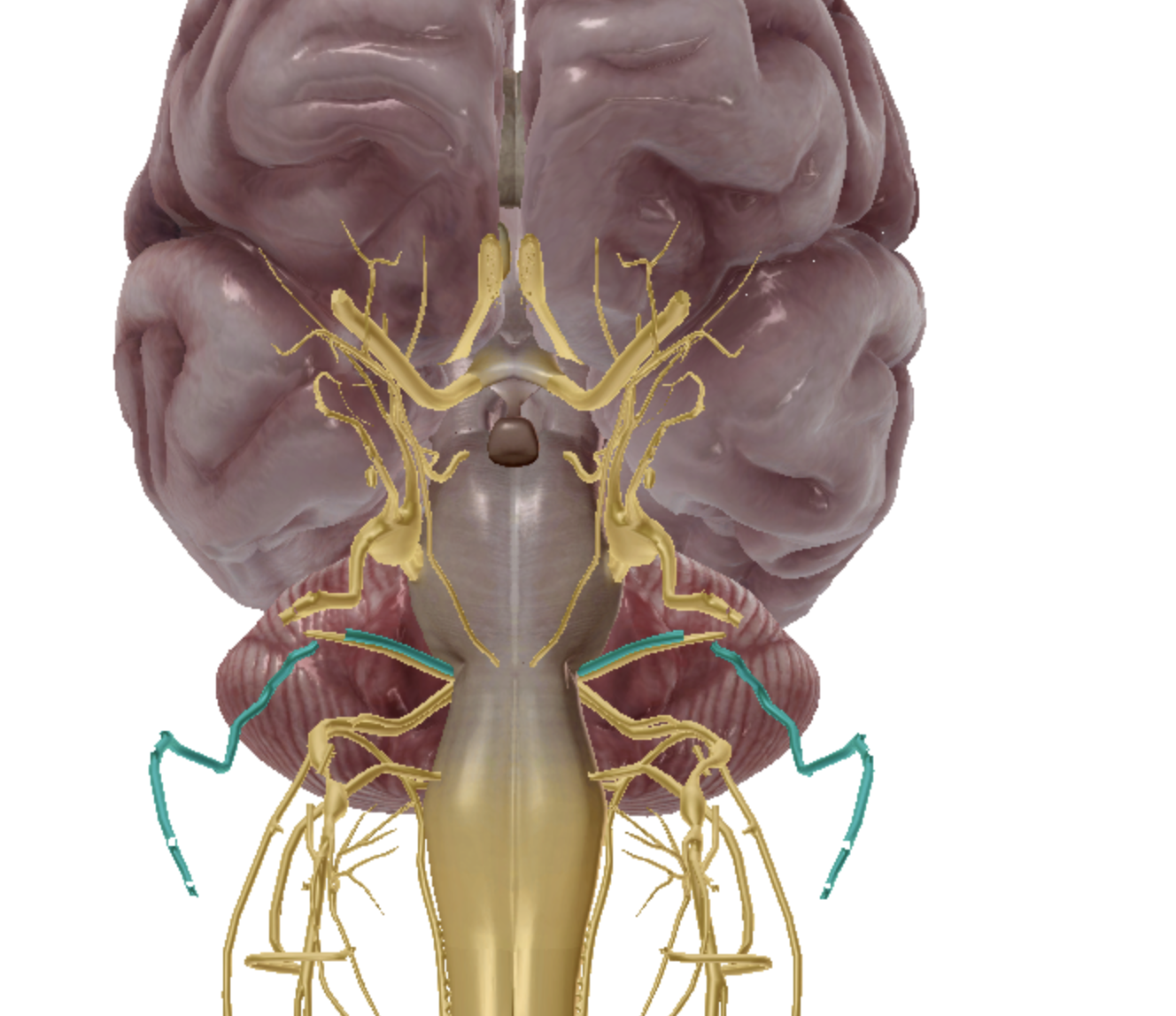
vagus CN X
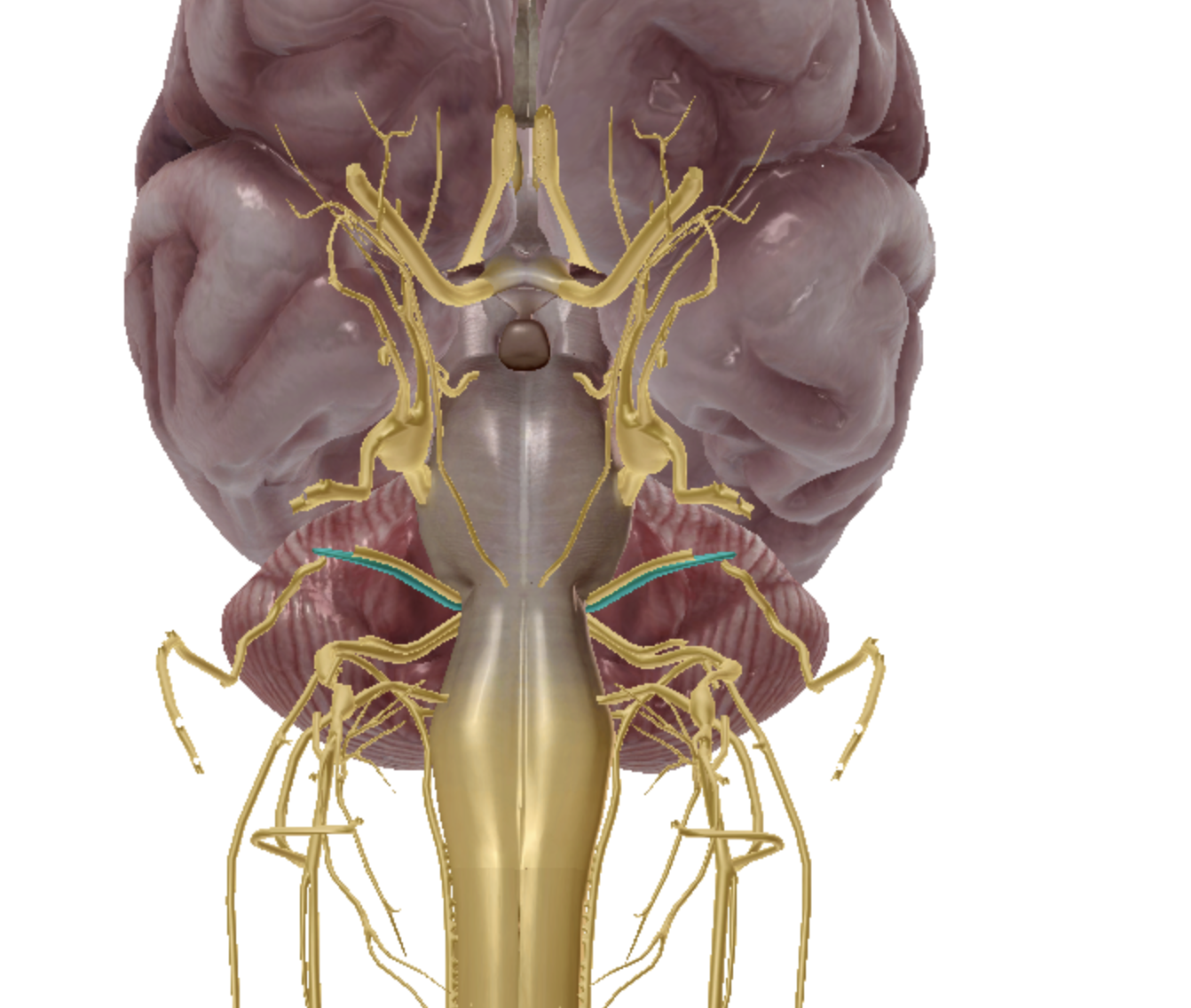
what is highlighted
spinal accessory CN XI
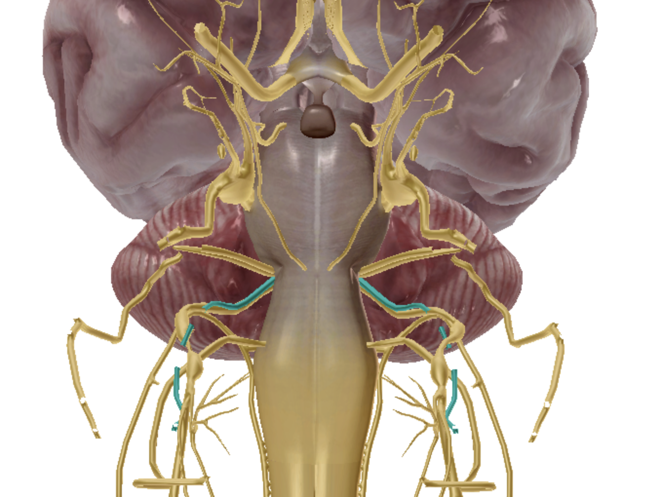
what is highlighted
hypoglossal CN XII
what is highlighted
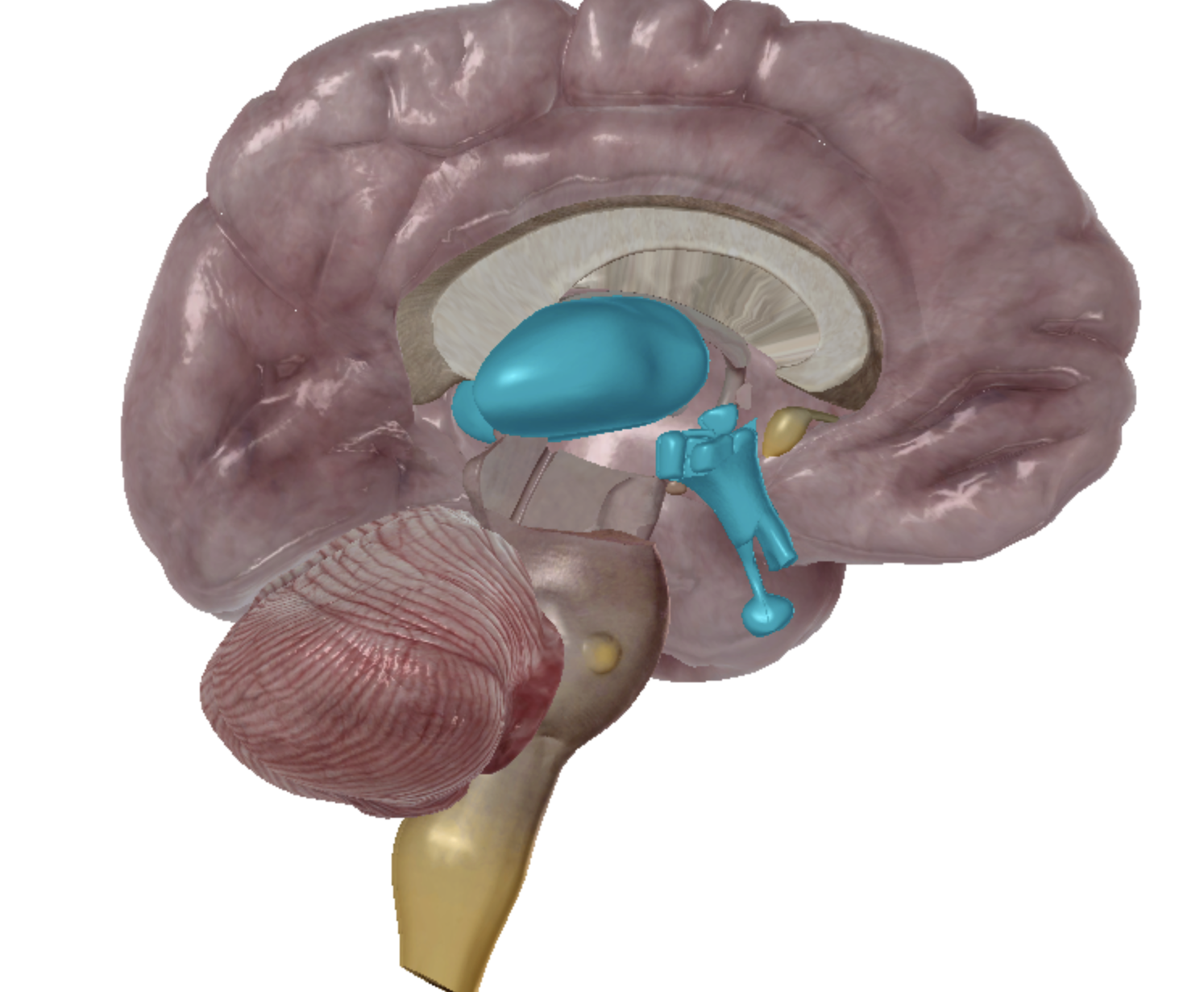
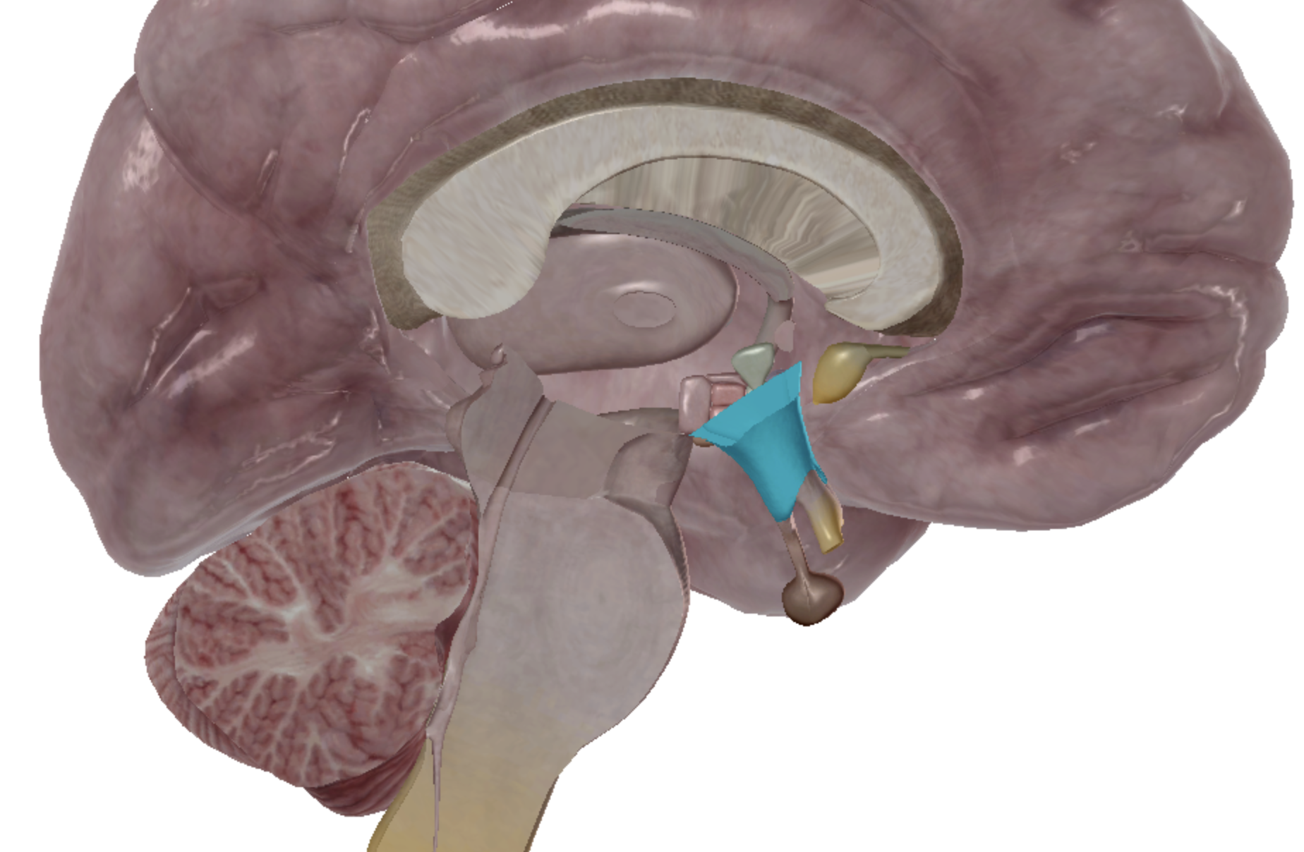
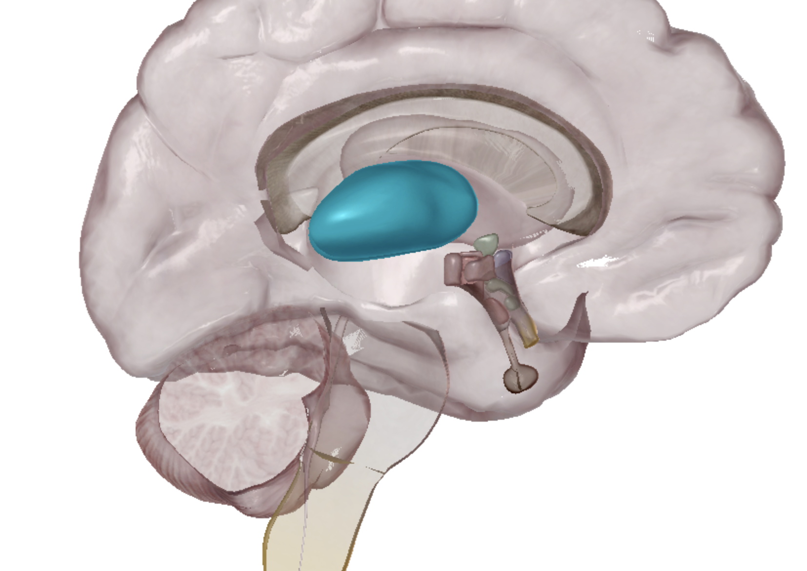
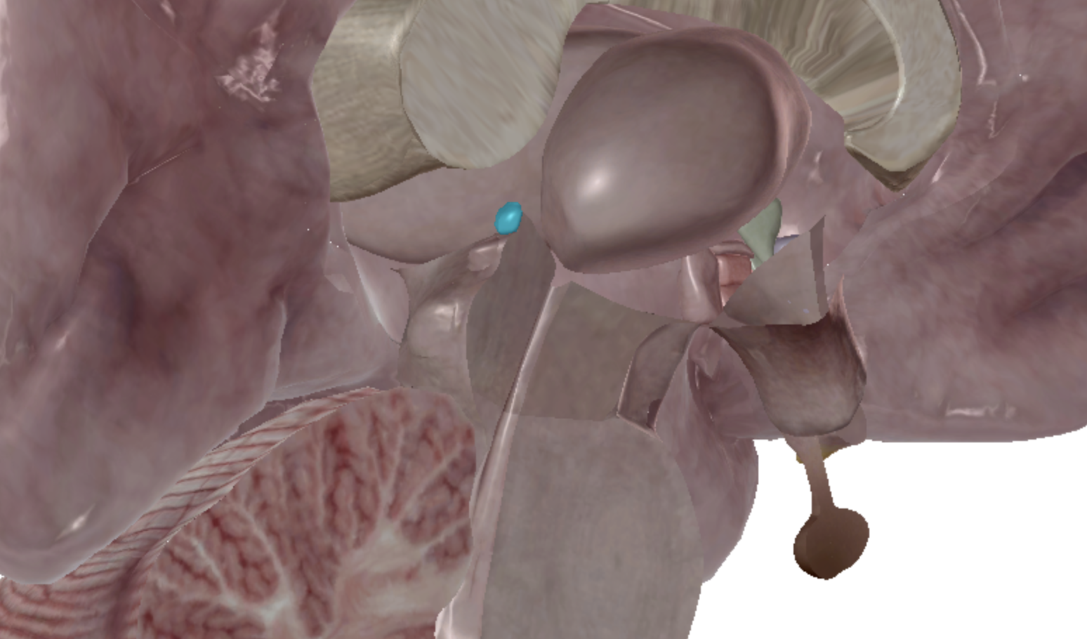
hypothalamus
what is highlighted
thalamus
what is highlighted
pineal gland
what is highlighted
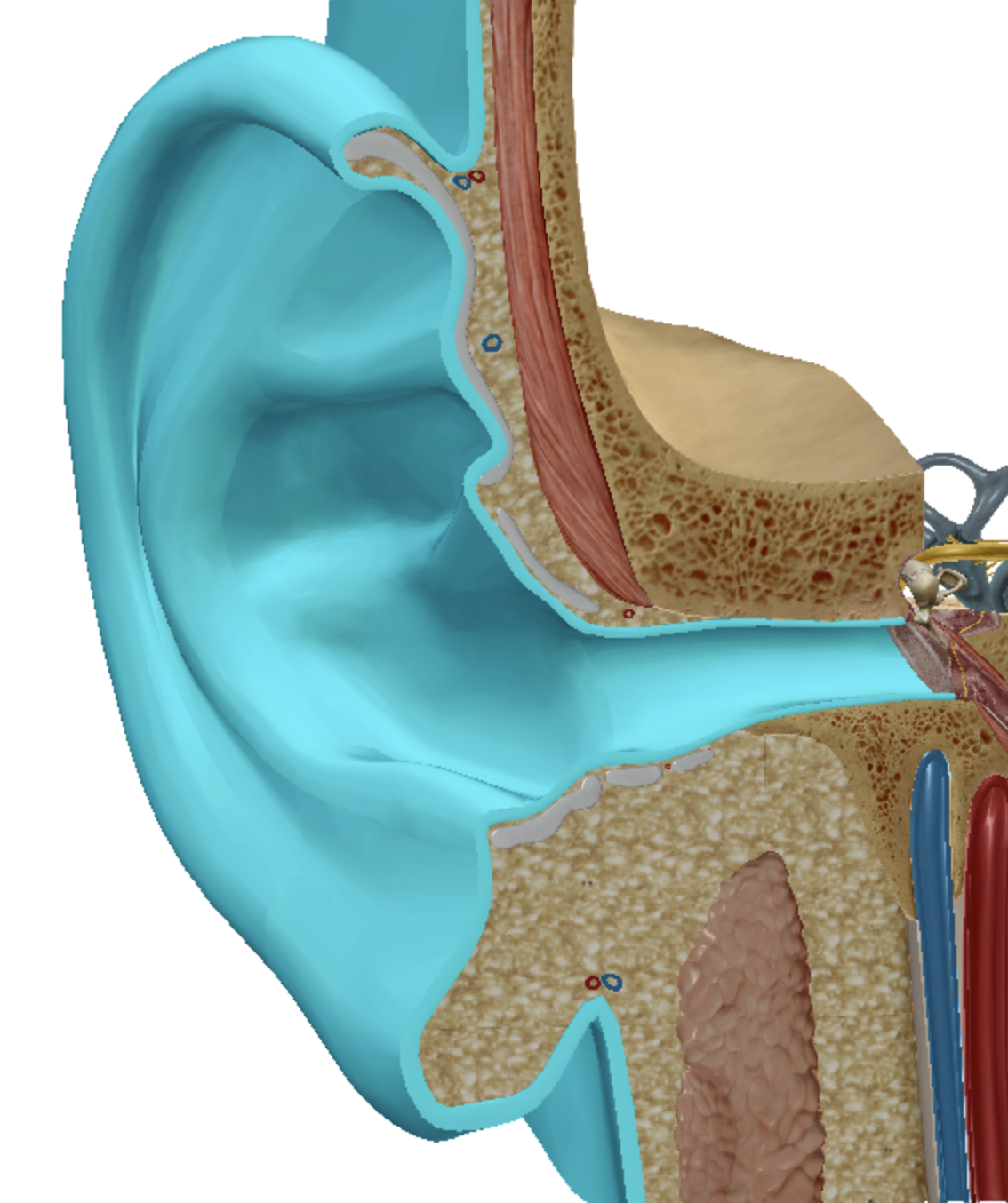
outer ear
what is highlighted
tympanic membrane
what is highlighted
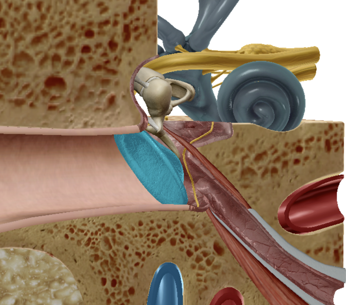
middle ear
what is highlighted
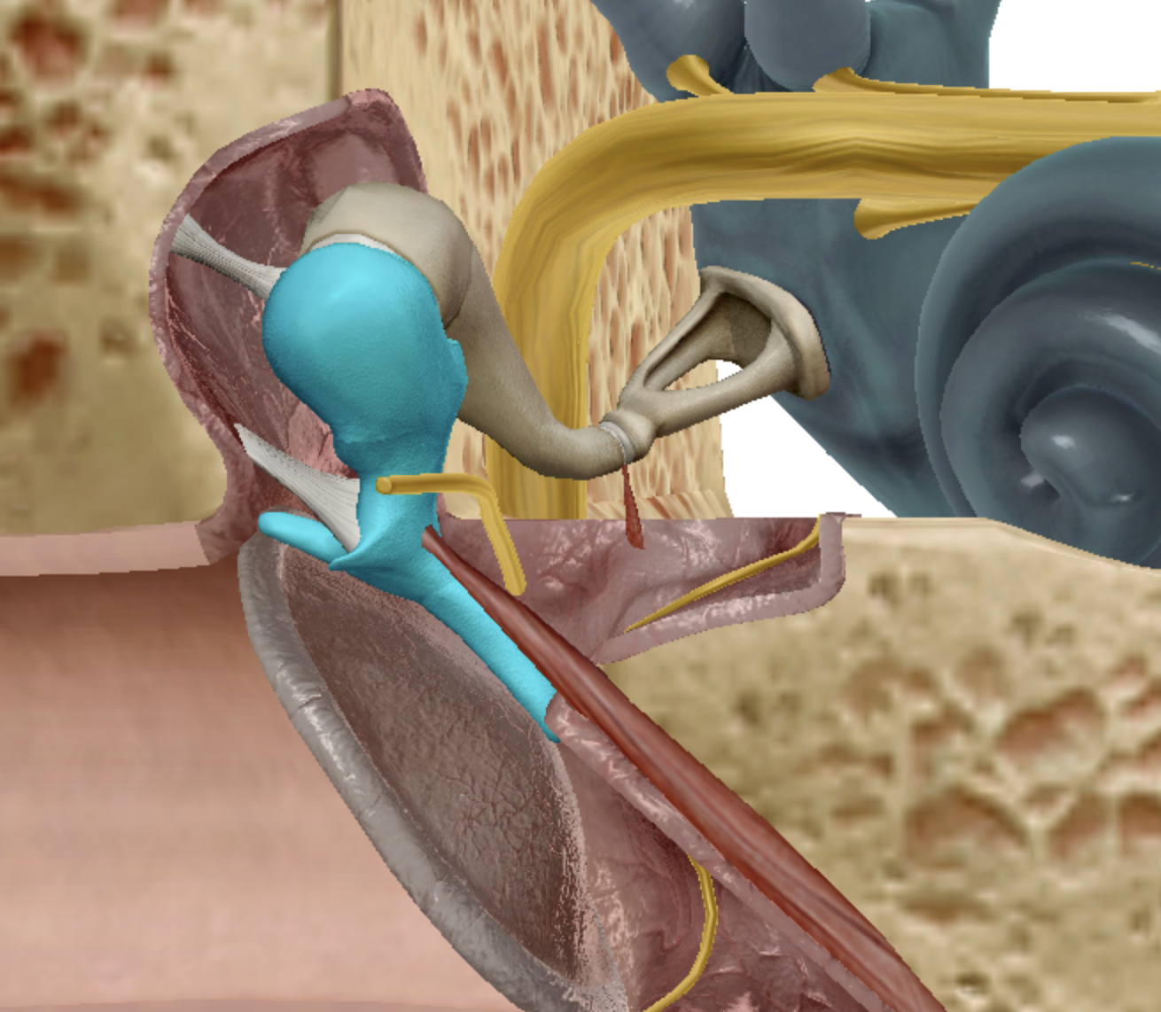
malleus
what is highlighted
incus
what is highlighted
stapes
what is highlighted
inner ear
what is highlighted
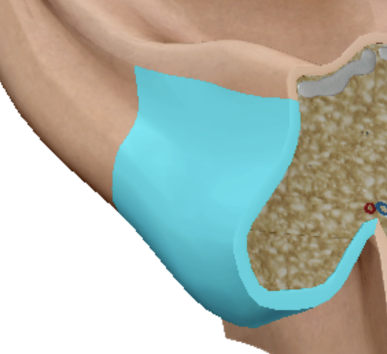
lobule
what is highlighted
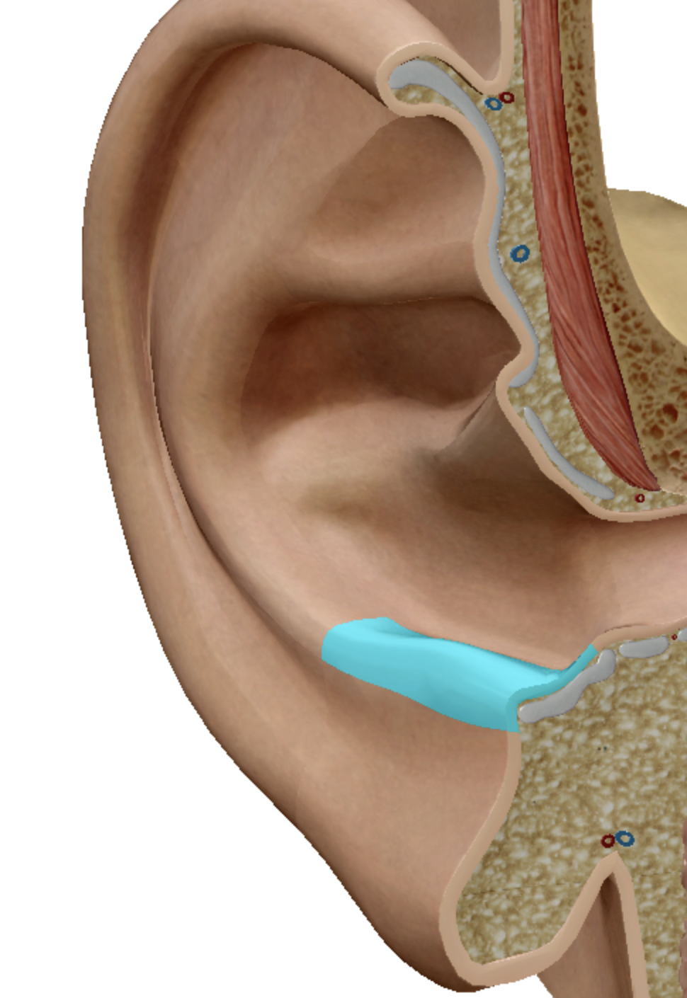
antitragus
what is highlighted
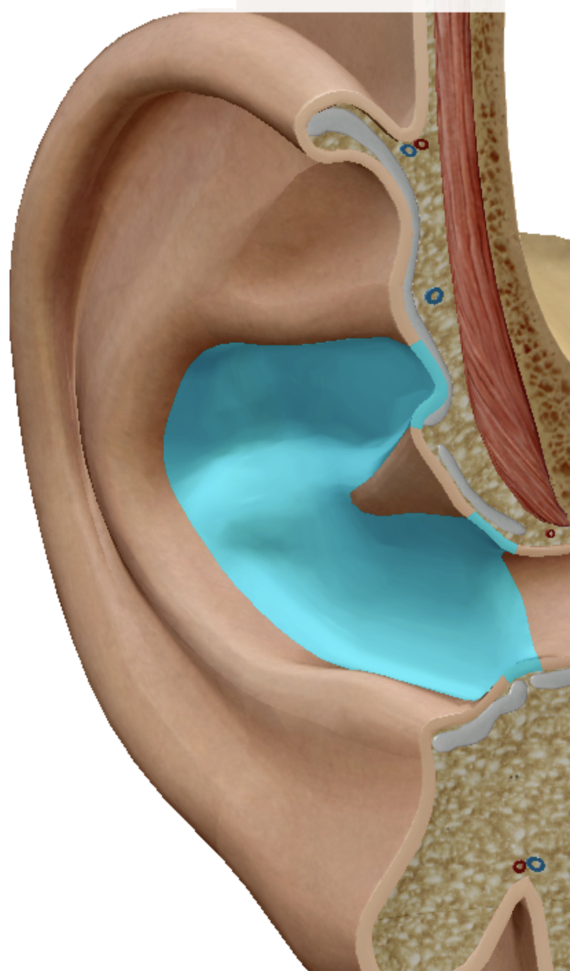
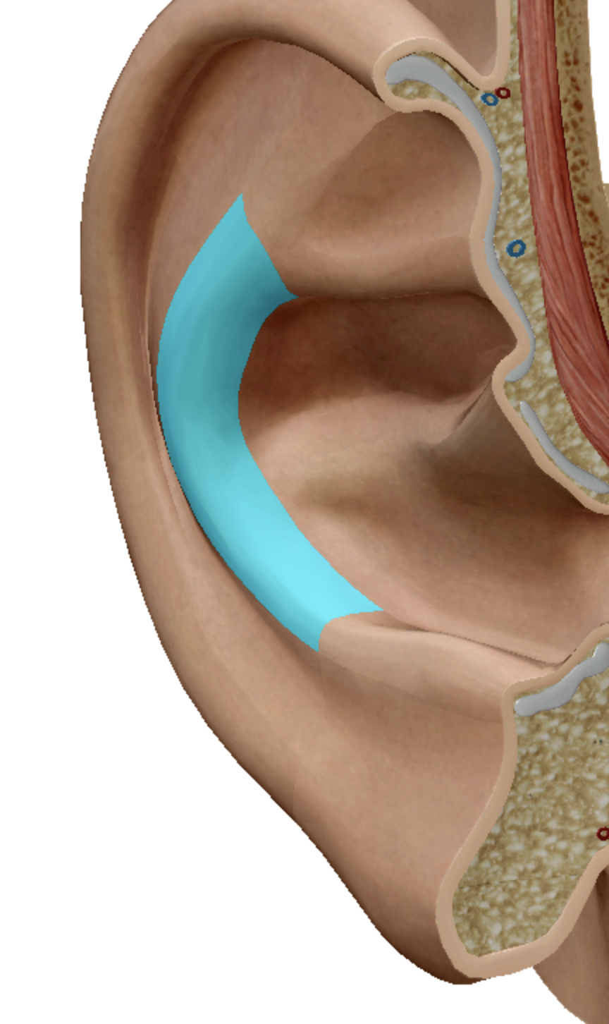
concha
what is highlighted
antihelix
what is highlighted
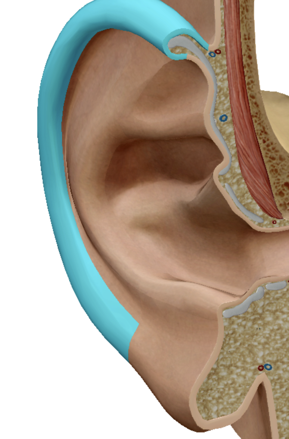
helix
what is highlighted
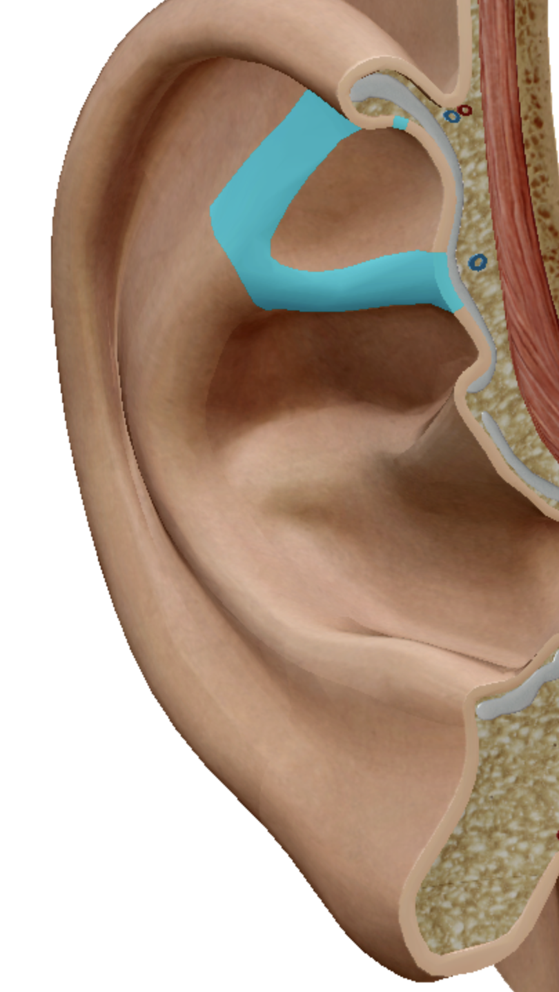
crura of antihelix
what is highlighted
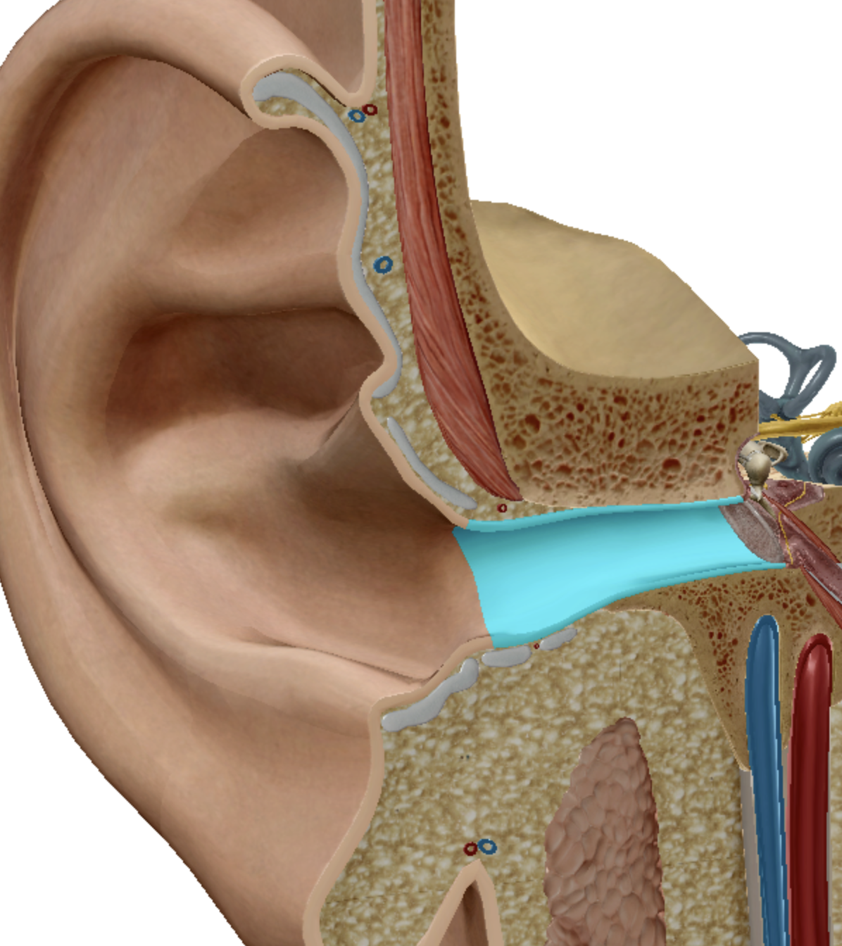
meatus
what is highlighted
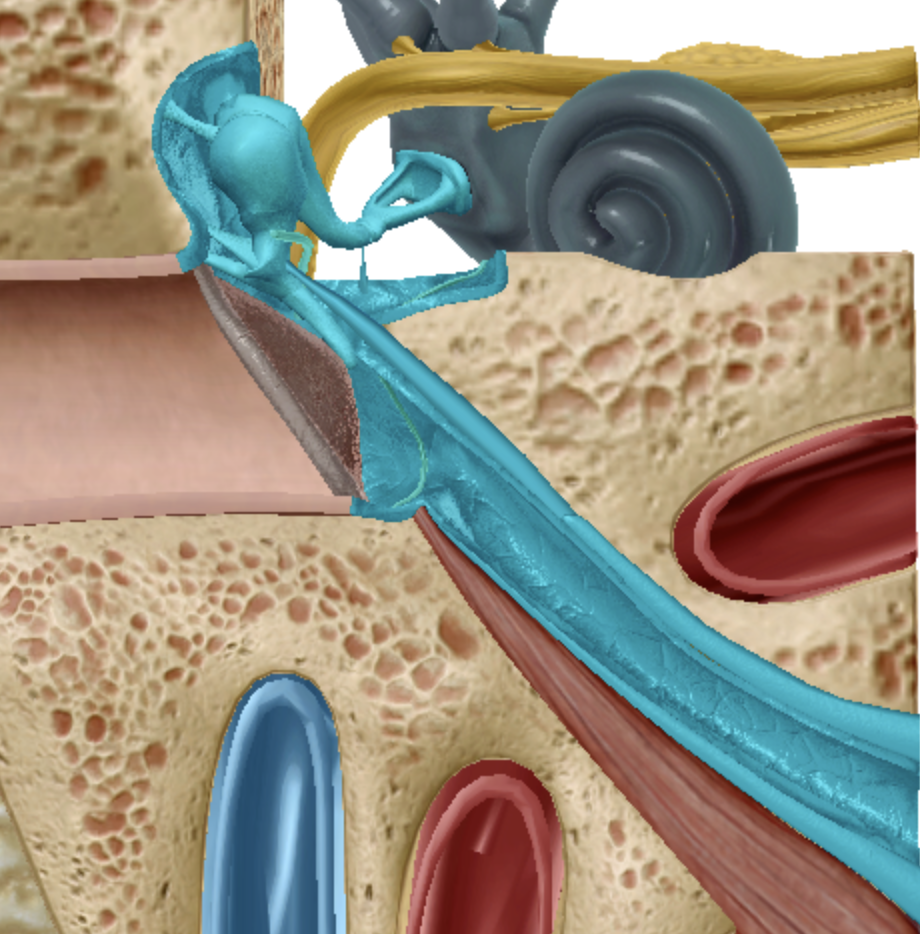
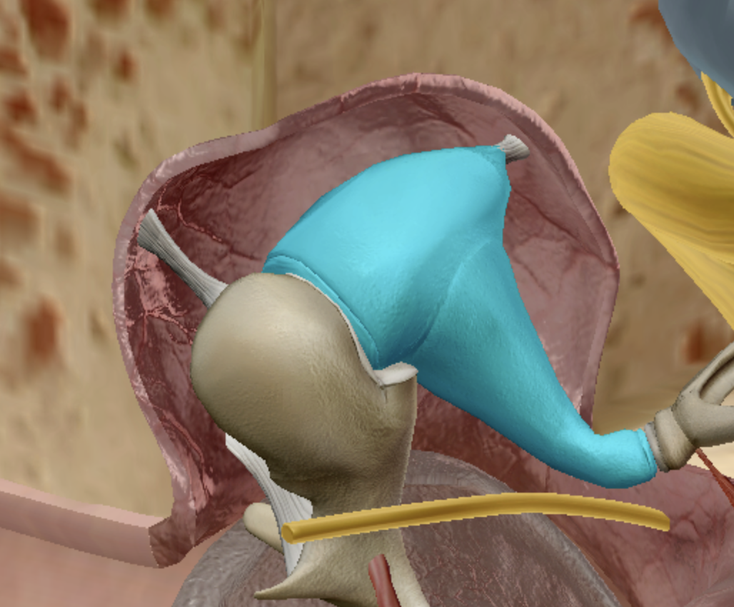
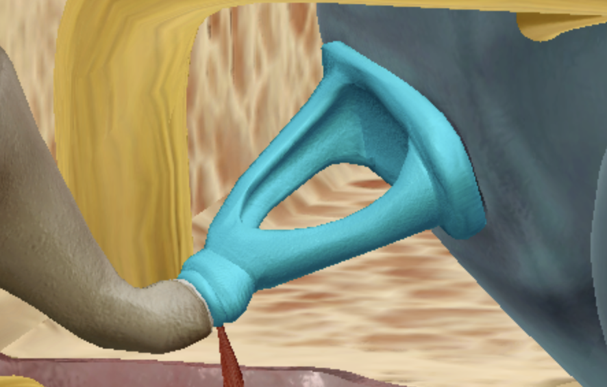
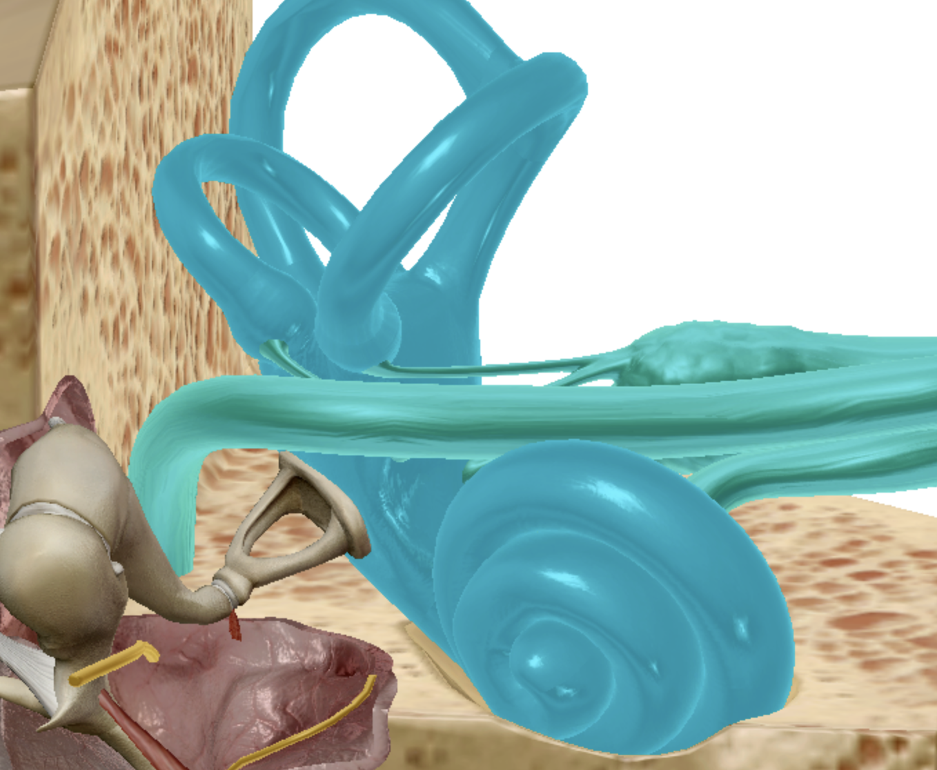
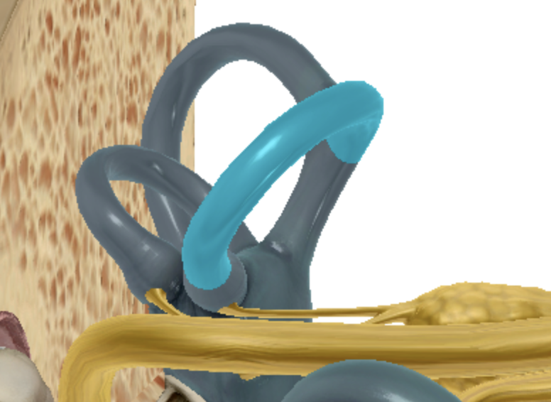
superior semicircular canal
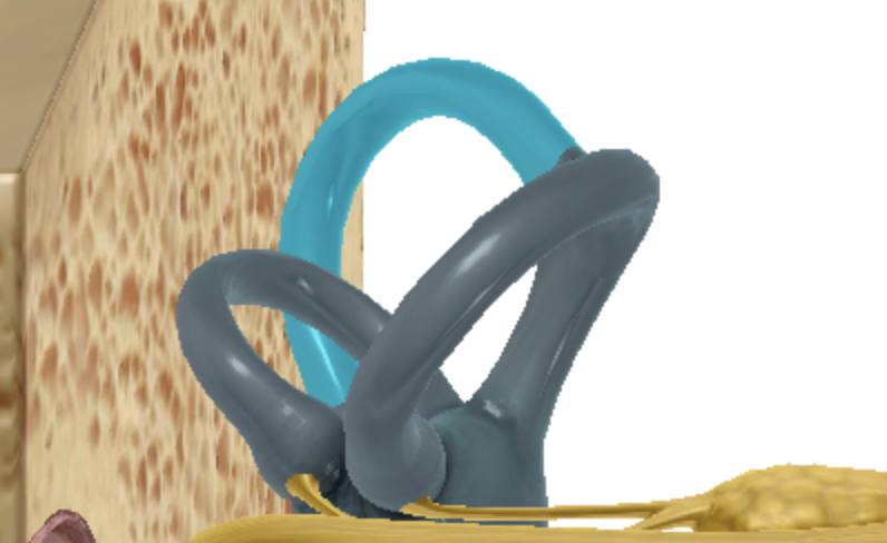
what is highlighted
posterior semicircular canal
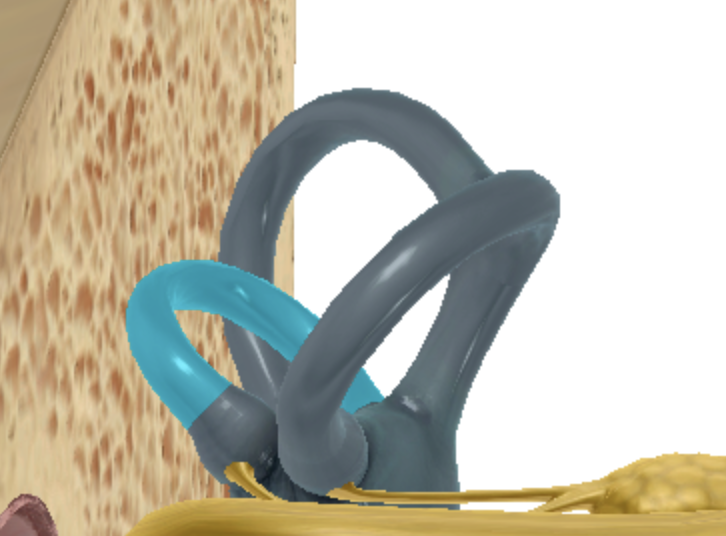
what is highlighted
lateral semicircular canal
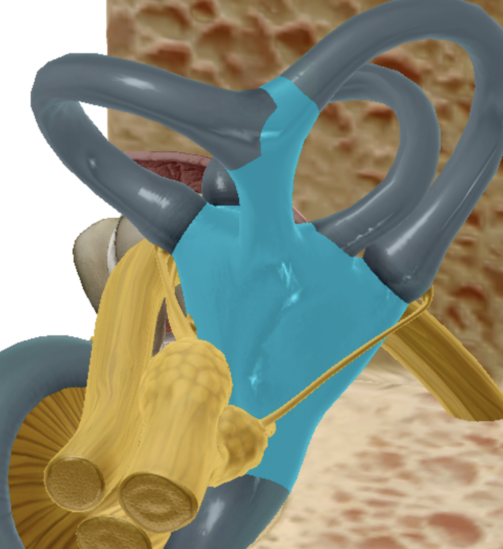
what is highlighted
vestibule
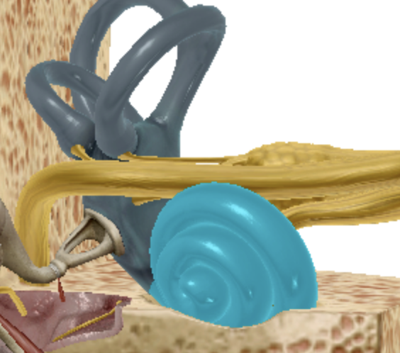
what is highlighted
cochlea
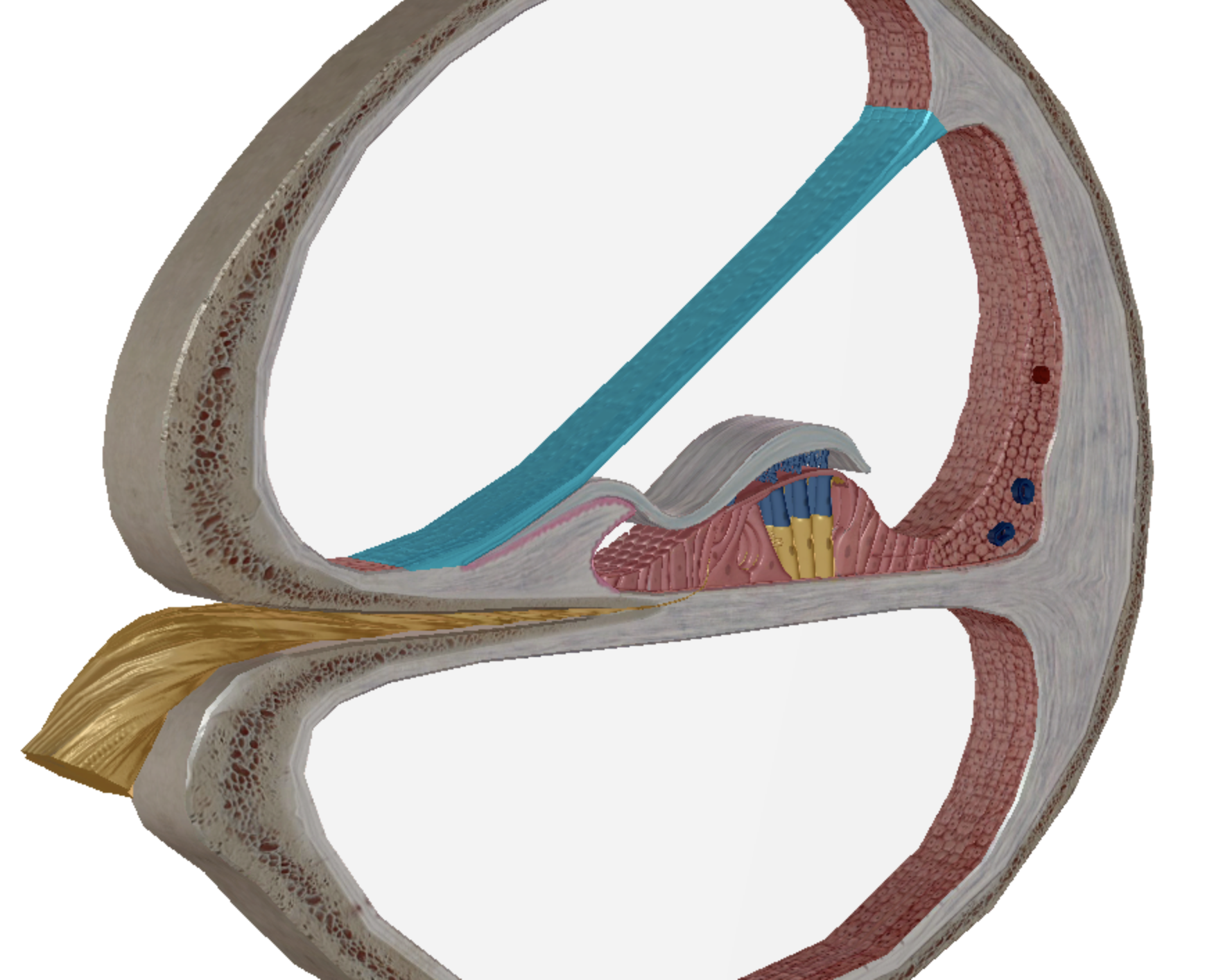
label what is highlighted
vestibular membrane
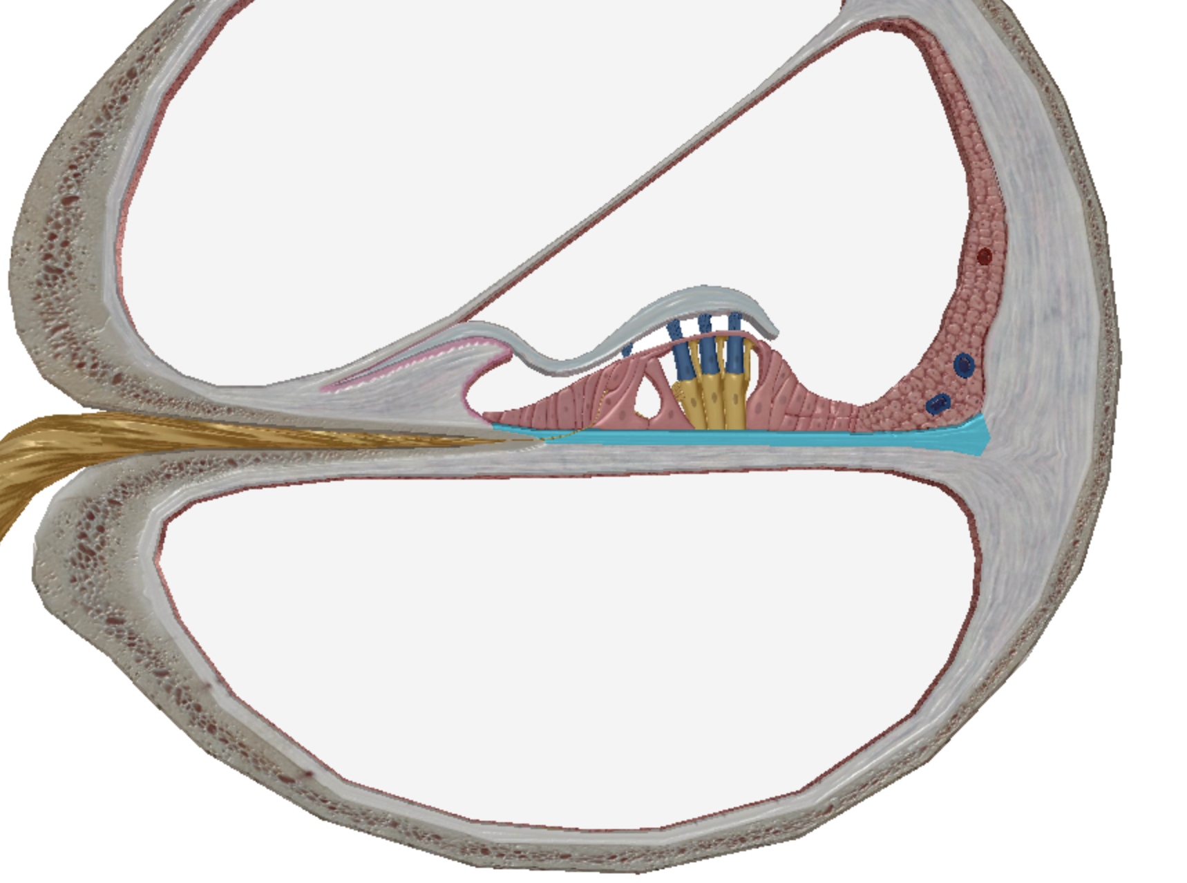
what is highlighted
basilar membrane
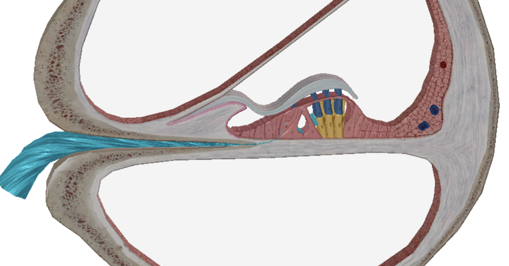
what is highlighted
cochlear nerve
what is highlighted
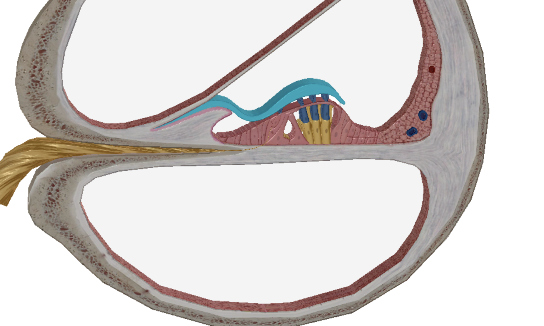
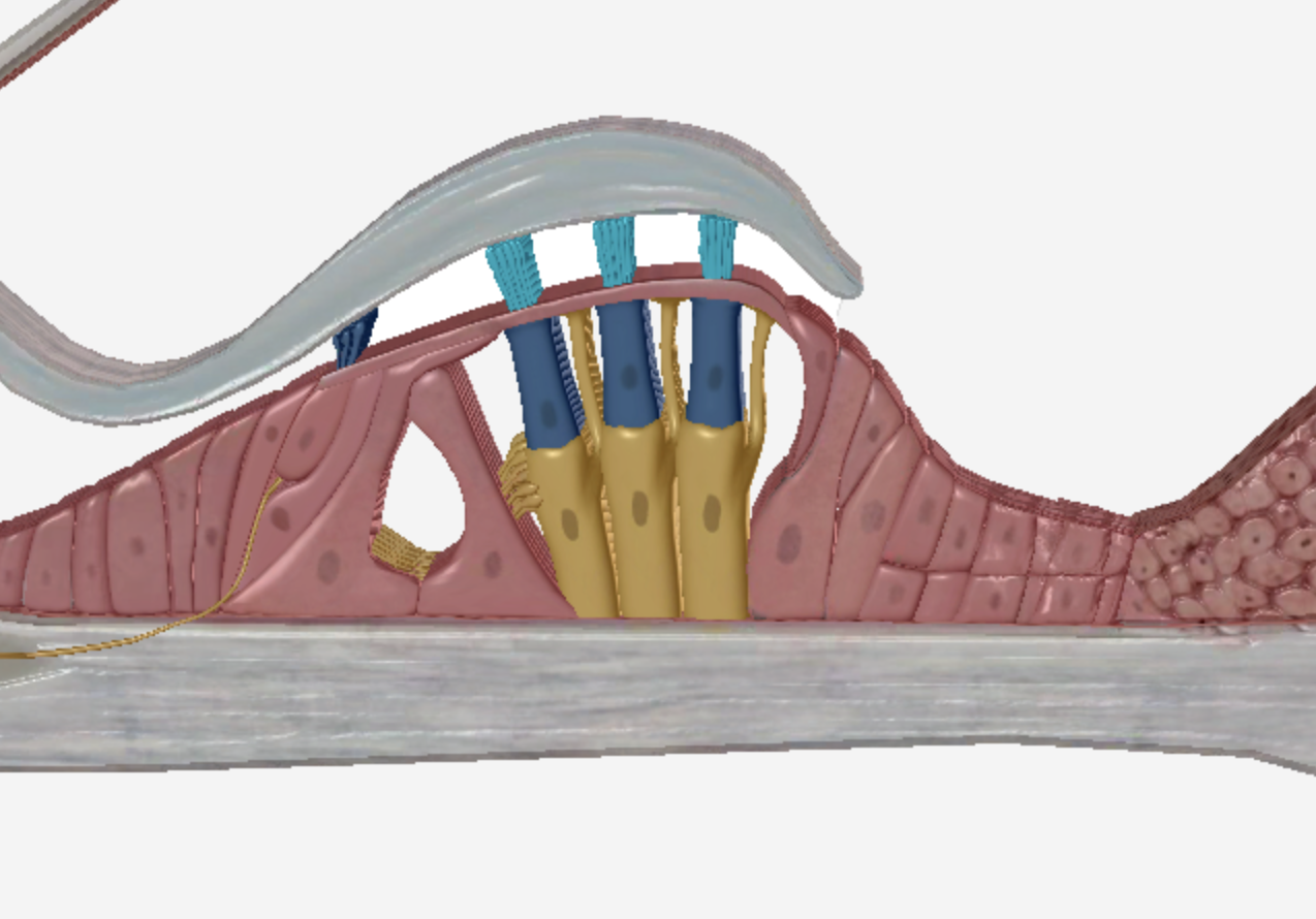
tectorial membrane
what is highlighted
hair cells
what is highlighted
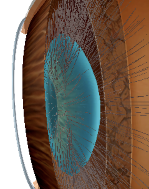
cornea
what is highlighted
lens
what is highlighted
retina
what is highlighted
ciliary muscles