Respiratory System
1/23
There's no tags or description
Looks like no tags are added yet.
Name | Mastery | Learn | Test | Matching | Spaced | Call with Kai |
|---|
No analytics yet
Send a link to your students to track their progress
24 Terms
Function of the respiratory system
Transport air into and out of the lungs
Allow O2 to diffuse from the lungs into the bloodstream
Remove CO2 from the bloodstream and expel it from the body
Non-vital function
Detecting odours
Speech production
Respiration system
Respiration system can be divided into 2 zones
Conducting zone
Respiratory zone
Conducting zone
Includes the organs and structures that transport air to the lungs but are not directly involved in gas exchange
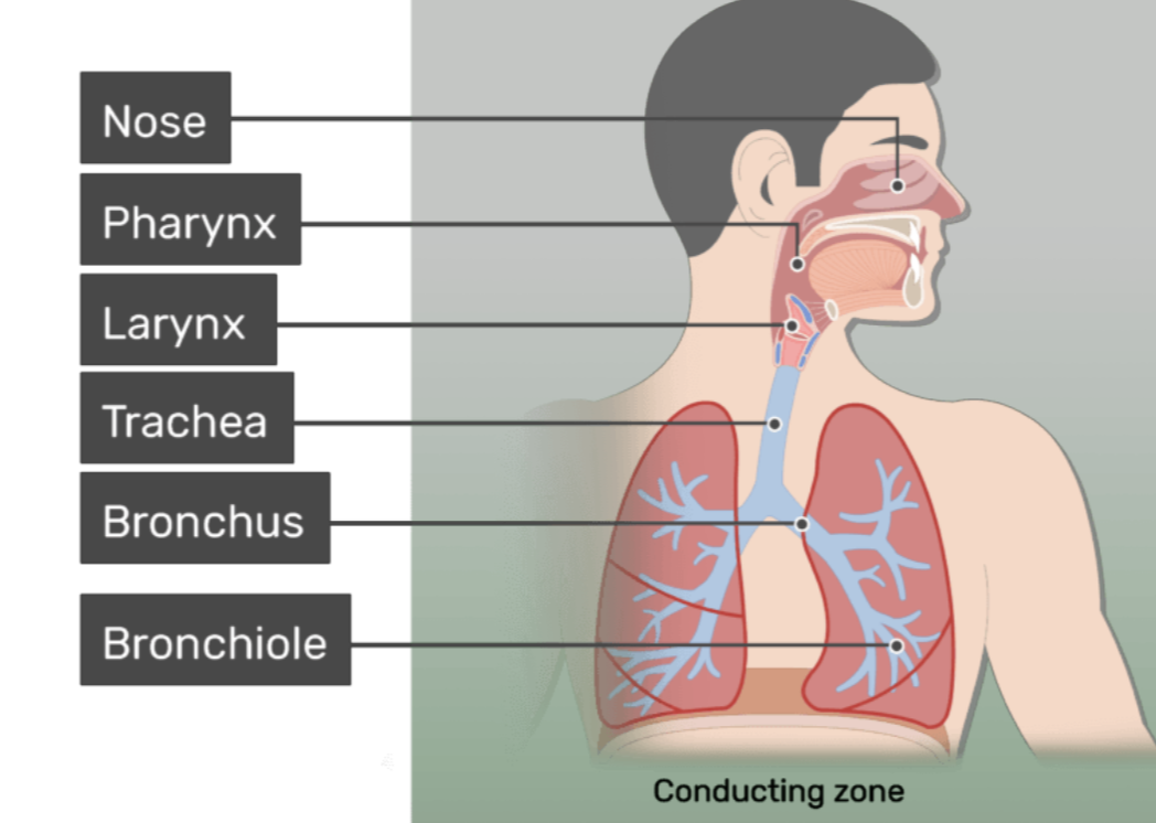
Respiratory zone
Where gas exchange occurs
Includes:
Respiratory bronchioles
Alveolar ducts
Alveoli
These structures allow O2 and CO2 to diffuse between the lungs and the bloodsteam
The journey of air
Nasal cavity → Pharynx → Larynx → Trachea → Bronchi → Bronchioles → Alveoli
Each structure has specialised features that help prepare air for gas exchange
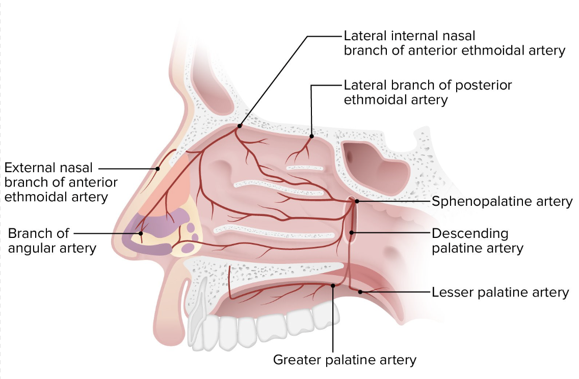
Nasal cavity
Air usually enters the respiratory system through the nostrils, which lead into the nasal cavity
Nasal cavity functions
Filters the air
warm the air
Moistens the air
These processes protect the delicate tissues of the lungs
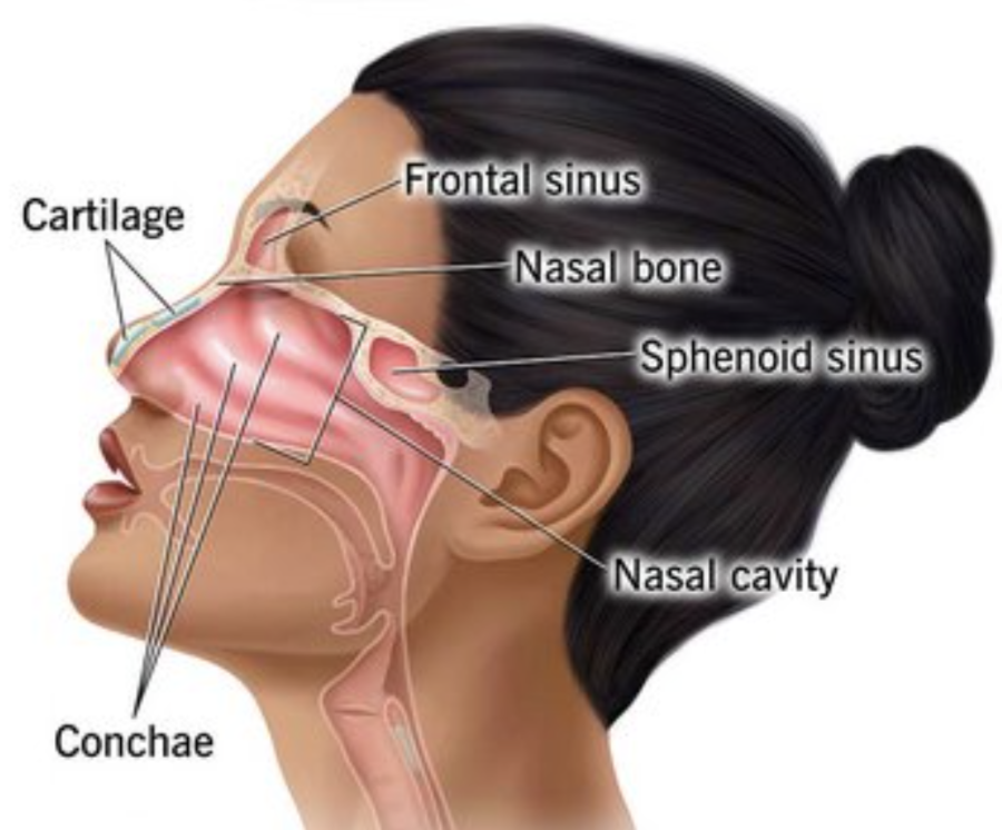
Structure of the nasal cavity
Divided into 2 chambers by nasal septum
Inside the nasal cavity are thre curved bony structures called nasal conchae ( turbinates )
These structures:
Increase the surface area
Create turbulence in airflow
Allow air to contact the lining of the nasal cavity
This improves the filtering, warming, and moistening of air
The nasal cavity also contains paranasal sinuses, which are air-filled spaces within the surrounding bones that help warm and misten incoming air
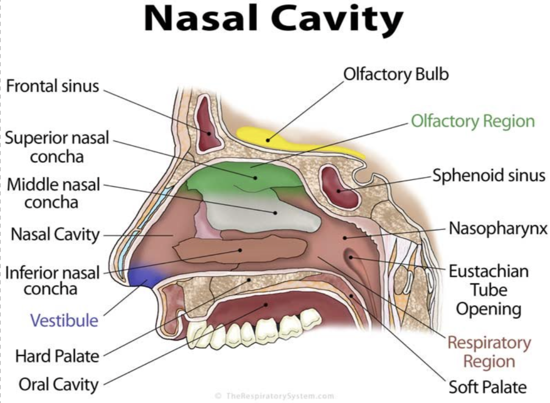
Nasal cavity lining
Lined with a mucous membrane containing specialised calls.
Goblet cells produce mucus, which traps:
Dust
Pollen
Microorganisms
Cilia which are hair-like projections that beat continously, moving mucus and trapped particles toward the throat where they can be swallowed
Blood supply
A dense network of capillaries beneath the lining helps warm incoming air
Oral cavity ( mounth )
Air can also enter through mouth:
Not filtered as effectively
Not warmed as efficiently
Not moistened as well
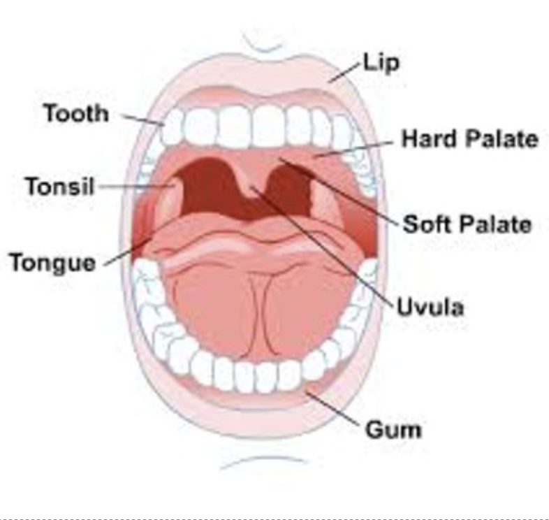
Pharynx ( throat )
A muscular passageway that connects the nasal cavity to the larynx
It allows the passage of:
Air
Food
Liquids
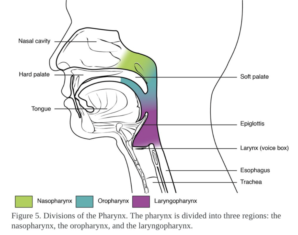
Nasopharynx ( region of the pharynx )
Located behind the nasal cavity
Lined with ciliated mucous membrane
Cilia move mucus and trapped particles toward the throat
Oropharynx ( region of the pharynx )
Located behind the nasal cavity
Lined with ciliated mucous membrane
Cilia move mucucs and trapped particles toward the throat
Laryngopharynx ( region of the pharynx )
The lowest region of the pharynx
Directs air toward the larynx
Directs food and liquids toward the oesophagus
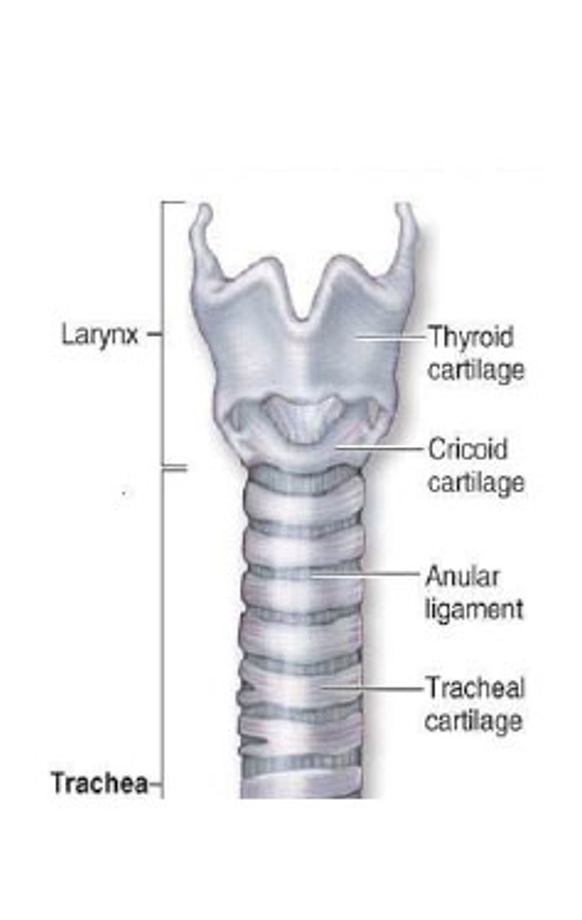
Larynx ( voice box )
Made of 9 cartilages
Epiglottis is one of the cartilages and is composed of elastic cartilage
Epiglottis is a flap of elastic cartilage:
Closes over the larynx during swallowing
Prevents food and liquid from entering the airway
The lining of the larynx contains cilia and mucus- secreting goblet ells, which trap and remove debris
Larynx functions
Keeps the airway open
Produces sound
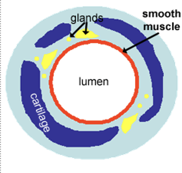
Trachea ( windpipe )
Carries air from the larynx to the bronchi
It is supported by 18-20 C-shaped rings of cartilage
These rings:
keep the airway open
prevent the trachea from collapsing
The open side of the cartilage rings contains smooth muscle, which allows the trachea to slightly change diameter
The lining of the trachea contains:
Goblet cells that produce mucus
Cilia that move mucus upward toward the pharynx
→ Remove dust and pathogens
Bronchi
Lower end of the trachea, it divides into 2 tubes called the bronchi
The right bronchus enters the right lung
The left bronchus enters the left lung
Contain:
Cartilage for structural support
Ciliated epithelium
Mucus-producing goblet cells
Bronchial tree
Once inside the lungs, the bronchi divide repeatedly into smaller branches, forming the bronchial free
The main bronchi branch into lobar bronchi, each supplying a lobe of the lung
The right lung has 3 lobes
The left lung has 2 lobes
The left lung has fewer lobes due to the position of the heart
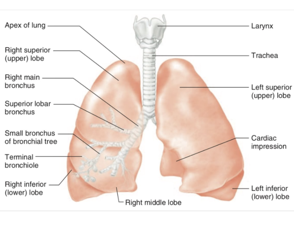
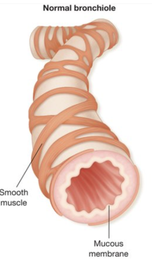
Bronchioles
The bronchi continue to divide into smaller tubes called bronchioles:
Approximately 1mm in diameter
Branch into smaller terminal bronchioles
Don’t contain cartilage
Have smooth muscle in their walls
Can contract, relax, controlling airflow through the lungs