Axial Skeleton - BYU Anatomy
1/111
There's no tags or description
Looks like no tags are added yet.
Name | Mastery | Learn | Test | Matching | Spaced | Call with Kai |
|---|
No analytics yet
Send a link to your students to track their progress
112 Terms
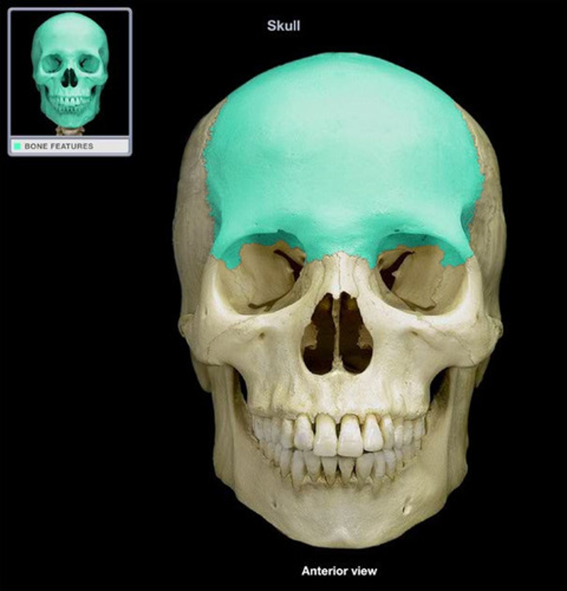
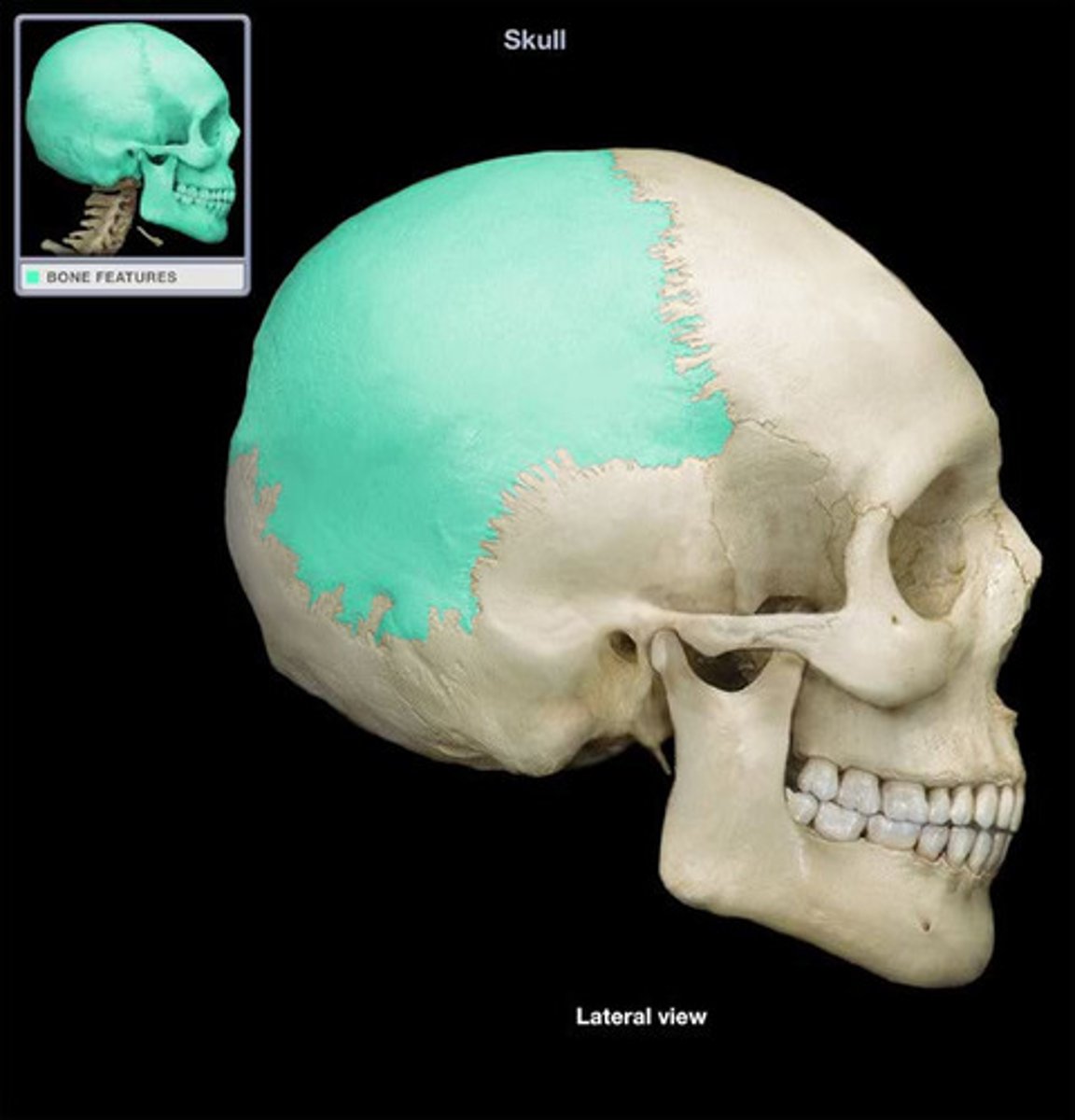
Frontal bone
bone that forms the forehead
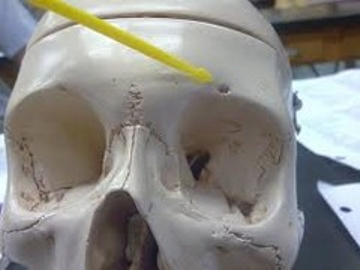
supraorbital notch
paired opening or notch superior to the orbit
superciliary arch
the ridge above the eye socket indicating the location of the frontal sinus

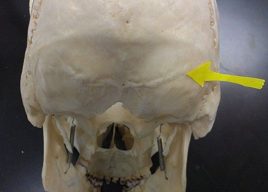
parietal bone
large paired bone located in the superior portion of the skull
coronal suture
runs laterally joining the frontal and parietal bones
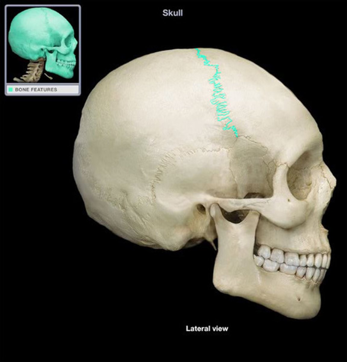
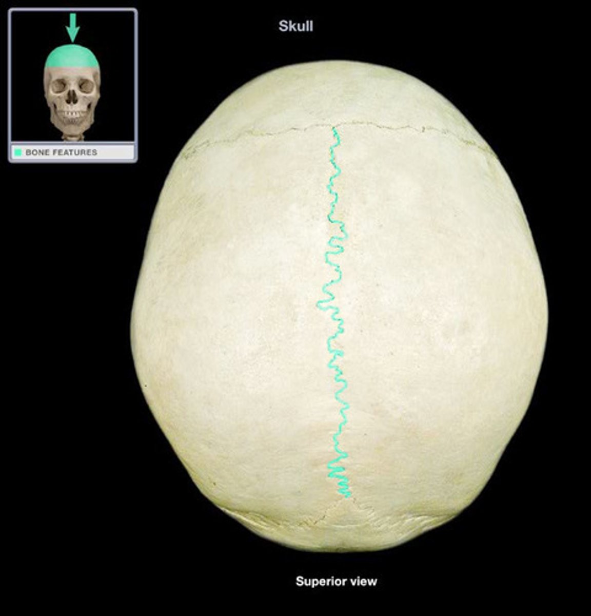
sagittal suture
runs anteroposteriorly joining the temporal and parietal bones
temporal bone
paired bone located on the lateral sides of the skull
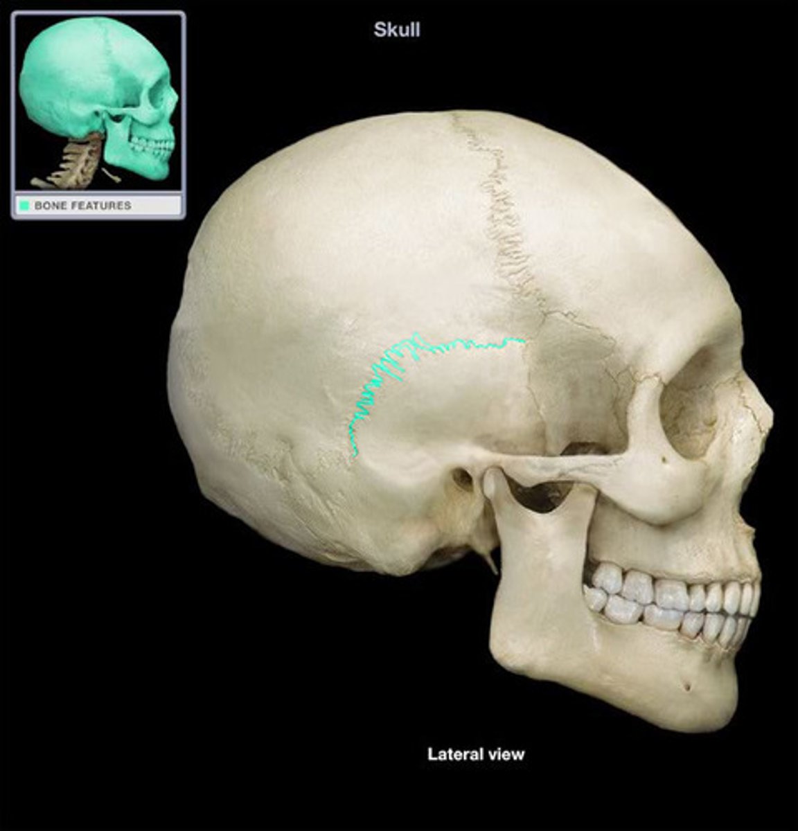
squamous suture
runs anteroposteriorly joining the temporal and parietal bones
mandibular fossa
articulates with the condylar process of the mandible
external auditory canal
opening in the temporal bone extending to the inner ear
petrous part
dense bony ridge on interior surface of temporal bone, location of middle and inner ear cavities
internal auditory canal
short canal in petrous part of temporal bone, extending laterally towards the inner ear.
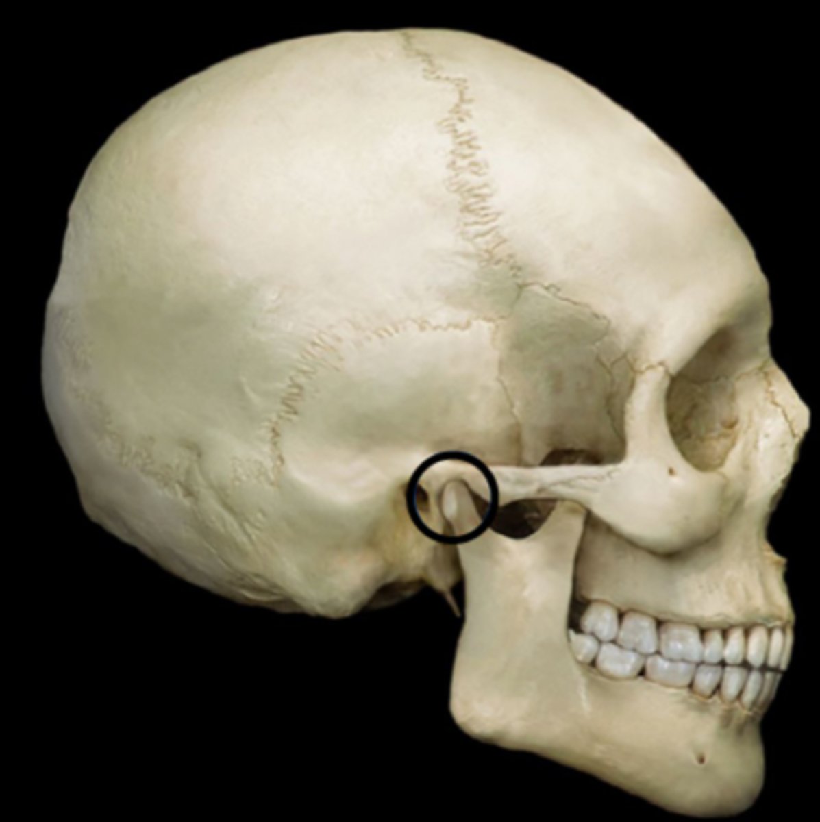
styloid process
long sharp process of the temporal bone extending inferiorly
mastoid process
rounded process of the temporal bone posterior to the styloid process and extending inferiorly.

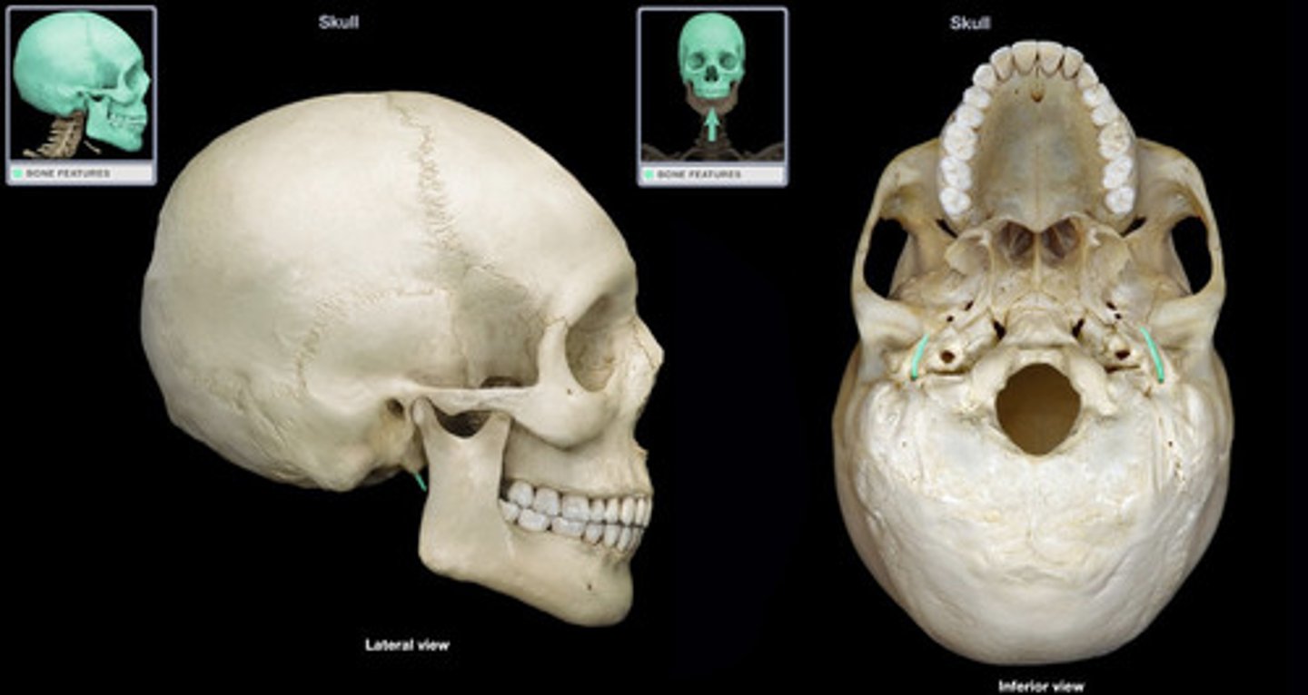
stylomastoid foramen
located between the styloid and mastoid processes; passageway for facial nerve

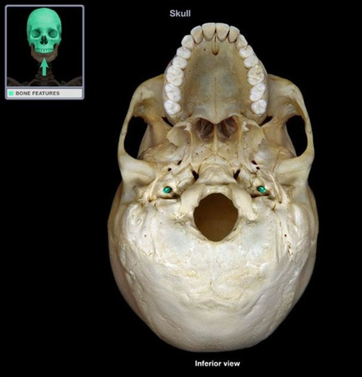
carotid canal
located on petrous part of the temporal bone anterior to the jugular foramen
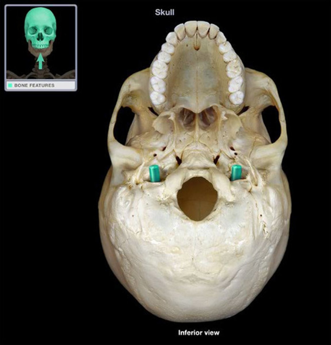
jugular foramen
located on inferior petrous part of the temporal bone posterior to the carotid canal
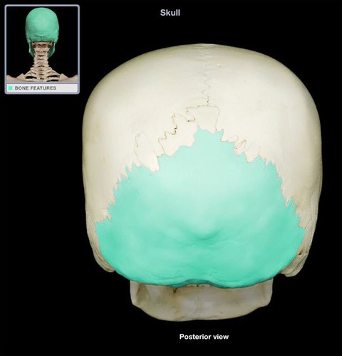
occipital bone
most posterior and inferior bone of the skull
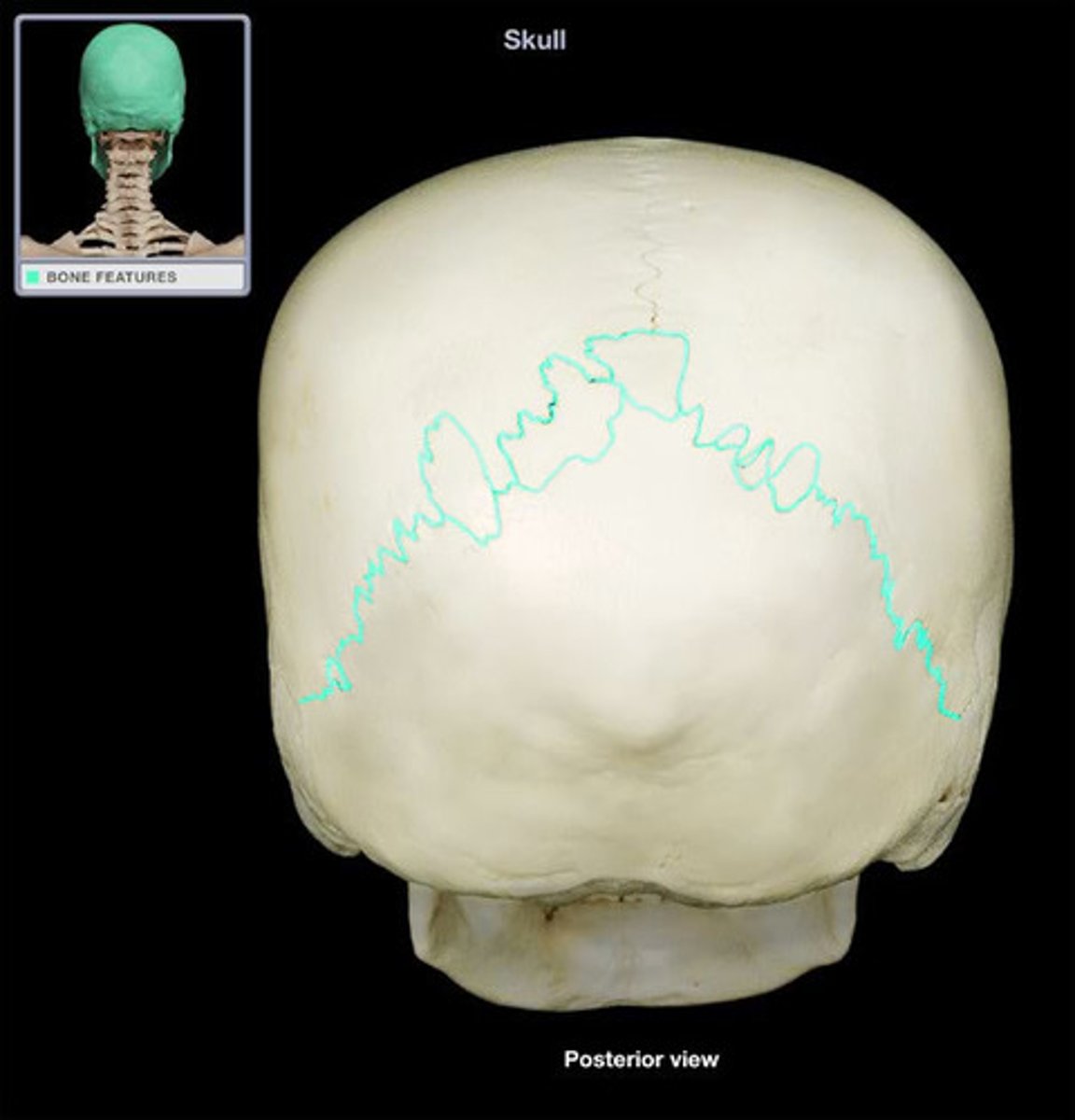
lambdoid suture
runs transversely joining the occipital and parietal bones
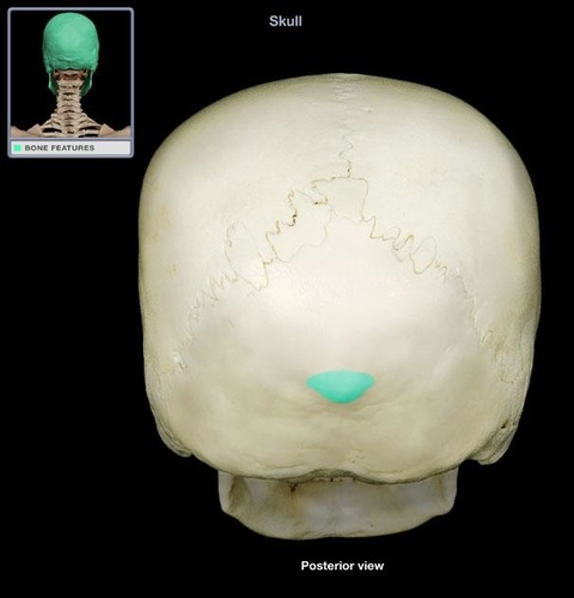
external occipital protuberance
median protrusion on the occipital bone posterior and superior to foramen magnum
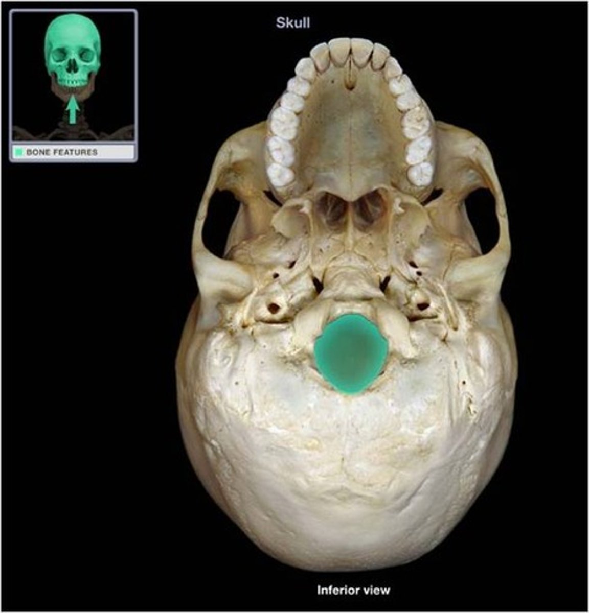
foramen magnum
large opening on the inferior part of the skull
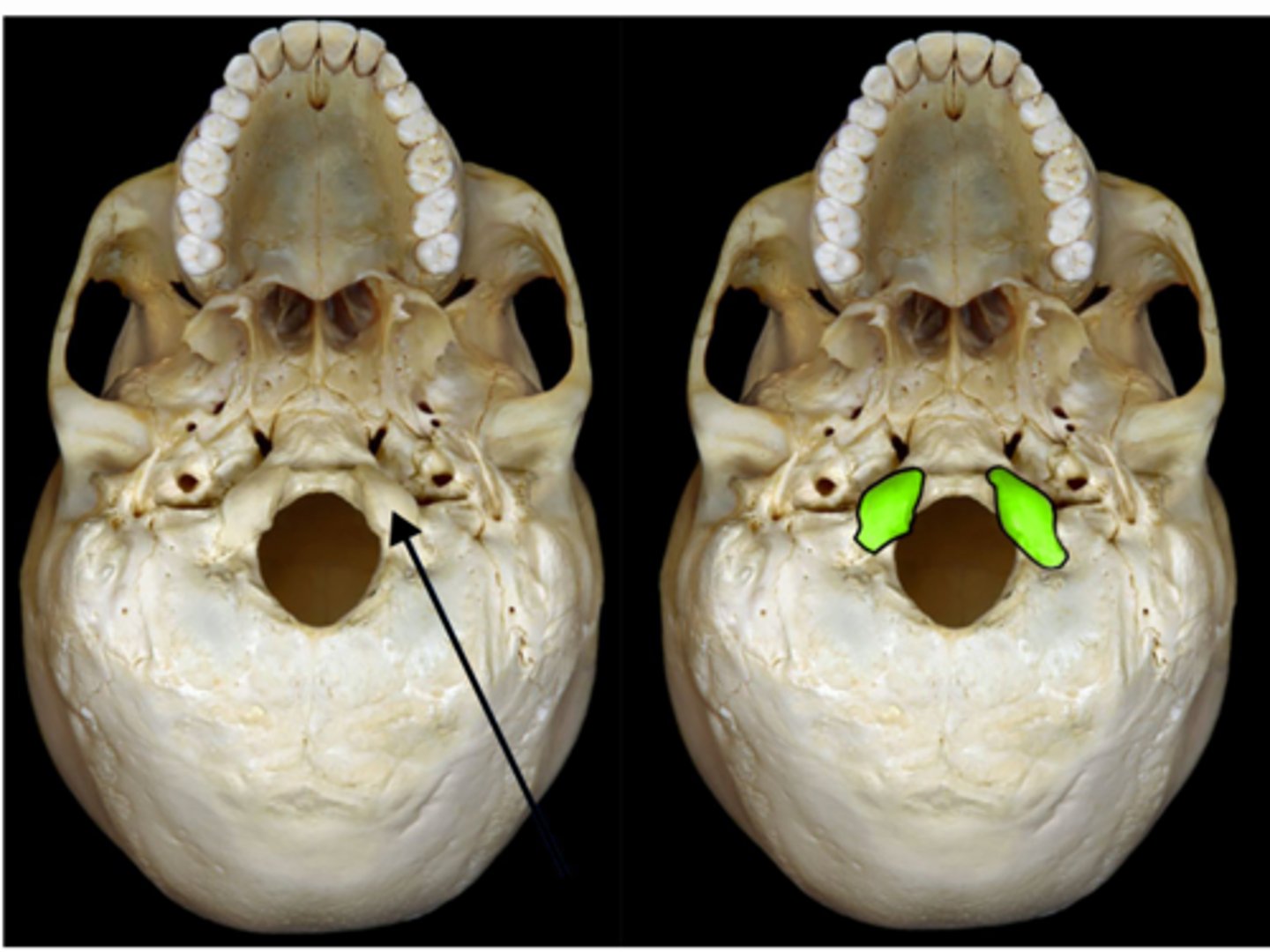
occipital condyle
paired rounded process lateral to the foramen magnum
hypoglossal canal
paired canal superior to occipital condyle
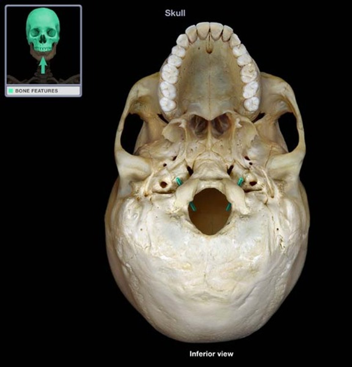
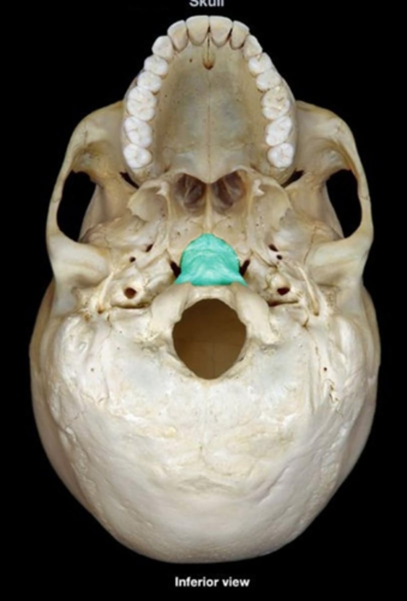
basilar part
region of the occipital bone anterior to foramen magnum
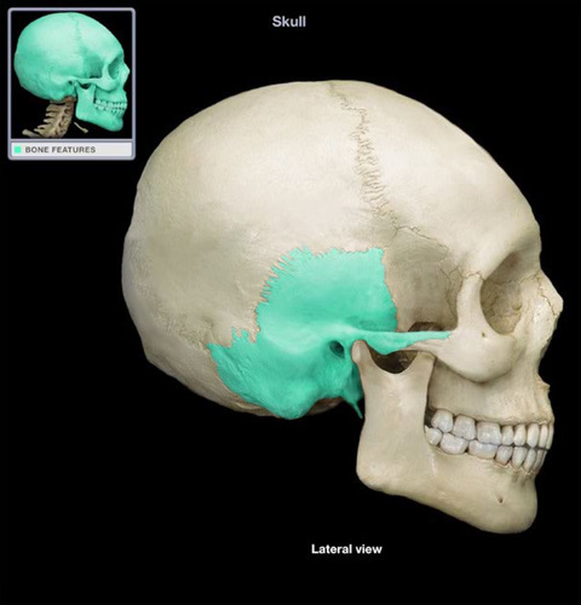
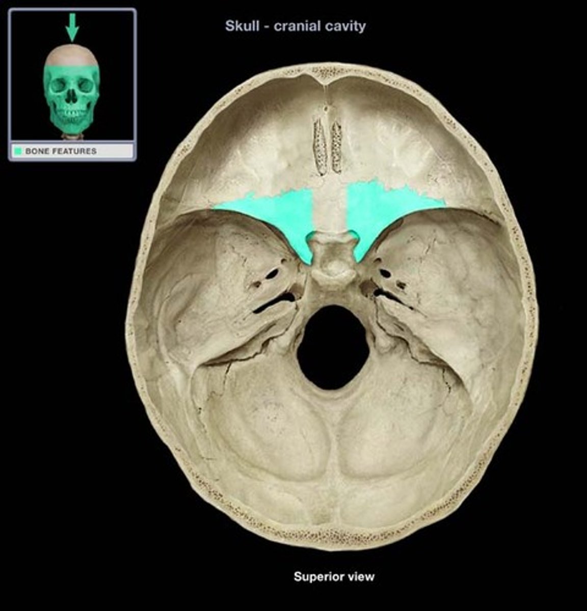
sphenoid bone
butterfly-shaped bone forming the floor of the cranium
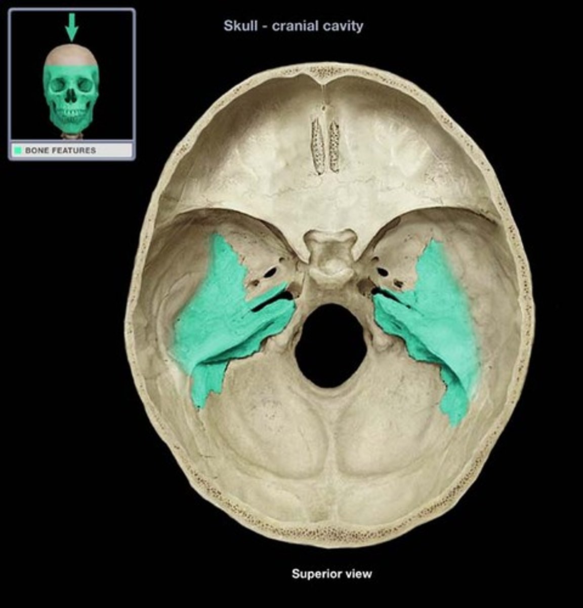
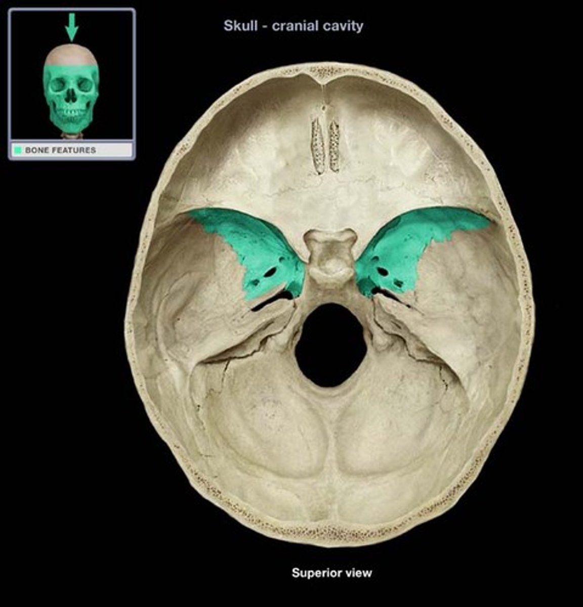
greater wing
posterior and inferior to lesser wing on the interior of the skull, also visible in the posterior orbit and lateral side of the skull
lesser wing
anterior and superior to greater wing on the interior of the skull
superior orbital fissure
fissure between greater and lesser wings of sphenoid visible from the orbits
Inferior orbital fissure
fissure in the orbit floor between maxilla and greater wing of sphenoid
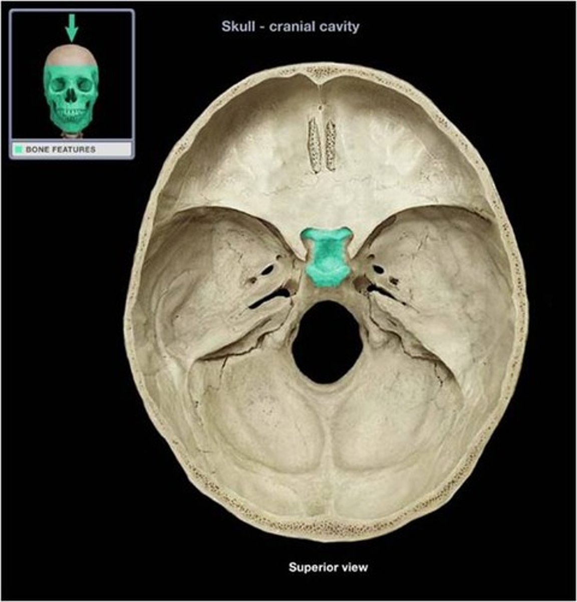
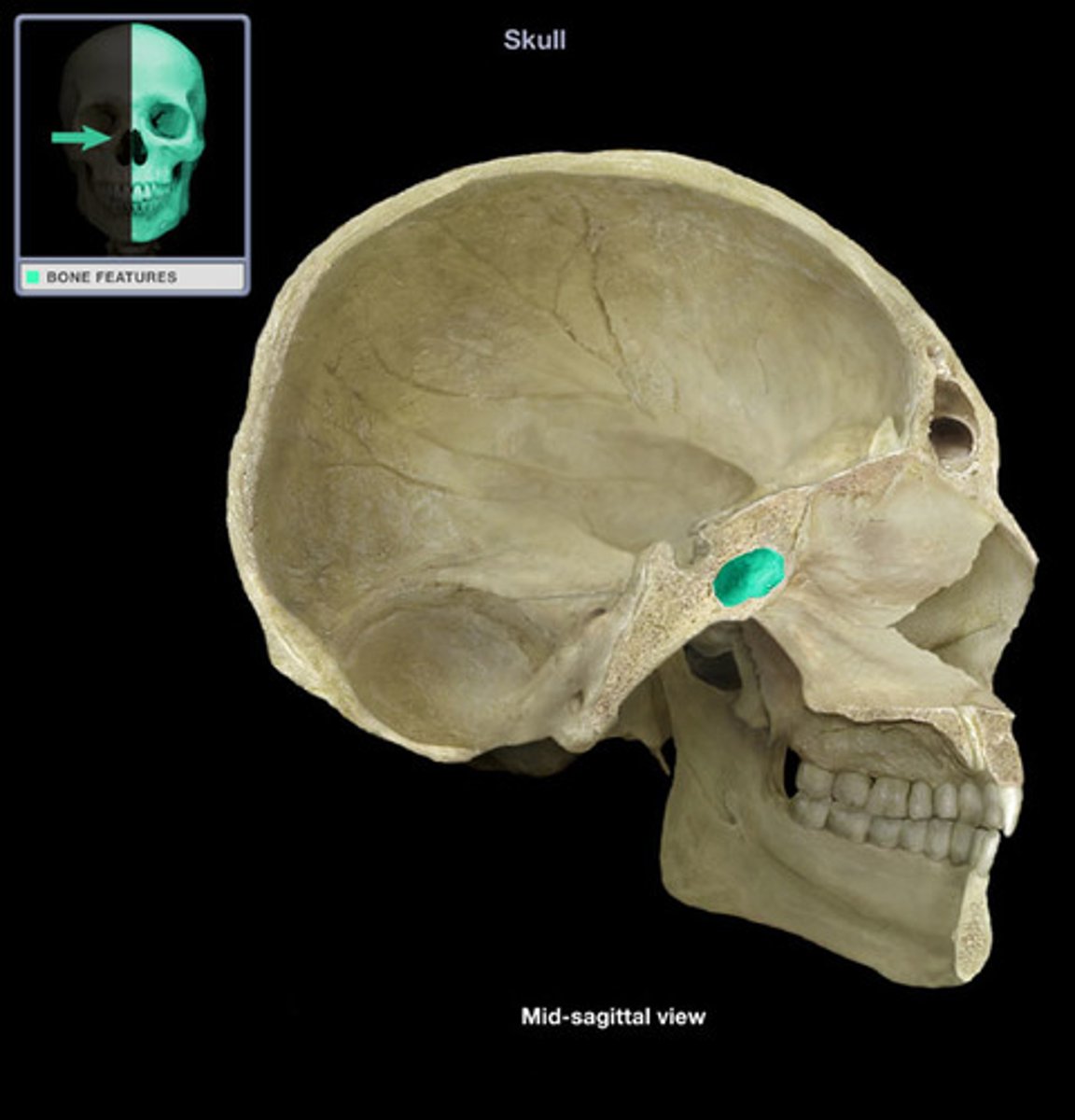
sella turcica
saddle-like depression posterior to lesser wing
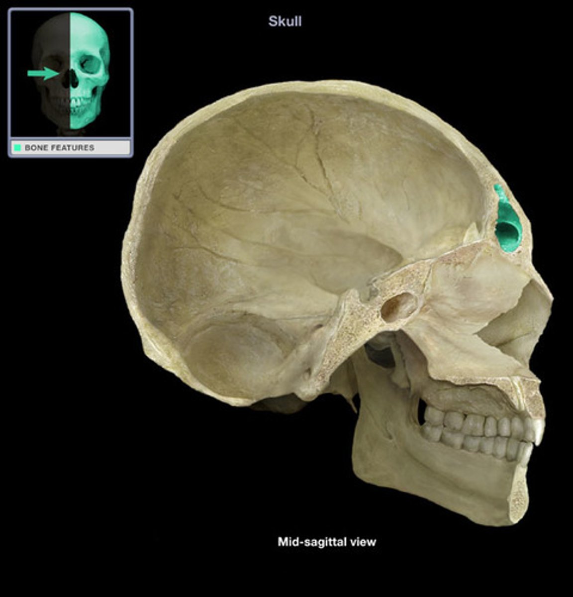
optic canal
paired opening lateral to the sella turcica

foramen lacerum
paired opening lateral to the sella turcica

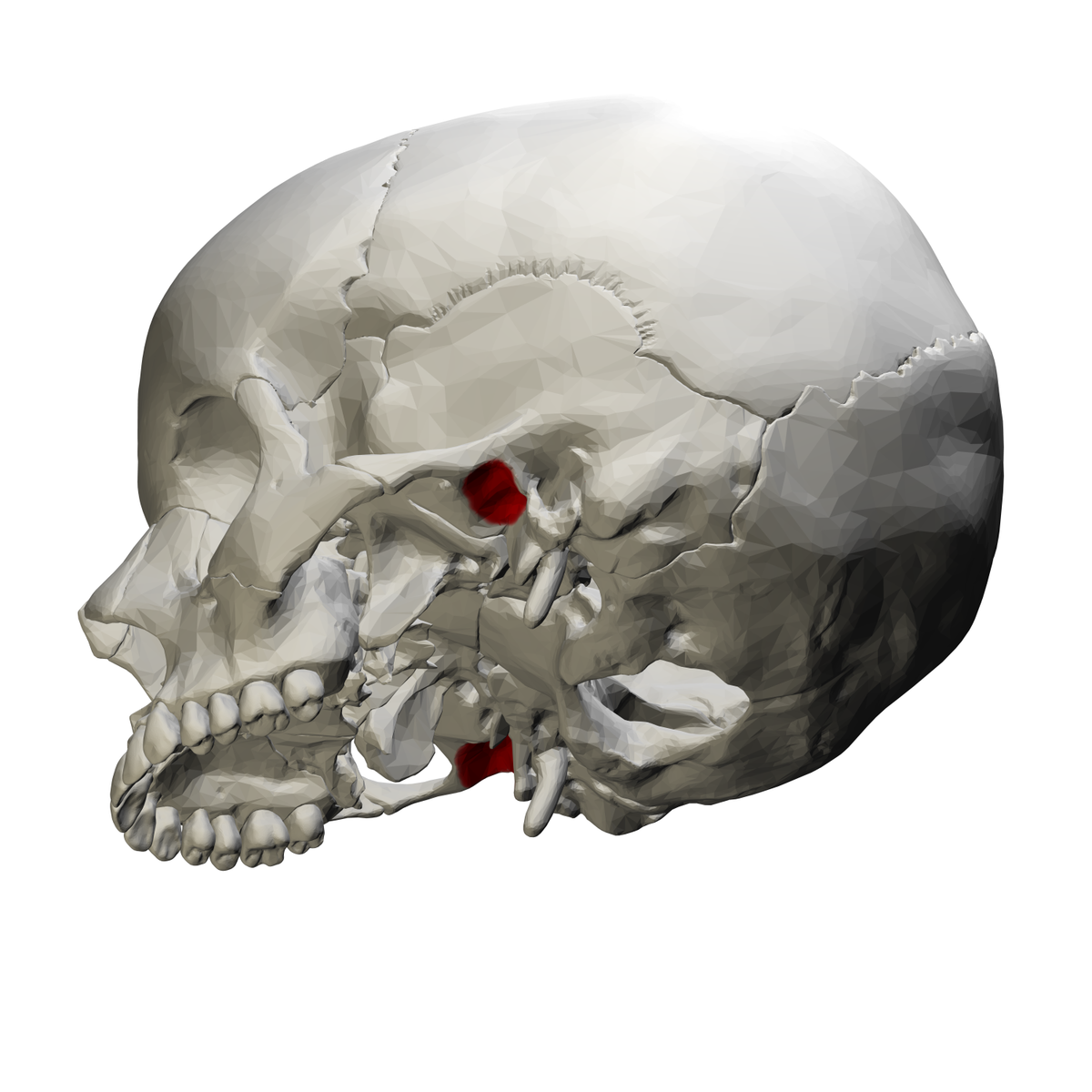
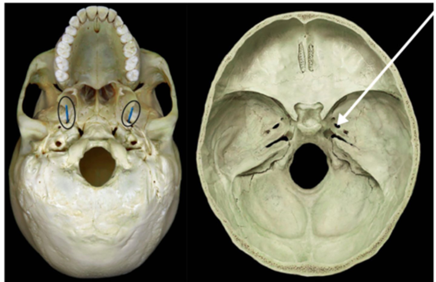
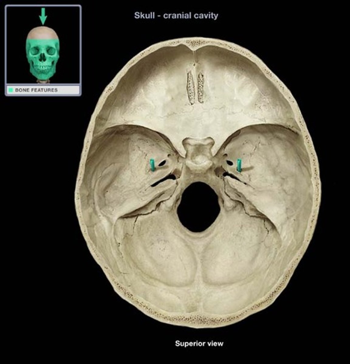
foramen ovale
paired opening lateral to the base of the sella turcica
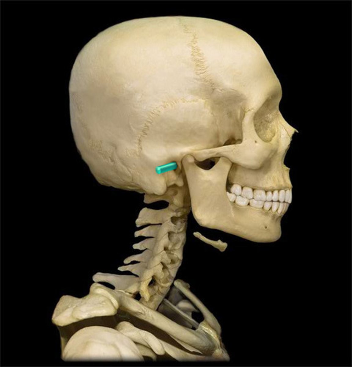
foramen spinosum
paired opening at the posterior margin of the sphenoid
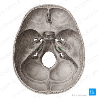
foramen rotundum
paired opening anterior to the foramen ovale

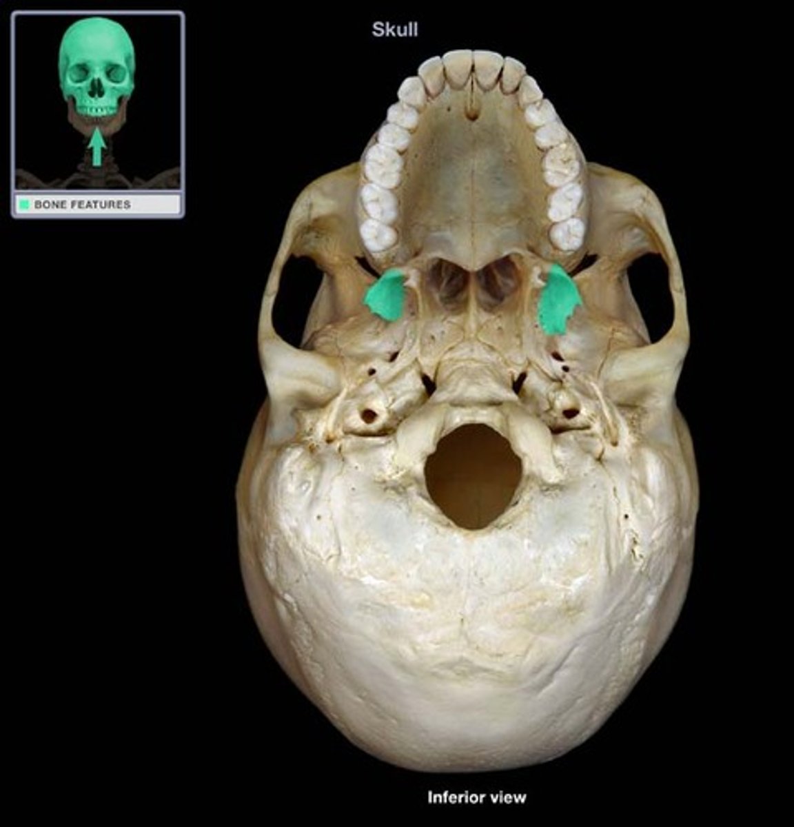
lateral pterygoid plate
sharp process that laterally and inferiorly projects from the sphenoid
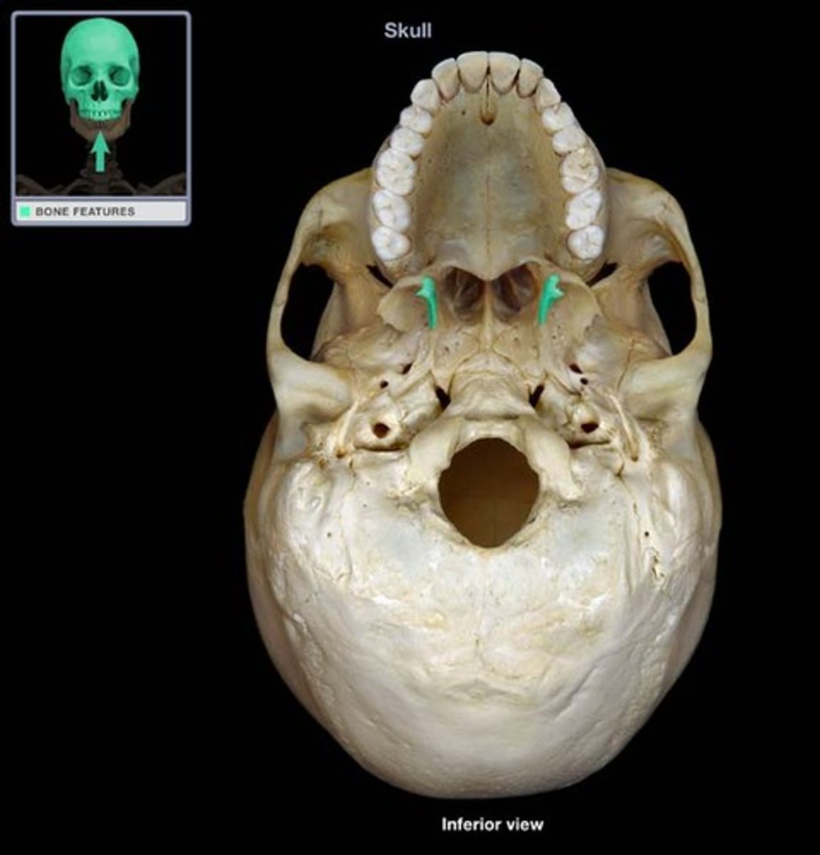
medial pterygoid plate
sharp process that medially and inferiorly projects from the sphenoid
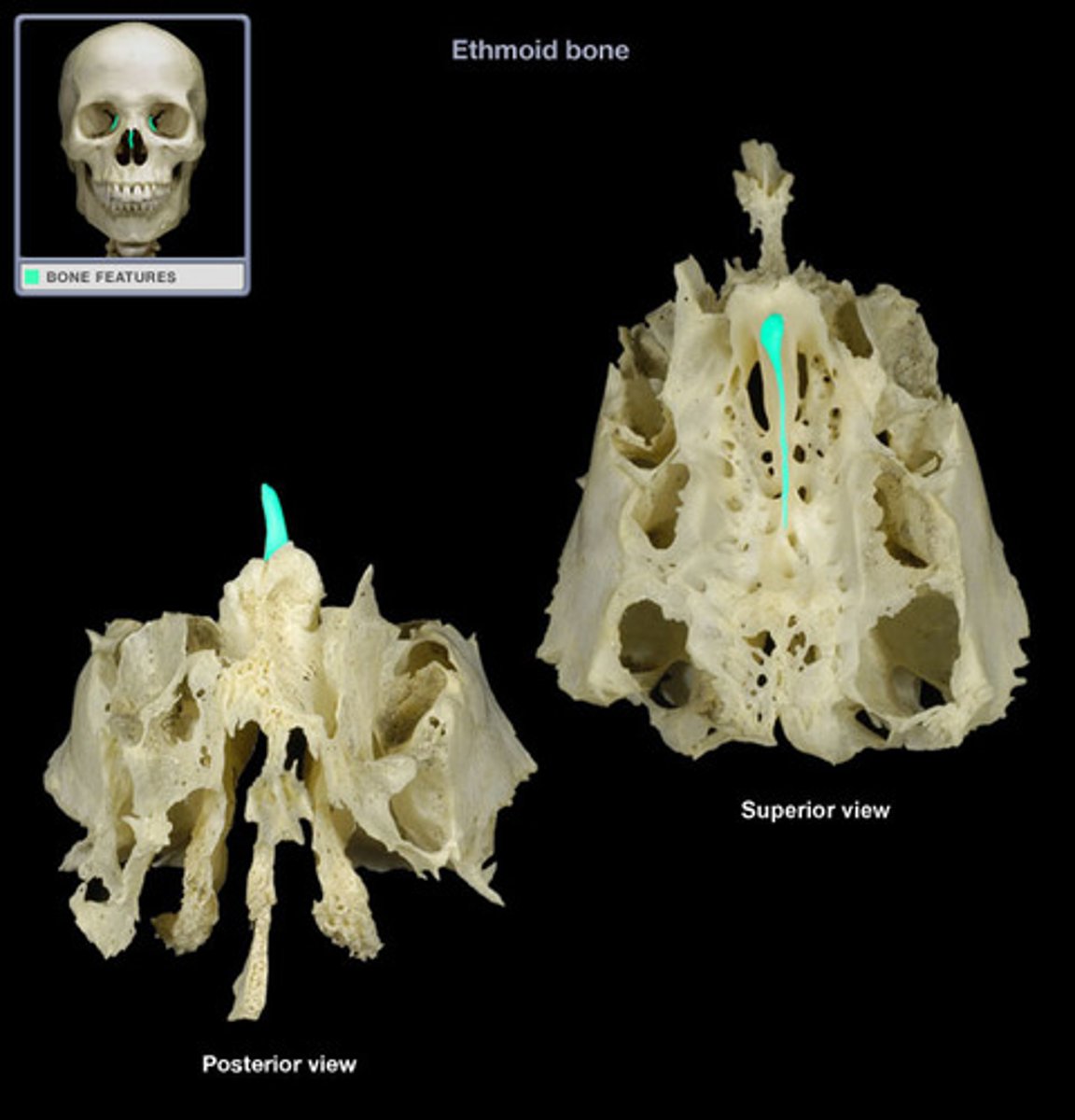
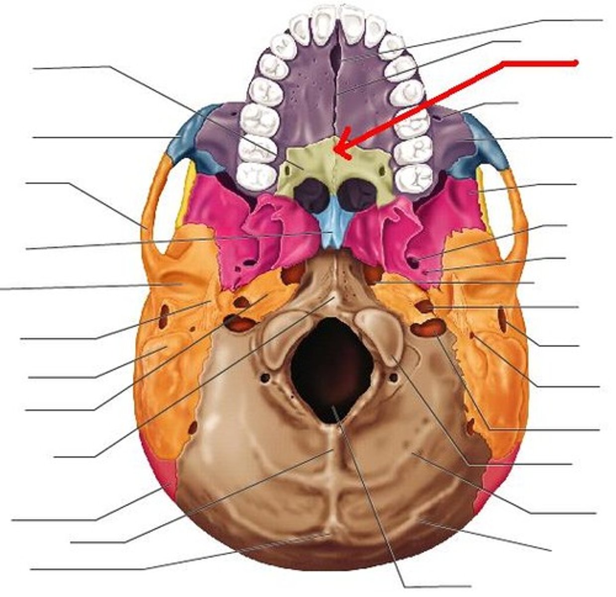
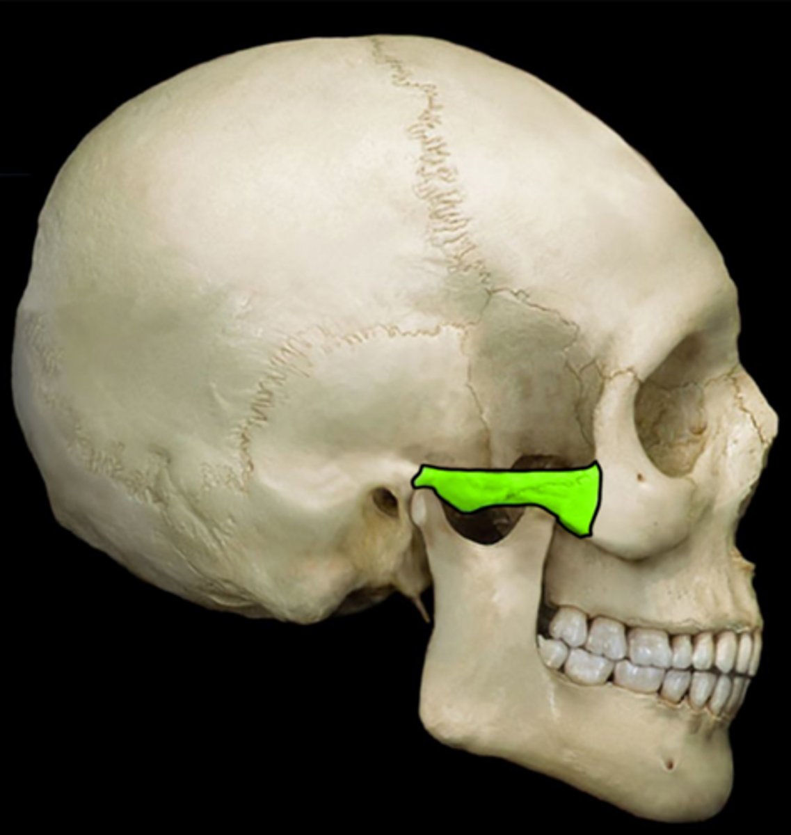
ethmoid bone
located between the orbits and between sphenoid and nasal bones; forms roof of nasal cavity
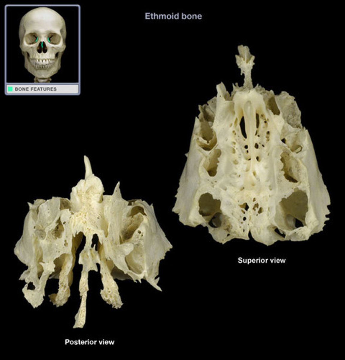
cribriform plate
porous bone surrounding the crista galli in the floor of the cranium through which olfactory nerves transverse
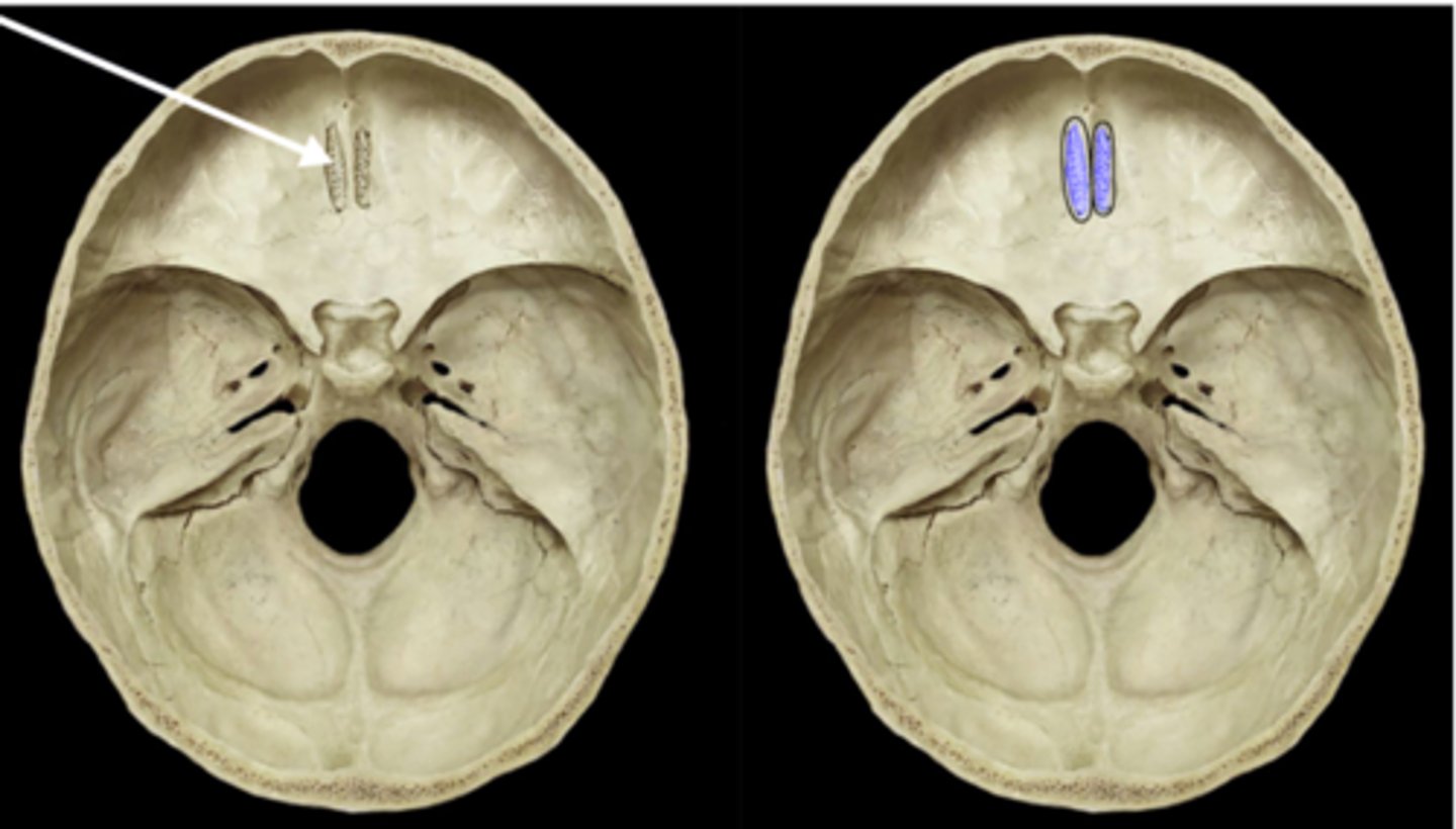
crista galli
superior projection in the middle of the cribriform plate of the ethmoid bone
perpendicular plate
median inferior projection of the ethmoid; forms superior nasal septum
vomer
bone forming the inferior and posterior portion of the nasal septum
inferior nasal concha
paired bone that protrudes into the inferior part of the nasal cavity
maxilla
paired fused bone of the upper jaw and central face
infraorbital foramen
paired opening inferior to the orbit

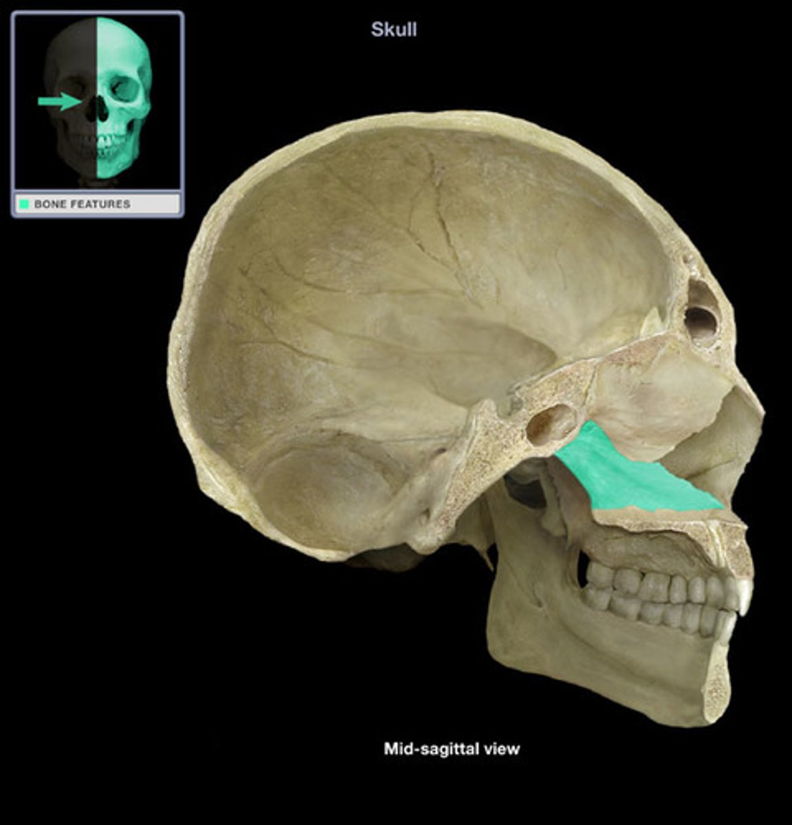
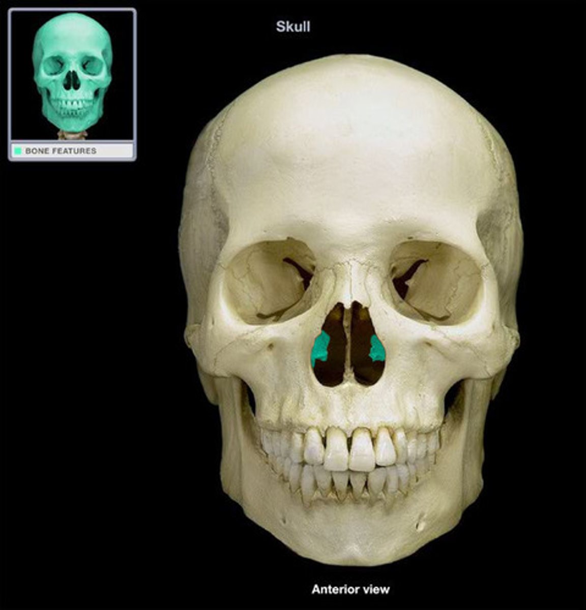
palatine process
anterior portion of the hard palate
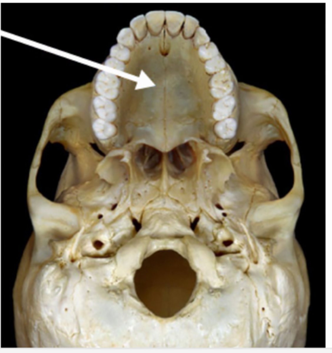
palatine bone
situated at the back part of the nasal cavity between the maxilla and sphenoid bones
nasal bone
paired bone composing the superior portion (bridge) of the nose
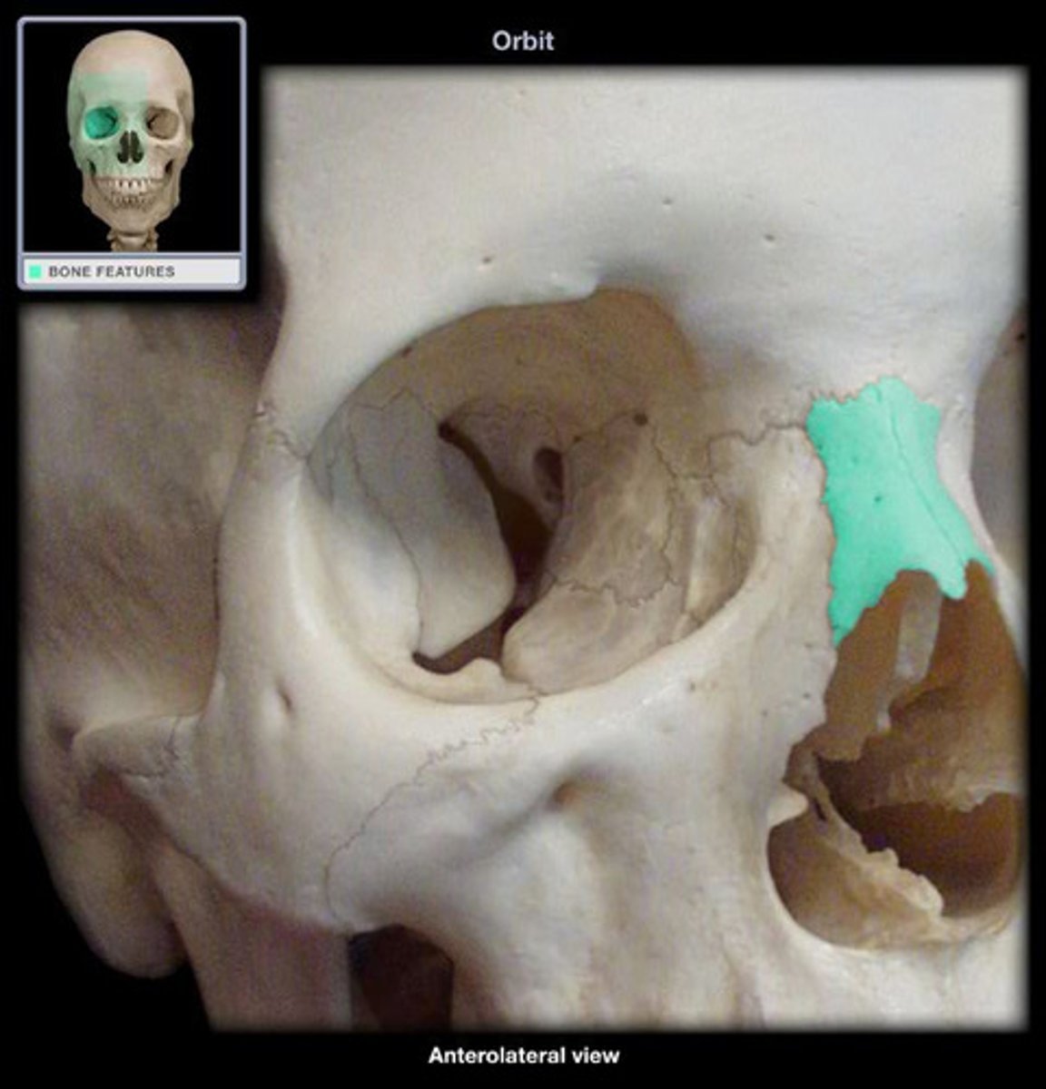
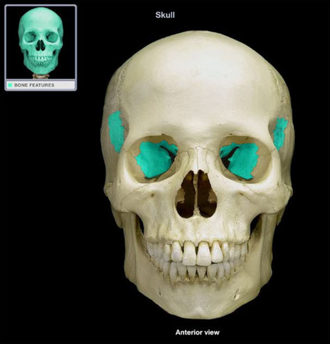
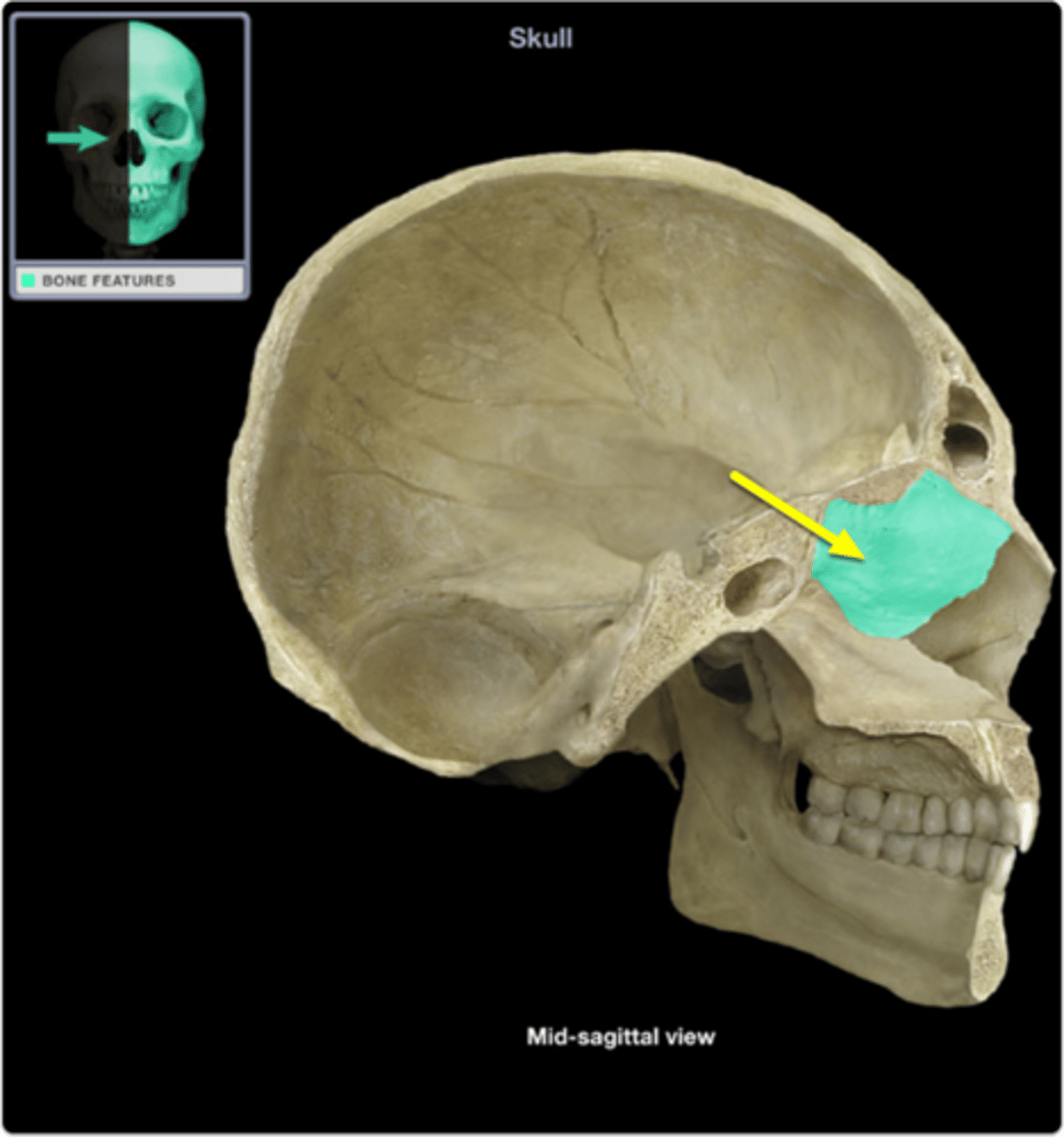
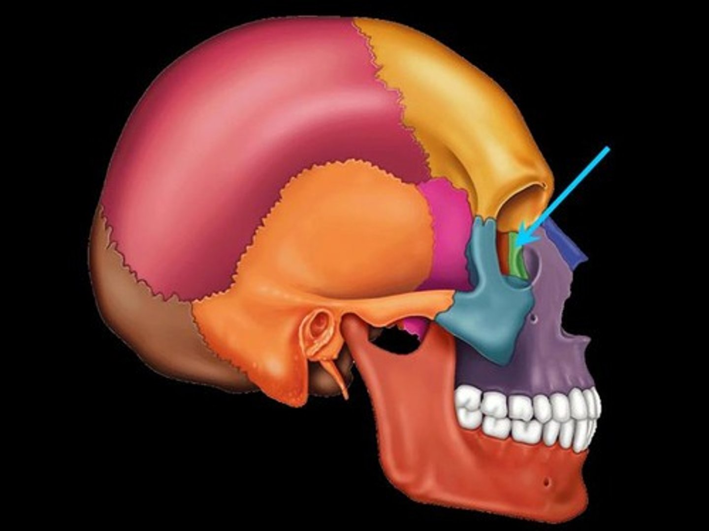
lacrimal bone
paired bone forming part of the medial wall of the orbit
zygomatic bone
paired bone which forms the prominence of the cheek
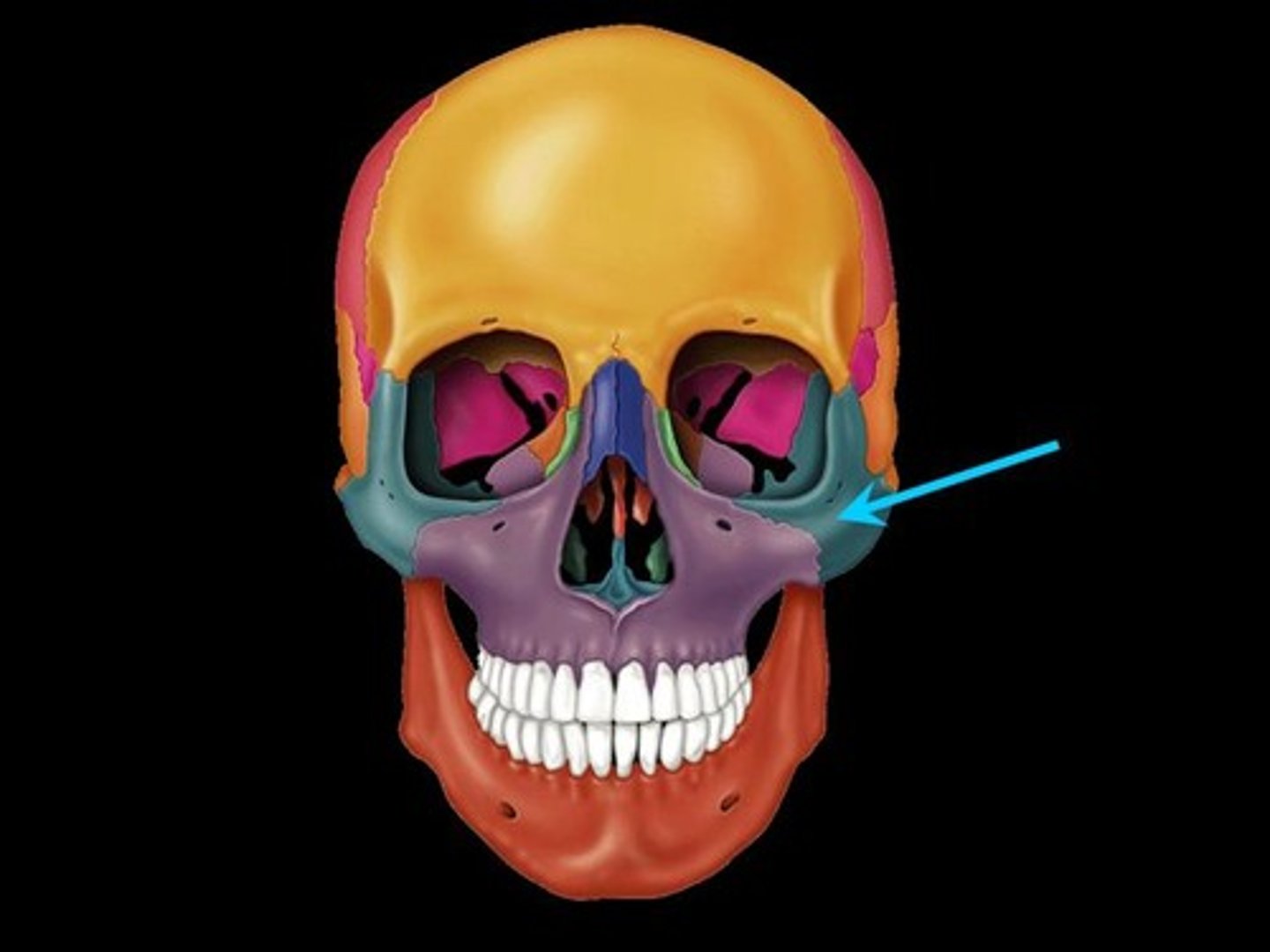
zygomatic arch
formed by union of the zygomatic bone and the temporal bone
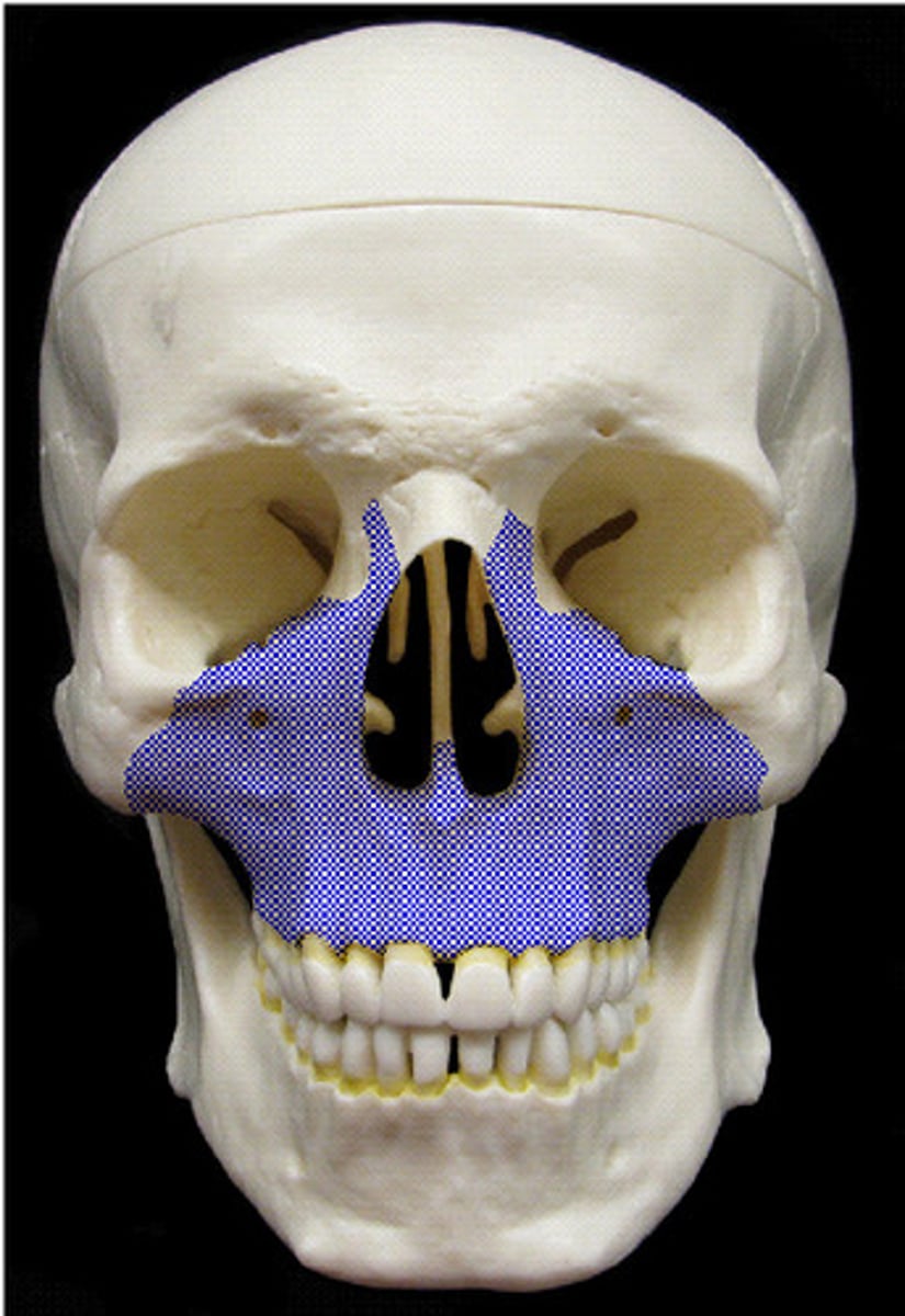
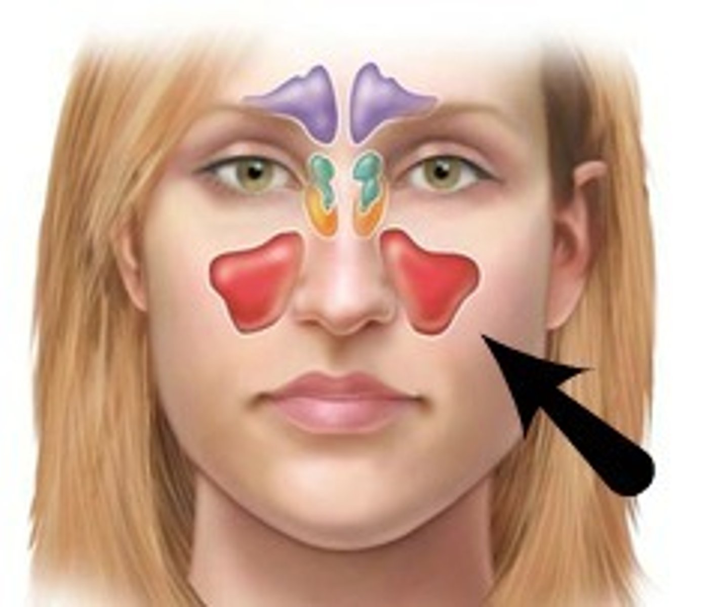
frontal sinus
cavity within the frontal bone
(purple in image)
sphenoidal sinus
paired cavity located within the sphenoid, inferior to the sella turcica
ethmoidal sinus
numerous cavities within the ethmoid bone
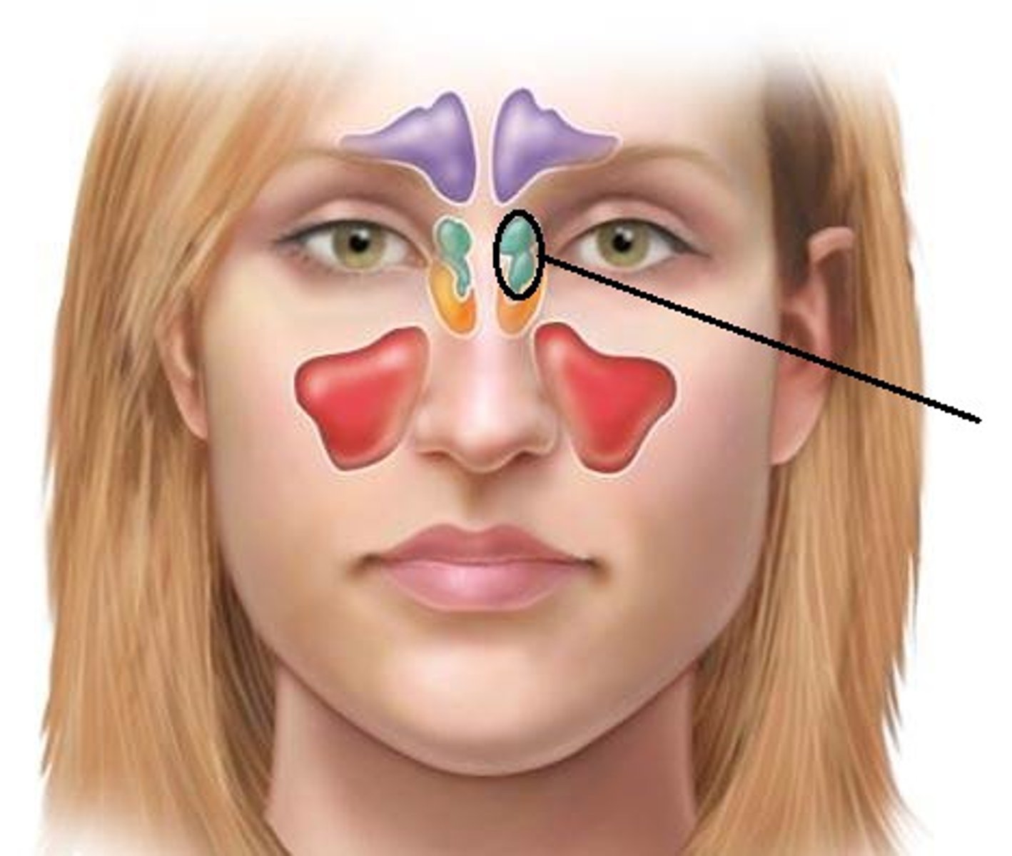
maxillary sinus
paired cavity found within the maxilla
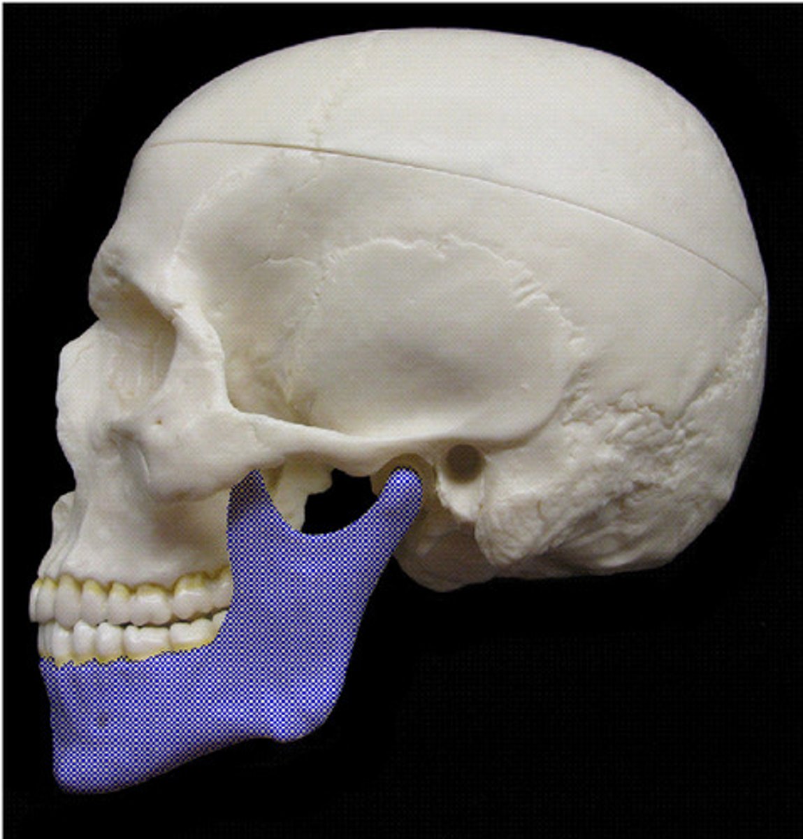
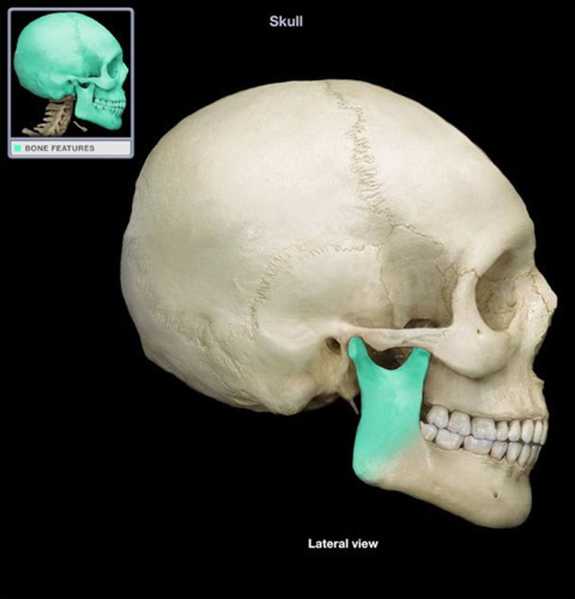
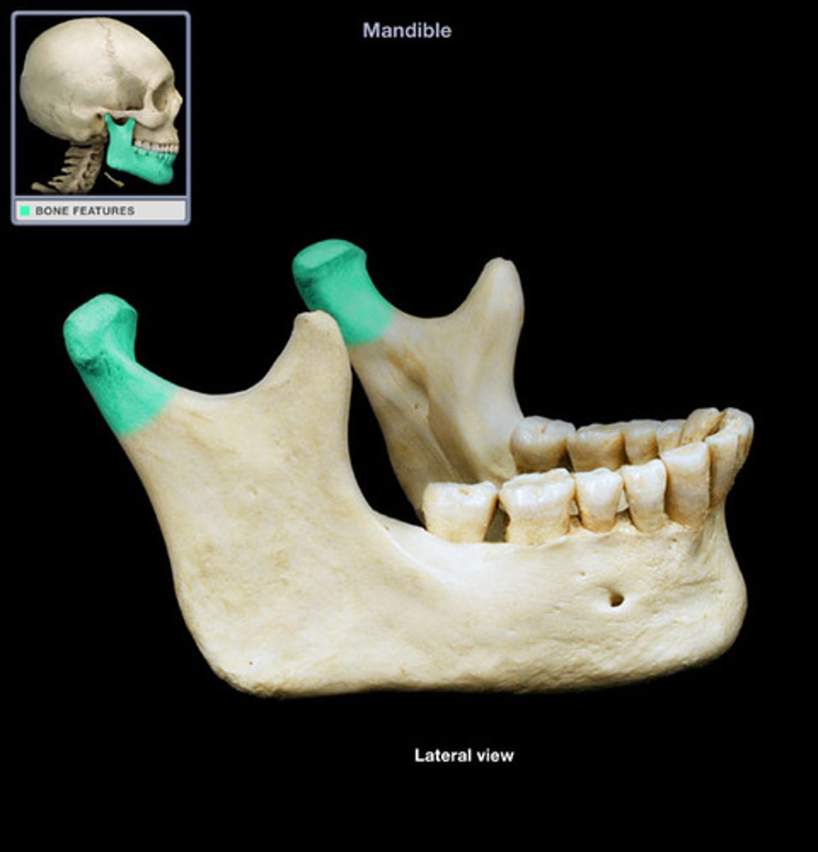
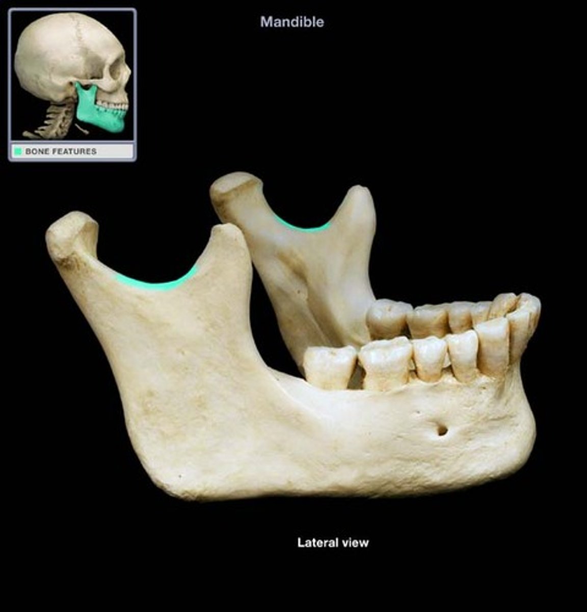
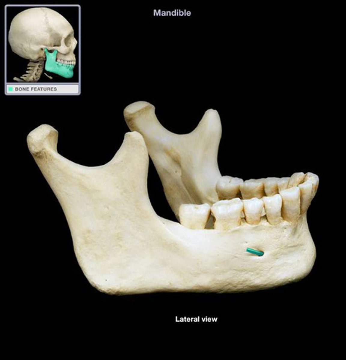
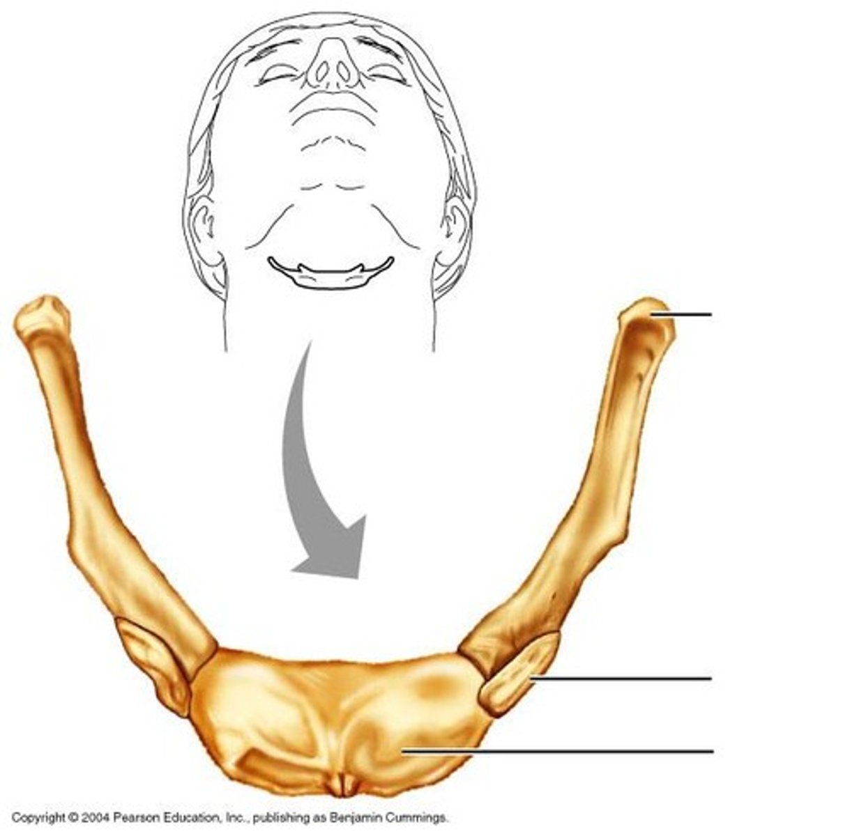
mandible
lower jaw bone
body
most anterior part extending horizontally to the angle of the mandible
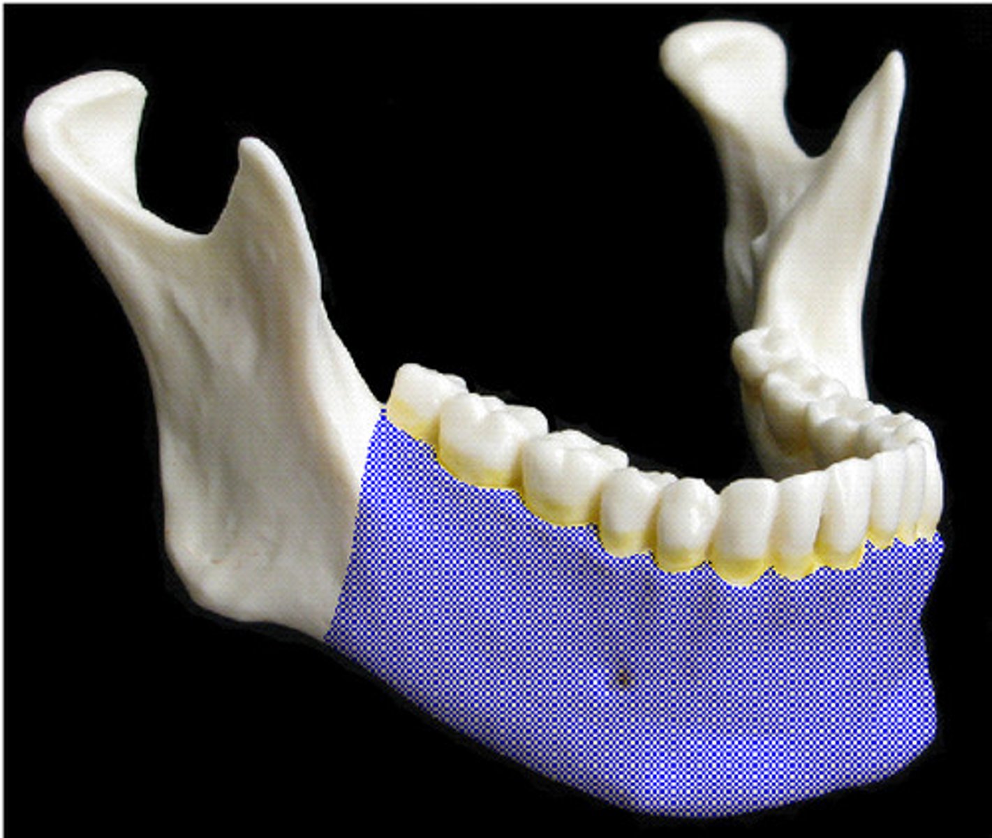
angle
where the body and ramus of the mandible meet
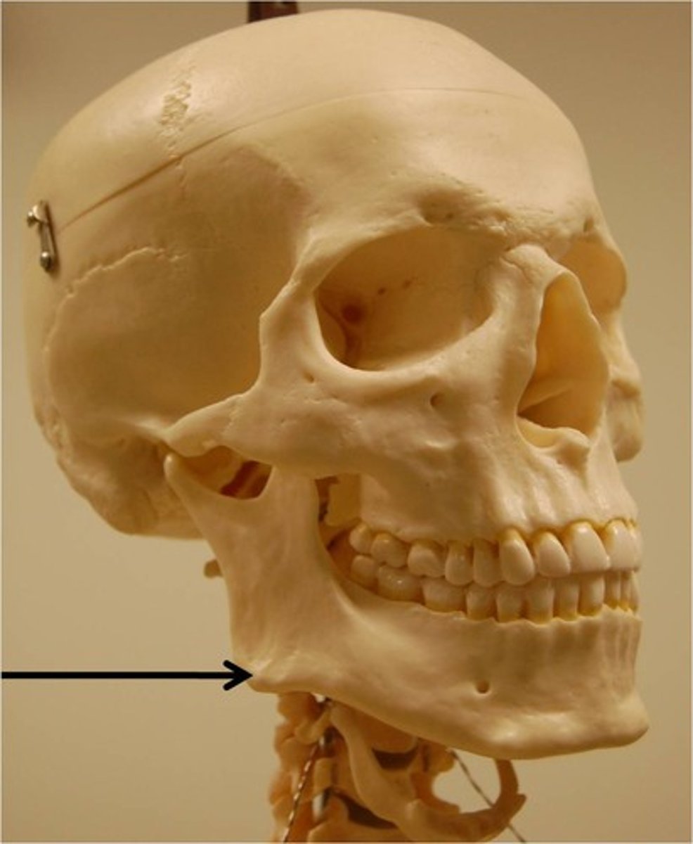
ramus
the verticle portion of the mandible extending from the angle
coronoid process
anterior process of the ramus
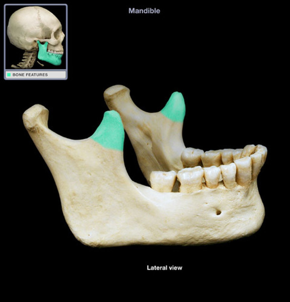
condylar process
The most posterior process of the ramus
mandibular notch
separates the coronoid from the condylar process
mandibular foramen
paired opening on the medial surface of the ramus

mental foramen
paired opening on the anterior aspect of the body
hyoid bone
U-shaped bone in the neck superior to the larynx
intervertebral discs
fibrocartilage cushion between the vertebral bodies
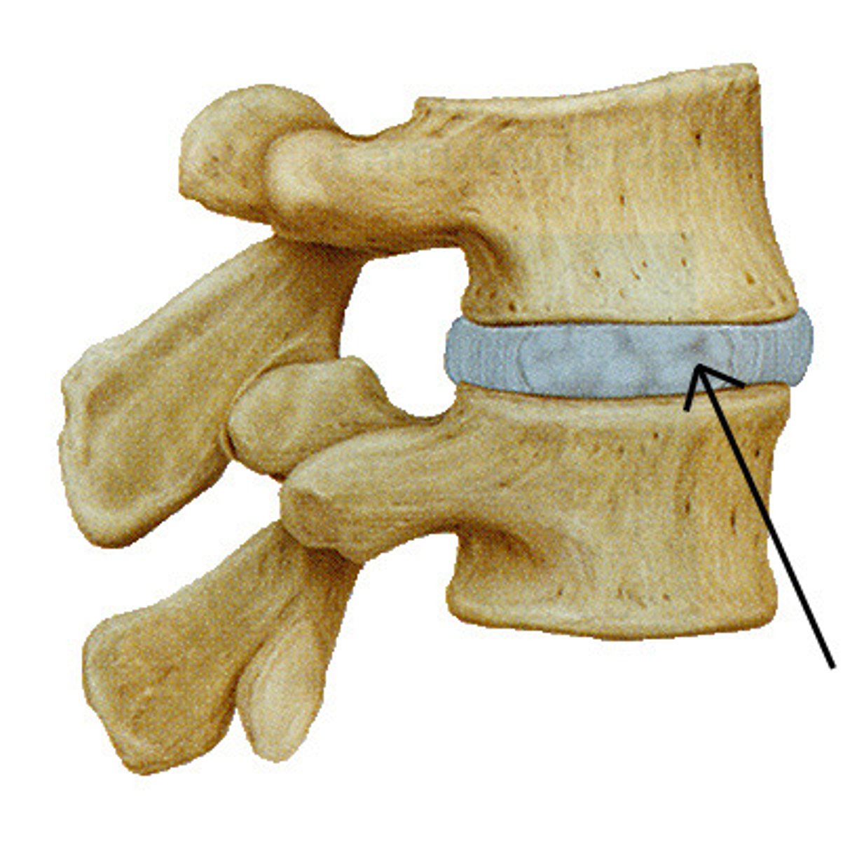
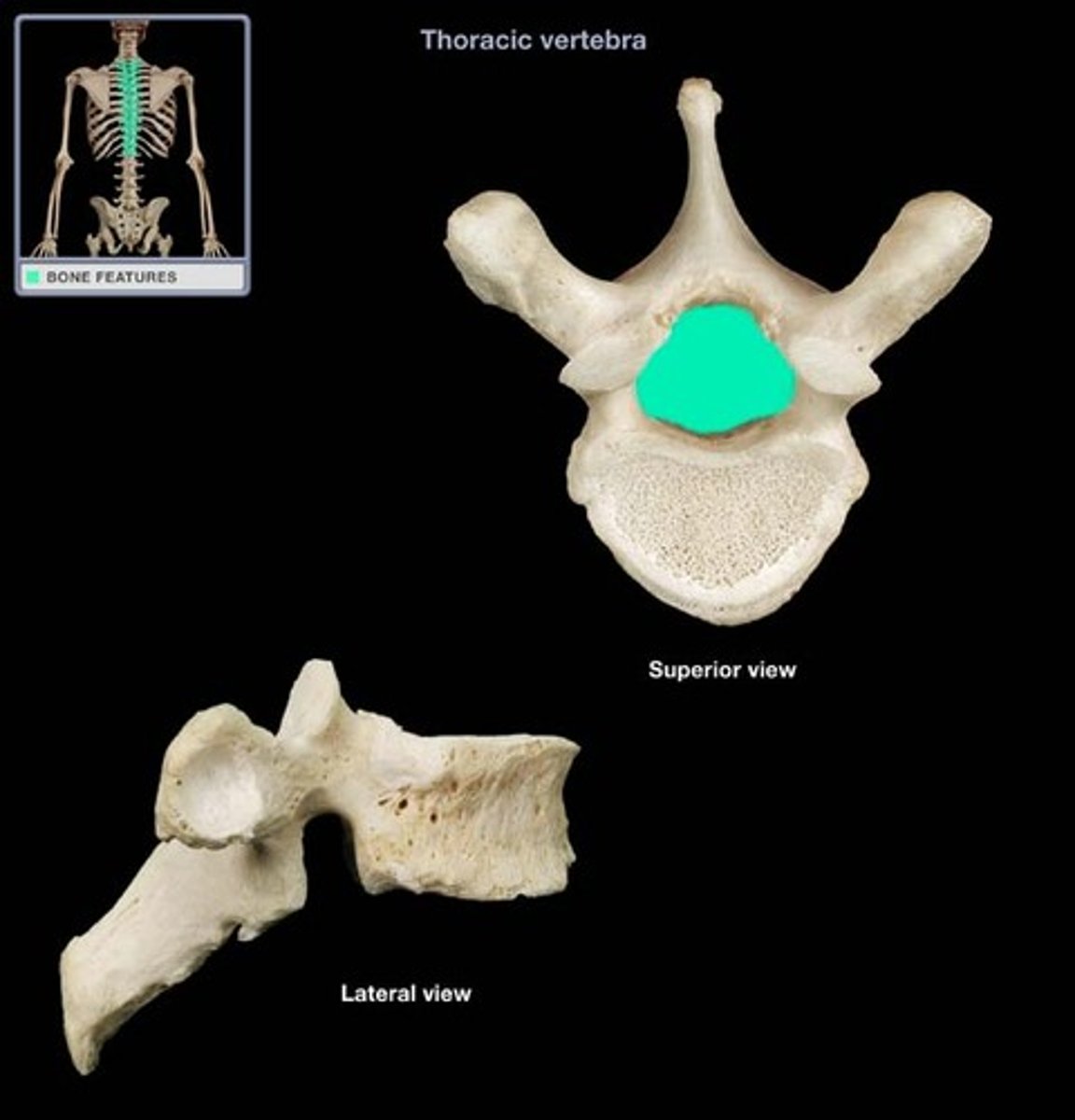
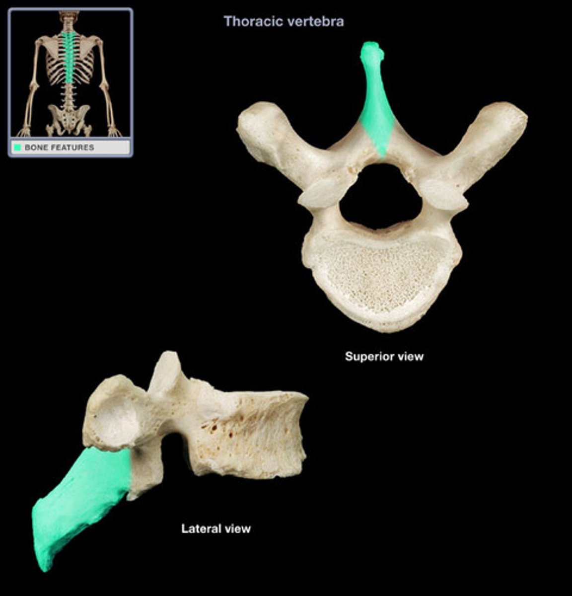
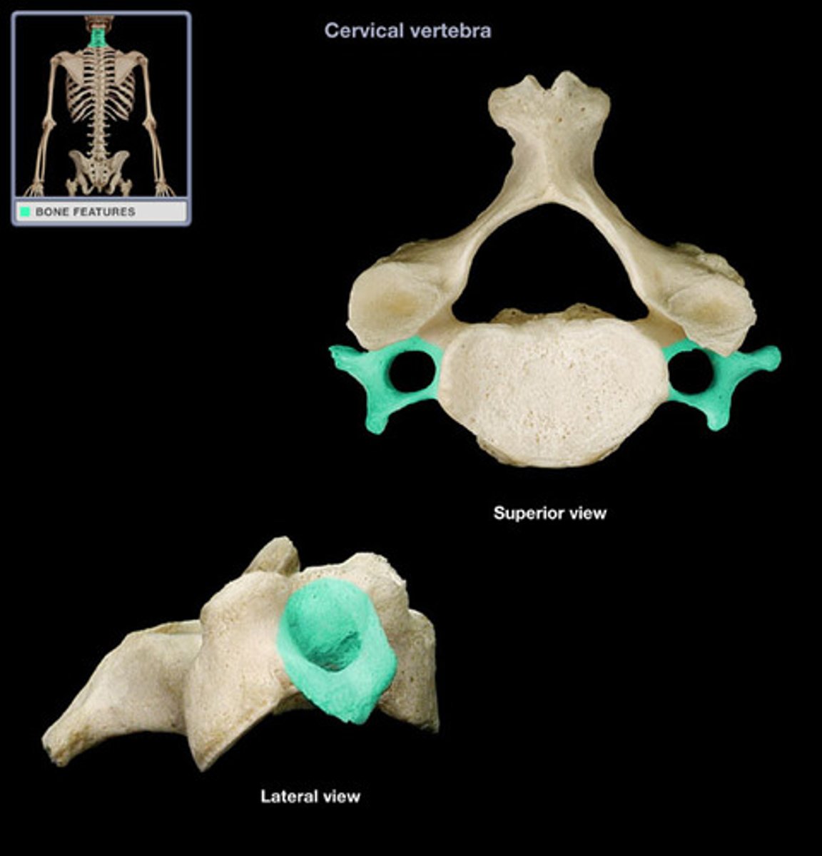
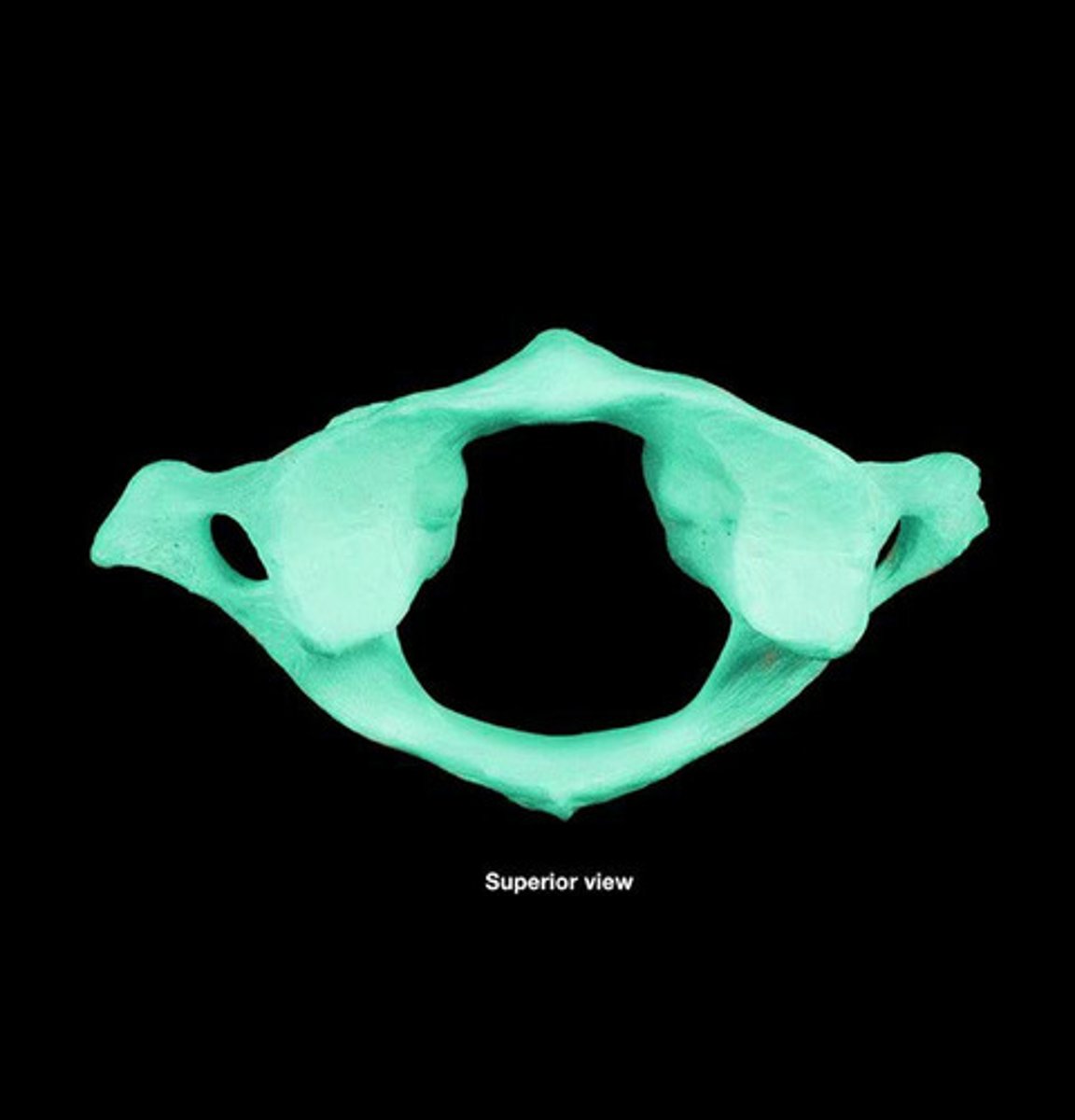
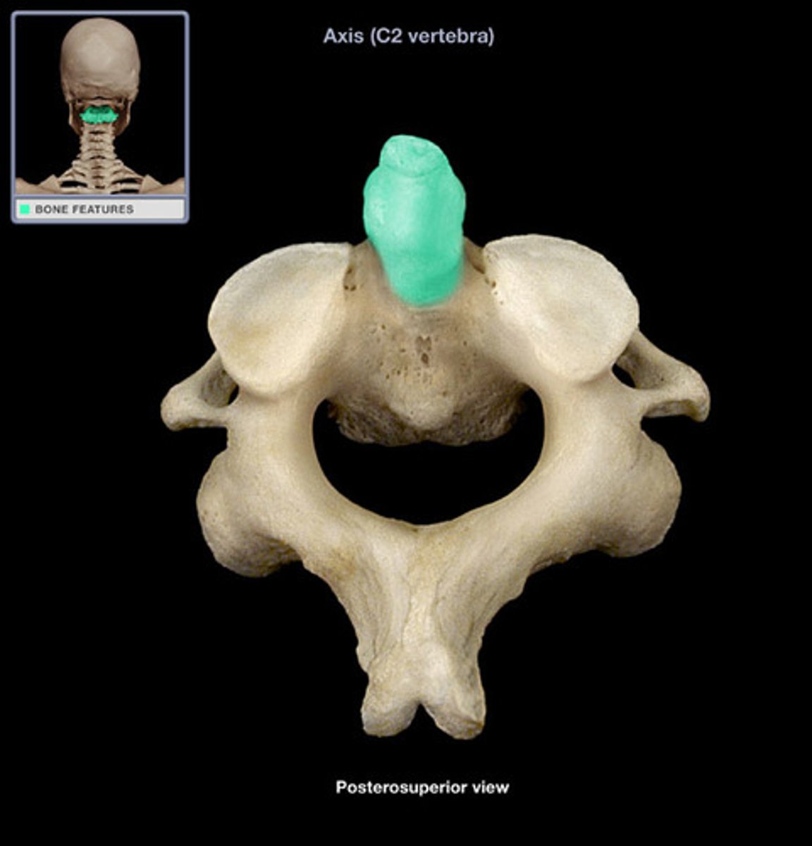
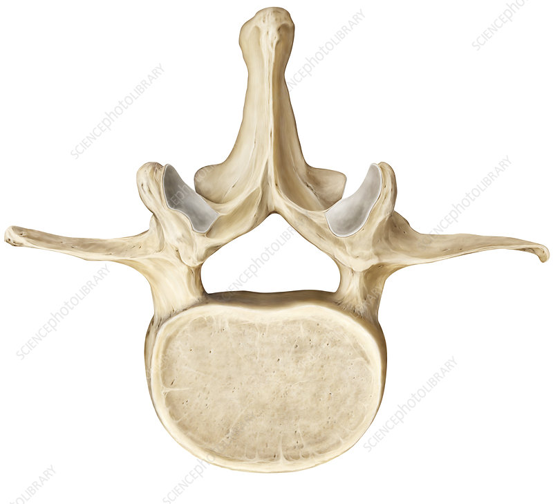
vertebra
basic bone unit of the vertebral column; all types of vertebrae (cervical, thoracic, and lumbar)
body
anterior portion of the vertebra
vertebral foramen
opening posterior to vertebral body through which the spinal cord passes
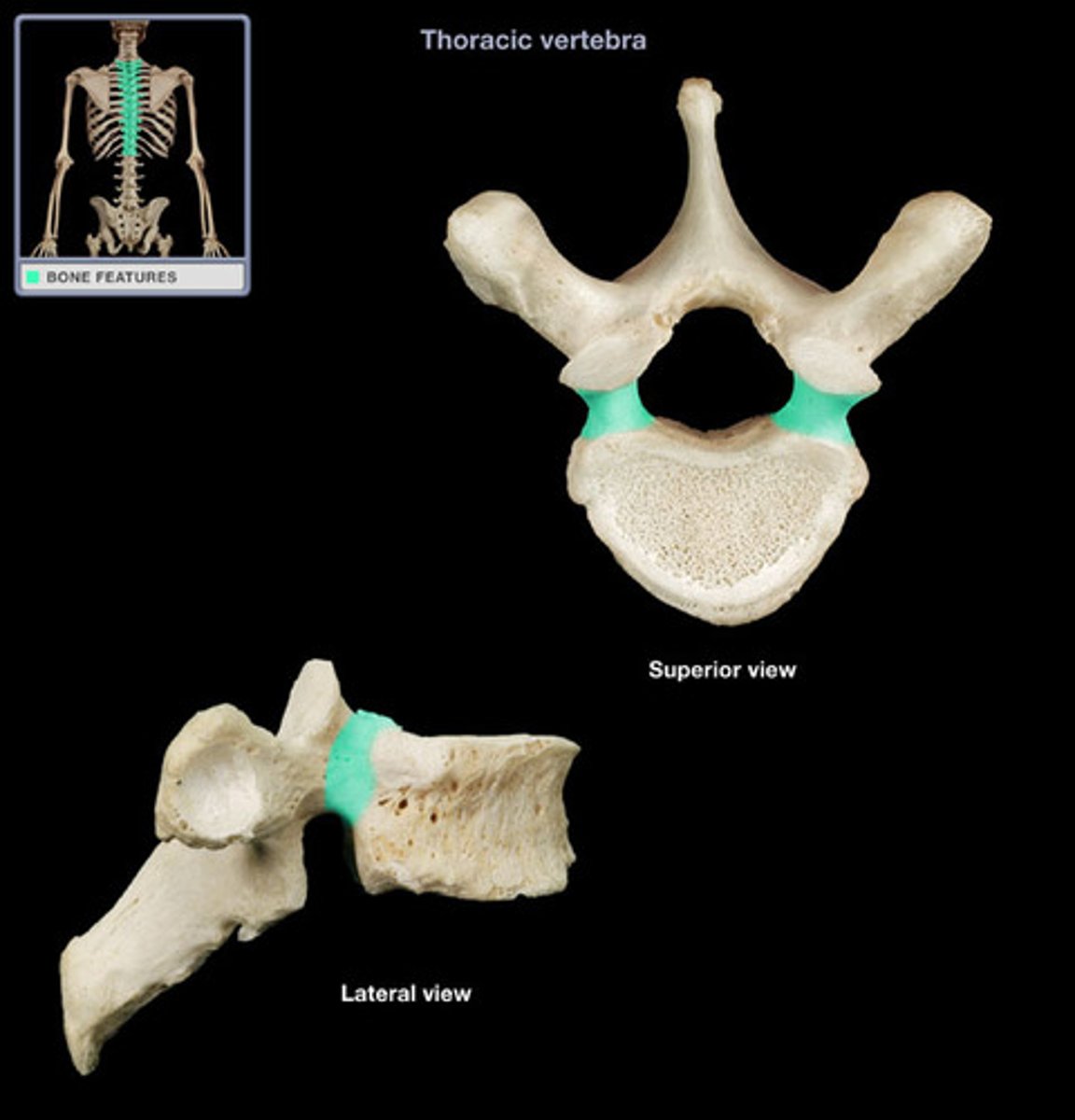
pedicle
posterior projection from vertebral body that supports the lamina
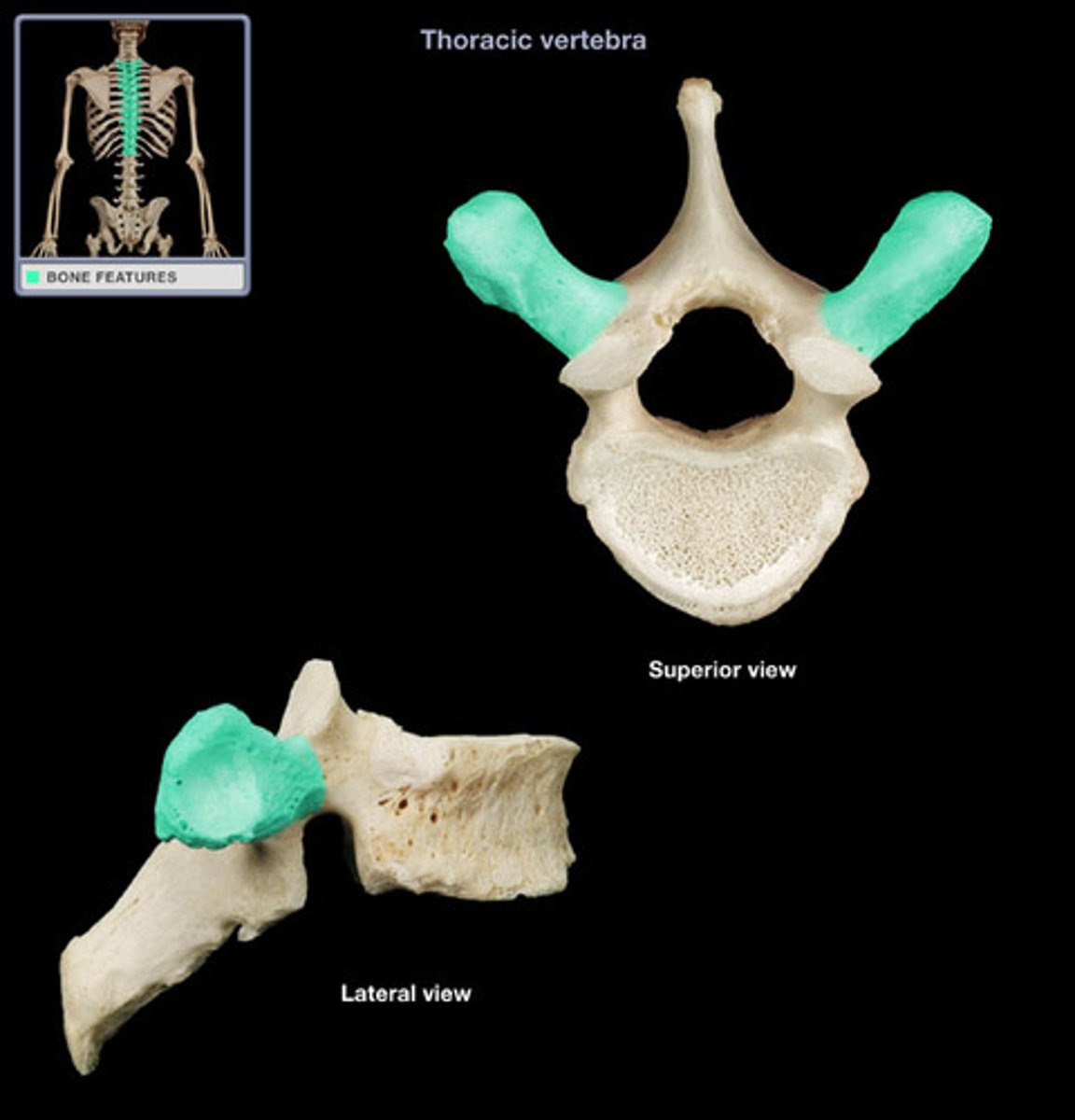
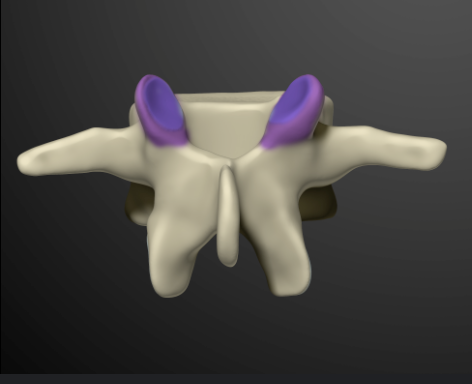
transverse process
lateral projections of the vertebra for muscle/ligament attachment
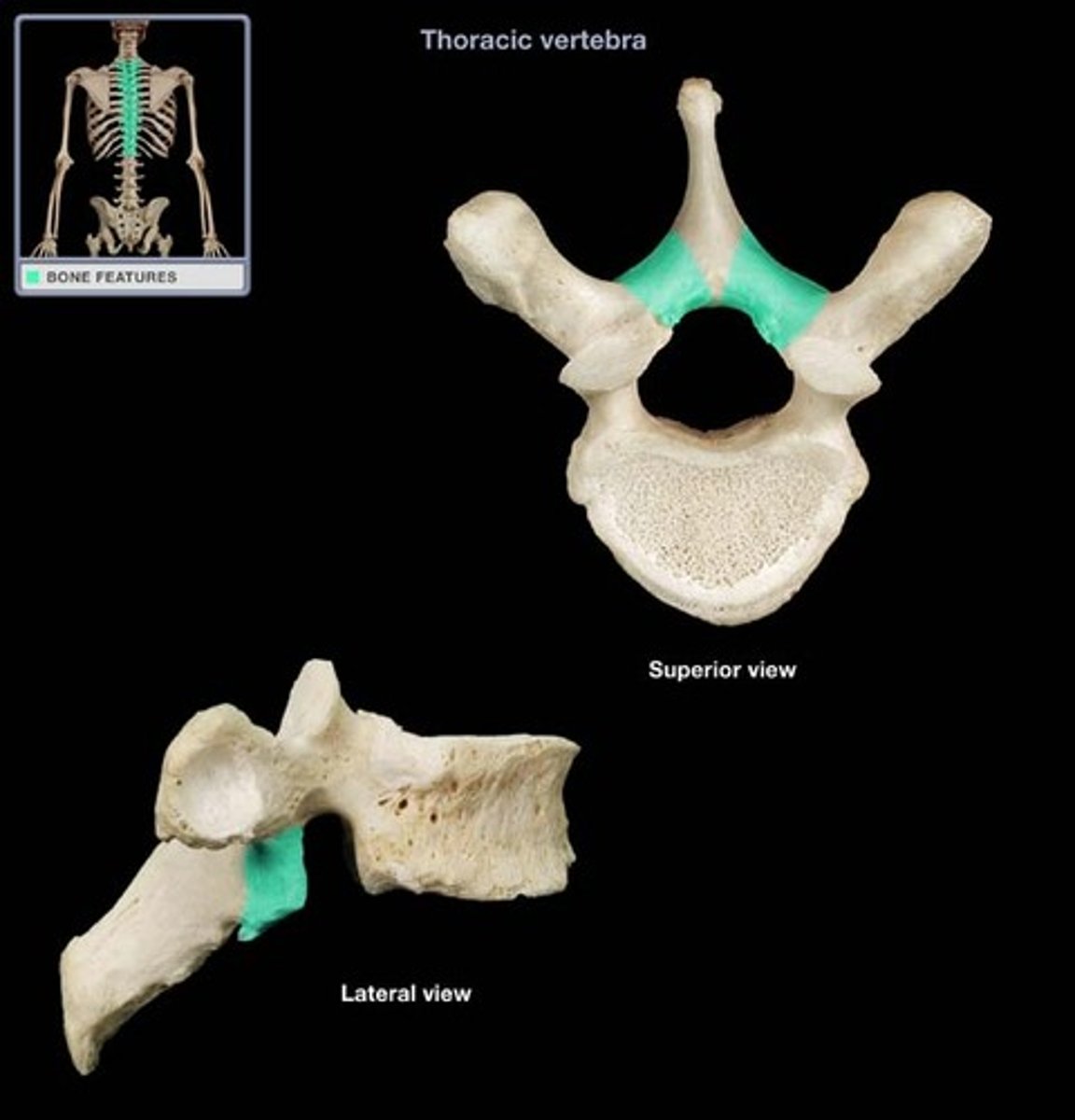
lamina
paired arches supported by the pedicles located posterior to the vertebral body
spinous process
most posterior projection of the vertebra for muscle/ligament attachment
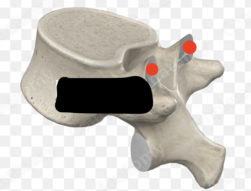
superior articular process
paired (superior and inferior) process which articulates with process of adjacent vertebrae
superior articular facet
smooth surface of articular processes; superior and inferior
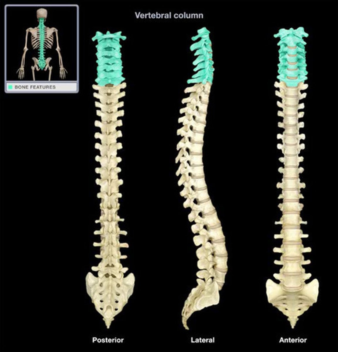
cervical vertebrae
transverse foramen
Atlas
(C1) supports the head
axis
(C2) serves as pivotal point for atlas and skull
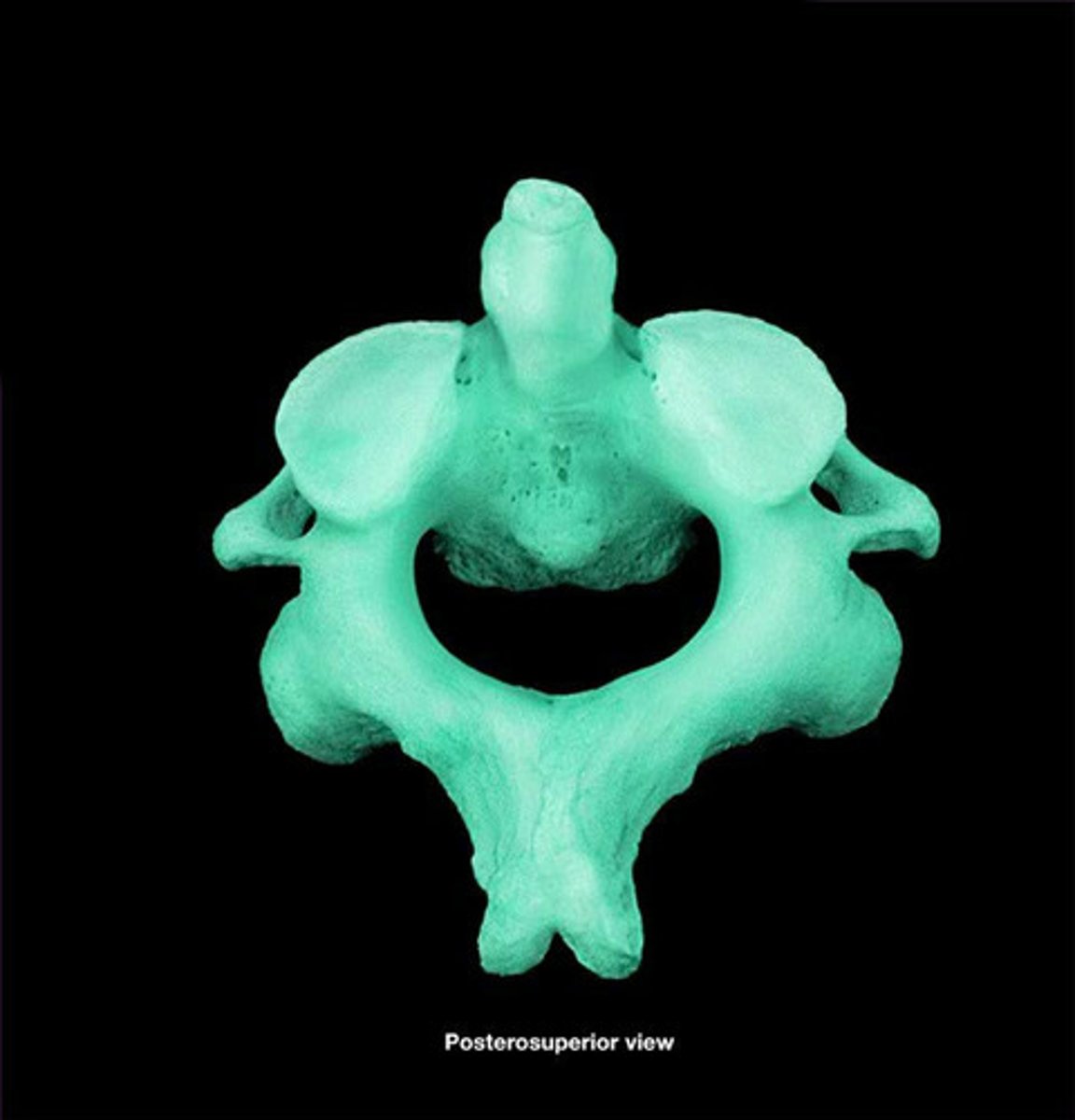
dens
process of the axis which passes through the vertebral foramen of the atlas
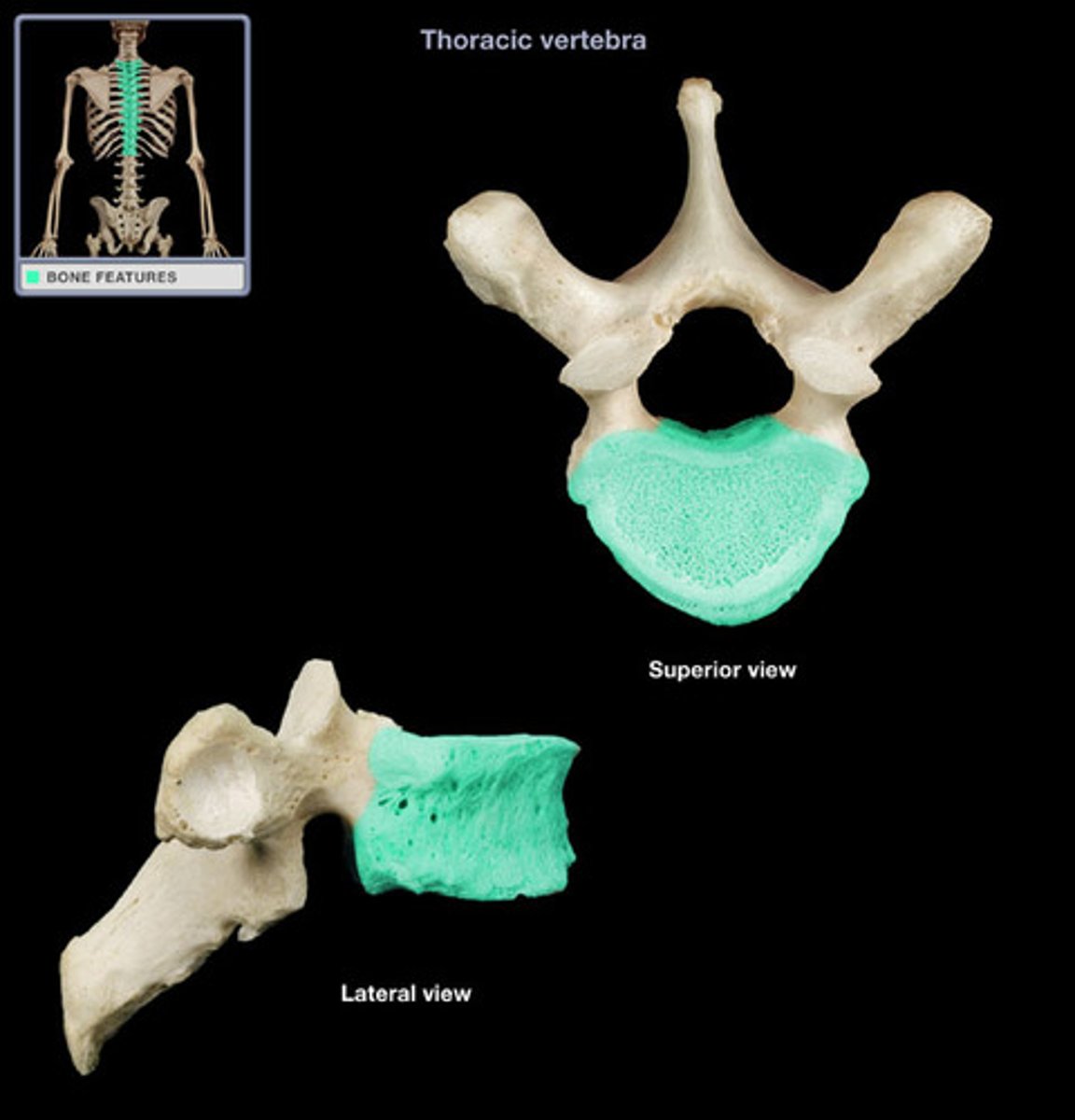
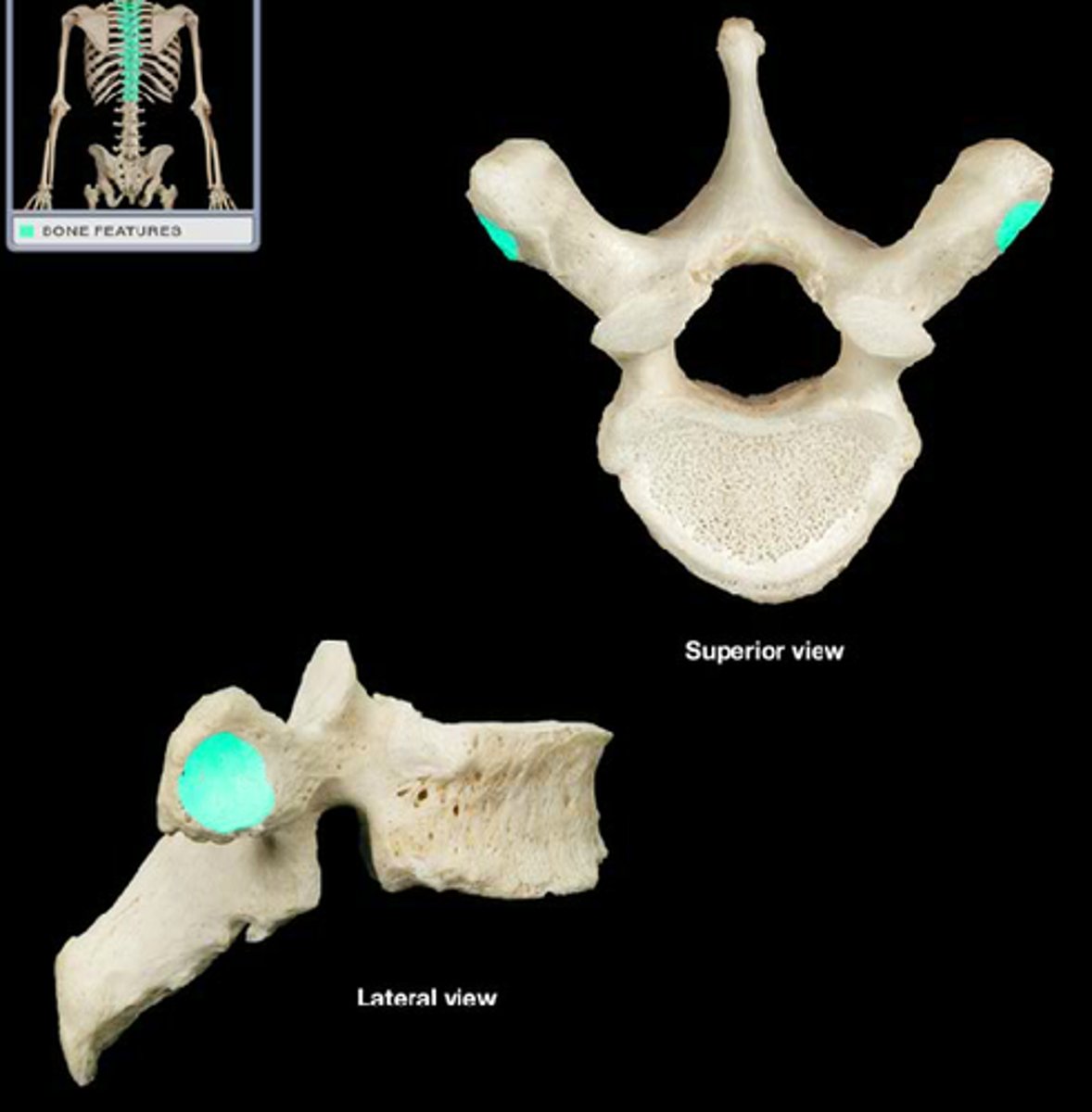
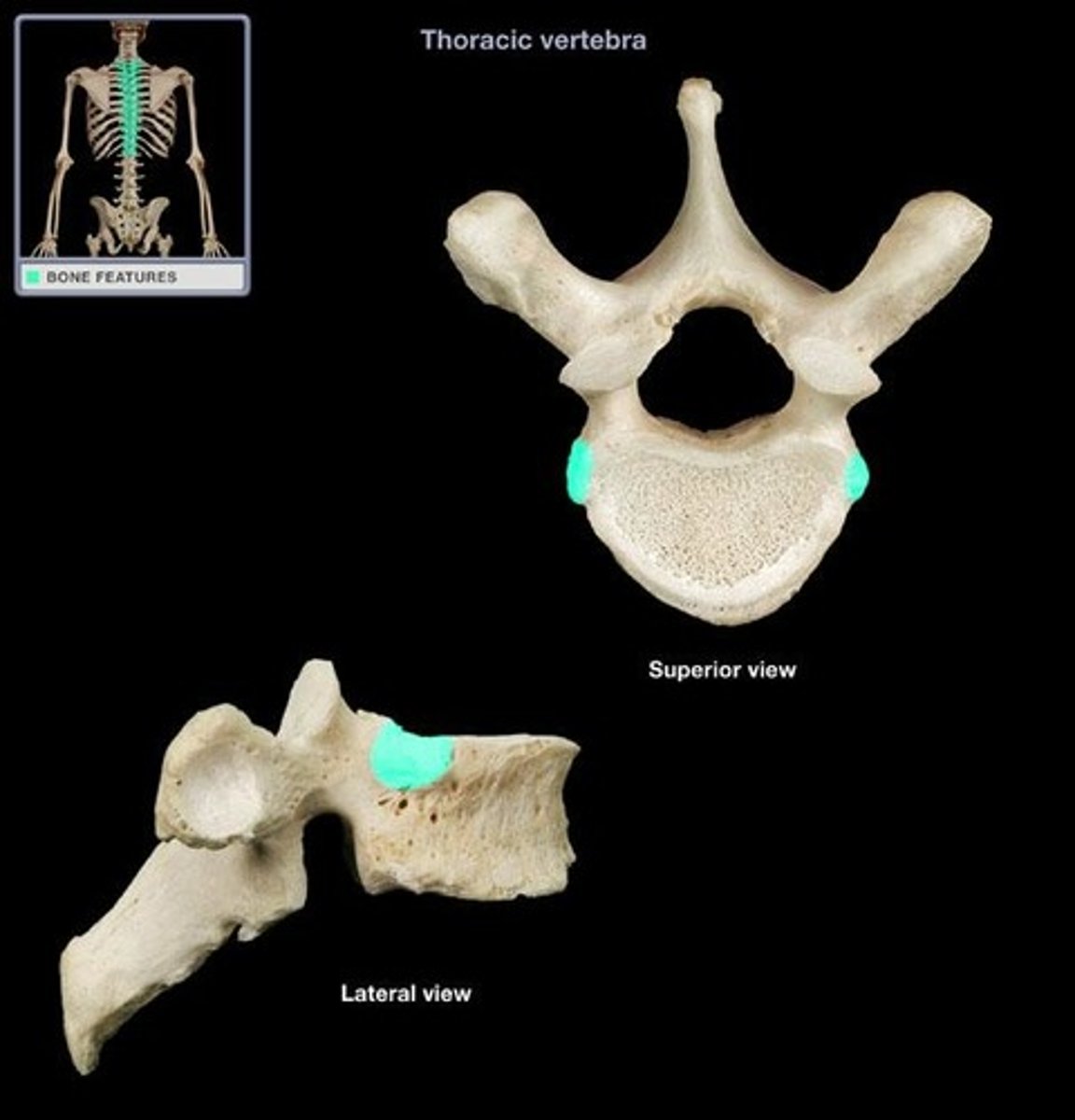
thoracic vertebrae
12 total (T 1-T 12), vertebrae with circular vertebral foramen and long spinous process
costal facet
depression on transverse process; articulates with the tubercle of the ribs; also named transverse facet of thoracic vertebra in APR
costal demifacet
half facet on superior and inferior edges of each vertebral body; articulates with head of ribs; also named superior and inferior costal facet of thoracic vertebra in APR
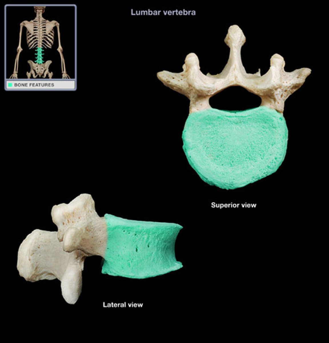
lumbar vertebrae
5 total (L 1 - L 5), vertebrae with strongly built components for weight bearing
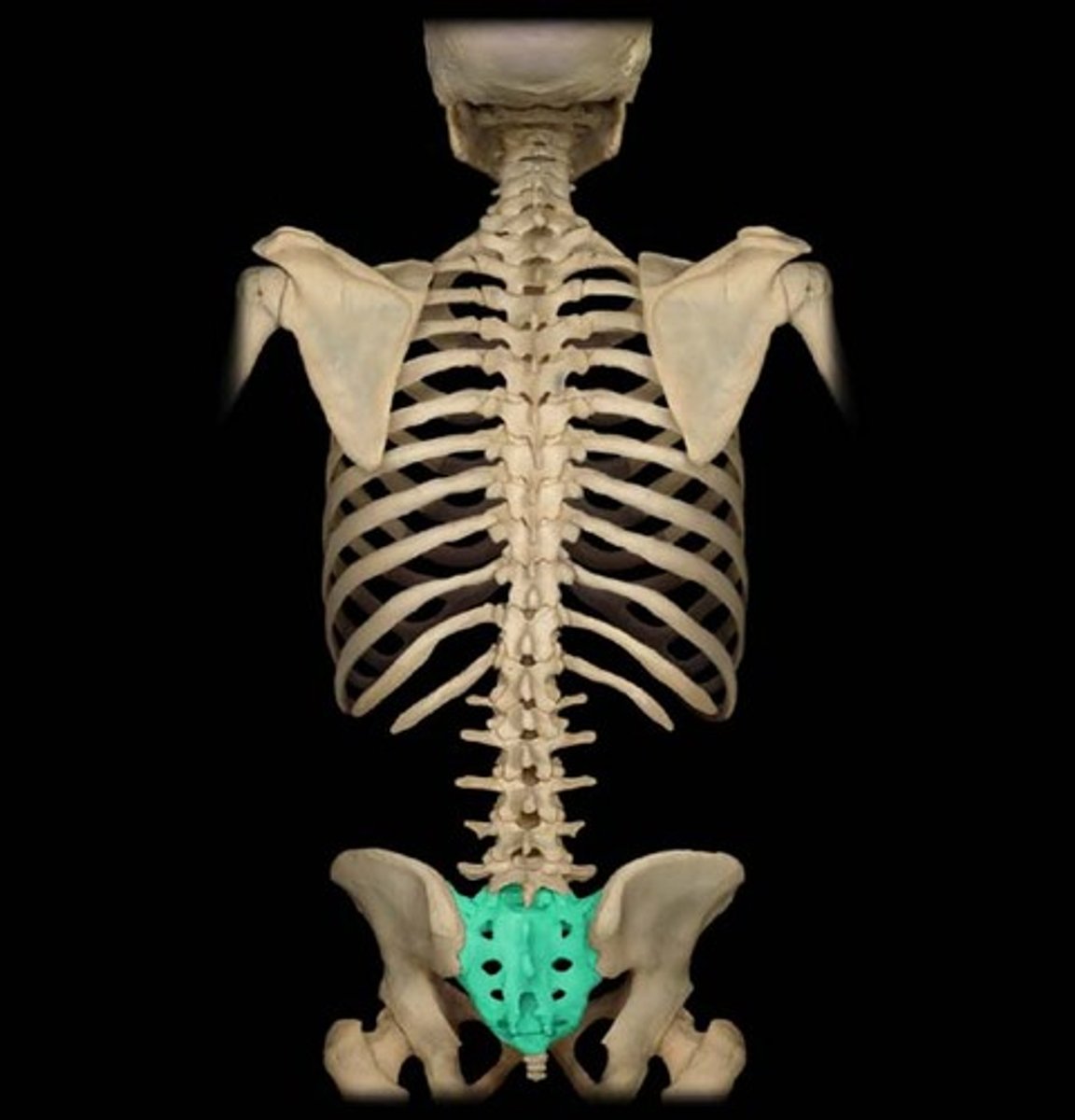
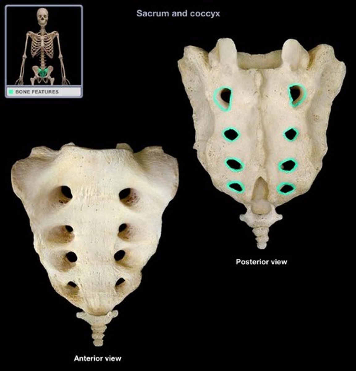
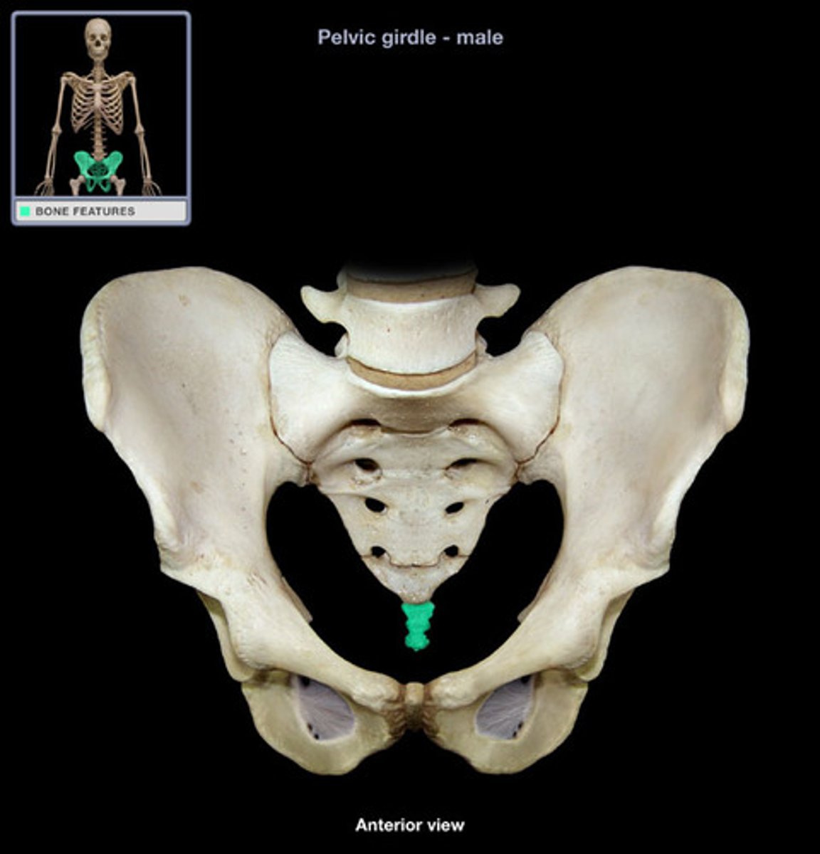
sacrum
(S1 - S5), composed of 5 fused vertebrae forming posterior wall of pelvis
auricular surface
lateral part that articulates with the os coxa through the sacroiliac joint of pelvis
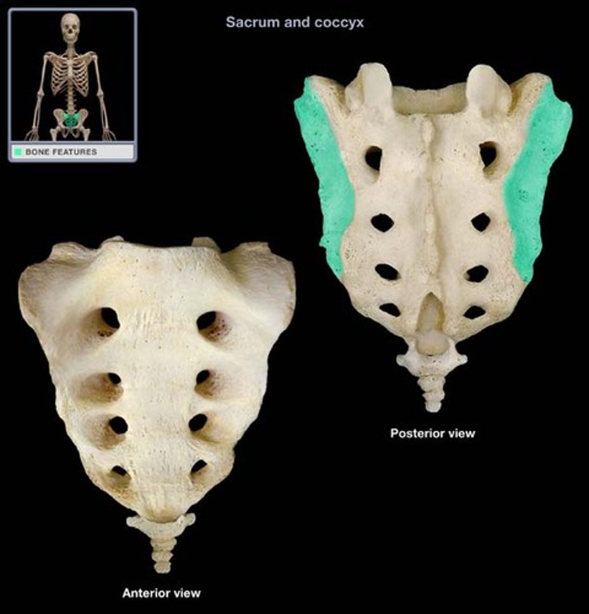
sacral foramina
anterior openings in the sacrum
coccyx
4 fused vertebrae forming the triangular "tailbone"
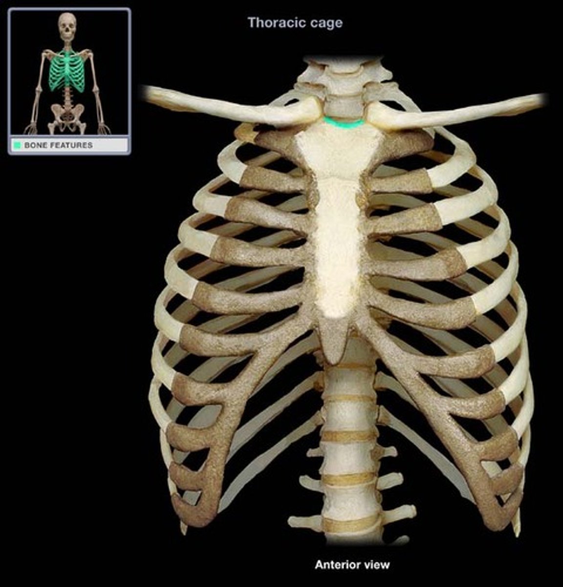
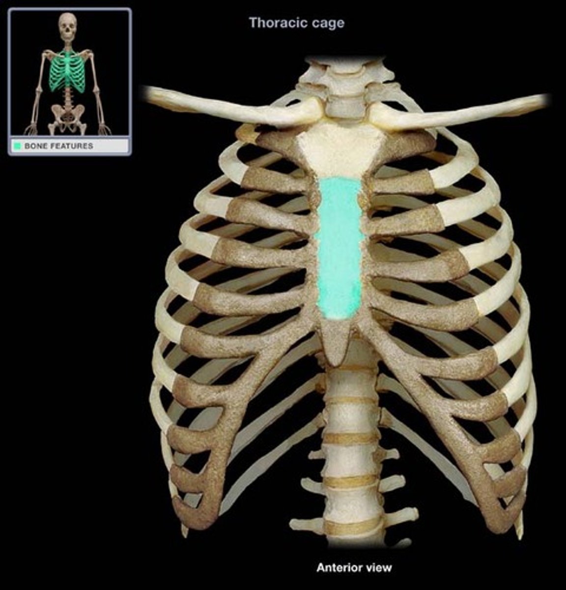
sternum
anterior midline anchor for the ribs; composed of three bones
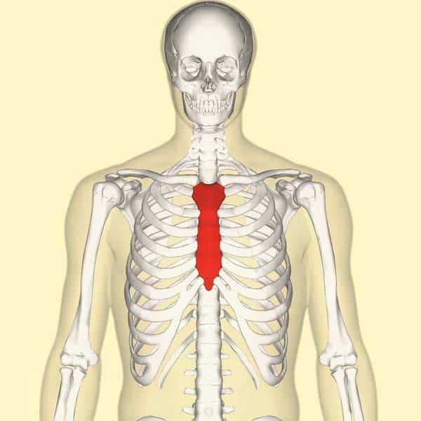
manubrium
superior portion of the sternum that articulates with the clavicle at its clavicular notch
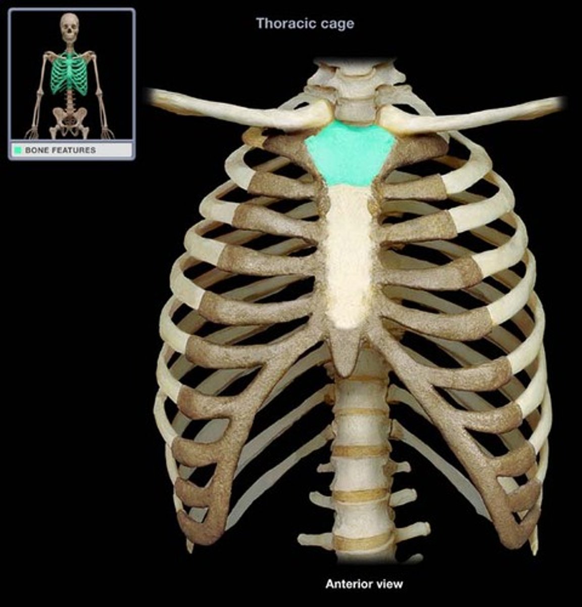
jugular notch
important anatomical landmark located in the superior portion of the manubrium
body
middle portion of the sternum that articulates with T2-T7 rib cartilage
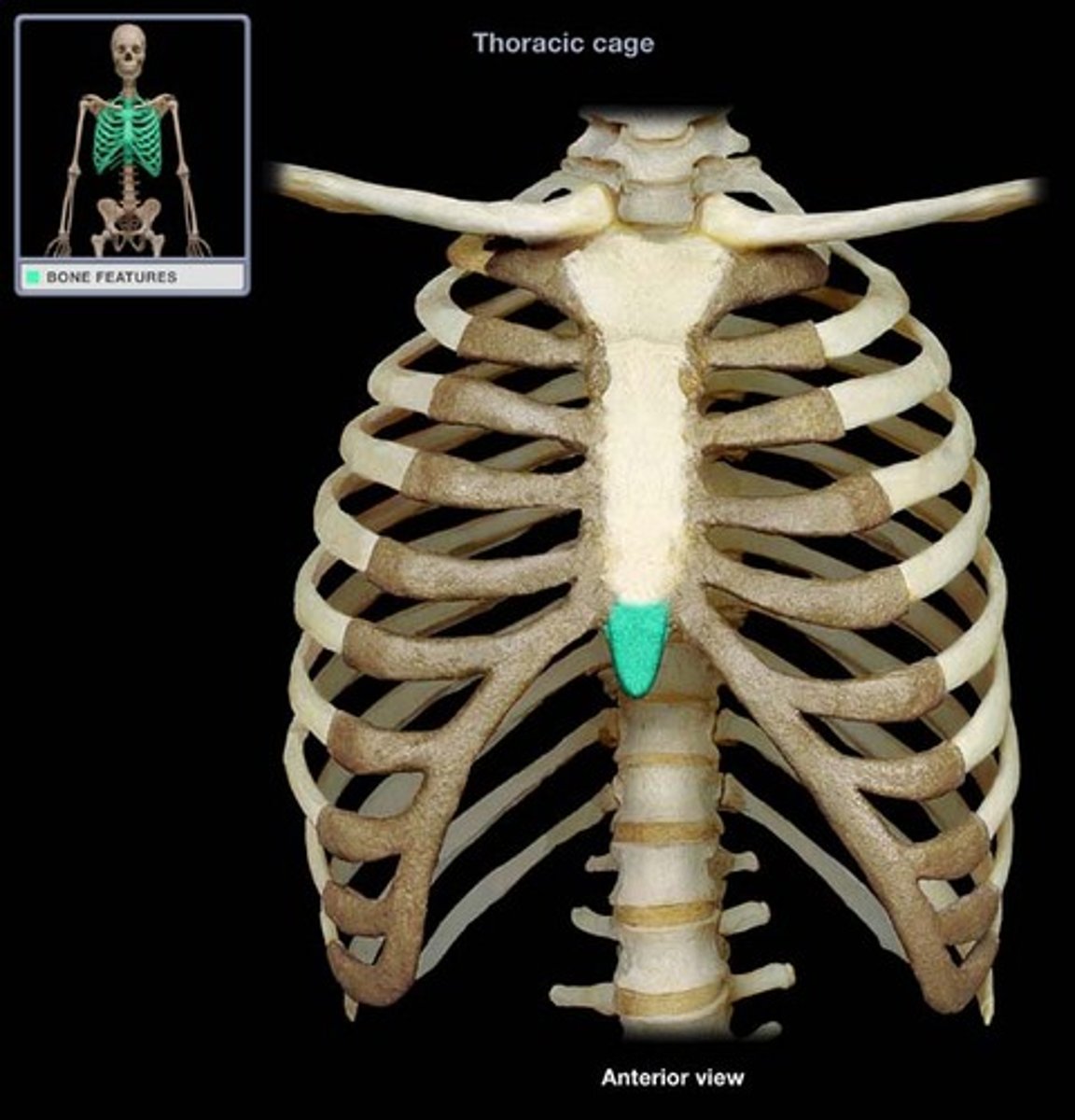
xiphoid process
small inferior portion of the sternum
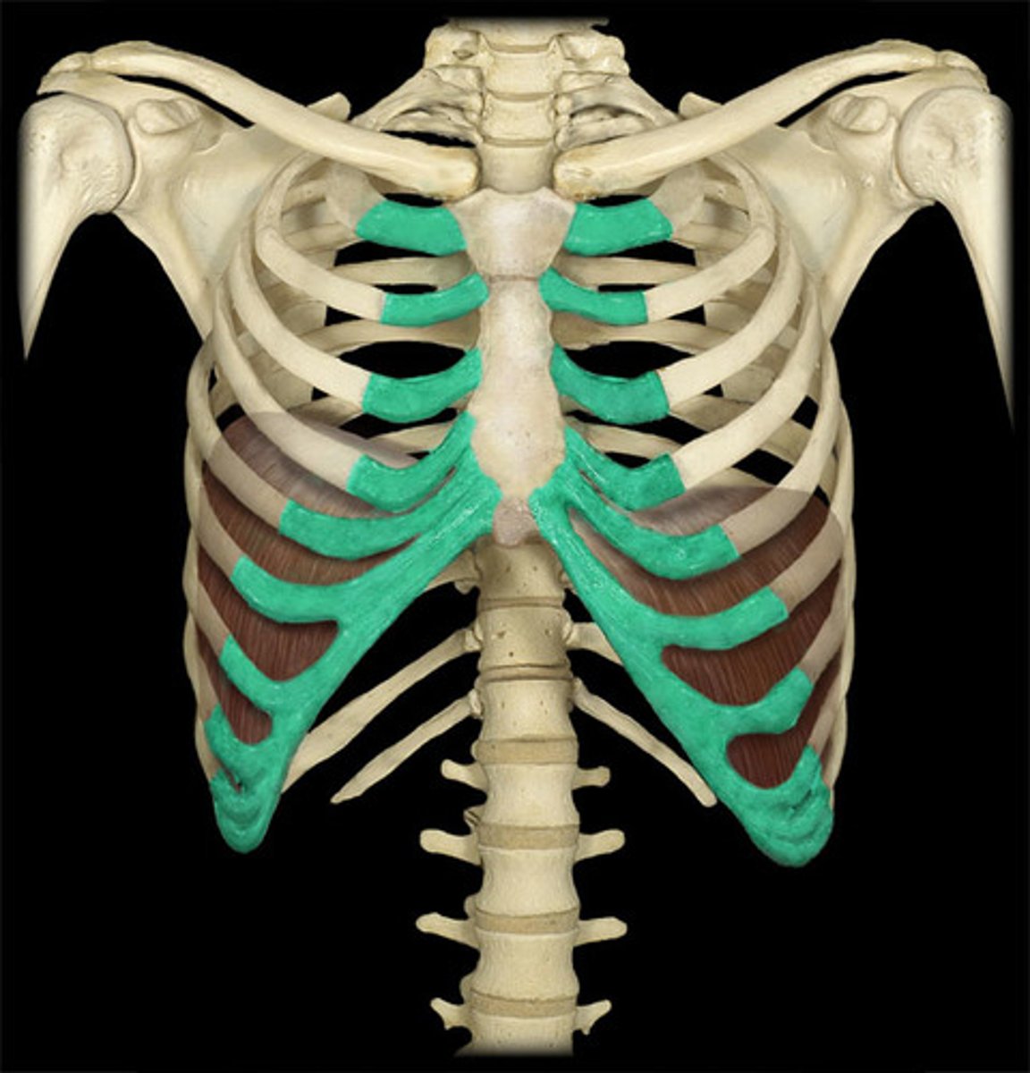
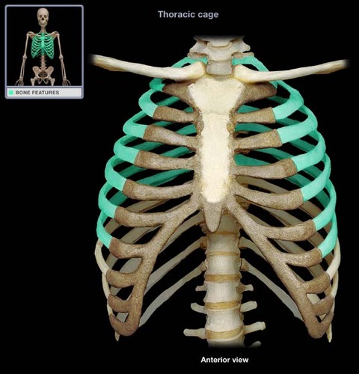
costal cartilage
connective tissue that connects the ribs to the sternum
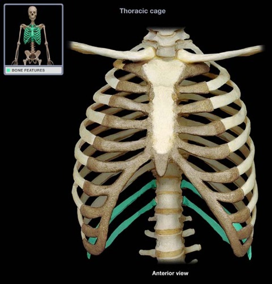
ribs
12 pairs, attached to T1 - T12 thoracic vertebrae
true rib
attached posteriorly to T1 - T7 and attached directly anteriorly to the sternum via costal cartilage
False rib
attached posteriorly to vertebrae T8 - T12; they are not directly attached to the sternum
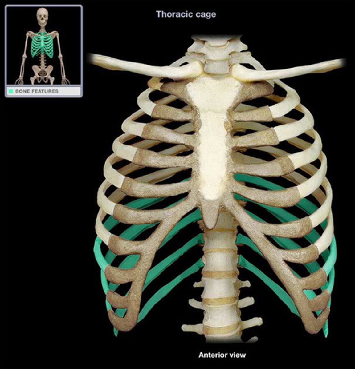
floating rib
false ribs attached posteriorly to vertebrae T11-T12 but not attached to the sternum
temporomandibular joint
Superior nuchal line