biology module 3 transport in animals
1/49
There's no tags or description
Looks like no tags are added yet.
Name | Mastery | Learn | Test | Matching | Spaced |
|---|
No study sessions yet.
50 Terms
why do living organisms have the need to exchange substances with their surrounding environment
They need to take oxygen and nutrients in
Waste products generated need to be released
what is mass flow
the bulk movement of materials
what do mass transport systems help do
Bring substances quickly from one exchange site to another
Maintain the diffusion gradients at exchange sites and between cells and their fluid surroundings
Ensure effective cell activity by keeping the immediate fluid environment of cells within a suitable metabolic range
difference between single and double circulatory systems
In a single circulatory system, the blood passes through the heart once during one complete circuit of the body
In a double circulatory system, the blood passes through the heart twice during one complete circuit of the body
single circulatory system in fish
Deoxygenated blood is pumped to the gills from the heart
The gills are the exchange site where oxygen and carbon dioxide are exchanged with the atmosphere and the blood
The oxygenated blood flows from the gills to the rest of the body
It travels through the capillaries in organs, delivering oxygen and nutrients
The blood returns to the heart
The heart only has one atrium and one ventricle
Double circulatory system in mammals
In mammals the blood passes throught the heart twice during a single circuit of the body
As a result the mammalian heart has a left side and right side with a wall (septum) dividing the two
The left side contains oxygenated blood and the right side contains deoxygenated blood
Blood in the right side of the heart leaves and travels to the lungs
The blood returns to the left side of the heart before being pumped around the rest of the body
Once the blood has passed through all the other organs and tissues it returns to the right side of the heart
In general, any blood that has just passed through an organ goes straight back to the heart, not to another organ
The hepatic portal vein is the exception to this rule, it allows blood from the gut to flow to the liver
the heart
a hollow, muscular organ located in the chest cavity which pumps blood. cardiac muscle tissue is specialised for repeated involuntary contraction without rest
arteries
blood vessels which carry blood away from the heart. the walls of the arteries contain lots of muscle and elastic tissue and a narrow lumen, to maintain high blood pressure. arteries range from 0.4 to 2.5 cm in diameter
arterioles
small arteries which branch from larger arteries and connect to capillaries.
capillaries
tiny blood vessels which connect arterioles and venules. their size means they pass directly past cells and tissues and perform gas exchange and exchange of substances such as glucose
venules
small veins which join capillaries to larger veins
veins
blood vessels which carry blood back towards the heart. thin walls in comparison to arteries, having less muscle and elastic tissue but a wider lumen. valves help maintain blood flow back towards the heart
advantages of double circulation
When blood enters a capillary network the pressure and speed drops significantly
In a single circulatory system, the blood has to pass through two capillary networks before returning to the heart
In a double circulatory system, the blood only passes through one capillary network before returning to the heart
As a result, the double circulation maintains higher blood pressure and average speed of flow
This increased pressure and speed helps to maintain a steeper concentration gradient which allows for the efficient exchange of nutrients and waste with the surrounding tissues
closed vs open circulatory systems
In a closed circulatory system, blood is pumped around the body and is always contained within a network of blood vessels
All vertebrates and many invertebrates have closed circulatory systems
In an open circulatory system, blood is not contained within blood vessels but is pumped directly into body cavities
circulatory systems in humans
Humans have a closed double circulatory system
The right side of the heart pumps blood deoxygenated blood to the lungs for gas exchange; this is the pulmonary circulatory system
Blood then returns to the left side of the heart, so that oxygenated blood can be pumped efficiently (at high pressure) around the body; this is the systemic circulatory system
Circulatory system in insects
Insects have one main blood vessel - the dorsal vessel
The tubular heart in the abdomen pumps haemolymph (this is what blood in insects is called) into the dorsal vessel
The dorsal vessel delivers the haemolymph into the haemocoel (body cavity)
Haemolymph surrounds the organs and eventually reenters the heart via one-way valves called ostia
Unlike the blood in a mammals circulatory system, the haemolymph is not specifically directed towards any organs in an insect
Insects are able to survive with this less efficient circulatory system because oxygen is delivered directly to their tissues via tracheae (a system of tubes) that connect directly to the outside
what is plasma
a straw-coloured liquid that constitutes around 55 % of the blood
Plasma is largely composed of water (95 %) and because water is a good solvent many substances can dissolve in it, allowing them to be transported around the body
how is tissue fluid formed
As blood passes through capillaries some plasma leaks out through gaps in the walls of the capillary to surround the cells of the body
Hydrostatic pressure
This is the pressure exerted by a fluid, e.g. blood
The hydrostatic pressure in this example is the blood pressure, generated by the contraction of the heart muscle
oncotic pressure
This is the osmotic pressure exerted by plasma proteins within a blood vessel
Plasma proteins lower the water potential within the blood vessel, causing water to move into the blood vessel by osmosis
what happens at the arterial end
When blood is at the arterial end of a capillary the hydrostatic pressure is great enough to force fluid out of the capillary
Proteins remain in the blood as they are too large to pass through the pores in the capillary wall
The increased protein content creates a water potential gradient (osmotic pressure) between the capillary and the tissue fluid
At the arterial end the hydrostatic pressure is greater than the osmotic pressure so the net movement of water is out of the capillaries into the tissue fluid
what happens at the venous end
At the venous end of the capillary the hydrostatic pressure within the capillary is reduced due to increased distance from the heart and the slowing of blood flow as it passes through the capillaries
The water potential gradient between the capillary and the tissue fluid remains the same as at the arterial end
At the venous end the osmotic pressure is greater than the hydrostatic pressure and water begins to flow back into the capillary from the tissue fluid
Roughly 90 % of the fluid lost at the arterial end of the capillary is reabsorbed at the venous end
The other 10 % remains as tissue fluid and is eventually collected by lymph vessels and returned to the circulatory system
what happens if the blood pressure is high
If blood pressure is high (hypertension) then the pressure at the arterial end is even greater
This pushes more fluid out of the capillary and fluid begins to accumulate around the tissues. This is called oedema
formation of lymph
Some tissue fluid reenters the capillaries while some enters the lymph vessels
The lymph vessels are separate from the circulatory system
They have closed ends and large pores that allow large molecules to pass through
Larger molecules that are not able to pass through the capillary wall enter the lymphatic system as lymph
Small valves in the vessel walls are the entry point to the lymphatic system
The liquid moves along the larger vessels of this system by compression caused by body movement. Any backflow is prevented by valves
This is why people who have been sedentary on planes can experience swollen lower limbs
The lymph eventually reenters the bloodstream through veins located close to the heart
Any plasma proteins that have escaped from the blood are returned to the blood via the lymph capillaries
If plasma proteins were not removed from tissue fluid they could lower the water potential (of the tissue fluid) and prevent the reabsorption of water into the blood in the capillaries
After digestion lipids are transported from the intestines to the bloodstream by the lymph system
when do valves in the heart open and close
Open when the pressure of blood behind them is greater than the pressure in front of them
Close when the pressure of blood in front of them is greater than the pressure behind them
why are valves important
keeping blood flowing forward in the right direction and stopping it flowing backwards. They are also important for maintaining the correct pressure in the chambers of the heart
what are the right atrium and right ventricle separated by
the atrioventricular valve, which is otherwise known as the tricuspid valve
what are the right ventricle and pulmonary artery separated by
the pulmonary valve
what are the left atrium and the left ventricle separated by
the aortic valve
coronary arteries
The heart is a muscle and so requires its own blood supply for aerobic respiration
The heart receives blood through arteries on its surface, called coronary arteries
It’s important that these arteries remain clear of plaques, as this could lead to angina or a heart attack (myocardial infarction)
what is the cardiac cycle
the series of events that take place in one heart beat, including muscle contraction and relaxation
what is systole
contraction of the heart
what is diastole
relaxation of the heart
how do volume changes in the heart occur
Contraction of the heart muscle causes a decrease in volume in the corresponding chamber of the heart, which then increases again when the muscle relaxes
how are volume changes linked to pressure changes
When volume decreases, pressure increases
When volume increases, pressure decreases
events of the cardiac cycle - atrial systole
The walls of the atria contract
Atrial volume decreases
Atrial pressure increases
The pressure in the atria rises above that in the ventricles, forcing the atrioventricular (AV) valves open
Blood is forced into the ventricles
There is a slight increase in ventricular pressure and chamber volume as the ventricles receive the blood from the atria
The ventricles are relaxed at this point; ventricular diastole coincides with atrial systole
events of the cardiac cycle - ventricular systole
The walls of the ventricles contract
Ventricular volume decreases
Ventricular pressure increases
The pressure in the ventricles rises above that in the atria
This forces the AV valves to close, preventing back flow of blood
The pressure in the ventricles rises above that in the aorta and pulmonary artery
This forces the semilunar (SL) valves open so blood is forced into the arteries and out of the heart
During this period, the atria are relaxing; atrial diastole coincides with ventricular systole
The blood flow to the heart continues, so the relaxed atria begin to fill with blood again
events of the cardiac cycle - diastole
The ventricles and atria are both relaxed
The pressure in the ventricles drops below that in the aorta and pulmonary artery, forcing the SL valves to close
The atria continue to fill with blood
Blood returns to the heart via the vena cava and pulmonary vein
Pressure in the atria rises above that in the ventricles, forcing the AV valves open
Blood flows passively into the ventricles without need of atrial systole
The cycle then begins again with atrial systole
Valve movements during the cardiac cycle
Stage in cardiac cycle Atrioventricular valves Semilunar valves
Atrial systole Open Closed
Ventricular systole Closed Open
Diastole Open Closed
what does it mean when they say the control of the basic heartbeat is myogenic
the heart will beat without any external stimulus
stages of cardiac cycle
sinoatrial node sends out a wave of extinction
atria contract
atrioventricular node sends out a wave of excitation
purkyne tissue conducts the wave of extinction
ventricles contract
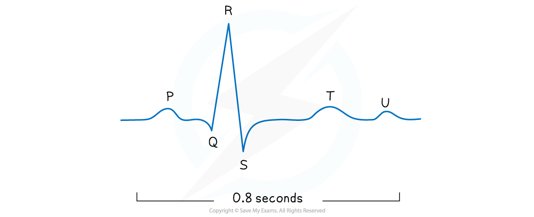
explain the letters in this diagram
The P wave
Caused by the depolarisation of the atria, which results in atrial contraction (systole)
The QRS complex
Caused by the depolarisation of the ventricles, which results in ventricular contraction (systole)
This is the largest wave because the ventricles have the largest muscle mass
The T wave
Caused by the repolarisation of the ventricles, which results in ventricular relaxation (diastole)
The U wave
Scientists are still uncertain of the cause of the U wave, some think it is caused by the repolarisation of the Purkyne fibres
tachychardia
When the heart beats too fast it is tachycardic
An individual with a resting heart rate of over 100 bpm is said to have tachycardia
Bradycardia
When the heart beats too slow it is bradycardic
An individual with a resting heart rate below 60 bpm is said to have bradycardia
A lot of fit individuals or athletes tend to have lower heart rates and it is usually not dangerous
Ectopic heartbeat
This condition is caused by an early heartbeat followed by a pause
It is common in the population and usually requires no treatment unless very severe
fibrillation
An irregular heartbeat will disrupt the rhythm of the heart
Severe cases of fibrillation can be very dangerous, even fatal
the role of haemoglobin - transport of oxygen
The majority of oxygen transported around the body is bound to the protein haemoglobin in red blood cells
Red blood cells are also known as erythrocytes
Each molecule of haemoglobin contains four haem groups, each able to bond with one molecule of oxygen
This means that each molecule of haemoglobin can carry four oxygen molecules, or eight oxygen atoms in total
role of haemoglobin - carbon dioxide transport
Waste carbon dioxide produced during respiration diffuses from the tissues into the blood
There are three main ways in which carbon dioxide is transported around the body
A very small percentage of carbon dioxide dissolves directly in the blood plasma and is transported in solution
Carbon dioxide can bind to haemoglobin, forming carbaminohaemoglobin
A much larger percentage of carbon dioxide is transported in the form of hydrogen carbonate ions (HCO3-)
formation of hydrogen carbonate ions
Carbon dioxide diffuses from the plasma into red blood cells
Inside red blood cells carbon dioxide combines with water to form H2CO3
CO2 + H2O ⇌ H2CO3
Red blood cells contain the enzyme carbonic anhydrase which catalyses the reaction between carbon dioxide and water
Without carbonic anhydrase this reaction proceeds very slowly
The plasma contains very little carbonic anhydrase hence H2CO3 forms more slowly in plasma than in the cytoplasm of red blood cells
Carbonic acid dissociates readily into H+ and HCO3- ions
H2CO3 ⇌ HCO3– + H+
Hydrogen ions can combine with haemoglobin, forming haemoglobinic acid and preventing the H+ ions from lowering the pH of the red blood cell
Haemoglobin is said to act as a buffer in this situation
The hydrogen carbonate ions diffuse out of the red blood cell into the blood plasma where they are transported in solution
the chloride shift
The chloride shift is the movement of chloride ions into red blood cells that occurs when hydrogen carbonate ions are formed
Hydrogen carbonate ions are formed by the following process
Carbon dioxide diffuses into red blood cells
The enzyme carbonic anhydrase catalyses the combining of carbon dioxide and water to form carbonic acid (H2CO3)
CO2 + H2O ⇌ H2CO3
Carbonic acid dissociates to form hydrogen carbonate ions and hydrogen ions
H2CO3 ⇌ HCO3- + H+
Negatively charged hydrogen carbonate ions formed from the dissociation of carbonic acid are transported out of red blood cells via a transport protein in the membrane
To prevent an electrical imbalance, negatively charged chloride ions are transported into the red blood cells via the same transport protein
If this did not occur then red blood cells would become positively charged as a result of a buildup of hydrogen ions formed from the dissociation of carbonic acid