exam 1 oral H & E
1/145
There's no tags or description
Looks like no tags are added yet.
Name | Mastery | Learn | Test | Matching | Spaced | Call with Kai |
|---|
No analytics yet
Send a link to your students to track their progress
146 Terms
fertilization, cleavage, morula, blastocyst, implantation
order of events in week 1
greater horn and lower rim of hyoid
formed from ectomesenchyme in the 3rd branchial arch
bilaminar disc, syncytiotrophoblast formation, prochordal plate formation
order of key developments during week 2 of embryonic development.
primitive node/streak/pit, gastrulation (endoderm and mesoderm), notochord, neurulation, neural plate
order of key processes during week 3 of embryonic development
neural tube, head folding, stomodeum, mandibular process, branchial archs/grooves/pouches, mand process join, buccopharyngeal mem rupture, nasal placodes form
order of major developments during week 4 of embryonic development.
end of week 4
when does odontogenic epithelium form
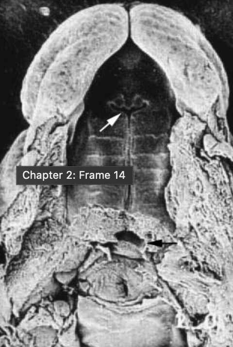
merging and fusion of facial processes, naso-optic and bucconasal groove, nasal fin, oronasal membrane
key developments during week 6
nasal fin, week 6
what is this structure and when does it form
oronasal membrane, week 6 after nasal fin
what is this structure, when does it form
end of week 6
when does the odontogenic epithelia merge to form primary epithelial bands
week 7-8
when does formation of secondary palate begin (merging of palatine shelves)
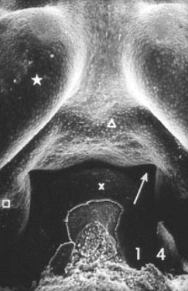
nasal septum, frontonasal process, palatine shelves
what three structures merge together during weeks 7-9
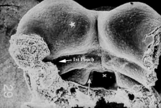
1st arch/mandibular process
what structure is represented by the star
weeks 5-8
when does pouch/neck development occur
week 5
when do auricular hillocks form
weeks 4-9
when does tongue development occur
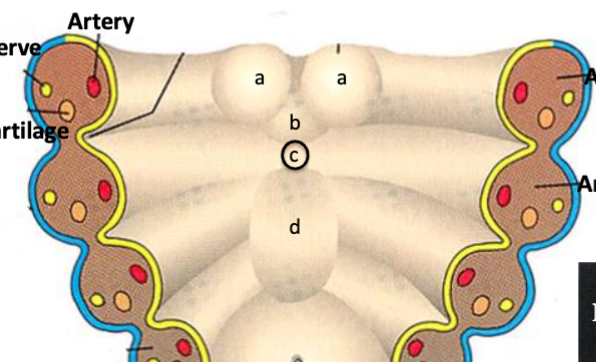
artery, vein,nerve, branchial cartilage (NCC)
what are the common structures within each brachial arch
cervical sinus
A temporary structure formed during embryonic development from branchial grooves 2-4
branchial pouches
Structures that develop internally leading to the formation of various organs and tissues, including the thymus and parathyroid glands.
branchial grooves
The external depressions between the branchial arches during embryonic development, which contribute to the formation of the neck and structures of the head.
rhombomeres 3-8
where is the hox gene family expreessed
Msx gene family
which genes are responsible for midbrain and rhombomeres 1 and 2
paraxial mesoderm
the region of mesoderm located on either side of the notochord that gives rise to SOMITES, which contribute to the development of the vertebral column and associated musculature.
Hox
what gene controls branchial/pharyngeal development (branchial arches 2-6)
1
branchial arches controlled by NCCs from rhombomeres 1, 2 and midbrain
2
branchial arches controlled by NCCs from rhombomere 4
3
branchial arches controlled by NCCs from rhombomere 6
4-6
branchial arches controlled by NCCs from rhombomere 7
foramen cecum
what is C
branchial mesenchyme
mix of NCC ectomesenchyme and paraxial mesoderm mesenchyme
NCC ectomesenchyme
forms branchial cartilages and associated bone
face and neck muscles, blood vessels (branchial arteries and veins)
structures derived from paraxial mesoderm
1st branchial pouch
what forms auditory tube, middle ear cavity (with contributions from 2nd), inner layer of tympanic membrane
1st branchial groove
forms external auditory meatus
palatine tonsil (CN VII), supra-tonsillar fossa
formed from 2nd branchial pouch
inferior parathyroid (dorsal wings) and thymus gland (ventral wing) (CN IX)
formed from 3rd branchial pouch
superior parathyroid and ultimobranchial body (parafollicular cells of thyroid gland)
formed from 4th branchial pouch
thyroid C cells
formed from 5th branchial pouch
cranial nerve ganglia
formed from neural crest cells from ectodermal placodes
CN V
formed from ectodermal placode in 1st branchial arch
CN VII
formed from ectodermal placode in the 2nd branchial arch
CN IX
formed from ectodermal placode in the 3rd branchial arch
CN X
formed from ectodermal placode in the 4th-6th branchial arch
meckel’s cartilage: incus,malleus, sphenomalleolar, sphenomandibular ligaments
ectomesenchyme from NCCs gives rise to what structures in 1st branchial arch
stapes, styloid process, stylohyoid ligament, lesser horns and upper rim of hyoid
formed from ectomesenchyme in the 2nd branchial arch
laryngeal cartilages (thyroid, crycoid, aryteenoid)
develop from the fourth and sixth branchial arches ectomesenchyme.
lingual swelling
develops into body of tongue
primarily 1st arch
what branchial arch does the tongue develop from
inner ear
what does otic placode develop into
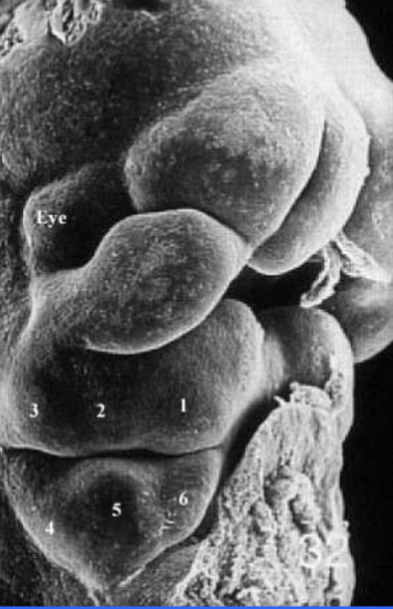
auricular hillocks of the 1st and 2nd branchial arches
what structures give rise to external ear
auricular hillocks
what structures are shown here labeled 1-6
complete branchial fistula
A congenital abnormality resulting from incomplete closure brachial grooves 2-4 leading to a connection between the pharynx and the skin.
maxillary
artery of 1st branchial arch
hyoid, stapedia
artery of 2nd branchial arch
internal carotid
artery of 3rd branchial arch
right subclavian, aorta
artery of 4th branchial arch
muscles of mastication
muscle of 1st branchial arch
muscles of facial expression
muscles derived from the 2nd branchial arch
stylopharyngeus
muscle derived from the 3rd branchial arch
pharyngeal and laryngeal musculature
muscles derived from the 4th and 6th branchial arches
NCC migration to ventral pharynx
provides ectomesenchyme for formation of branchial/pharyngeal structures
mid week 3-end week 4
when does anterior-posterior folding occur
neurocranium
surrounds brain composed of cartilages and placodes
dermocranium
the part of the skull that forms a protective shell around the chondrocranium
chondrocranium
the cartilaginous part of the skull that supports the brain and forms the base of the skull.
viscerocranium
the portion of the skull that forms the facial skeleton, including the jaw and other structures.
ectomesenchyme
derived from NCCs, develops into dental and skeletal tissues of oral cavity, responsible for early development of face
mesenchyme
a type of embryonic connective tissue that develops into various types of tissues including bones and cartilage.
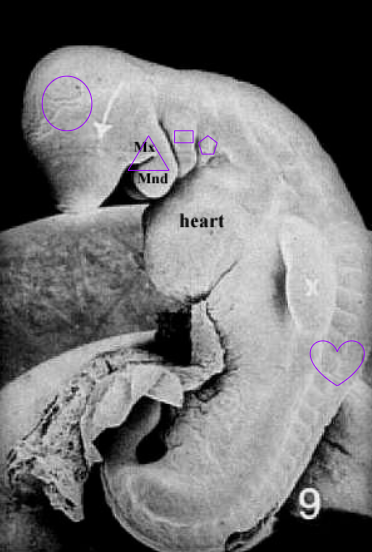
frontal,maxillary,stomodeum, mandibular, heart
processes that define the embryonic mouth
odontogenic epithelium
the epithelial tissue involved in tooth development, originating from the ectoderm.
frontal process
(A)
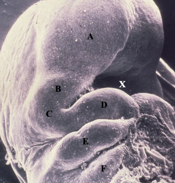
maxillary process
(B)
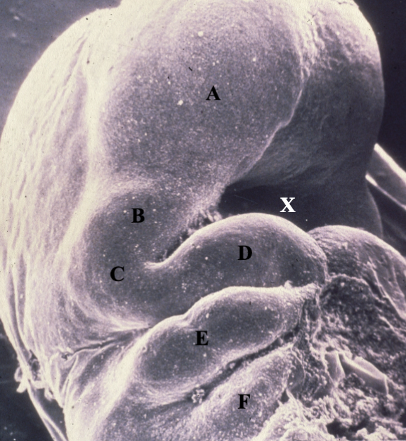
mandibular arch (1st)
(C)
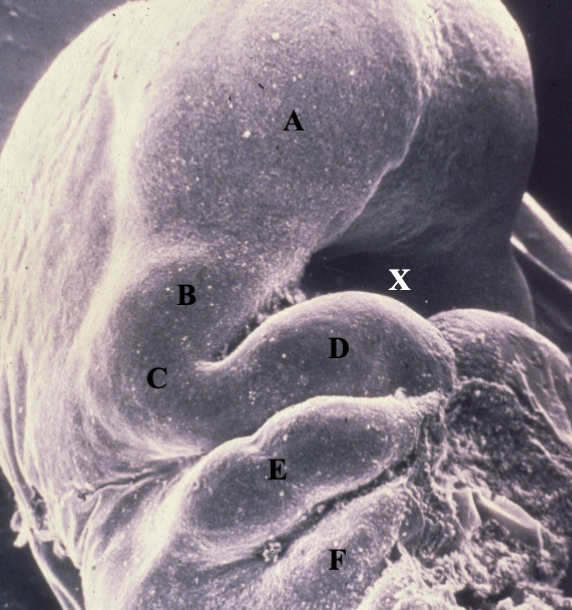
mandibular process
(D)
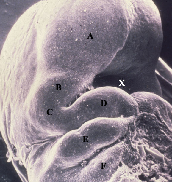
hyoid arch (2nd)
(E)
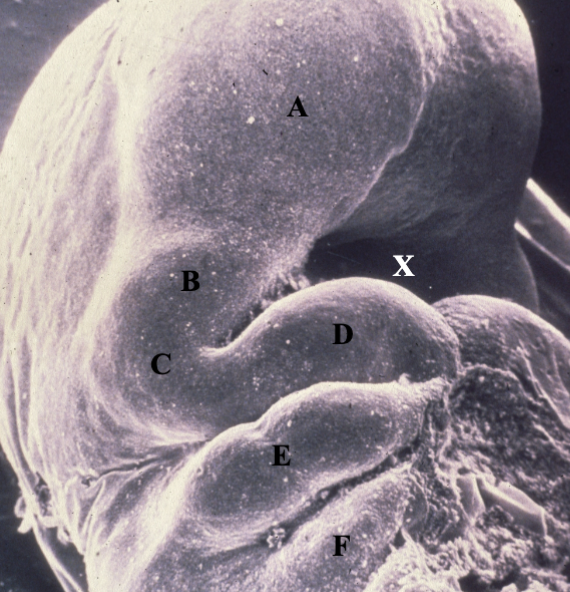
3rd branchial arch
(F)
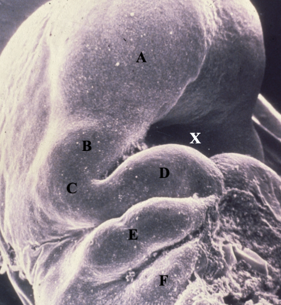
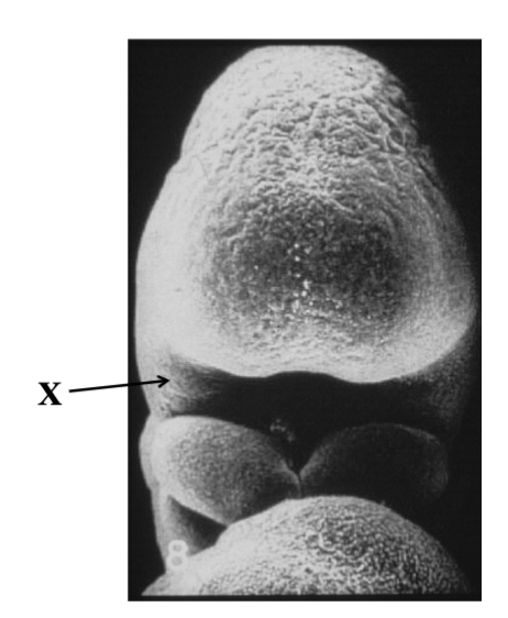
stomadeum
(X)

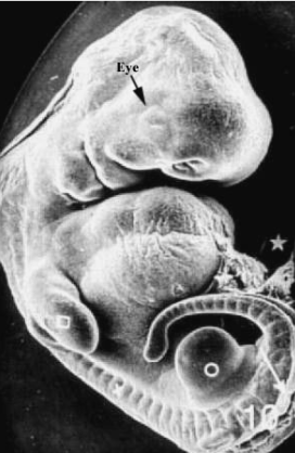
end of week 4 (28 days)
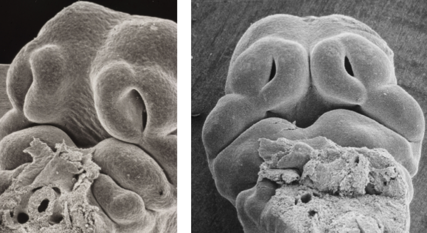
olfactory placodes (nasal placodes) begin to develop
placodes
thickening of ectoderm that forms sensory organs
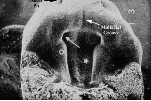
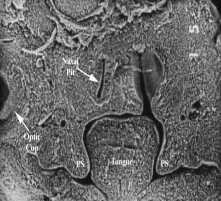
nasal pit formation
sign of week 5 development
medial nasal process
give rise to mid-portion of nose, upper lip, premaxilla and primary palate
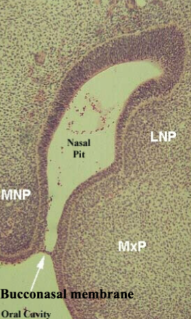
primary choanae (interenal nasal opening)
what is a sign of week 6 development
trabecular cartilages (NCC)
responsible for development of ethmoid bone
fusing of trabecular cartilage to midline of ethmoid
essential for fusion of facial processes in the midline
week 4
what week of development?
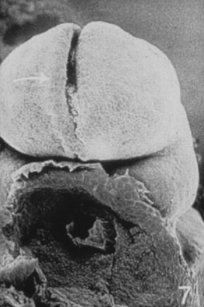
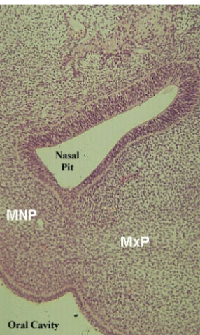
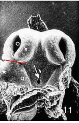
maxillary process
identify region marked by X
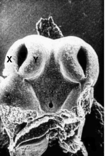
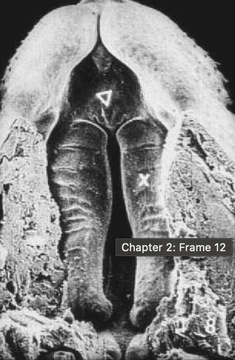
X:lateral nasal process, Y:medial nasal process
label X and Y
medial nasal process
forms into primary palate and premaxilla
maxillary process
forms secondary palate
week 7
tongue lowers to allow the fusion of the palatal shelves
week 8
palatal shelves begin to merge
week 9
palate shelves fuse with each other and the nasal septum
late week 4 (no nose)
what stage of development
week 5 (nasal pit present)
what stage of development?
week 5 (nose and no choanae)
stage of development?
week 6 (primary choanae present
what stage?
mid week 6 (tongue in way of palatal shelves)
what stage?
mid week 6 (tongue not retracted)
what stage?
week 8 (palate begins to merge)
late week 9 (palatal shelves joined but fusion incomplete)
what stage?